III. EMERGENCE OF ORDER IN TISSUES AND ORGANS Communication through Cell Junctions. Implications
in Growth Control and Differentiation
WERNER R. LOEWENSTEIN
Cell Physics Laboratory, Department of Physiology, College of Physicians and Surgeons, Columbia University, New York, New York 10032
When Alice came to Wonderland, the King asked her:
"What do you know about this business?"
"Nothing/' said Alice.
"Nothing whatever?" persisted the King.
"Nothing whatever," said Alice.
"That's very important," the King said.
Cells may, in principle, communicate with each other in three ways: (1) they may have information signals and receptor processes for these signals on their surface membrane; (2) they may release signals to the exterior and these may reach receptor processes of other cells via the intercellular spaces (hormonal systems); (3) the signals may pass directly from one cell interior to another. In an extreme form of kind (3), the cells communicate through macroscopic holes ( e. g., plant desmosomatas ). In another form, which has recently come to light, surface membranes of contiguous cells form junctions so structured as to allow flow of substances between cell interiors without appreciable leakage to the exterior. I shall explore here some of the possible developmental implications of this form of communication and deal with the related physicochemical mechanisms.
PHYSICOCHEMICAL ASPECTS
My story starts with a chance observation. In 1962, Y. Kanno and I were working on permeability properties of nuclear membranes in Drosophila salivary gland cells. We noticed to our surprise that when an ion current was passed between a point source inside the nucleus of one cell and the cell exterior, the voltage resulting inside a con
tiguous cell was nearly as high as that in the cell containing the 151
point source. It looked as though the membrane regions of cell contact {functional membranes) offered little resistance to ion flow (Loewen- stein and Kanno, 1963). By now it seems that intercellular com
munication through permeable junctional membranes is a rather general feature of tissues that do not normally carry electrical signals.
Salivary glands (Loewenstein and Kanno, 1963, 1964), neuroglia (Kuifler and Potter, 1964), renal tubules, certain sensory epithelia, urinary bladder (Loewenstein et al., 1965), liver (Penn, 1966), skin (Loewenstein and Penn, 1967), sponges (Loewenstein, 1967a), brown fat (Revel and Sheridan, 1968), thyroid (Jamakosmanovic and Loe
wenstein, 1968), stomach epithelium (Kanno and Matsui, 1968), and embryonic tissues (Potter et al., 1966; Ito and Hori, 1966; Sheridan, 1968; Ito and Loewenstein, 1968) are among the examples of tissues in which junctional communication has been shown.
Junctional Elements
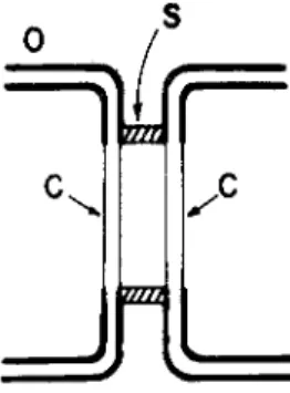
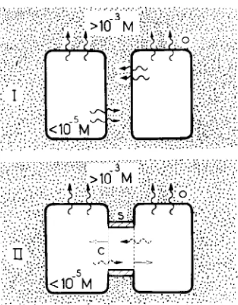
At the cell junctions of these tissues, one can distinguish three elements with clearly different resistances to ion diffusion (Fig. 1);
an element O, with a rather high resistance; an element C, which has a much lower resistance than that of O and which is spatially matched with a similar element on the contiguous cell; an element
^ fczza
S
FIG. 1. Elements of a cell junction. Diagram of a junctional unit: O, portion of nonjunctional surface membrane; C, junctional membrane; S, perijunctional insulation.
S, perijunctional, with a resistance higher than that of C. The scheme of Fig. 1 specifies the three elements only in terms of their resistive character and in terms of their spatial interrelationship. It says nothing about their structure or about their dimensions. O and C are mem-
brane structures. O represents the nonjunctional membrane portion with its well-known low ion permeability. C is the junctional mem
brane with ion permeability generally several orders of magnitude greater than that of O. S, the peri junctional insulation element, is not necessarily a membrane structure. The electrical measurements merely specify it as a barrier restricting ion flow between the interior of the cell system and the exterior at the level of the junction. Such a barrier may conceivably result, for instance, through close apposition of O-element portions. The separation between the C elements in the diagram is meant to indicate that the two C elements and the S element are all separate entities, not that there is necessarily a com
partment between the C-elements. (Possible structural correlates and related morphological literature are discussed in Loewenstein and Kanno, 1964; Loewenstein, 1966; Bullivant and Loewenstein, 1968.) In summary, the three elements form a functional unit in which diffusion is restricted in the direction of cell interior to exterior and relatively unrestricted in the direction of interior to interior.
Cell-to-Cell Flow of Small Ions
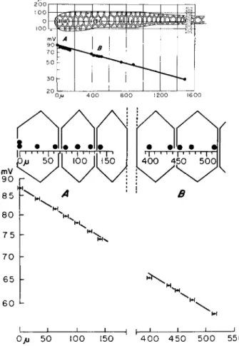
The foregoing picture is based on electrical measurements in a variety of junctional cell systems. The two main types of measure
ments are illustrated in Figs. 2 and 3, The low cell-to-cell attenuation of voltage in the measurement of Fig. 3 gives the general information that the relation between the resistances (r) for small ions of the three elements is rc< <r0; rc<rs. This information is available for most of the communicating systems listed above. The ratio of mem
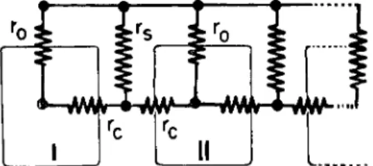
brane voltages (V) in contiguous cells (I,II), resulting from a current injected into one of the cells ( I ) , provides a convenient index of communication. In a longitudinal chain of cells (Fig. 4), the relation between the resistances and the quality of communication is then given by
Vu rv rs\ rp/
where rp represents the combination of nonjunctional membrane re
sistances r0 in cell II in parallel with the network to the right of II (Loewenstein, 1966). The continuity of the voltage attenuation curve in the measurement of Fig. 3 provides the special information that rs
> r0. This information is available so far only for the salivary gland of
1 Λ"
M r r
Kn
4r1 \
20
1 i i
io JP /r"
4
1 Œ) 1 p
ff
$ - ki> i r•
H
r80mV
- 6 0
-40
J
7/
-20g
El i
|
- 2 0
- 4 0
- 6 0
A ftB
__| ( | 10 20
xlCT7 AMP
FIG. 2. A basic measurement of voltage attenuation showing functional com
munication in Drosophila salivary gland cells. An electrical current (abscissas) is passed between a microelectrode inside cell A and the extracellular medium (grounded), and the resulting drops in membrane voltage (ordinates) are meas
ured inside cell A and a contiguous cell B. Outward current, at right; depolariza
tion, upward. The degree of voltage attenuation provides information on the cur
rent fractions passing between interior of A and exterior, and between interiors of A and B. From Loewenstein and Kanno ( 1964).
Drosophila, where favorable anatomical conditions allow measure
ments of sufficient spatial resolution.
Cell-to-Cell Flow of Larger Particles
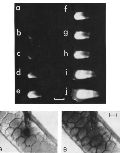
Another type of experiment, in which fluorescent particles are in
jected into the cell system, provides further information on the nature
of the C element. It shows that particles larger than the small cellular ions—in the case illustrated in Fig. 5, the anion fluorescein of 300 molecular weight—pass rapidly from one cell interior to another with negligible losses to the exterior, presumably through the same diffusion path as the small ions carrying the current in the electrical measure-
O/j 50 100 150 H h
4 0 0 4 5 0 5 0 0 550
FIG. 3. A measurement of voltage attenuation along a cell chain (Drosophih salivary gland). Current is passed between a point source (distance 0) and the exterior, and the resulting voltages are recorded intracellularly at a depth of up to 1 μ from the surface membrane at points near the surface centers and at the edges of the cell junctions. Ordinates: mean voltages of various successive measure
ments; standard error less than 0.3%. Abscissas: distance; bars subtend standard error. From Loewenstein and Kanno ( 1964 ),
ments. This finding (Loewenstein and Kanno, 1964) was, in fact, the decisive point that moved junctional communication into the realm of developmental biology.
Our knowledge of the size range of particles permeating the junc
tions is as yet scant. Particles of 103 MW pass through the junctions of Drosophila salivary gland cells (third instar); particles of 10Γ) MW do not pass (Loewenstein, 1966). The upper limit is probably of the order of 104 MW in this cell system. However, it is advisable to use
FIG. 4. Equivalent circuit of a cell chain. r0, non junctional membrane re
sistance; rc, junctional membrane resistance; r«, perijunctional resistance. The rela
tively low resistance of the extracellular medium has been omitted. From Loewen
stein (1966).
for the moment the conservative value of about 3 X 10³ MW; frag
mentation of the tracer molecules, their binding to cytoplasm, and several other technical problems in dealing with diffusion of large molecules, will have to be solved before one should venture to give more absolute upper limits. Other cell junctions have recently been shown to allow passage of large particles too: junctions in squid embryos pass dyes of 900 MW (Potter et al., 1966), and junctions of certain crayfish axons (Bennett, 1966), and those of certain tissue- cultured fibroblasts, pass fluorescein (Furshpan and Potter, personal communication ).
Genesis of Junctional Communication:
Factors Controlling Junctional Permeability
Junctional communication is a property of adhering cells. It does not occur in cells artificially separated or in tissues that lack close cell junctions (e.g., skeletal muscle). Only when cells form cohesive aggre
gates, do they establish junctional communication.
How does the communication come about? In trying to construct a
FIG. 5. Cell-to-cell diffusion. Top: 10~° ml of fluorescein-Na (332 M W ) are injected into a cell of Drosophila salivary gland with a micropipette. Ultraviolet rays are projected obliquely onto the gland, and the scattered fluorescence is photographed at equal exposure time, a, at the moment of injection, and h-j, every 2 minutes after injection. [The low intensities of fluorescence at the higher concentrations (a-c) are due to fluorescence quenching] Calibration, 3 0 0μ . From Loewenstein and Kanno, 1964. Bottom: Injection (10~8 ml) of the dye azure-B ( 305 M W ) into a cell of Drosophila salivary gland. Bright-field photograph. A, just after injection; B, 3 minutes after injection. The movement of both tracer mole
cules is from cell interior to interior without appreciable leakage to the exterior Calibration 50 μ.
8 0
60
4 0
20
0 L
I·,
0.4
0.2
0.0
\à
• · · VT
cP o
o 00
X o
X
o vn
x v
m
0 15 30 45
MINUTES
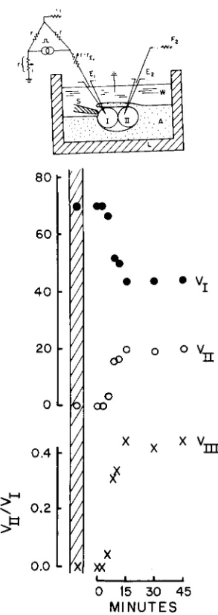
FIG. 6. Establishment of junctional communication between a pair of isolated sponge cells (Haliclona). Current is passed between interior and exterior of one cell ( I ) , and the resulting voltage ( V, ordinates, relative units) are recorded in that cell and simultaneously in an adjacent one II, as depicted in inset. The two cells were micromanipulated into contact (time zero) without selection of contact regions. Hatched area on graph gives the corresponding values when the cells were
picture of the formation of junctional communication, I have been guided by the following experimental points: (1) the junctional membrane regions appear not to be predetermined; (2) junctional membrane permeability depends on Ca2+; (3) junctional membrane permeability depends on perijunctional insulation; (4) junctional per
meability depends on cellular metabolism.
1. The functional membrane region is undetermined. This point fol
lows from experiments on sponge cells. With sponge cells one can do the almost schematic experiment of assembling a junction, starting with a pair of isolated cells; and cell membrane (input) resistance and electrical coupling between the pair can be monitored continuously, throughout the experiment. When the cells are separated, their entire surface membrane is of low permeability. When the cells are brought into adhesive contact, they form within minutes a communicating sys
tem: a perijunctional insulation is formed and the junctional membrane regions become more permeable than the rest of the cell membrane (Fig. 6) (Loewenstein, 1967a). The latter transformation is particu
larly striking, since it involves a permeability differentiation of several orders of magnitude in the membrane. In these experiments, the cells are paired at random and with little chance of selection of the junc
tional regions. Thus, it is clear that elements of high permeability can arise anywhere in the cell surface membrane; the membrane regions which are to become C elements, appear not to be predetermined, at least not at the scale of the light microscope.
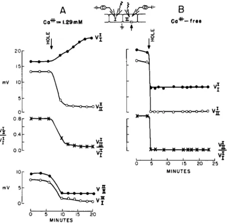
2. Junctional membrane permeability dependence on Ca2+. This dependence is shown by experiments on epithelial tissues in which the cell interiors are allowed to equilibrate with extracellular media of known Ca2+ concentration through leaks in the nonjunctional mem
branes or in the perijunctional insulations (Loewenstein et al.y 1967).
A convenient way to do this, is to make a hole in the nonjunctional cell surface membrane. Upon exposure to media with Ca2+ concentra
tions above 10~5 M, junctional membrane permeability falls rapidly, approaching the low values of the nonjunctional membranes at 10~3 M concentrations (Fig. 7A). Thus, at least in the direction high-to-low, permeability can change 3-4 orders of magnitude over the span of
fully separated. Microelectrode connected to a balanced bridge circuit serves here for both current passing and voltage recording in cell I. From Loewenstein (1967a).
Ca2+ concentrations normally prevailing in cytoplasm ( < 1 0 , ;M) and cell exterior ( > 10-3 M ).
Figure 7 also illustrates a phenomenon of much physiological inter
est. In the example shown, the cell was injured in a medium containing Ca2+ at a concentration similar to that in normal extracellular fluid;
and at this concentration, the normally highly permeable junctional
A B
Co"***-free
10
o IX
>-¥+-* « · · · Vj
6-ΟΌ-Τ3- ■ o o o K H W V«;
XX X—X XXX X * X
4
10 15 20 25 MINUTES
mV
5 10 15 MINUTES
FIG. 7. Junctional membrane permeability dependence on Ca2+. Current is pulsed between cell interior I and exterior, and, alternately, between cell interior II and interior; the resulting voltages V are recorded in the two cells. The super
scripts denote the corresponding current-passing electrodes, and the subscripts, the voltage-recording electrodes. At arrow a hole of 2-4 μ is made in the surface membrane of cell II. (A) Results in extracellular medium with normal ion com
position containing 10 :f M Ca2+; ( B ) in Ca2+-free medium. From Loewenstein et al (1967).
membranes were in effect sealed in the injured cell. This self-sealing capacity is a widespread, if not general property of connected cell systems. It has been observed in adult tissues, such as salivary glands, liver ( Loewenstein, 1966; Loewenstein et al., 1967), skin (Loewen- stein and Penn, 1967), and various embryonic tissues (Potter et al., 1966; Ito and Loewenstein, 1968). It is indeed, a remarkable functional adaptation. All the elements required for such a sealing reaction are built into the normal system and are critically poised: the Ca2+ concen
tration profile falls off steeply across the cell membrane in the inward direction, and the junctional membranes are capable of sealing when exposed to sufficiently high Ca2+. All that is required to set this reac
tion in motion is a discontinuity in the nonjunctional membrane or perijunctional insulation.
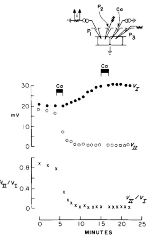
Cell injury itself is not a sufficient cause for junctional membrane sealing: no sealing ensues if the injury is made in Ca2+-free medium (Fig. 7B). A certain minimal cytoplasmic Ca2+ concentration, on the other hand, is a sufficient cause. Junctional membrane sealing ensues promptly when Ca2+ is injected into the cell system with a fine micro- pipette that causes no membrane leaks detectable by measurements of membrane impedance (equivalent injections of other ions such as K+ produce no sealing) (Fig. 8) (Loewenstein, 1966; Loewenstein et al., 1967).
3. Junctional membrane permeability dependence on perijunctional insulation. The resistances to ion diffusion of the elements C and S are inversely related, when the cell systems are in normal extracellular media. Whenever and wherever the perijunctional insulation is good, the underlying membrane regions transform into permeable elements;
and conversely, whenever the insulation is poor, the membranes return to the impermeable state.
One aspect of this relation is clearly seen in sponge cell aggregates in which some of the determining factors of perijunctional insulation can be controlled. As is perhaps not too surprising, these factors are the same as those that determine cell adhesion, namely Ca2+ and Humphreys' (1963) and Moscona's (1963) adhesive factor. [The role of Ca2+ in perijunctional insulation is entirely independent of that in junctional membrane permeability; and the limiting Ca2+ concentra
tions are different for the two processes ( Loewenstein, 1967b, Loewen
stein et al, 1967).] When these factors are provided to a pair of sponge cells, a good insulation is formed around the junction, and this is in-
3 0
2 0 m V
10
0 . 8
Co
Ca
PI
# # · · · · ·*ν
Oo O Q O O O O O O O O O O O I C
V /v
0.4
XXXX X X X X X X X X X I I I
vn/vi
10 15 2 0 MINUTES
25
FIG. 8. Action of Ca2+ on junctional membrane permeability. Ca2+ is injected into a cell of Chironomus salivary gland by iontophoresis (micropipette, C a ) , while junctional communication is probed by passing current between the interior of one cell (microelectrode Pi) and the exterior (ground potential), and the resulting voltages are recorded in this cell, Vj (microelectrode P2) and in an adjacent cell, Vn ( Ps ). The black bars mark the periods of iontophoresis in which 5 X 10~14 mole of Ca2+ are injected in each period. From Loewenstein et al. ( 1967).
variably accompanied by the junctional membranes becoming highly permeable. Conversely, when a formed perijunctional insulation is rendered leaky to influx of sufficient Ca2+ from the junctional side, the junctional membranes become as impermeable as the nonjunctional ones (Loewenstein, 1967a). The latter aspect can be shown in many other communicating cell systems as well (Loewenstein et al., 1967).
4. Junctional membrane permeability dependence on cellular me-
tabolism. The normal Ca2+ concentrations in cytoplasm, as determined in a variety of cells, is several orders of magnitude lower than in the normal extracellular media (Hodgkin and Keynes, 1957; Gilbert and Fenn, 1957; Portzehl et al, 1964; Hagiwara and Nakajima, 1966). This situation is maintained far from electrochemical equilibrium through
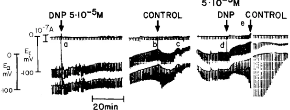
out the life of a cell in spite of some inward leakage of Ca2+. Thus Ca2+ must be extruded from the cell and this extrusion must ultimately depend on metabolic energy. Since junctional membrane permeability depends in turn on. the maintenance of a low cytoplasmic level of Ca2+ (point 2), it follows that junctional membrane permeability must ultimately depend on supply of metabolic energy too. This, in fact, has now been shown. Exposure of Chironomus salivary glands to low temperature or to various chemical metabolic inhibitors, such as dinitrophenol, cyanide, oligomycin, and N-ethylmaleimide, causes de
pression of junctional membrane permeability to levels of virtual interruption of communication (Fig. 9). (Ouabain, the specific in
hibitor of Na+ and K+-activated ATPase, causes no depression. ) Intra- cellular injection of ATP prevents the depression in the case of dinitro
phenol (Politoff et al, 1967,1968).
DNP 5·Ι(Γ5Μ
I 0 "7A ?
"r T miiiiiimitmiimiiimiiinfliflnrii'ft
mV -100 -*-
mV
■100-"-
5 I 0 ~5M
DNP CONTROL
* iHnnnmnitfn'iMiuMf ΤΤΜΙΙΙΙΙΙΠΠΙΠΤΠΠ
dl
I 1 20min
FIG. 9. Junctional uncoupling by metabolic inhibition. Dinitrophenol ( D N P ) . Electrical arrangement for monitoring junctional communication as in inset of Fig. 2. Upper trace is the storage oscilloscope record of membrane current (out
ward current pulses 2 X 10"8 A; 100 msec duration; 1/min ). Middle and bottom traces are the records of Vn and V,, respectively; their baselines give the cell rest
ing potentials (En, Ei) and their downstrokes from baseline, the peak values of Vn and Vi on same voltage scale as En and E7. Cell system is initially bathed in control medium; subsequent changes of medium are indicated by vertical marker lines. Control medium is restored at b as a junctional uncoupling trend became noticeable; complete uncoupling followed the second application (d) of DNP- medium. From Politoff et al ( 1968).
π·4
<10"5M
M-,
(m)
FIG. 10. Top; Permeability conversion during junctional formation. Hypothe
sis. Intracellular Ca2+ concentrations are kept below 10"5 by energized extrusion of Ca2+ (wiggled arrows). I, unconnected cells: The entire cell membrane faces the extracellular compartment with Ca2+ concentrations above 10~3 M; Ca2+ attach to membrane binding sites (impermeable state). II, Connected cells: The membrane portion (C) circumscribed by the elements S are insulated from the extracellular compartment; Ca2+ detach (straight arrows) from the membrane (permeable state ).
Bottom: Three causes of interruption of junctional communication ( uncoupling ) : (i) slowing of Ca2+ extrusion; (ii) fall in diffusion resistance to Ca2+ in S; or (iii) in O. Permeability of C falls as Cau concentration rises in the intracellular compartment.
Permeability Conversion Hypothesis
On the basis of these four experimental points, one may now con
struct a picture of the permeability transformation during junctional formation. Once the perijunctional insulation is formed, the membrane portion circumscribed by it is incorporated into the intracellular com
partment, where it faces Ca2+ concentrations several orders of magni
tude lower than formerly when in contact with the extracellular me
dium. The chemical gradients now favor Ca2+ detachment from the membrane structure, and conversion from low to high permeability ensues (Fig. 10).
This hypothesis contains one point that is not directly shown by experiment, namely that the membrane permeability of Ca2+, which experiment shows to operate in the direction high-to-low permeability, holds also in the reverse direction. But such an assumption of reversi
bility of a reaction does not seem too risky. Stated in more specific terms, the assumption is that certain membrane binding sites occupied by Ca2+ in the impermeable state ( O state ) become free in the perme
able state (C state).
An attractive feature of this conversion picture is that it requires no special properties for the membrane sites that are to become junctional, nor special mechanisms for switching the permeability transformation on. The essential element starting the transformation, and circum
scribing it to a given membrane region, is the perijunctional insulation.
The driving force behind the permeability transformation (and its maintenance) is the metabolic force(s) that keeps the Ca2+ concen
tration low in the cytoplasm.
DEVELOPMENTAL ASPECTS
Possible Functional Adaptations of Junctional Communication I come now to the possible implications of junctional communica
tion in animal development. Here I am looking over the fence to a field in which I am a stranger.
What are the functional adaptations of this organized and efficient form of cellular communication? In dealing with this question, we are severely handicapped by our lack of knowledge of the identity of the communication signals. The best one can do at present is to guess at the general category of the signals. One of the most obvious functional
consequences of junctional communication is the establishment of a common intracellular milieu of small ions throughout the connected cell population. This follows directly from the finding of a high elec
trical junctional membrane conductance, which is principally the con
ductance to the smaller cellular ions K, Cl, etc. Thus, functions con
cerning these ions, such as their (nonjunctional) membrane transport, draw on a common pool and are probably concerted throughout the cell community.
Control of Growth and Differentiation
It is of particular interest that the common pool also includes larger particles. Many metabolites, hormones, and other cellular substances fall within the size range of particles (103 MW) permeating junctional membranes. There is thus ample possibility for cell-to-cell flow of substances exerting regulatory actions on cellular activities. An enticing possibility is that this includes substances regulating gene activity.
This is a possibility which will most immediately come to mind to the developmental biologist familiar with the findings of the past 10 years of microbial genetics, showing that metabolites and other relatively small molecules can regulate genes; and brought up with the results of the past 30-40 years of experimental embryology, showing that cellu
lar differentiation involves some form of close range interaction be
tween cells. Here we have in junctional communication an obvious candidate for such close range interaction: an organized close system allowing substances to flow from cell interior to interior with little loss, and, what is more, a system in which flow is channeled. Each inter
connected cell system constitutes a communication channel; the cyto
plasm plus the junctional membranes constitute the channel core, and the nonjunctional cell surface membranes plus the perijunctional insu
lations, the channel walls. A system of this kind has a good spatial resolving power.
An alternative is a hormonal system in which cells sputter signals into the open extracellular spaces. A communication system of this kind, aside from being wasteful, would have an extremely poor space resolving power. It would have to depend on some form of spatial attenuation of signals, with all the inaccuracies inherent in a system of very irregular geometry. Such a system would pose enormous logistic problems if it were used, for instance, in determining cellular position or in engineering something like the complex and highly local-
ized patterns of differentiation occurring in metazoa. On the other hand, a system of the channeled kind, like the junctional system, is capable of bringing forth patterns of differentiation with a precision of a few cell diameters (in some cases, perhaps, with a precision of 1 cell diameter) (Loewenstein et al., 1965).
There are two further reasons why junctional communication is a good candidate for the close range cell interactions involved in the control of growth and differentiation: junctional communication is altered in certain tissues with abnormal differentiation; and junctional communication is extensively present in embryonic tissues.
Alterations in communication in cancer cells. In normally differenti
ated adult tissues, there is junctional communication between cells of the same kind. This appears to be the general rule in epithelial tissues ( Loewenstein, 1966 ). By contrast, in certain abnormally differentiated tissues, such as various types of cancers, communication is altered.
Evidence to this effect is now available for various types of epithelial cancers growing in animals, such as several forms of liver cancer
(Loewenstein and Kanno, 1966, 1967), thyroid cancer (Jamakos- manovic and Loewenstein, 1968), and stomach cancer (Kanno and Matsui, 1968 ) ; as well as for two lines of liver cancer cells and certain x-ray transformed embryonic epithelioid cells in tissue culture (C.
Borek, S. Higashino, and W. R. Loewenstein, unpublished). No elec
trical communication is detectable between the cells of these cancerous tissues, under conditions in which their normal counterparts show good communication. The data on communication in regenerating liver, which has growth rates as fast or even faster than tissues with can
cerous growth, are particularly revealing. The cells of normally grow
ing liver, in contrast to the cells of cancerous liver, communicate through permeable junctions, regardless of their growth rate (Fig.
11) (Loewenstein and Penn, 1967).
Loss of junctional communication emerges from these results as a possible etiological factor of cancer. The junction is, indeed, a vulner
able bottleneck. As discussed before, junctional membrane permeabil
ity depends, among other factors, on the surface and perijunctional insulations, on cytoplasmic free Ca2+, and on cell adhesion. Alterations in any of these factors can lead to interruption of communication
(Loewenstein et al., 1967). Thus, if the junction is conveying sub
stances for growth control, cancerous growth could arise through defects in the mechanisms concerned with (i) the pumping of Ca2+
50 40 30l·
20h
mV
2h
°°1
4Δ
*Cancerous
• Normal + 2 Days A3 Days Δ7 Doys
•
A
i.
+-*-
-#- 100 200 3 0 0 μ _LFIG. 11. Junctional communication in normally growing liver cells and its absence in cancerous liver cells. Current is injected into a cell of the liver tissue, and the resulting voltages (ordinates) are measured inside other cells at varying distances (d, abscissa) from the point of source of current. Data obtained, under the same experimental conditions, in normal unoperated liver, in normal liver regenerating from an ablation of ca. 70% of its cell mass, and in cancerous liver (Morris' tumor). The measurements on regenerating liver were done 2 days, 3 days, and 7 days after ablation. Between days 2 and 3, the liver tissue is growing very rapidly ( 1-2 gm/day ). By the day 7, growth is down to 8% of the above rate (Higgins and Anderson, 1931), and the liver has regenerated to nearly its original mass. After Loewenstein and Kanno (1967) and Loewenstein and Penn (1967).
168
through the nonjunctional cell surfaces; and with (ii) barring the influx of Ca2+ at the perijunctional insulation, or (iii) the nonjunctional membranes (Fig. 10, bottom) (Loewenstein, 1968). The causative defect here could be (a) a genetic defect in any of these mechanisms or (b) a purely somatic defect in these mechanisms, giving rise to a permanent alteration in the gene processes concerned with growth control. An etiological mechanism of the b type has the interesting consequence that even a transient interruption of communication could give rise to a permanent defect in growth control. The safety margins for junctional communication are normally quite large. To produce interruption of communication, one must push the cellular environ
ment well beyond its normal physiological state, e.g., to pH > 9, or extracellular [Ca2+] to <10- 4 M, to tonicity to about 1.5 times normal.
But this by no means precludes the operation of an etiological mecha
nism of type b, since in pathological conditions, deviations may well go beyond the safety margins.
The apparently simple yes-or-no communication pattern of normal and cancerous epithelial tissues is not seen in fibroblasts in long-term tissue culture. Cultured fibroblasts transformed by polyoma and SV 40 virus (Potter et al., 1966), or by X-radiation (Higashino, Borek, and Loewenstein, unpublished), which do not show contact inhibition, are electrically communicating to a degree indistinguishable from normal fibroblasts.
The meaning of the differences between the communication patterns of cells in the epithelial cancers and in the transformed fibroblast cultures is not clear. One possibility, of course, is that different stages of growth regulation are affected in the two cell classes. It already seems a priori unlikely that the many forms of abnormalities of growth control lumped under the name of cancer would all have the same causes. The polyoma virus, or the transforming X-rays, for instance, may well act at stages completely unrelated to junctional communica
tion in the case of fibroblasts.
Another possibility is that communication is interrupted during an early phase of fibroblast transformation, and that it is reestablished later on in the same cells or in their descendants, whereas the defect in growth control is retained (e.g., by a b-type mechanism). Two recent findings are interesting in this connection. One, by Stoker (1968), shows that most of the polyoma-transformed cells revert to the untransformed state after a few generations; only few cells perpetuate the transformed character. ( The electrical measurements of communi-
cation were made on the descendants of these selected cells ). Another finding, by Rubin and Haitié (1968) emphasizes the fact that cells undergo profound changes in long-term tissue culture. All the meas
urements of electrical communication were done on cultured cells many generations removed from the cells originally transformed.
There is a further difficulty in interpreting the results on trans
formed tissue cultured systems. The result of "presence of communi
cation" has a rather poor informational content. Such a result, as shown by electrical measurements, means only that there is communication for the small ions that carry the current. It tells nothing about the communication for larger and perhaps more relevant particles. A result of "lack of communication" by such measurements, as obtained on the various tumors, provides much more information. It means total lack of communication for diffusible particles, small and large.
However, in terms of experimental certainty, the result of absence of electrical communication is the weaker. The state of high junctional membrane permeability is less stable than the state of low membrane permeability (Loewenstein, 1966). This raises the question whether the results of absence of communication reflect the actual state in the cancer tissues, or whether they reflect differences in safety margins for communication, particularly since the measurements were done on iso
lated tumors. It is true that both the cancerous and the normal tissues were isolated, and measurements on both were done under identical conditions. One can also safely discard surface injury as a possible cause of interruption of communication, because membrane (input) conductance was measured together with electrical coupling (see Loewenstein and Kanno, 1967). But there is no certainty as to whether, in respect to the isolation or measuring procedures, the safety margins for communication in the cancerous tissues are as high as those in their normal counterparts. At the moment, these are the weak points of the results. However, the points are amenable experimentally, and we may expect to have answers to them soon.
Junctional communication in embryonic tissues. The first results in our laboratory on communication between embryonic cells looked rather unpromising. During the summer of 1964, at Woods Hole, R.
Ashman, Y. Kanno, and I looked into the question of junctional com
munication between the first embryonic cells, the daughter cells of the egg of starfish. We found that all communication was interrupted at the end of egg cell division (Ashman et al, 1964). In these experi-
ments we had used urea to soften the fertilization membrane to allow microelectrode penetration, and so we thought that the uncoupling might have resulted from the urea treatment. The following summer, S. Ito and I did similar measurements on the first daughter cells of the eggs of sand dollars. This time we avoided the use of urea and elimi
nated the fertilization membrane by gentle shaking of the cell system in seawater. Junctional uncoupling ensued all the same upon the com
pletion of cleavage (Ito and Loewenstein; see Loewenstein, 1966).
Ito and N. Hori then examined intercellular communication at a later embryonic stage in the newt, and found an entirely different situation.
Many, if not all, blastomeres in the newt morula appeared to be elec
trically connected (Ito and Hori, 1966). At the same time, Potter, Furshpan, and Lennox (1966), working on squid embryos, and later Sheridan (1968) on chick embryos, discovered electrical interconnec
tions between a variety of cells even at later stages of development.
Recent work on isolated blastomere cells of newt morulae add another point to these results. Blastomeres (macromeres) isolated from the morula, and mechanically separated, will readily and rapidly reestab
lish communication when brought in contact (Fig. 12); moreover, rather small areas of membrane contact ( cross sections of the order of 1 /x) seem to suffice for the establishment of full junctional communi
cation ( Ito and Loewenstein, 1968 ).
In general conclusion then, there is a reasonably good chance that functional communication is instrumental in the flow of signals con- trolling gene activity concerned with growth and differentiation.
Interruption of Junctional Communication and Differentiation
The above conclusion implies that both establishment (coupling) and interruption of signal flow (uncoupling), may give rise to differ- entiation under certain conditions. Consider, for example, two sets of cells interacting through junctional communication, such as those dia
grammed in Fig. 13. One is the signal-producing set and the other the target set which undergoes changes in gene activity, that is, it differ
entiates, under the action of the signals when junctional communica
tion is established. The differentiation process—and in this process I include the changes in gene activity as well as the resulting somatic structural-functional changes—may be of three general kinds: (a) It may be fully reversible; the target systems go back to their former state of differentiation upon interruption of the signal flow, (b) It may
i ' . ^ ^ i
FIG. 12. Establishment of junctional communication between a pair of isolated embryonic cells. Two blastomere cells (macromeres) isolated by micromanipula
tion from a morula (newt) are first separated mechanically and then manipulated into contact again, while electrical communication between the cells is monitored continuously (electrical arrangement as in Fig. 2 inset), ( a ) Electrical communi
cation when the cells were in their unseparated state; ( b ) interruption of com
munication upon separation of the cells; (c) reestablishment of communication following cell readhesion. Current 3.5 X 1 0s A; 225 msec duration. Voltage cali
bration, 14 mV per grid division. Top: SL cell pair of this kind, communicating through small protruding contact regions. Calibration, 200 μ. From I to and Loewenstein (1968).
be irreversible; once initiated, differentiation is retained stably and independently of the signals, (c) It may be hysteretic; as in (a), maintenance of differentiation depends on signal flow, but when this is interrupted, the systems suffer hysteretic losses on their backslide and stabilize at a state of differentiation different from the former one.
The corresponding outcomes of uncoupling will then depend on the type of off-switch operating at the junction. The switch may work as
#n all-or-none device which cuts off virtually all flow of substances
>□ D HD HD HD
Π
m
• D
_J L_
K A
J L
aa s K
■ H D
π
III
• —
L±J—
=U-o
H 1 -ilfJ
FIG. 13. Patterns of differentiation by junctional coupling and uncoupling.
Top series: 7, unconnected state; 77, all-or-none coupling; 17J, all-or-none un
coupling; a, reversible; b, irreversible; c, mixed; d, hysteretic differentiation proc
esses. Bottom series: an example of selective coupling (711) with reversible differ
entiation processes. Key: Inducer cell, left; target cell, right. Inducer signals: # , X , # . Differentiation kind: □ , reversible; ■ , irreversible; crosshatched cross and rectangle, hysteretic (see text).
down to the smallest ions. This is the kind of switch we know to operate when the intracellular compartment is flooded with Ca2+
(Loewenstein et al., 1967; Loewenstein, 1967b). But the switch may also be a more subtle one allowing selective changes of junctional permeability; for instance, it may reduce the size limit for permeating particles. This would take the exploration of meaningful changes in communication beyond the reach of measurements of membrane volt
age attenuation and other tricks in the repertoire of the electrobiologist.
But it is a particularly interesting possibility, since such selective uncoupling sets the least restrictions on the differentiation processes.
All-or-none coupling gives rise to new forms of differentiation only
with hysteretic differentiation processes or with reversible processes concurring with irreversible ones in the same cell ( Fig. 13, schemes d and c), not, of course, with purely reversible or purely irreversible processes (Fig. 13, a, b). Selective uncoupling, on the other hand, may produce new differentiation even with purely reversible processes
(Fig. 13, e).
The schemes of Fig. 13 are readily fitted to models of cellular dif
ferentiation derived from studies of bacterial genetics. Thus, for in
stance, if the communicated state (II) represents two cell sets hooked up in one of Jacob and Monod's ( 1961 ) inducer or repressor circuits, with an inducer or a correpressor passing through the junction, then the uncoupled state (III) represents the situation when the circuit is cut off and the corresponding genes of each cell run on their own. The scheme fitting such circuits most immediately is that in which the junction operates as a selective switch (Fig. 13, e). In this scheme, uncoupling is instrumental in producing differentiation with reversible processes as they occur in bacteria. The hysteretic scheme is simpler, but it is still rather versatile. On a priori thermodynamic grounds, hysteresis is likely to occur in reactions involving macromolecules. In fact, as Katchalsky, Oplatka, and Litan ( 1966 ) so incisively showed, hysteresis with metastable states is a prominent feature of polyelectrolyte reac
tions. It confers on the reacting molecular system the capability of recognizing directionality in the reaction. Evolution has probably made use of it many times in fashioning biological processes.
In searching for support for the proposal of a role of junctional un
coupling in differentiation, I have looked for clues in cell systems, such as nerve, skeletal muscle, blood, and gamete cells, which are clearly uncoupled in their final state of differentiation. I have been guided mostly by the experimental findings on differentiation of amphibian ectoderm into neural tissue (neuralization), particularly by the pene
trating studies of Holtfreter (1944, 1945). Here one is struck immedi
ately by the variety of unspecific and seemingly unrelated agents that can initiate such neuralization: Ca2+ and Mg2+ sequestration, mechani
cal or chemically produced cell injury, anisotonicity, alkali (Holtfreter, 1944, 1945; Barth, 1941, 1959), méthylène blue (Waddington et al, 1936), and various metabolic inhibitors (Waddington, 1938; see also Runnström, 1933; Waddington et al, 1936; Hörstadius, 1953). All these disparate agents ("unnatural inducers") have one thing in common:
they are uncouplers of junctional communication. The first four of
these agents act, we know now, by opening the surface barrier to the influx of extracellular Ca2+ and Mg2+ (schemes it or in, Fig. 10, bot- tom) (Loewenstein et al, 1967); and the last two, by interfering with Ca2+extrusion through nonjunctional membranes (scheme i) (Loewen
stein, 1967b; Politoff et al, 1967, 1968). In either mode of action, the final result is the sealing of the junctional membranes.
There are also observations concerning cell adhesion which relate to the present argument. Among the earliest manifestations of differentiation of ectoderm into neural tissue are coarse changes in cellular contact relations. Such changes are observed in the intact embryo ( Giersberg, 1924 ) and more clearly in tissue culture, where the cells induced to neural differentiation move amoeba fashion after originally being in close contact. In contrast, noninduced ectodermal cells devel
oping into epidermal elements remain in close contact (cf. Holtfreter and Ham
burger, 1955; Townes and Holtfreter, 1955). An analogous situation is found in epithelial rudiments of mouse thymus. When differentiating into lymphoid cells, they lose their adhesiveness, but do not do so when they become epthilial (Auer- bach, 1961).
A fact that transpired early from the work with "unnatural inducers"
is that the inducers do not enter themselves into the chemical ma
chinery of differentiation (Holtfreter, 1944, 1945; Waddington et al, 1936; Waddington, 1962). Agents so entirely different are likely to act through a common mechanism, and the evidence points to a common trigger mechanism. A piece of ectoderm left alone may develop into epidermis; if acted upon by one of the inducing agents, it will differ
entiate into neural tissue (Holtfreter, 1932, 1945). An often-used figure in this relation is that the cell system is switched from one differentiation path to another. In the light of the present proposals, the cell junction provides just such a switch. In the on position, it establishes the information flow between inducer and target cells in the ectoderm that drives the system from ectodermal to epidermal differ
entiation. The target set may be made of strings or clusters of many connected cells (see Loewenstein, et al., 1965, for examples of the anatomy of cellular interconnections); relatively few inducer cells may thus be sufficient. In the off position, it interrupts this flow, and the system runs toward neural differentiation according to schemes c, d, or e, Fig. 13. There are no clues on the nature of the causal agent of the uncoupling step under natural conditions (for "unnatural in
ducers," see above), but the mode of operation may reasonably be expected to fall in with one of the schemes pictured in Fig. 10 (bot- tom). An interesting point that emerges in this connection from the
present considerations is that the natural agent for uncoupling need not be a substance external to the connected cell system, that is, an inducing substance in the usual embryological sense; any of the causes of junctional uncoupling pictured in Fig. 10 (bottom) may conceiv
ably be programmed into the connected cell system.
Interruption of junctional communication by lithium. I should like to mention at this point the results of experiments on the action of Li+ on junctional communication which Birgit Rose has recently obtained in our laboratory. I put these results here rather than in the section dealing with physicochemical mechanisms, because they may bear directly on earlier embryological work, the long-known "vegetalizing"
effect of Li+ on echinoderm embryos (Herbst, 1893; Hörstadius, 1937a,b). When this effect was viewed in the light of the Ca2+ hy
pothesis of permeability conversion (pp. 163), the possibility suggested itself that the effect of Li+ might be related to junctional uncoupling.
Ca2+ extrusion, which is dependent on Na+ in various cell systems, does not operate, or is markedly reduced, when Na+ is replaced by Li+ (Niedergerke, 1963; Baker et al, 1967; Baker and Blaustein, 1968).
As it turned out, Li+ does produce junctional uncoupling. When Li+ is substituted for Na+ in the extracellular medium of Chironomus salivary gland, junctional membrane permeability falls to so low a level that all communication between cells, including that for the smallest ions, is effectively interrupted (Rose and Loewenstein, 1968) (Fig. 14).
a
Λ^ —
E,°
"40-1 n
EÎÏ"
-40Jj
4.
P*
f i
m* £ MS n»w
Vjlm
Π Pr
■ | 1 1 1 >L,
< I ' » {
■U4*
b
f 1*φ\
'■—-'
n * .*-.*
i^i r
■ · - J
y i " ' τ Ρ 4ntr
FIG. 14. Junctional uncoupling by Li+ (Chironomus salivary gland). Current passing and voltage recording as in Fig. 9 ( current record is not shown ). Initially the gland is bathed by control medium. At a it is bathed by a medium in which Li+ substitutes for Na+ on a mole-to-mole basis, causing irreversible junctional uncoupling. At b, return to control medium. Current 10~8 A; 100 msec duration;
1/min. Voltage calibration, 25 mV/grid division. From Rose and Loewenstein (1968).
Junctional Models for Growth Regulation
I should like to end now by considering some simple possibilities of growth control in cell systems with junctional communication. I shall focus on the problems of how a cell population limits its size and of how cells determine their position within a population.
Information on cell population size. On the general premise that the control signals are diffusible substances, one operational advantage of a junctional system over a hormonal one is immediately apparent. The junctional system has a finite volume, that is, the cytoplasmic volume of the connected ensemble circumscribed by the nonjunctional mem
branes and perijunctional insulations. This confers on members of the system the potentiality for determining signal parameters in relation to this volume, i.e., concentrations. A cell member has thus, for in
stance, the potential for determining the number of cells in the ensem
ble by measuring the local signal concentration.
I shall consider two general cases. In one case each cell member (or member set) produces a different signal and senses this signal. In this case, the signal concentration (steady state) changes with the size of cell population in proportion to the volume of the interconnected cyto
plasms (Fig. 15A). There are no special conditions for the timing of signal output in this case; local signal concentrations provide here information on population size, regardless of the timing of the signal output among the cell members. In another case, all cells of the en
semble put out the same signal and are sensitive to this signal. This case puts the least demands on cellular differentiation. But it is more restrictive as to signal timing. Changes in population size do not give rise here to changes in signal concentration, unless the signal output of the various cells is sufficiently asynchronous (Fig. 15A, II). Under conditions of asynchrony, this case operates like the first: a fall in concentration signalizes increase of population size.
Information on cell position. Both cases have also inherent the potential for determining cell position within the population. Here the finite volume property of the junctional system is again the essen
tial element. Because the system is circumscribed by a diffusion bar
rier, the concentration profiles of the signal substances will depend on the location of the cell emitting (and sensing) the signals. Thus, local concentrations determined at different times may provide in
formation on cell position. Consider, for example, a central (4) and a
I
Π
l_l 2 ΓΠ
XX
* *
L_J J~~l
• ·
u
L J 4 ΓΠ
1_J Γ Ί
x ·—
• r
1 Y
1 •
* 1 1
r
~L_r~
ΓΊ
* 1
l_l ΓΊ
L J
B
ST
IL.
FIG. 15. Dilution models. (A) Information on size of cell population is given by the concentration (steady state) of signal substances. I, system with cell spe
cific signals (X, # ) ; II, system with cells producing the same signals ( # ) asyn
chronously. ( B ) Information on cell position is given by the rates of local concen
tration changes. The graph sketches the signal concentration (c) vs. time (t) relation for a central ( 4 ) and a marginal cell ( 2 ) of a chain. ( C ) Feedback loop in which the information above controls cellular activity, s, Signal substance; c, cells of the connected system (dilution factor); r-e, receptor-effector process.
peripheral (2) cell of a simple chain, such as in Fig. 15B, each pro
ducing equal bursts of signals at different times. At the moment the bursts begin, the rates of concentration change are equal in the two cells. But, at later times, concentration falls more rapidly in the central cell. Thus the time integral of the concentrations in each of the two cells provides information on their respective positions.
Control loops. Various kinds of feedback mechanisms may be en
visaged in which the above information on population size and cell position is used to control growth. A particularly simple kind of
mechanism is one in which the informational signal serves also as con
trol signal on the effector side of the feedback loop. All the elements for such a control loop are present in the aforegoing junctional sys
tems, if the effector processes are made dependent on the steady state concentrations or on the rate of changes in concentrations of the sig
nals. For instance, activation or inhibition of effector processes (cell division, synthesis of a molecule, etc.) may ensue when the cor
responding signal concentration reaches a critical level or when the rate of change in concentration of the corresponding signal is above a certain magnitude. The complete feedback loop is then given by: (1) the cell signals: (2) the cytoplasms of the connected system, providing the dilution factor and the continuity of a close circuit; and (3) the cell receptor-effector process (Fig. 15C). Regulation within the cell population would thus ensue when the size of the communicated cell population increases, and this regardless of whether the increase in population size is by cell proliferation or by establishment of junctional communication between previously noncommunicating cells. Contact inhibition of cells in tissue culture (Abercrombie and Heaysman, 1954, Abercrombie, 1966; P. Weiss, 1958), organ size control (e.g., Higgins and Anderson, 1931; P. Weiss, 1961; L. Weiss, 1967), and a variety of gradient phenomena in regenerating tissues (e.g., Locke, 1967;
Lawrence, 1966; Stumpf, 1966) are among the more obvious candi
dates for this kind of regulation.
The above regulation schemes are applicable to purely somatic processes as well as to processes involving gene activity. It is easy to see how these models can be fitted to a Jacob and Monod-type of circuit of gene regulation where an "inducer," a "correpressor," or a
"repressor" would be the signal diluted within the communicated cell system. A very attractive feature of the models is that they translate information about space into simple time sequences of chemical con
centrations. The latter is the familiar language of chemical reactions.
The conditions for signal timing in the schemes, as considered in Fig. 15B, were chosen so as to make them simplest for a qualitative presentation; the periods of signal activity in the various cells are per
fectly asynchronous, and the signal diffusion time is short in relation to the intervals between activity periods. In large cell populations and with the time constants of diffusion systems, there is likely to be over
lap in activity periods between the various cells. The models become then stochastic ones. The principle of operation remains the same; but
the information must now be averaged over several activity periods and extracted against a background of signal noise. Quantitative solu
tions for such cases will be given in later publications.
Experimental questions may now be formulated in terms of these models, and we may soon know how close they come to reality. In any event, it was fun to walk through Wonderland.
REFERENCES
ABERCROMBIE, M : (1966). Contact Inhibition: The phenomenon and its biological implications. Nation. Cancer Inst. Monograph 26, 249-273.
ABERCROMBIE, M., and HEAYSMAN, E . M. (1954). Social behaviour of cells in tis
sue culture. II. Monolayering of fibroblasts. Exptl. Cell. Res. 6, 293-306.
ASHMAN, R. F., KANNO, Y., and LOEWENSTEIN, W . R. ( 1 9 6 4 ) . T h e intercellular
electrical coupling at a forming membrane junction in a dividing cell. Science 145, 604-605.
AUERBACH, R. (1961). Genetic control of thymus lymphoid differentiation. Proc.
Natl. Acad. Sei. U. S. 47, 1175-1181.
BAKER, P. F., and BLAUSTEIN, M. P. (1968). Sodium-dependent uptake of calcium by crab nerve. Biochim. Biophys. Ada 150, 167-170.
BAKER, P. F., BLAUSTEIN, M. P., HODGKIN, A. L., and STEINHARDT, R. L. ( 1 9 6 7 ) .
The effect of sodium concentration on calcium movements in giant axons of Loligo forbesi. J. Physiol. (London) 192, 43P.
BARTH, L. G. (1941). Neural differentiation without organizer. J. Exptl. Zool. 87, 371-383.
BARTH, L. C , and BARTH, L. J. ( 1959). Differentiation of cells of the Rana pipiens gastrula in unconditioned medium. J. Emhryol. Exptl. Morphol. 7, 210.
BENNETT, M. V. L. (1966). Physiology of electrofonic junctions. Conf. Biol. Mem- branes, Recent Progr., Ann. N. Y. Acad. Sei. 137, 509-539.
BRÄCHET, J. ( 1960). "The Biochemistry of Development." Pergamon Press, London.
BULLIVANT, S., and LOEWENSTEIN, W . R. (1968). Structure of coupled and un
coupled cell membrane junctions. J. Cell. Biol. 37, 621-632.
GIERSBERG, H . (1924). Beiträge zur Entwicklungs Physiologie der Amphibien.
II. Neurulation bei Rana und Triton. Roux Arch. Entwicklungsmech. Organ.
103, 387-424.
GILBERT, D., and FENN, W . O. (1959). Calcium equilibrium in muscle. / . Gen.
Physiol. 40, 393-408.
GROBSTEIN, C. (1964). Cytodifferentiation and its controls. Science 143, 643-650.
HAGIWARA, S., and NAKAJIMA, S. (1966). Effects of the intracellular Ca ion con
centration upon the excitability of the muscle fiber membrane of a barnacle. / . Gen. Physiol 49, 807-818.
HERBST, C. (1893). Experimentelle Untersuchungen über den Einfluss der verän
derten chemischen Zusammensetzung des umgebenden Mediums auf die En
twicklung der Thiere. I I . Weiteres über die morphologische Wirkung der Lithiumsalze und ihre theoretische Bedeutung. Mitt. Zool. Stat. Neapel I I , 136- 220.