Accepted Manuscript
Presence of cardiomyocytes exhibiting Purkinje-type morphology and prominent connexin 45 immunoreactivity in the myocardial sleeves of cardiac veins
Szilvia Kugler, MD, Nándor Nagy, PhD, Gergely Rácz, MD, PhD, Anna-Mária Tőkés, PhD, Bence Dorogi, MD, Ágnes Nemeskéri, MD, PhD
PII: S1547-5271(17)31192-X DOI: 10.1016/j.hrthm.2017.09.044 Reference: HRTHM 7333
To appear in: Heart Rhythm Received Date: 31 January 2017
Please cite this article as: Kugler S, Nagy N, Rácz G, Tőkés A-M, Dorogi B, Nemeskéri Á, Presence of cardiomyocytes exhibiting Purkinje-type morphology and prominent connexin 45 immunoreactivity in the myocardial sleeves of cardiac veins, Heart Rhythm (2017), doi: 10.1016/j.hrthm.2017.09.044.
This is a PDF file of an unedited manuscript that has been accepted for publication. As a service to our customers we are providing this early version of the manuscript. The manuscript will undergo copyediting, typesetting, and review of the resulting proof before it is published in its final form. Please note that during the production process errors may be discovered which could affect the content, and all legal disclaimers that apply to the journal pertain.
M AN US CR IP T
AC CE PT ED
M AN US CR IP T
AC CE PT ED
1
Presence of cardiomyocytes exhibiting Purkinje-type morphology and prominent connexin 45 1
immunoreactivity in the myocardial sleeves of cardiac veins 2
3
Short title: Immunohistology of cardiac veins’ myocardium 4
5
Szilvia Kugler MDa, Nándor Nagy PhDa, Gergely Rácz MD, PhDb, Anna-Mária Tőkés PhDc, 6
Bence Dorogi MDa, Ágnes Nemeskéri MD, PhDa 7
8
aDepartment of Anatomy, Histology and Embryology, Faculty of Medicine, Semmelweis 9
University, H-1094 Budapest, Tűzoltó u. 58., Hungary 10
b1st Department of Pathology and Experimental Cancer Research, Faculty of Medicine, 11
Semmelweis University, H-1085 Budapest, Üllői út 26., Hungary 12
c2nd Department of Pathology, Faculty of Medicine, Semmelweis University, H-1091 13
Budapest, Üllői út 93., Hungary 14
15
Correspondence to Ágnes Nemeskéri MD, PhD, Semmelweis University, H-1085 Budapest, 16
Üllői út 26, Hungary; e-mail address: nemeskeri.agnes@med.semmelweis-univ.hu; telephone 17
number: 0036 1 215 69 20 18
19
Conflict of interest: none.
20
21
Word count: 3585 22
23
24
25
M AN US CR IP T
AC CE PT ED
2 Abstract
26
Background Pulmonary vein (PV) myocardium is a known source of atrial fibrillation. A 27
debated question is whether myocardial extensions into caval veins and coronary sinus (CS) 28
have similar properties. No studies have documented specific pacemaker and/or conducting 29
properties for the human extracardiac myocardium.
30
Objective The aim was to characterize the histology and immunohistochemical features of 31
myocardial sleeves in the wall of cardiac veins.
32
Methods Sections of 32 human hearts were investigated. Specimens of PVs, superior caval 33
vein (SVC), CS, sinoatrial and atrioventricular nodes and left ventricle were stained with 34
Best’s Carmine for selective staining of intracellular glycogen. Anti-connexin (Cx)-45 and 35
Cx43 specific antibodies were used to determine the conduction properties of extracardiac 36
myocardium.
37
Results Myocardial sleeve was found in the wall of PVs of 15/16 hearts, 21/22 SVCs and 8/8 38
CSs. Bundles of glycogen positive cardiomyocytes exhibiting pale cytoplasm and peripheral 39
myofibrils were observed in the venous sleeves. Strong Cx45 and weak Cx43 labeling was 40
detected in the extracardiac myocardium. Similar staining pattern was observed at the 41
pacemaker and conduction system, while ventricular myocardium exhibited prominent Cx43 42
and no Cx45 immunoreactivity.
43
Conclusion Myocardial fibers of PVs, SVC and CS exhibit similar morphology to that of 44
Purkinje fibers and are enriched in glycogen. We provide data for the first time, on the 45
prominent positive staining for Cx45 in the extracardiac myocardium, indicating its potential 46
pacemaker and/or conducting nature.
47
Keywords: pulmonary vein; caval vein; coronary sinus; cardiac muscle sleeve; Purkinje-type 48
morphology; glycogen; connexin 45; immunohistochemistry 49
50
M AN US CR IP T
AC CE PT ED
3 Introduction
51
Myocardial sleeve of pulmonary veins (PV) play a critical role in the mechanism of 52
atrial fibrillation (AF). Macroscopic features of these areas were described previously.1-3 53
During the last decade, growing attention has prompted to the microscopic properties of the 54
extracardiac myocardial sleeves.2,4-6 In the wall of human PVs, Perez-Lugones et al.7 55
documented the presence of cardiomyocytes exhibiting ultrastructural morphology of P-cells 56
and Purkinje fibers (PFs). Although accepted that atrial tachyarrhythmias are frequently 57
triggered from caval veins and coronary sinus (CS)8-12, limited data has been published about 58
macroscopic3 and microscopic morphology13,14 of the myocardial sleeves of these regions.
59
Moreover, there is a general lack of research in the immunohistochemical characterization of 60
caval and CS myocardial sleeves.
61
Immunohistochemical markers to distinguish working myocardium, pacemaker or 62
conducting cells are established. Besides several determinants of conduction in the heart, such 63
as HCN4 and HNK-1, connexin (Cx) isoforms, of which gap junction channels are comprised 64
are also characteristic proteins of cardiac pacemaker tissues. Cx40, Cx43 and Cx45 are found 65
differentially expressed in cardiomyocytes at various sites, which determines the 66
characteristics in conduction velocity.15 Cx43 is present throughout the working myocardium, 67
whereas Cx40 is confined to the atrial myocardium and the ventricular conduction system.
68
Cx45 is predominantly expressed in the impulse generating and conduction system, while it is 69
present in substantially lower amounts in the working myocardium.16,17 70
71
Methods 72
Human tissues 73
Thirty two adult human hearts were removed from cadavers at 12-72 hours 74
postmortem age kept at 1-5 oC until fixation. The ages of deceased individuals ranged 75
M AN US CR IP T
AC CE PT ED
4
between 60 and 81 years. Their medical histories were unknown. Prior to death donors gave 76
written consent for the use of their bodies for education and research {Willed (Whole) Body 77
Program - WWBP}. The work has been ethically approved by the Regional and Institutional 78
Committee of Science and Research Ethics, Semmelweis University (Research Ethics 79
committee approval 122/2016).
80
Due to technical reasons, the heart could be excised together with PVs only in 16/33 81
and with SVC in 22/33 subjects. The excision was extended into the lung hilum in the case of 82
PVs, above the level of azygos vein regarding SVC and as far as the orifice of great cardiac 83
vein in the case of CS. The veins were then separated from the atria at the level of their ostia 84
and were cut transversely. CSs of 8/33 subjects were suitable for further tissue processing.
85
Tissue samples were obtained from the sinoatrial and atrioventricular nodes, the atria, anterior 86
wall of the left ventricle and the interventricular septum. Specimens were either fixed in 4%
87
formaldehyde, or in 70% ethanol or in methanol. After dehydration in graded concentrations 88
of alcohol, tissue samples were embedded in paraffin and 3-4 µm sections were made.
89
Tissue processing for histology 90
For general histology, paraffin sections were stained with hematoxylin and eosin (HE) 91
or trichrome. Intracellular glycogen was demonstrated by Best’s Carmine stain, which is a 92
specific stain for glycogen content. Best’s Carmine stain was performed as described.
93
Immunohistochemistry 94
For Cx45 immunohistochemistry, specimens were fixed in ethanol/methanol and 95
embedded in paraffin. After deparaffinization and rehydration through graded alcohols, the 96
slides were washed three times in phosphate-buffered saline (PBS). Heat-induced antigen 97
retrieval method was applied using Tris-based (Target Retrieval Solution pH-9; Dako) or 98
citrate-based (Sigma; H-3300) antigen unmasking solution, respectively. For Cx43 99
immunohistochemistry, frozen sections were prepared. Specimens were embedded in 100
M AN US CR IP T
AC CE PT ED
5
Cryomatrix (Shandon), frozen in liquid nitrogen and stored in deep freezer (-80 oC). 10 µm 101
thin cryosections were mounted on poly-L-lysine coated slides, fixed in cold (+4 oC) acetone 102
for 10 minutes and air dried. Before immunostaining the slides were rehydrated in PBS and 103
permeabilization with 0,3% Triton X-100 was carried out for 40 minutes.
104
Cx45 and Cx43 immunostainings were prepared as follows. Protein blocking was 105
carried out for 15 minutes with 1% BSA in PBS. It was followed by overnight incubation at 4 106
oC with primary antibodies: Cx45 was detected with a rabbit polyclonal antibody (Santa Cruz 107
Biotechnology, Inc.; sc-25716; dilution 1:100), Cx43 was detected with a goat polyclonal 108
antibody (Santa Cruz Biotechnology, Inc.; sc-6560; dilution 1:50). Secondary antibodies, 109
which included biotinylated goat anti-rabbit IgG and biotinylated horse anti-goat IgG (Vector 110
Labs) were used and followed by endogenous peroxidase activity quenching step using 3%
111
hydrogen peroxide (Sigma) in PBS. After formation of the avidin-biotinylated peroxidase 112
complex (Vectastain Elite ABC kit; Vector), the binding sites of the primary antibodies were 113
visualized by 4-chloro-1-naphthol (Sigma).
114
The sections were covered by aqueous Poly/Mount (Polyscience, Inc., Warrington, 115
PA) and examined by Zeiss Axiophot photomicroscope. An automated 3D-Histotech whole 116
slide imaging system was used to image the sections.
117
118
Results 119
Myocardial sleeve of the pulmonary veins 120
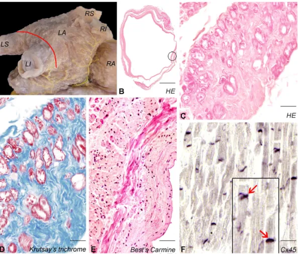
Extensions of left atrial myocardium could be observed in the PVs of 15/16 (94%) 121
hearts and formed bundles displaying various course (Fig. 1A,B). Bundles of large cardiac 122
cells (median diameter 18,1 (IQR: 16,5 – 19,7) µm) resembling of PFs due to their lightly 123
stained cytoplasm and peripheral myofibrils were detected in the PVs of 14 hearts. Among 124
these cardiomyocytes, dense network of fine collagen bundles was present (Fig. 1C,D). Best’s 125
M AN US CR IP T
AC CE PT ED
6
Carmine staining confirmed that PV myocardium was enriched in cardiomyocytes containing 126
abundant glycogen (Fig. 1E). Intense Cx45 labeling was observed in the myocardial sleeve of 127
PVs. Connexins were clustered in intercalated discs (Fig. 1F).
128
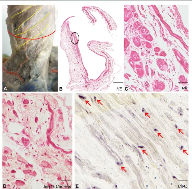
Myocardial sleeve of the superior caval vein 129
Myocardial sleeve composed of fibers displaying mainly spiral course was found 130
around 21 of 22 (95%) SVCs (Fig. 2A,B). In one case, some groups of myocardial fibers were 131
present at the root of the azygos vein but no cardiac cells were found in the portion distal to 132
this point. Bundles of Purkinje-like cardiomyocytes (median diameter 29,4 (IQR: 27,9 – 32,5) 133
µm) embedded in connective tissue were identified in 20 SVCs (Fig. 2C). Similar to PV 134
abundant intracellular glycogen content was found in the SVC myocardium (Fig. 2D). We 135
observed intense Cx45 positivity with a similar pattern as we described in Fig. 1F (Fig. 2E).
136
No Cx43 staining in the sinoatrial node, sparse labeling in the vicinity of sinoatrial node 137
(mixed population of atrial and pacemaker cells) and marked positivity were detected in the 138
atrial working myocardium (data not shown).
139
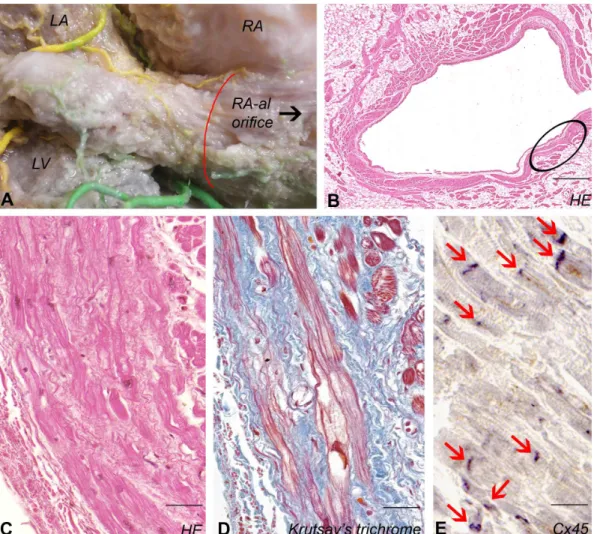
Myocardial sleeve of the coronary sinus 140
Cardiac muscle was present in 8 of 8 (100%) CS specimens (Fig. 3A). The course of 141
cardiomyocyte-bundles was spiral closer to the venous lumen and predominantly longitudinal 142
at the outer circumference (Fig. 3B). Purkinje-like myocardial fibers (median diameter 22,7 143
(IQR: 20,7 – 25,5) µm) embedded in network of collagen fibers were identified in 7 CSs (Fig.
144
3C,D). Immunostaining revealed that Cx45 labelings are as prominent in intercalated discs as 145
were described in PV and SVC (Fig. 3E). Cx43 labeling could barely be observed in the 146
myocardial sleeve of CS.
147
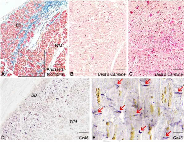
Histology of the working myocardium and conduction system of the heart 148
The ventricular conducting cells were rich in glycogen contrary to the ventricular 149
working myocardium (Fig. 4A,B,C). As compared to the extracardiac myocardium, no Cx45 150
M AN US CR IP T
AC CE PT ED
7
signal could be detected in the working myocardium, while conducting cells proved to be 151
strongly positive (Fig. 4D). Weak Cx45 immunoreactivity was present in the atria. Cx43 label 152
was marked throughout the ventricular working myocardium (Fig. 4E). At the region of 153
sinoatrial node prominent Cx45 immunoreactivity was detected. Cx45 was found to be 154
present at the atrioventricular nodal region and in the atrioventricular bundle as well.
155
156
Discussion 157
Characteristics of myocardial extensions around pulmonary veins 158
It has been demonstrated that left atrial myocardium extends into the wall of PVs in a 159
68-100% proportion.2,4-6 No specialized cells were observed except Perez-Lugones et al.7 who 160
analyzed electron microscopic images of human PV myocardium and reported the presence of 161
P cells and PFs. Nguyen et al.18 found PAS-positive cells, further supporting specialized 162
characteristics of PVs. In the current study bundles of cardiomyocytes displaying the 163
characteristic features of PFs were identified in almost all PV, SVC and CS samples. These 164
cells contained high amount of glycogen in their cytoplasm and they were found to be 165
embedded in a dense network of fine collagen fibers.
166
Potential arrhythmogenic role of caval veins and coronary sinus 167
Increasing interest has been recently devoted to non-PV ectopic beats that proved to be 168
responsible for 20% - 32% of all AF cases.11,12 Among patients with non-PV-initiating AF, 169
SVC triggers were found in about 40%.9,11,12 In the wall of SVC, myocardial sleeve was 170
detected in 76% - 78% of all cases.3,14 CS area were recognized as sites of tachyarrhythmias 171
in 1% - 17%11,12 of all non-PV ectopies. Two studies noted the presence of myocardium in all 172
CSs examined19,20 while DeSimone et al.3 reported that only 7% of the CSs contained 173
myocardial extensions from the right atrium. To the best of our knowledge, the present study 174
demonstrates for the first time that cells displaying PF morphology are present in human SVC 175
M AN US CR IP T
AC CE PT ED
8
and CS myocardium. The question arises whether this may indicate possible 176
arrhythmogenicity of these regions. We intend to investigate hearts removed from deceased 177
patients who possessed evidenced extracardiac loci of atrial arrhythmias to provide data for 178
the clarification of this issue.
179
Immunohistochemical characterization of extracardiac myocardial sleeves 180
In order to characterize the immunophenotype of the extracardiac myocardium, we 181
carried out immunostainings. Until now, positivity for HNK-1, which is an antigen to the 182
developing conduction system21, and reactivity for the cardiac pacemaker antigen HCN4 were 183
detected by human studies.18,22 184
According to previous data Cx45 was detected at very low levels in atrial and 185
ventricular working myocardium,16 whereas distinct positive signal was found at the 186
atrioventricular node of human adults.17 Therefore Cx45 seems to be a specific marker of the 187
conduction system. During the current study, strong staining for Cx45 was observed 188
throughout the impulse generating and conduction system of the heart, whereas almost no 189
immunopositivity could be detected in the working myocardium.
190
Data on the connexin expression patterns of cardiac veins are based on animal 191
researches. Differences regarding distinct species were published. In canine SVC, the 192
presence of all cardiac connexins including Cx45 were reported, with distinct areas 193
characterized by abundance of Cx43 in the center and diffuse Cx40 signals in the periphery.
194
Such areas of atypical connexin expression were mainly present in the proximal portion of the 195
SVC, usually in the outer circumference of the myocardial sleeve.23 Both Cx40 and Cx43 196
were observed in isolated cardiac cells from canine great veins, with a higher amount of Cx43 197
in the SVC than in the PVs. The absence of Cx45 signal was presumably caused by 198
paraformaldehyde fixation or injury of the cell membrane.24 In rat, a nodal-like tissue looping 199
around the junction of right atrium and SVC was reported. From the junction lightly stained 200
M AN US CR IP T
AC CE PT ED
9
cells extended both next to the crista terminalis and the interatrial groove. While nodal-like 201
cells proved to be strongly positive for Cx45 and negative for Cx43, atrial walls and PV 202
myocardium exhibited intense Cx43 but no Cx45 immunostaining in rat.25 203
The current study documents for the first time the presence of Cx45 positive 204
myocardial fibers in the wall of human PVs, SVC and CS. Based on the difference between 205
Cx45 immunopositivity of the working myocardium and the pacemaker and/or conducting 206
structures, our findings regarding the prominent Cx45 staining of the extracardiac myocardial 207
fibers might provide some support for the presumed specialized nature of these areas. Since 208
Cx43 labeling was weak in the myocardial sleeves, it can be hypothesized that extracardiac 209
myocardium in PVs, SVC and CS contains predominantly cardiomyocytes with pacemaker 210
and/or conducting nature.
211
Limitation 212
Immunohistochemistry is not an adequate method for reporting amounts of protein 213
expression. Therefore application of quantitative western blot analysis which is suitable for 214
determining relative abundance of distinct proteins would add weight to our observations.
215
216
Conclusions 217
1. Presence of Purkinje-like cardiomyocytes exhibiting strong glycogen positivity was 218
documented in the myocardial sleeves of human PVs, SVC and CS.
219
2. This research is the first to demonstrate pronounced Cx45 positivity of the 220
extracardiac myocardium which might provide some support for the presumed 221
arrhythmogenicity of the myocardial sleeves ensheathing PVs, caval veins and CS.
222
223
224
225
M AN US CR IP T
AC CE PT ED
10 Funding
226
This work was supported by institutional funds and by István Apáthy Foundation’s Research 227
Grant (2014-2016).
228
229
Acknowledgements 230
We thank the technical staff for their important contribution.
231
232
233
234
235
236
237
238
239
240
241
242
243
244
245
246
247
248
249
250
M AN US CR IP T
AC CE PT ED
11 References
251
1. Nathan H, Eliakim M. The junction between the left atrium and the pulmonary veins:
252
An anatomic study of human hearts. Circulation 1966;34:412-422.
253
2. Saito T, Waki K, Becker AE. Left atrial myocardial extensions onto pulmonary veins 254
in humans: Anatomic observations relevant for atrial arrhythmias. J Cardiovasc 255
Electrophysiol 2000;11:888-894.
256
3. DeSimone CV, Noheria A, Lachman N, Edwards WD, Gami AS, Maleszewski JJ, 257
Friedman PA, Munger TM, Hammill SC, Packer DL, Asirvatham SJ. Myocardium of 258
the superior vena cava, coronary sinus, vein of Marshall, and the pulmonary vein 259
ostia: Gross anatomic studies in 620 hearts. J Cardiovasc Electrophysiol 260
2012;23:1304-1309.
261
4. Ho SY, Cabrera JA, Tran VH, Farré J, Anderson RH, Sánchez-Quintana D.
262
Architecture of the pulmonary veins: relevance to radiofrequency ablation. Heart 263
2001;86:265-270.
264
5. Kholová I, Kautzner J. Anatomic characteristics of extensions of atrial myocardium 265
into pulmonary veins in subjects with and without atrial fibrillation. Pacing Clin 266
Electrophysiol 2003;26:1348-1355.
267
6. Hassink RJ, Aretz HT, Ruskin J, Keane D. Morphology of atrial myocardium in 268
human pulmonary veins. A postmortem analysis in patients with and without atrial 269
fibrillation. J Am Coll Cardiol 2003;42:1108-1114.
270
7. Perez-Lugones A, McMahon JT, Ratliff NB, Saliba WI, Schweikert RA, Marrouche 271
NF, Saad EB, Navia JL, McCarthy PM, Tchou P, Gillinov AM, Natale A. Evidence of 272
specialized conduction cells in human pulmonary veins of patients with atrial 273
fibrillation. J Cardiovasc Electrophysiol 2003;14:803-809.
274
M AN US CR IP T
AC CE PT ED
12
8. Tsai CF, Tai CT, Hsieh MH, Lin WS, Yu WC, Ueng KC, Ding YA, Chang MS, Chen 275
SA. Initiation of atrial fibrillation by ectopic beats originating from the superior vena 276
cava. Circulation 2000;102:67-74.
277
9. Lin WS, Tai CT, Hsieh MH, Tsai CF, Lin YK, Tsao HM, Huang JL, Yu WC, Yang 278
SP, Ding YA, Chang MS, Chen SA. Catheter ablation of paroxysmal atrial fibrillation 279
initiated by non–pulmonary vein ectopy. Circulation 2003;107:3176-3183.
280
10. Katsivas AG, Manolis AG, Vassilopoulos C, Ioannidis P, Giotopoulou A, Kyriakides 281
Z. Electroanatomical mapping of a right atrial tachycardia originating within the 282
inferior vena cava. Hellenic J Cardiol 2004;45:187-190.
283
11. Lee SH, Tai CT, Hsieh MH, Tsao HM, Lin YJ, Chang SL, Huang JL, Lee KT, Chen 284
YJ, Cheng JJ, Chen SA. Predictors of non-pulmonary vein ectopic beats initiating 285
paroxysmal atrial fibrillation: Implication for catheter ablation. J Am Coll Cardiol 286
2005;46:1054-1059.
287
12. Chang HY, Lo LW, Lin YJ et al. Long-term outcome of catheter ablation in patients 288
with atrial fibrillation originating from nonpulmonary vein ectopy. J Cardiovasc 289
Electrophysiol 2013;24:250-258.
290
13. Hashizume H, Ushiki T, Ahe K. A histological study of the cardiac muscle of the 291
human superior and inferior venae cavae. Arch Histol Cytol 1995;58:457-464.
292
14. Kholová I, Kautzner J. Morphology of atrial myocardial extensions into human caval 293
veins: A postmortem study in patients with and without atrial fibrillation. Circulation 294
2004;110:483-488.
295
15. Severs NJ, Rothery S, Dupont E, Coppen SR, Yeh HI, Ko YS, Matsushita T, Kaba R, 296
Halliday D. Immunocytochemical analysis of connexin expression in the healthy and 297
diseased cardiovascular system. Microsc Res Tech 2001;52:301-322.
298
M AN US CR IP T
AC CE PT ED
13
16. Vozzi C, Dupont E, Coppen SR, Yeh HI, Severs NJ. Chamber-related differences in 299
connexin expression in the human heart. J Mol Cell Cardiol 1999;31:991-1003.
300
17. Kreuzberg MM, Liebermann M, Segschneider S, Dobrowolski R, Dobrzynski H, Kaba 301
R, Rowlinson G, Dupont E, Severs NJ, Willecke K. Human connexin31.9, unlike its 302
orthologous protein connexin30.2 in the mouse, is not detectable in the human cardiac 303
conduction system. J Mol Cell Cardiol 2009;46:553-559.
304
18. Nguyen BL, Fishbein MC, Chen LS, Chen PS, Masroor S. Histopathological substrate 305
for chronic atrial fibrillation in humans. Heart Rhythm. 2009;6:454-460.
306
19. Lüdinghausen M, Ohmachi N, Boot C. Myocardial coverage of the coronary sinus and 307
related veins. Clin Anat 1992;5:1-15.
308
20. Chauvin M, Shah DC, Haїssaguerre M, Marcellin L, Brechenmacher C. The anatomic 309
basis of connections between the coronary sinus musculature and the left atrium in 310
humans. Circulation 2000;101:647-652.
311
21. Blom NA, Gittenberger-de Groot AC, DeRuiter MC, Poelmann RE, Mentink MMT, 312
Ottenkamp J. Development of the cardiac conduction tissue in human embryos using 313
HNK-1 antigen expression: Possible relevance for understanding of abnormal atrial 314
automaticity. Circulation 1999;99:800-806.
315
22. Kholová I, Niessen HWM, Kautzner J. Expression of Leu-7 in myocardial sleeves 316
around human pulmonary veins. Cardiovasc Pathol 2003;12:263-266.
317
23. Yeh HI, Lai YJ, Lee SH, Lee YN, Ko YS, Chen SA, Severs NJ, Tsai CH.
318
Heterogeneity of myocardial sleeve morphology and gap junctions in canine superior 319
vena cava. Circulation 2001;104:3152-3157.
320
24. Yeh HI, Lai YJ, Lee YN, Chen YJ, Chen YC, Chen CC, Chen SA, Lin CI, Tsai CH.
321
Differential expression of connexin43 gap junctions in cardiomyocytes isolated from 322
canine thoracic veins. J Histochem Cytochem 2003;51:259-266.
323
M AN US CR IP T
AC CE PT ED
14
25. Yamamoto M, Dobrzynski H, Tellez J, Niwa R, Billeter R, Honjo H, Kodama I, 324
Boyett MR. Extended atrial conduction system characterised by the expression of the 325
HCN4 channel and connexin45. Cardiovasc Res 2006;72:271-281.
326
327
328
329
330
331
332
333
334
335
336
337
338
339
340
341
342
343
344
345
346
347
348
M AN US CR IP T
AC CE PT ED
15
Figure 1. Histology of the PV myocardium. Myocardial extensions into the wall of PVs (LS, LI, 349
RS, RI). LA and RA= left and right atria. Circumflex artery is filled up by yellow resin mixture.
350
Red line shows the area which was cut out for histology (A). Transverse section of a PV with 351
myocardial sleeve (B). Large cardiomyocytes with lightly staining cytoplasm at the area signed by 352
circle at picture B (C). Trichrome staining shows that myocardial fibers (red) are isolated by 353
fibrous tissue (blue) (D). A bundle of myocardial cells containing much glycogen (E). Prominent 354
Cx45 positivity in intercalated discs (inset, arrows) (F). Scale bar: 5000 µm (B); 40 µm (C); 30 355
µm (D); 70 µm (E); 30 µm (F); 20 µm (F-inset).
356
357
358
359
360
M AN US CR IP T
AC CE PT ED
16
Figure 2. Histology of the SVC myocardium. Cardiomyocyte bundles with spiral course (yellow 361
lines). Sinoatrial nodal artery is filled up by red resin mixture. Red line shows the area which was 362
cut out for histology (A). Transverse section of SVC with myocardial sleeve (B). Purkinje-like 363
myocardial cells at the area signed by circle at picture B (C). Glycogen containing 364
cardiomyocytes (D). In intercalated discs, Cx45 immunoreactivity is prominent (E, arrows). Scale 365
bar: 1750 µm (B); 30 µm (C); 30 µm (D); 20 µm (E).
366
367
368
369
370
371
M AN US CR IP T
AC CE PT ED
17
Figure 3. Histology of the CS myocardium. Myocardial sleeve around the wall of CS. RA=right 372
atrium, LA and LV= left atrium and ventricle. Right coronary artery (green) and circumflex artery 373
(yellow) are filled up by synthetic resin. Red line shows the area which was cut out for histology 374
(A). Cross section of the CS orifice (B). Bundles of Purkinje-like cardiomyocytes run around the 375
lumen at the area signed by circle at picture B (C). Trichrome staining shows that myocardial 376
fibers having lightly staining cytoplasm (red) are separated by connective tissue (blue) (D). Cx45 377
immunoreactivity is prominent at intercalated discs (E, arrows). Scale bar: 900 µm (B); 45 µm 378
(C); 40 µm (D); 30 µm (E).
379
380
381
382
383
M AN US CR IP T
AC CE PT ED
18
Figure 4. Histology of the left ventricular myocardium. Trichrome staining of the working 384
myocardium (WM) and a bundle branch (BB, inset) composed of cardiomyocytes with pale 385
cytoplasm (A). WM shows almost no sign with glycogen specific Best’s Carmine staining (B), 386
while much glycogen is recognized at the BB (C). Cx45 immunoreactivity is prominent at the BB 387
but barely detectable in the WM (D). At the WM, marked positivity for Cx43 is detectable in 388
intercalated discs (arrows). Yellow-brown intracellular granules in the vicinity of nuclei are 389
lipofuscin pigments (E). Scale bar: 80 µm (A); 40 µm (A-inset); 160 µm (B); 100 µm (C); 90 µm 390
(D); 12 µm (E).
391