1
Cite as: Janacsek, K., Shattuck, K. F., Lum, J. A. G., Tagarelli, K. M., Turkeltaub, P. E. & Ullman, M. T. (2020). Sequence learning in the human brain: A functional neuroanatomical meta-analysis of SRT studies. NeuroImage, 207, 116387. https://doi.org/10.1016/j.neuroimage.2019.116387
Sequence learning in the human brain: A functional neuroanatomical meta-analysis of serial reaction time studies
Karolina Janacsek1,2,3*, Kyle F. Shattuck4, Kaitlyn M. Tagarelli4, Jarrad A.G. Lum5, Peter E.
Turkeltaub6, Michael T. Ullman4*
1Brain, Memory and Language Lab, Institute of Cognitive Neuroscience and Psychology, Research Centre for Natural Sciences, Hungarian Academy of Sciences, Magyar Tudósok Körútja 2, H-1117 Budapest, Hungary
2Institute of Psychology, ELTE Eotvos Lorand University, Damjanich utca 41-43, H-1071 Budapest, Hungary
3 School of Human Sciences, Faculty of Education, Health and Human Sciences, University of Greenwich, Old Royal Naval College, 150 Dreadnought, London, United Kingdom
4 Department of Neuroscience, Georgetown University, EP-04 New Research Building, 20007 Washington DC, USA
5 School of Psychology, Deakin University, Melbourne Burwood Campus, Burwood, Victoria, Australia
6 Center for Brain Plasticity and Recovery, Department of Neurology, Georgetown University, 3800 Reservoir Rd NW, 20007 Washington DC, USA
* Corresponding authors
2 Abstract
Sequence learning underlies numerous motor, cognitive, and social skills. Previous models and empirical investigations of sequence learning in humans and non-human animals have implicated cortico-basal ganglia-cerebellar circuitry as well as other structures. To systematically examine the functional neuroanatomy of sequence learning in humans, we conducted a series of neuroanatomical meta-analyses. We focused on the serial reaction time (SRT) task. This task, which is the most widely used paradigm for probing sequence learning in humans, allows for the rigorous control of visual, motor, and other factors. Controlling for these factors (in sequence-random block contrasts), sequence learning yielded consistent activation only in the basal ganglia, across the striatum (anterior/mid caudate nucleus and putamen) and the globus pallidus. In contrast, when visual, motor, and other factors were not controlled for (in a global analysis with all sequence-baseline contrasts, not just sequence- random contrasts), premotor cortical and cerebellar activation were additionally observed.
The study provides solid evidence that, at least as tested with the visuo-motor SRT task, sequence learning in humans relies on the basal ganglia, whereas cerebellar and premotor regions appear to contribute to aspects of the task not related to sequence learning itself. The findings have both basic research and translational implications.
Keywords: sequence learning, implicit learning, procedural memory, serial reaction time (SRT) task, basal ganglia, striatum
3 Introduction
Sequence learning underlies a range of motor, cognitive, and social skills that are integral to our everyday life. For example, when we dance, type, use our smartphones, play video games, play a musical instrument, perform arithmetic operations, produce or understand language, or interact with others in social contexts, we seem to be relying at least in part on sequential knowledge (Evans and Ullman, 2016; Landau and D'Esposito, 2006; Lieberman, 2000; Nemeth et al., 2011; Norman and Price, 2012; Romano Bergstrom et al., 2012; Ullman, 2004, 2016). The acquisition of this knowledge generally occurs gradually and implicitly through practice, resulting in rapid and reliable processing that is characteristic of automatized skills (Ashby et al., 2010; Berry and Dienes, 1993; Penhune and Steele, 2012; Squire, 1986;
Ullman, 2004).
Sequence learning has been most widely studied in task paradigms examining the learning of (visuo)motor sequences, such as in the serial reaction time (SRT) task. In such task paradigms, human or non-human animal participants (implicitly or explicitly) acquire motor skills through repeated sequences of motor responses, often in response to visual stimuli, although the exact nature of the tasks can vary (Doyon et al., 2009; Hikosaka et al., 2002; Janacsek and Nemeth, 2012; Penhune and Steele, 2012; Robertson, 2007). In these task paradigms, the sequences of (visual stimuli and) motor responses follow predetermined (predictable) orders that can be acquired through repeated practice. In SRT studies, participants are typically not informed about the sequence order, and their acquired sequence knowledge generally remains largely implicit (inaccessible to consciousness). In contrast to such implicit SRT task paradigms, in some SRT studies participants are told the sequence (or are instructed to learn it) and thus they gain explicit sequential knowledge.
Previous functional neuroanatomical models and empirical investigations of animals and humans have suggested that such motor skill learning critically relies on cortico-basal ganglia-cerebellar circuitry (Bischoff-Grethe et al., 2001; Daselaar et al., 2003; Doyon et al., 2009; Graybiel, 2008; Hikosaka et al., 2002; Keele et al., 2003; Penhune and Steele, 2012;
Rauch et al., 1997a; Willingham, 1998; Willingham et al., 2002). Within cortex, (pre)motor and parietal regions have been particularly implicated, as has the striatum within the basal ganglia. Additionally, some human studies have implicated other (neo)cortical structures, including the dorsolateral prefrontal cortex and the medial temporal lobe (Albouy et al., 2013;
Clark and Lum, 2017; Grafton et al., 1995; Schendan et al., 2003; Willingham et al., 2002);
also see Discussion.
4
However, the exact roles of each of these structures in (visuo)motor sequence learning tasks remain unclear, in part because the tasks encompass a number of different functions.
These include not only the acquisition of predictable sequential associations (the order of the sequential stimuli) – that is, sequence-specific learning – but also the sensorimotor integration of visual stimuli and motor responses, and the formation of internal models to control movements (Grafton et al., 2008; Penhune and Steele, 2012; Seidler et al., 2002; Wolpert et al., 1998). Additionally, sequence-specific learning itself may be implicit or explicit (see above), with different accompanying functions (e.g., working memory for explicit learning;
Janacsek and Nemeth, 2013, 2015). Thus, exactly which brain structures support (implicit or explicit) sequence learning itself, as opposed to other functions, has been difficult to pin down, including in human sequence learning (Fletcher et al., 2005; Schendan et al., 2003;
Willingham et al., 2002).
Human functional neuroimaging studies may elucidate this issue. Indeed, over the past two decades or so, an increasing number of neuroimaging studies of (visuo)motor sequence learning have been published. However, the tasks, task contrasts and other factors, such as whether learning involved explicit knowledge or not, have varied across studies, and the results have been at least somewhat inconsistent. It has therefore been difficult to synthesize the data from these studies, and thus to ascertain the pattern of brain activation associated with (visuo)motor sequence learning tasks, let alone sequence-specific learning, in humans.
One approach that could substantially clarify the functional neuroanatomy of sequence learning is to conduct neuroanatomical meta-analyses of previous functional neuroimaging studies. A quantitative meta-analytical approach can rigorously synthesize the existing literature and reveal any consistent patterns of activation. Meta-analyses have substantial power because they examine a much larger number of participants than individual studies.
Thus, meta-analysis results are likely more reliable and generalizable than single study findings. Additionally, they can be more objective than qualitative reviews.
The present study
In the present study we attempted to elucidate the functional neuroanatomy of human sequence learning by conducting a series of Activation Likelihood Estimation (ALE) meta- analyses. ALE, which is the most widely used neuroanatomical meta-analytic technique, determines areas of significant spatial convergence based on peak activation coordinates reported in previous studies (Eickhoff et al., 2009; Laird et al., 2005; Turkeltaub et al., 2002;
5
Turkeltaub et al., 2012). We followed the guidelines for neuroimaging meta-analyses recently laid out by Müller et al. (2017).
We focused on neuroimaging studies using the SRT task (see Discussion for neuroanatomical meta-analyses examining motor learning more generally, i.e., Hardwick et al., 2013; Lohse et al., 2014). In this task, participants are typically presented with a series of visuo-spatial stimuli (e.g., across four locations on the screen) that follow a sequence, and are asked to respond to each stimulus position with a corresponding button. Generally in neuroimaging studies, ‘sequence blocks’ in which the sequence is repeatedly presented alternate with ‘baseline blocks’ (generally of randomly ordered stimuli), with learning operationalized as the performance difference between sequence and baseline blocks. The SRT task is of particular interest because it is the most widely used paradigm employed to examine (implicit) sequence learning in humans. Importantly (and, for example, in contrast to typical finger sequence tapping tasks; Janacsek and Nemeth, 2012; Walker et al., 2003), the SRT task allows for rigorous control of perceptuo-motor and other confounds, in sequence- random block comparisons, providing a clear means to probe sequence-specific learning.
Moreover (and unlike finger sequence tapping), the task is designed to allow one to specifically examine either implicit or explicit sequence-specific learning.
We performed separate ALE meta-analyses on three sets of SRT studies. First, we ran a global ALE analysis on all SRT neuroimaging contrasts that found more brain activation in sequence blocks than baseline blocks. This analysis included all such contrasts, irrespective of the baseline task. This ALE analysis was designed to reveal the functional neuroanatomy of sequence learning overall, without specifically controlling for visual, motor, and other confounds. Second, we performed an analogous ALE analysis that only included sequence- baseline contrasts with a random order of items as the baseline. This can shed light on the neural substrates of sequence-specific learning, by holding constant a range of visual, motor, and other potentially confounding factors. Third, we conducted an even more specific ALE analysis, on the subset of these sequence-specific contrasts in which participants had apparently acquired only implicit sequential knowledge – that is, in which explicit knowledge was tested but was not found. This analysis can elucidate which neural structures are involved in implicit sequence-specific learning, although it does not preclude the possibility that the same structures may also be involved in explicit sequence-specific learning. Note that there was insufficient power to separately examine the learning of explicit sequential knowledge, or to directly compare the activation between implicit and explicit sequence-specific learning.
6
Thus, although we can identify activation involved in the implicit acquisition of sequential knowledge, in the absence of a direct comparison between implicit and explicit sequence learning, we cannot identify any activation that is unique to implicit (vs. explicit) sequence- specific learning. Overall, the present study was designed to unravel aspects of the functional neuroanatomy of human sequence learning as tested in the SRT task.
Methods
Ethics statement
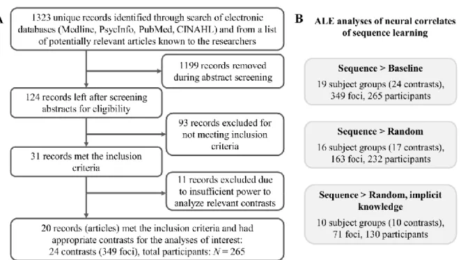
The study was conducted according to the Declaration of Helsinki and the Ethical Principles of Psychologists and Code of Conduct (APA, 2017) that outlines guidelines for conducting and reporting analyses, including meta-analyses. Also see the PRISMA flowchart (Figure 1A).
Data used for the ALE meta-analysis Search and selection of studies
A systematic search for papers in PubMed, Medline, PsycINFO, and CINAHL, together with a list of potentially relevant papers known to the authors, yielded 1323 unique records (papers) as of September 11, 2017. These included investigations that reported an original piece of research that had been published or had been accepted for publication, as well as master’s theses and doctoral dissertations. There were no restrictions on publication date. We used a variety of search strings to be able to identify all possibly relevant papers.
The search results contained at least one search string related to sequence learning AND one search string related to neuroimaging (see Table 1). After screening abstracts and evaluating the full text of articles (process summarized in the PRISMA flowchart shown in Figure 1A), 31 papers were identified as meeting the study inclusion criteria, with 100% agreement between the first, second and last authors. Due to the exclusion of 11 papers due to insufficient power to analyze the relevant contrasts (see below), the final analyses were based on 20 papers overall.
7
Table 1. The syntax for database searches, which were carried out simultaneously in the following fields: study title, abstract, keywords, and subject terms.
Search syntax Related to
sequence learning
((functional magnetic resonance imaging) OR (fMRI) OR (positron emission tomography) OR (PET) OR (regional cerebral blood flow) OR (rCBF) OR (blood oxygen level dependent) OR (BOLD) OR (functional imaging) OR (neuroimaging) OR (brain imaging))
AND
Related to neuroimaging
((implicit memory) OR (implicit learning) OR (serial reaction) OR (serial learn*) OR (sequence learning) OR (implicit sequence) OR (implicit learn*) OR (implicit visuo*spatial) OR (implicit visuospatial) OR (procedural learn*) OR (procedural mem*) OR (srt) OR (srtt) OR (motor skill learning) OR (triplets))
Study inclusion criteria
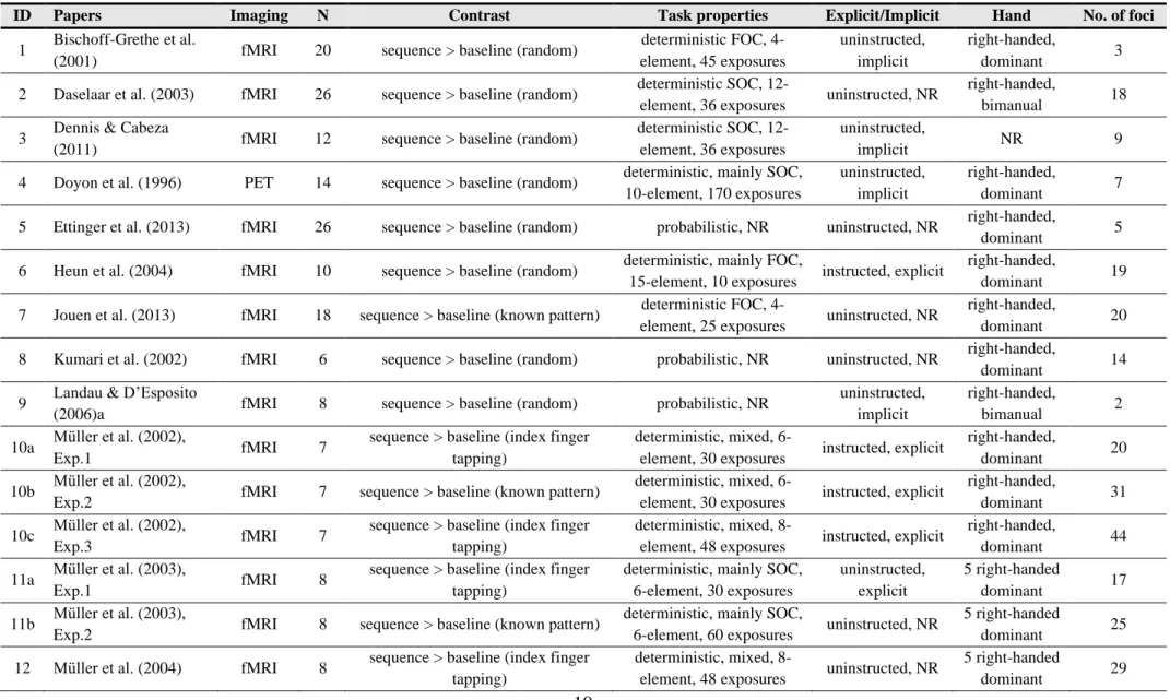
Studies were included only if they met all the following criteria. 1) They were functional neuroimaging studies (fMRI or PET; no relevant SPECT studies were found) that examined neural activation during tasks probing sequence learning, using whole-brain analysis, and reported peak coordinates in standard reference space (Talairach/Tournoux or MNI). 2) They presented data from healthy (non-elderly) adults; the age range in the papers included in the meta-analyses was between 18 and 52 years. 3) The sequence learning task was a form of the classic visuomotor SRT task, with one-to-one stimulus-response mapping. Thus, in these tasks participants were asked to respond – press the corresponding button – when a visual stimulus was serially (sequentially) presented to them, and different response buttons corresponded to each of the different stimuli (e.g., four response buttons corresponding to four stimulus locations). Finally, of the 31 fMRI/PET papers that met these inclusion criteria, we excluded 11 papers, leaving 20 for the ALE meta-analyses reported below. These 11 papers were excluded because they either reported only activation increases and/or decreases over the course of learning, and/or only less activation for sequence than baseline. Perhaps due to the relatively low number of subject groups in each of these cases (9, 7, and 6, respectively), ALE analyses on these contrasts yielded no significant ALE results (not reported). We thus focused on the 20 papers that met all inclusion criteria and reported the key contrast of greater activation for sequence compared to baseline, since this contrast is most frequently reported in SRT studies, and is well-suited to reveal the neural correlates of sequence learning (see Table 2).
8
Figure 1. A) PRISMA flowchart showing the process of identifying the articles included in the meta-analyses. B) ALE analyses for specific neural contrasts extracted from the articles.
Contrasts of interest in the ALE meta-analyses
To examine the neural correlates of sequence learning, we included all relevant contrasts examining greater activation in sequence than baseline conditions. Consistent with classic SRT task design, in all included studies participants were presented with a series of sequence and baseline blocks, with each block composed of multiple trials. In the sequence blocks, the presentation order of the trials followed a particular sequential order (e.g., 124313241423, where the numbers correspond to four locations on the screen). Each specific stimulus was associated with a response button, which corresponded to that stimulus only.
The sequence was repeated multiple times in each block to enable learning of the sequence.
Brain activation while performing these sequence blocks was contrasted with brain activation while performing baseline blocks, in which the predetermined stimulus order was removed. Baseline blocks were implemented differently in different studies. First, in some studies (e.g., Muller et al., 2004; Muller et al., 2003; Muller et al., 2002), in the baseline condition participants had to respond only to one location using one response button, always using the index finger (referred to as index finger tapping in Table 2). Second, in some studies (e.g., Jouen et al., 2013; Muller et al., 2003), baseline blocks contained a known pattern (e.g., 12341234), which was assumed to be familiar to the participants. Thus, it was assumed that
9
they could respond rapidly and accurately with no additional learning. These blocks serve as a better baseline compared to the one location–one response button baseline condition because they control for visual and motor demands more precisely (i.e., the same number of locations are presented on the screen and the same number of fingers are used as in the sequence blocks). Third, the majority of the studies used blocks with random stimulus order as a baseline. This type of baseline is generally considered to be the most appropriate for controlling for visual and motor demands: in this case the only difference between the sequence and baseline blocks is that the sequence blocks contain a particular serial order of stimuli practiced repeatedly while the baseline blocks contain no such predetermined order (we refer to these type of blocks as random blocks) (Dennis and Cabeza, 2011; Janacsek and Nemeth, 2012).
In Table 2, for each paper we report which of these sequence vs. baseline contrasts were analyzed: 1) sequence vs. index finger tapping, 2) sequence vs. known pattern, or 3) sequence vs. random blocks. As described above, all included papers (and contrasts) tested and reported greater neural activation for the sequence blocks compared to the baseline blocks (shown as 'sequence > baseline' in Table 2). Note that all contrasts in our ALE analyses probed neural activation during the first day of training, and thus may be considered as occurring during early phases of sequence learning (Doyon et al., 2009; Ullman et al., in press, 2020).
Since explicit sequence learning may rely on different neurocognitive substrates than implicit sequence learning, we coded, for each contrast, 1) whether participants were explicitly instructed regarding the existence of a sequence (instructed) or not (uninstructed);
and 2) whether the participants showed evidence of having explicit knowledge of the sequence (explicit) or not (implicit), if tested for explicit knowledge after sequence learning;
see Table 2 (if testing was not reported, this is indicated with NR, i.e., not reported). Because the explicit vs. implicit knowledge acquired is more likely to affect the neurocognition of sequence learning than the method of instruction, we performed separate ALE analyses, where possible, on only those contrasts where participants had or had not acquired explicit knowledge.
Additional task and subject characteristics (e.g., handedness) are displayed for each contrast in Table 2. These variables are displayed for completeness and transparency but are not of primary interest in the current study, and were not analyzed.
10
Table 2. Papers and contrasts included in the meta-analyses
ID Papers Imaging N Contrast Task properties Explicit/Implicit Hand No. of foci
1 Bischoff-Grethe et al.
(2001) fMRI 20 sequence > baseline (random) deterministic FOC, 4-
element, 45 exposures
uninstructed, implicit
right-handed,
dominant 3
2 Daselaar et al. (2003) fMRI 26 sequence > baseline (random) deterministic SOC, 12-
element, 36 exposures uninstructed, NR right-handed,
bimanual 18
3 Dennis & Cabeza
(2011) fMRI 12 sequence > baseline (random) deterministic SOC, 12- element, 36 exposures
uninstructed,
implicit NR 9
4 Doyon et al. (1996) PET 14 sequence > baseline (random) deterministic, mainly SOC, 10-element, 170 exposures
uninstructed, implicit
right-handed,
dominant 7
5 Ettinger et al. (2013) fMRI 26 sequence > baseline (random) probabilistic, NR uninstructed, NR right-handed,
dominant 5
6 Heun et al. (2004) fMRI 10 sequence > baseline (random) deterministic, mainly FOC,
15-element, 10 exposures instructed, explicit right-handed,
dominant 19
7 Jouen et al. (2013) fMRI 18 sequence > baseline (known pattern) deterministic FOC, 4-
element, 25 exposures uninstructed, NR right-handed,
dominant 20
8 Kumari et al. (2002) fMRI 6 sequence > baseline (random) probabilistic, NR uninstructed, NR right-handed,
dominant 14
9 Landau & D’Esposito
(2006)a fMRI 8 sequence > baseline (random) probabilistic, NR uninstructed,
implicit
right-handed,
bimanual 2
10a Müller et al. (2002),
Exp.1 fMRI 7 sequence > baseline (index finger
tapping)
deterministic, mixed, 6-
element, 30 exposures instructed, explicit right-handed,
dominant 20
10b Müller et al. (2002),
Exp.2 fMRI 7 sequence > baseline (known pattern) deterministic, mixed, 6-
element, 30 exposures instructed, explicit right-handed,
dominant 31
10c Müller et al. (2002),
Exp.3 fMRI 7 sequence > baseline (index finger
tapping)
deterministic, mixed, 8-
element, 48 exposures instructed, explicit right-handed,
dominant 44
11a Müller et al. (2003),
Exp.1 fMRI 8 sequence > baseline (index finger
tapping)
deterministic, mainly SOC, 6-element, 30 exposures
uninstructed, explicit
5 right-handed
dominant 17
11b Müller et al. (2003),
Exp.2 fMRI 8 sequence > baseline (known pattern) deterministic, mainly SOC,
6-element, 60 exposures uninstructed, NR 5 right-handed
dominant 25
12 Müller et al. (2004) fMRI 8 sequence > baseline (index finger tapping)
deterministic, mixed, 8-
element, 48 exposures uninstructed, NR 5 right-handed
dominant 29
11
13 Naismith et al. (2010) fMRI 20 sequence > baseline (random) deterministic SOC, 12-
element, 36 exposures uninstructed, NR NR 21
14 Poldrack et al. (2005) fMRI 14 sequence > baseline (random) deterministic SOC?, 12-
element, 30 exposures uninstructed, NR NR 1
15 Purdon et al. (2011) fMRI 17 sequence > baseline (random) deterministic SOC, 12- element, 60 exposures
uninstructed, implicit
right-handed,
bimanual 13
16 Rauch, Savage, et al.
(1997a) PET 9 sequence > baseline (random) deterministic SOC?, 12- element, 36 exposures
uninstructed, implicit
right-handed,
bimanual 6
17 Rauch, Whalen, et al.
(1997b) fMRI 10 sequence > baseline (random) deterministic SOC, 12- element, 36 exposures
uninstructed, implicit
right-handed,
bimanual 10
18 Werheid et al. (2003) fMRI 7 sequence > baseline (random) deterministic SOC, 12- element, 192 exposures
uninstructed, implicit
right-handed,
bimanual 7
19a Willingham et al.
(2002) fMRI 18 sequence > baseline (random) deterministic SOC?, 12- element, 96 exposures
uninstructed, implicit
right-handed,
bimanual 4
19b Willingham et al.
(2002) fMRI 18 sequence > baseline (random) deterministic SOC?, 12-
element, 96 exposures instructed, explicit right-handed,
bimanual 14
20 Zedkova et al. (2006) fMRI 15 sequence > baseline (random) deterministic SOC?, 12- element, 66 exposures
uninstructed, implicit
right-handed,
bimanual 10
Note. Rows reflect distinct contrasts. Since a given paper may have two or more contrasts, more than one row is displayed for some papers. All characteristics refer to the specific contrast on that row. N: number of participants. Contrasts: ‘sequence > baseline’: greater activation for the sequence blocks compared to the baseline blocks.
Characteristics of the baseline blocks are indicated in parentheses. For example, baseline stimuli presented in random order are indicated with ‘(random)’. See main text for more information. Note that in almost all cases different papers examined different subject groups (with one exception: papers 11 and 12 examined the same subject group), and in all cases each paper examined only one subject group; thus whereas there are 20 papers, there are 19 subject groups. Task properties: sequence type (deterministic or probabilistic); first order conditional or second order conditional sequence, or mixed (FOC, SOC, mixed; SOC? refers to sequences that appear to be SOC, but the article does not report enough information to make a clear determination); length of the sequence (how many elements were in the sequence); and the number of exposures to the sequence (how many times the sequence occurred in total, over all sequence blocks). Explicit/implicit: whether or not participants were explicitly instructed to learn the sequence (instructed vs. uninstructed), as well as whether or not they gained explicit knowledge about the sequence, if this was tested (explicit vs. implicit); if such testing was not reported, this is indicated with NR. Hand: indicates both participant handedness (almost all right-handed) and whether the dominant or non-dominant hand or both hands (bimanual) were used to respond to (both types of blocks) in the task. NR – not reported in the paper.
12 ALE analysis procedure
All analyses were conducted using GingerALE 2.3.6 (downloaded from www.brainmap.org/ale on 16 September, 2017; Eickhoff et al., 2009; Laird et al., 2005). The ALE algorithm tests against the null hypothesis that activation foci in the dataset are distributed uniformly across the brain, and thus tests for regions with an above-chance concentration of activity across experiments (Eickhoff et al., 2012; Laird et al., 2005). We used the algorithm described in Turkeltaub et al. (2012), which organizes the foci by subject group (as opposed to experimental affiliation), and thus prevents subject groups with multiple experiments or tasks in a given ALE meta-analysis from influencing ALE values more than others.
Because all of the foci must be in the same coordinate space, foci that were reported in Talairach space were transformed to MNI space. Specifically, foci reported in Talairach space that were transformed from MNI space in the original study using the Brett transform (mni2tal) were converted back to MNI space using the inverse of the Brett transform. Those that were transformed in the original study into Talairach space using the Lancaster transform (icbm2tal) were transformed back into MNI space using the inverse of the Lancaster transform (Laird et al., 2010; Lancaster et al., 2007). In cases where data were initially normalized into Talairach space, the Lancaster icbm2tal transform was used to convert these foci into MNI space (see Bernard and Seidler, 2013). All transformations were performed using the “convert foci” tool in GingerALE.
Foci in MNI space were entered into GingerALE, which computes the ALE values for every voxel in the brain using an automatically determined full-width half-maximum (FWHM) value (Eickhoff et al., 2009). The resulting ALE map was thresholded using cluster- level family-wise error (cFWE) correction, with a cluster-level requirement of p < 0.05 and a voxel-level requirement of p < 0.001 (Eickhoff et al., 2016). The significance levels of ALE values were determined by comparing the resulting ALE statistics to a null distribution generated from 10,000 permutations (Acikalin et al., 2017; Eickhoff et al., 2012; Eickhoff et al., 2016). It has been shown that the cFWE correction method that we used in our analyses is the most sensitive approach to reveal the true effects (neural activation) with high power and, at the same time, to control for false positives, compared to other correction methods (Eickhoff et al., 2016).
The power to detect a true activation effect in ALE depends on the proportion of experiments/subject groups showing that effect (i.e., the effect size). Following Eickhoff et al.
13
(2016), if about one-third of the included experiments show the effect of interest (corresponding to an effect size of 0.33), 17 experiments are sufficient to achieve a desired power of 80% using the cFWE correction, though, in general, at least about 20 experiments are recommended (Eickhoff et al., 2016). However, it is also possible to achieve 80% power with a smaller number of experiments if a larger proportion of those experiments show the effect of interest (that is, if the effect size is larger). In the current study, we report three ALE analyses, with 19, 16 and 10 subject groups. The number of subject groups in the first two analyses should yield enough power to detect true effects with an effect size of ~0.33 or above (Eickhoff et al., 2016). Our analysis with 10 subject groups, however, is underpowered for effects with an effect size of ~0.33. Thus, it is likely that effects of this size cannot be detected in this analysis, though larger effect sizes (0.45 and above) should achieve the desired power of 80% (Figure 8 in Eickhoff et al., 2016). To address the issue of power, we estimated the observed (a posteriori) power for each peak in each ALE analysis, as a function of their effect sizes: that is, as a function of the proportion of contributing subject groups for each ALE peak (Eickhoff et al., 2016). Contributing subject groups were determined to be those with coordinates within 3 standard deviations (SDs) of the coordinates of the peak activations in each ALE analysis (Turkeltaub et al., 2011). We report all (sub)peaks, irrespective of how many subject groups contributed to them; however, no (sub)peak had fewer than 3 contributing subject groups (see Table 3).
Results were visualized using MRICron and overlaid on an MNI brain template. In the results table (Table 3), we present anatomical labels corresponding to each cluster peak and subpeak(s). In the text, we point out instances when clusters extended to other brain structures. This is particularly relevant here because ALE analyses with cFWE correction tend to identify larger clusters, compared to other correction methods (Eickhoff et al., 2016), and these large clusters can encompass several distinct brain regions that may be of interest (e.g., within the basal ganglia). The identification of anatomical structures containing (sub)peaks, and areas of extended activation, were performed both based on the Neuromorphometrics atlas and by visual inspection of the AAL template and the MNI brain template in MRICron.
ALE analyses to reveal the neural correlates of sequence learning
Sequence > Baseline: First, we conducted an ALE analysis over all contrasts that found more brain activation in sequence blocks than baseline blocks, irrespective both of the type of baseline and of whether the participants had or had not acquired explicit knowledge of
14
the sequence. This global analysis gives us a general picture of all the potential differences in brain activation between sequence and baseline blocks, but does not separate neural activation that is specific to sequence learning vs. other factors (e.g., different visual and motor processing demands between sequence and baseline blocks). Nineteen subject groups (that corresponded to 24 contrasts) were included in this analysis. For a summary of the number of subject groups, contrasts, foci, and participants in this and other ALE analyses, see Figure 1B.
Sequence > Random: To control for a variety of potentially confounding factors, including visual and motor processing demands, we conducted a second ALE analysis over all contrasts that reported greater activation for sequence than random blocks, irrespective of whether the participants had or had not acquired explicit knowledge of the sequence. These blocks differed only in that stimuli were presented in a sequential or a random order, with the number of stimulus locations and the number of response buttons, as well as the stimulus- response mappings, matched between these conditions. This analysis can reveal neural activation of sequence-specific learning while controlling for confounding factors of visual, motor and other processing demands. Sixteen subject groups (17 contrasts) were included in this analysis.
Sequence > Random, implicit knowledge: In the next step, we focused only on those sequence vs. random comparisons where explicit knowledge of the sequence structure was tested for and reported, and participants were not found to show any evidence of such knowledge. Note that in none of these studies was any explicit instruction of the sequence provided. This analysis can shed light on the neural structures involved in the implicit acquisition of sequential knowledge—although, as highlighted in the Introduction, it does not preclude the possibility that the same neural structures may also be involved in explicit sequence-specific learning. Ten subject groups (10 contrasts) were included in this analysis.
Of the 16 subject groups examined in sequence vs. random comparisons, there were only two in which participants were tested for and showed explicit knowledge about the sequence structure (the presence of explicit knowledge was not reported in four subject groups [corresponding to five contrasts]; see Table 2). Therefore, no separate ALE analyses were performed specifically on Sequence > Random with explicit knowledge, or on comparisons between implicit and explicit knowledge for Sequence > Random. We emphasize that without the direct comparison of implicit vs. explicit sequence learning conditions, we cannot reveal activation that is unique to implicit (vs. explicit) sequence-specific learning; see Discussion.
15 Data code and availability statement
The data supporting the findings of this study are available from the first author (KJ) on request.
Results
Neural correlates of sequence learning
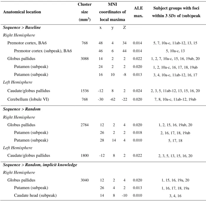
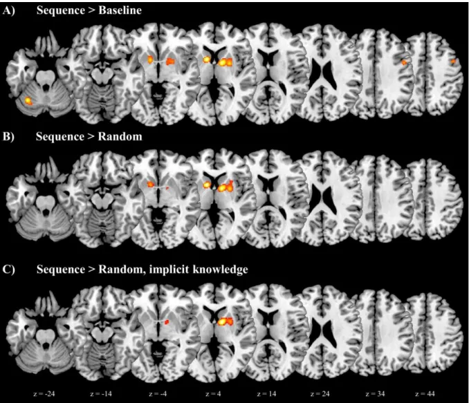
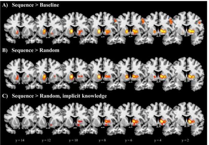
Table 3 shows, for each ALE analysis of sequence learning, the anatomical location, cluster size, MNI coordinates, ALE maximum, and number of contributing subject groups for each peak and subpeak. Figure 2 presents the related ALE maps; Figure 3 focuses on the basal ganglia.
First, we conducted a global ALE analysis that included all contrasts yielding greater activation in sequence than baseline blocks, irrespective of the type of baseline or whether the participants were tested for or acquired explicit knowledge of the sequence (see Methods). In this Sequence > Baseline analysis four areas showed significant convergence across studies:
1) right ventral premotor cortex; 2) the right globus pallidus (apparently across both the internal and external segments, as with other right globus pallidus activation in these analyses), with subpeaks in the (anterior-to-mid) putamen, and activation extending slightly to the (body and head of the) caudate; 3) the left caudate/globus pallidus, including both the head and body of the caudate, with activation extending to the (anterior) putamen and nucleus accumbens (in the ventral striatum); and 4) the left cerebellum (lobule VI). All foci entered into this ALE analysis, as well as those included in the two other analyses that yielded significant ALE results (see just below), were from early stages of learning (within the first day of training); thus the results pertain to early rather than late sequence learning.
The next ALE analysis focused only on those studies with greater activation during sequence blocks than random blocks (i.e., with only random blocks constituting the baseline), again irrespective of whether explicit knowledge was tested for or was acquired (Sequence >
Random). This analysis can reveal brain activation that is related to the acquisition of sequential information, while controlling for visual, motor, and other factors. Significant convergence was observed in the basal ganglia only: 1) the right globus pallidus, with subpeaks in the (anterior-to-mid) putamen, and activation extending to (at least the body of) the caudate nucleus; and 2) the left caudate/globus pallidus, including both the head and body
16
of the caudate, with activation extending to the (anterior-to-mid) putamen and the nucleus accumbens.
We then conducted a third ALE analysis on only those studies with greater activation in sequence than random blocks, but specifically where participants did not show evidence of explicit knowledge of the sequence structure, when tested (Sequence > Random, implicit knowledge). Only the right basal ganglia showed significant convergence across studies, specifically in the right globus pallidus, with subpeaks in the (anterior-to-mid) putamen and the caudate head, and activation extending to the caudate body and the nucleus accumbens.
Table 3. Results from the ALE analyses of sequence learning.
Anatomical location
Cluster size (mm3)
MNI coordinates of
local maxima
ALE max.
Subject groups with foci within 3 SDs of (sub)peak
Sequence > Baseline x y Z
Right Hemisphere
Premotor cortex, BA6 768 48 4 34 0.014 5, 7, 10a-c, 11ab-12, 13, 15 Premotor cortex (subpeak), BA6 46 6 44 0.014 5, 10a-c, 13
Globus pallidus 3088 14 2 2 0.022 1, 2, 7, 10a-c, 15, 16, 19ab, 20 Putamen (subpeak) 24 2 2 0.020 1, 2, 10a-c, 16, 17, 18, 19ab Putamen (subpeak) 16 10 -8 0.013 3, 4, 10a-c, 11ab-12, 16, 17 Left Hemisphere
Caudate/globus pallidus 1536 -12 8 2 0.024 2, 3, 5, 11ab-12, 13, 15, 16, 20 Cerebellum (lobule VI) 768 -30 -62 -22 0.020 7, 8, 10a-c, 11ab-12, 19ab Sequence > Random
Right Hemisphere
Globus pallidus 2784 12 2 4 0.020 1, 2, 15, 16, 19ab, 20
Putamen (subpeak) 26 2 2 0.018 2, 16, 17, 18, 19ab
Putamen (subpeak) 28 14 4 0.010 5, 17, 18
Left Hemisphere
Caudate/globus pallidus 1800 -12 8 2 0.022 2, 3, 5, 13, 15, 16, 20 Sequence > Random, implicit knowledge
Right Hemisphere
Globus pallidus 3040 12 2 4 0.020 1, 15, 16, 19a, 20
Putamen (subpeak) 26 4 2 0.013 1, 16, 17, 18, 19a
Caudate head (subpeak) 14 8 -10 0.010 3, 4, 16
17
Note. Anatomical locations (for peaks and, indented, for subpeaks), cluster size in mm3, and MNI coordinates of local maxima (i.e., of peaks and subpeaks), together with their associated ALE maxima, and their contributing subject groups, that is, the subject groups with foci within 3 standard deviations (SD) of the (sub)peak (paper/contrast numbers correspond to the list of studies presented in Table 2). Note that we report all (sub)peaks, irrespective of how many subject groups contributed to them; however, no (sub)peak had fewer than 3 contributing subject groups. Distinct subject groups are separated by commas; thus, for example, 11ab-12 are combined in the Table without a separating comma, because in this case different contrasts were analyzed on data from the same subject group.
Figure 2. ALE analysis results for the neural correlates of sequence learning (horizontal views). Cluster extents in the basal ganglia are shown in more detail in the coronal images shown in Figure 3. Images are displayed according to neurological convention (the left side of each slice represents the left hemisphere).
18
Figure 3. Neural correlates of sequence learning: clusters in the basal ganglia (coronal views). Images are displayed according to neurological convention (the left side of each slice represents the left hemisphere).
To estimate the observed (a posteriori) power of each peak in each ALE analysis, we first determined their effect sizes (the proportion of contributing subject groups for each ALE peak), and then used those effect sizes to estimate the power (see Figure 7 in Eickhoff et al., 2016). In the Sequence > Baseline analysis, the right premotor peak yielded an effect size of 0.32 (6 contributing subject groups out of 19, see Table 3), resulting in observed power of
~85%; the left cerebellum peak yielded an effect size of 0.26 (5 contributing subject groups out of 19), resulting in observed power of ~80%; and both the left and right basal ganglia peaks yielded effect sizes of 0.42 (8/19 contributing subject groups), resulting in observed power of ~95%. The observed power was similar in the Sequence > Random analysis, with
~85% for the right basal ganglia peak (effect size: 0.37, 6/16 contributing subject groups) and
~95% for the left basal ganglia (effect size: 0.44, 7/16 contributing subject groups). The observed power for the right basal ganglia peak in the Sequence > Random, implicit knowledge analysis was ~85% as well (effect size of 0.5, 5/10 contributing subject groups).
19
Overall, this suggests that all three of our ALE analyses had at least 80% power to detect relatively consistent neural activations. Nevertheless, it is likely that these ALE analyses were not well-suited to detect less consistent neural activation (smaller effects) with similarly high power.
Discussion Interpretation
Sequence-specific learning analysis
We focus first on the results from the key analysis examining sequence-specific learning, that is, when visual, motor, and other confounds were controlled for, in sequence >
random contrasts. In this analysis the only structures that showed consistent activation across studies were in the basal ganglia. (For simplicity, below we refer to consistent activation across studies simply as ‘activation’.) Specifically, (bilateral) activation was found 1) in more anterior than posterior portions of the striatum, in particular in anterior-to-mid portions of both the caudate nucleus and putamen, extending somewhat to the nucleus accumbens; and 2) in the globus pallidus, apparently in both the internal and external segments. Together, these structures thus appear to play key roles in sequence-specific learning, that is, in the learning of sequential information.
The implication of the striatum in sequence learning is broadly consistent with previous models (Hikosaka et al., 2002; Penhune and Steele, 2012), as well as with empirical findings not only in neuroimaging studies (Bischoff-Grethe et al., 2001; Daselaar et al., 2003;
Rauch et al., 1997a; Willingham et al., 2002), but also in lesion studies (Sefcsik et al., 2009;
Vakil et al., 2000). For example, lesions studies of patients with Parkinson’s disease highlight the importance of the striatum, and more broadly of the basal ganglia, in sequence learning (Clark et al., 2014). Overall, previous models and empirical studies suggest that the striatum is involved in learning predictable sequential associations (i.e., the order of the sequential stimuli). This striatal involvement seems to hold across various types of sequences, including not only perceptual-motor sequences, but also purely perceptual sequences (e.g., in learning auditory or visual sequences without any accompanying motor responses) (e.g., Karuza et al., 2013; Turk-Browne et al., 2009; Yang and Li, 2012), and is observed in both language- and music-related sequences (Chan et al., 2013; Ullman et al., in press, 2020).
20
The fact that our analyses yielded activation in anterior-to-mid (but not posterior) portions of the caudate and putamen, as well as in the nucleus accumbens, is also consistent with previous models and findings. In particular, more anterior portions of the striatum (specifically, anterior caudate/putamen and perhaps the ventral striatum) seem to be more important for earlier phases of basal ganglia-based learning of sequential associations, while posterior portions (posterior caudate/putamen) play a larger role in later phases (Doyon et al., 2009; Hikosaka et al., 2002; Penhune and Steele, 2012; Ullman et al., in press, 2020). Since every contrast in our sequence-specific analysis examined early phases of learning (occurring the first day of training; see Methods), our results are consistent with these claims. Note that the reasons for this observed anterior/posterior striatal distinction for earlier/later learning remain unclear, but may be related to the different parallel circuits that pass through the basal ganglia (Alexander et al., 1986; Draganski et al., 2008; Postuma and Dagher, 2006). Anterior striatal circuits may support aspects of motivation (linked to ventral striatal circuitry) as well as early-stage prediction-feedback learning of associations (linked to anterior caudate/putamen); in contrast, posterior portions may underlie aspects of motor and/or visual learning (motor and visual circuits rely on more posterior putamen/caudate) that may take place during the fine-tuning of performance in later stages of acquisition, or perhaps even in the processing of eventually emergent automatized associations (Doyon et al., 2009; Ullman et al., in press, 2020). Thus, consistent with the broader literature, the striatal activation in the present study suggests that more anterior (but not posterior) portions of the caudate nucleus and the putamen, and perhaps the nucleus accumbens, play an important role in early phases of human sequence-specific learning.
Our study also strongly implicates the globus pallidus in sequence-specific learning.
Indeed, this was the only basal ganglia structure present in the main peak activation on the right side, and one of two structures in the peak activation on the left. This clear involvement of the globus pallidus may at first blush seem somewhat surprising, given that previous models and empirical studies of sequence learning have generally focused on the striatum within the basal ganglia, and have largely ignored the globus pallidus (Doyon et al., 2009;
Hikosaka et al., 2002; Penhune and Steele, 2012). However, the observed globus pallidus activation seems less surprising if we consider its strong anatomical and functional connections with the striatum. The globus pallidus has a central anatomical and functional position in the basal ganglia, with both the external and internal segments playing key intermediary roles in projections between the striatum and (the thalamus and) cortex
21
(Alexander et al., 1986; Postuma and Dagher, 2006). In line with such roles, a number of empirical investigations (which indeed were included in our analyses and contributed to the observed globus pallidus activation) have reported globus pallidus involvement during sequence learning, though again without much discussion (e.g., Daselaar et al., 2003; Muller et al., 2002; Purdon et al., 2011; Rauch et al., 1997a; Willingham et al., 2002; Zedkova et al., 2006). Thus, although the particular functional role(s) of the globus pallidus in sequence learning remain to be elucidated, our findings clearly indicate that this structure (apparently including both the internal and external segments) is involved in sequence-specific learning.
Therefore, it seems warranted that future theoretical and empirical investigations extend their basal ganglia focus beyond the striatum to also include the globus pallidus.
Implicit sequence-specific learning analysis
The analysis examining implicit sequence-specific learning included only those studies with a random baseline (sequence > random) that tested for and found no evidence of explicit sequential knowledge. This analysis yielded only (right) basal ganglia activation, in particular in the globus pallidus (apparently including both the internal and external segments), with subpeaks in the anterior-to-mid putamen and caudate head, with activation extending somewhat to the caudate body and the nucleus accumbens.
Thus, these structures appear to play key roles in implicit sequence-specific learning in humans. This result is difficult to compare with some important previous models, since those discuss implicit sequence learning in the context of later stages of automatization (regarding fine-tuning of performance after extended practice), even when earlier stages may have involved explicit instruction and consciously accessible sequence knowledge, for example as tested in finger sequence tapping tasks in humans or similar tasks in non-human animals (Hikosaka et al., 2002; Penhune and Steele, 2012). In contrast, our findings reveal the neural substrates of early stages of learning in implicit learning contexts, that is, when learning occurs without apparent conscious access to what was learned or that learning occurred (Cleeremans et al., 1998; Reber, 1996). This type of implicit learning is generally examined in humans in tasks in which no explicit instruction is given prior to learning and no relevant explicit knowledge seems to be acquired after learning (Janacsek and Nemeth, 2012; Reber, 2013) – as in the case of sequence learning in the SRT tasks included in this analysis. Thus, the present study suggests that the (right) basal ganglia, in particular more anterior portions of the caudate and putamen, and perhaps the nucleus accumbens, as well as (both segments of)
22
the globus pallidus, play key roles in such early-stage implicit sequence-specific learning. The roles of at least these striatal structures in implicit sequence-specific learning may be related to early-stage prediction-feedback learning of associations, as well as aspects of motivation (see above).
Note however that the results of this analysis should be interpreted with caution, for the following reasons. First, although the inclusion of only ten subject groups in the analysis appeared to be sufficient to detect the observed ALE result, due to its relatively consistent activation (yielding a large effect size, of 0.5) with high power (~85%), the analysis was underpowered to detect effect sizes under 0.45 (Eickhoff et al., 2016). Consequently, while our analysis clearly shows that the basal ganglia are indeed involved in the implicit acquisition of sequences, there may be other structures (with smaller effect sizes) that are also involved, which the analysis with ten subject groups could not detect. Second, since no direct comparison between implicit and explicit sequence learning conditions could be performed (due to too few subject groups in the latter condition), we cannot conclude that the basal ganglia are only involved in implicit (and not in explicit) learning conditions. Intriguingly, some evidence (including from studies not meeting the inclusion criteria in this paper) suggests that sequence learning conditions that involve explicit knowledge also show basal ganglia activation. In particular, evidence suggests that although other structures and neurocognitive mechanisms (e.g., including the cerebellum; see below) seem to underlie the acquisition (and processing) of explicit knowledge, the basal ganglia simultaneously acquire the same or similar knowledge implicitly (Schendan et al., 2003; Ullman et al., in press, 2020;
Willingham et al., 2002). In other words, the basal ganglia seem to be involved in acquiring sequence knowledge both when explicit knowledge is acquired and when it is not, though in both cases they seem to underlie implicit learning, which occurs even while explicit learning may occur at the same time. Finally, note that it is possible that the basal ganglia are in fact involved bilaterally in implicit sequence learning, and the lack of left basal ganglia activation in this analysis (as compared to sequence learning more generally; see above) is due to a lack of power. Further research seems warranted to examine these issues.
Global analysis of sequence learning
The global analysis of sequence learning, which was our least restrictive analysis examining the neural correlates of this function, did not control for visual, motor, attention, or other potential confounds. This analysis yielded not only the pattern of basal ganglia
23
activation obtained in the more restrictive analyses, but also activation in (right) ventral premotor cortex and the (left) cerebellum (lobule VI), which were not observed in the analyses examining sequence-specific learning. This, together with their proposed roles in previous research, suggests that these structures are involved not in sequence learning itself (i.e., the acquisition of sequential order), but rather in other functions that support sequence learning and were not controlled for in at least some tasks probing sequence learning in the global analysis.
Premotor activation: The ventral premotor activation may be best explained by aspects of sensorimotor integration. Ventral premotor cortex has been independently implicated in this function, that is, in studies not examining sequence learning (Avenanti et al., 2012; Hoshi and Tanji, 2004; Kakei et al., 2001). Unlike in our sequence-specific analyses, sensorimotor integration was likely not well-controlled for in the global analysis, given the different baselines in contrasts included in this analysis, such as index finger tapping and known patterns: These baselines likely involve different visual and motor processing demands compared to the sequence blocks; for example, responding to only one stimulus with a single finger in the baseline of index finger tapping, or performing a known pattern, seem to be less demanding than responding to multiple visual stimuli with the corresponding response buttons in the sequence blocks. Indeed, sequence-learning studies finding ventral premotor activation employed such baselines, and attributed this activation to aspects of sensorimotor integration, such as transitioning a movement from a spatial representation to a motoric representation, rather than to sequence-specific learning (Muller et al., 2004; Muller et al., 2002). Note that a recent neuroanatomical meta-analysis of motor learning suggested dorsal premotor cortex as a “motor learning core” (Hardwick et al., 2013; see below), which can be distinguished from the sensorimotor functions that seem to depend on ventral premotor cortex. Thus, the results both from our study and from previous research emphasize roles for premotor cortex in functions other than sequence-specific learning. In particular, premotor cortex appears to underlie various motor-related functions, perhaps including aspects of motor learning itself (dorsal premotor cortex) as well as sensorimotor integration (ventral premotor cortex). However, since we did not directly compare neural activation associated with different baselines, further studies are needed to provide direct evidence for this interpretation.
Cerebellar activation: Based on previous research, we speculate that the observed cerebellar lobule VI activation may reflect the involvement of executive, working memory, or
24
other attention-related functions in sequence learning. Independent evidence suggests that lobule VI subserves just such cognitive functions, rather than motor functions (Bernard and Seidler, 2013; Stoodley and Schmahmann, 2016). Indeed, these cognitive functions were likely not adequately controlled for in at least some studies included in the global analysis, given the variety of baselines employed. Such functions may be particularly associated with explicit sequential knowledge. Explicit knowledge is closely linked to working memory and attention (Cowan, 1999; Ullman, 2004, 2016), and thus any explicit knowledge of the sequences could have contributed to lobule VI activation via the engagement of these functions. Consistent with this view, all five subject groups that contributed to lobule VI activation in our global analysis either found evidence for explicit sequential knowledge (Muller et al., 2003; 2002; Willingham et al., 2002) or did not test for it (Jouen et al., 2013;
Kumari et al., 2002), and thus such knowledge could have been present.
Further supporting the implication of lobule VI in explicit sequential knowledge, as well as in executive functions (particularly working memory) more broadly, a recent neuroanatomical meta-analysis of sequence learning focusing on the cerebellum reported (right and left) lobule VI activation, which was attributed (respectively) to explicit sequence learning and spatial working memory (Bernard and Seidler, 2013). The implication of spatial working memory is specifically consistent with sequence learning in the SRT task, which involves spatially-dependent sequences. Any aspects of the sequence that are noticed and maintained during this task would be expected to engage spatial working memory (Janacsek and Nemeth, 2013, 2015). Thus, lobule VI activation might be expected even in sequence- specific contrasts (i.e., with random blocks as the baseline), as long as sequential knowledge is noticed and maintained in working memory during the sequence blocks. On this view, such activation would not implicate sequence learning itself, but rather working memory and attention-related functions underlying explicit sequential knowledge. As indicated above, due to the small number of studies reporting explicit sequence knowledge, we were not able to directly contrast implicit and explicit sequence learning, and thus could not directly test this possibility. However, this seems worthwhile investigating in future studies.
Thus overall, our findings here, together with evidence from previous research, suggest that cerebellar lobule VI activation in sequence learning may reflect working memory or other attention-related functions, perhaps in particular related to explicit knowledge of the sequences, rather than motor or sensorimotor related functions or the acquisition of the sequential order itself.
25 Previous neuroanatomical meta-analyses
As mentioned in the Introduction, we are aware of two previous whole-brain neuroanatomical meta-analyses (both using ALE) that examined motor learning (Hardwick et al., 2013; Lohse et al., 2014). Lohse et al. (2014) investigated how short, medium, and long time-scales of practice affect brain activity associated with motor skill learning in general.
They included a large variety of motor learning tasks, and did not focus specifically on sequence learning. Hardwick et al. (2013) examined the learning of novel movement kinematics and dynamics (i.e., in motor adaptation tasks; Doyon et al., 2009) as well as the learning of various aspects of sequential motor behaviors. Three levels of analysis reported on the latter are of greatest relevance here. First, in a global analysis that included a wide range of sequence learning tasks, they found activation in sensorimotor cortex, primary motor cortex, dorsal premotor cortex, the superior parietal lobule, the thalamus, and the cerebellum in lobule VI. Basal ganglia activation was not observed. This analysis included not only classic SRT tasks (the focus of the present study), but also other SRT variants and apparently finger tapping tasks. Studies examining both earlier and later stages of learning as well as those testing for increases over the time course of learning were included. There were no baseline restrictions; e.g., some studies had a baseline of rest. They also performed two subanalyses of particular interest here. First, they ran a subanalysis examining “movement controlled” sequence learning, which yielded activation in dorsal premotor cortex, the thalamus, and the cerebellum in lobule VI, but again not in the basal ganglia. In a second subanalysis they probed implicit (vs. explicit) sequence learning. This analysis yielded activation in the (left) caudate head/body, which extended to the thalamus (see text on page 286 and Figure 5 in Hardwick et al.).
Comparison between these subanalyses and the somewhat analogous sequence- specific and implicit sequence-specific learning analyses reported here is difficult. It was unclear exactly which studies were included in the two subanalyses in Hardwick et al., and what their specific inclusion/exclusion criteria were. Moreover, it appears that their first and second subanalyses were not limited to studies with random baselines, and that their second subanalysis was not limited to studies without (or with) explicit knowledge. Indeed, of all the sequence learning studies included in Hardwick et al., only six were included in our sequence- specific (sequence > random) analysis (in contrast, they included 24 in their first subanalysis).
Similarly, of all the studies included in Hardwick et al., only two were included in our implicit