This appendix has been provided by the authors to give readers additional information about their work.
Supplement to: Sands BE, Peyrin-Biroulet L, Loftus EV Jr, et al. Vedolizumab versus adalimumab for moderate- to-severe ulcerative colitis. N Engl J Med 2019;381:1215-26. DOI: 10.1056/NEJMoa1905725
(PDF updated on November 27, 2019.)
Online Supplement Table of Contents
List of Study Investigators and Sites (VARSITY Study Group) List of Highest Recruiting Sites
VARSITY Steering Committee Data Safety Monitoring Board
Progressive Multifocal Leukoencephalopathy (PML) Adjudication Committee Concomitant Corticosteroid Discontinuation/Tapering
PML Monitoring
Monitoring and Auditing of Study Procedures and Data Quality Patient Eligibility Criteria
Prespecified Additional Outcomes in Protocol
Prespecified Exploratory Outcomes in Statistical Analysis Plan Statistical Analysis
Figure S1 Patient Disposition.
Figure S2 Clinical Remission at Week 52 in Subgroups by Patient Demographic and Baseline Characteristics.
Figure S3 Clinical Remission and Endoscopic Improvement at Week 52 by Baseline Concomitant Medication Use.
Figure S4 Clinical Remission at Week 14 in the Overall population and Subgroups by Prior TNF Inhibitor Therapy.
Figure S5 Endoscopic Improvement at Week 52 by Patient Demographic and Baseline Characteristics.
Figure S6 Corticosteroid-free Clinical Remission at Week 52 by Patient Demographic and Baseline Characteristics.
Figure S7 Oral Corticosteroid Use.
Figure S8 Histological Remission at Week 52 in Subgroups by Prior TNF inhibitor Therapy.
Figure S9 Histological Remission at Week 14.
Figure S10 Minimal Histologic Disease Activity at Week 52.
Figure S11 Clinical Response based on Total Score on the Mayo Scale at Week 14 in the Overall Population and Subgroups by Prior TNF Inhibitor Therapy.
Table S1 Schedule of Trial Procedures.
Table S2 Baseline Disease Characteristics.
Table S3 Post Hoc Analysis of Clinical Remission and Endoscopic Improvement at Week 52, Using the Weighted Cochran–Mantel–Haenszel Method.
Table S4 Sensitivity Analysis to Assess the Impact of Dropouts Under Different Missing Data Mechanisms for Primary and Secondary Outcomes.
Table S5 Results from Prespecified Analyses of Efficacy Outcomes.
Table S6 Most Frequent Adverse Events.
Table S7 Serious Adverse Events.
List of Trial Investigators and Sites *
Argentina Dr Sebastian Calvo; Dr Edgardo Gimenez; Dr Jorge Hector Resk†; Dr Emiliano Pablo Tron
Australia Dr Mohammed Al-Ansari†; Dr Jane Andrews; Dr Peter Bampton†; Dr Henry Debinski†; Dr Peter Hendy; Dr Gerald Holtmann†; Dr Rupert Leong; Dr Gregory Moore†
Belgium Dr Pieter Hindryckx Bosnia and
Herzegovina
Dr Tatjana Barac
Bulgaria Dr Raina Draganova; Dr Iskren Kotzev; Dr Ivanka Marinova†; Dr Mario Markov†; Dr Yordan Mihaylov†; Dr Dimitar Pavlov; Dr Mariana
Penkova†; Dr Asen Petrov; Dr Zoia Spassova; Dr Daniela Stoyanova; Dr Emilian Velev†; Dr Borislav Vladimirov; Dr Feliks Yanev†
Canada Dr Jeffrey Axler; Dr Richard Fedorak‡; Dr Sharyle Fowler; Dr Smita Halder†; Dr Vipul Jairath†; Dr Terry Ponich; Dr Karen Wong
Colombia Dr Edwin Baez†; Dr Juan Ricardo Marquez Velasquez
Croatia Dr Marko Banic; Dr Ivan Bogadi†; Dr Vladimir Borzan; Dr Jadranka Cetina Zgrablic; Dr Marko Duvnjak; Dr Maja Gusej†; Dr Zeljko Krznaric Czech Republic Dr Ivana Cechova†; Dr Tomas Hala; Dr Lubos Janu; Dr Pavel Kohout;
Dr Milan Lukas; Dr Martina Machkova; Dr Jan Matous; Dr Jan Ulbrych†; Dr Tomas Vanasek
Denmark Dr Lone Klinge; Dr Terje Rannem†; Dr Klaus Theede Estonia Dr Julia Borissova†; Dr Karin Kull†
France Dr Morgane Amil; Dr Yoram Bouhnik†; Dr Cyrielle Gilletta; Dr Mathurin Fumery; Dr Jean-Charles Grimaud; Dr Xavier Hebuterne; Dr David Laharie†; Dr Jacques Moreau; Dr Laurent Peyrin-Biroulet; Dr Xavier Roblin†
Germany Dr Daniel Baumgart†; Dr Carsten Buening; Dr Johanna Dinter†; Dr Annika Gauss†; Dr Tanja Kuehbacher; Dr Khan Fareed Rahman†; Dr
Ingolf Schiefke; Dr Stefan Schreiber; Dr Ulrike Von Arnim†; Dr Stefan Zeuzem†
Hong Kong Dr Wai Keung Leung; Dr Michael Kin Kong Li
Hungary Dr Istvan Altorjay; Dr Laszlo Bene†‡; Dr Peter Lakatos†; Dr Tamas Molnar; Dr Agnes Salamon; Dr Robert Schnabel; Dr Zsolt Tulassay Israel Dr Yona Avni; Dr Olga Barkay; Dr Sigal Fishman; Dr Eran Goldin; Dr
Dan Keret†; Dr Adi Lahat-Zok; Dr Ehud Melzer; Dr Timna Naftali; Dr Assy Nimer; Dr Baruch Ovadia; Dr Eran Zittan
Italy Dr Angelo Andriulli; Dr Maria Cappello†; Dr Fabiana Castiglione†; Dr Silvio Danese; Dr Walter Fries†; Dr Paolo Gionchetti; Dr Anna Kohn; Dr Giovanni Maconi; Dr Marchi Santino; Dr Edoardo Savarino; Dr
Antonino Privitera†; Dr Marco Romano†; Dr Maurizio Vecchi†; Dr Erica Villa
Korea, Republic of
Dr Jae Hee Cheon†; Dr Dong Soo Han†; Dr Byung-ik Jang; Yoon-Tae Jeen†; Dr Joo Sung Kim; Dr Sung Kook Kim; Dr Hyo-Jong Kim; Dr YoungHo Kim; Dr Kang-Moon Lee; Dr Bo In Lee; Dr Dong il Park; Dr Young Soo Park; Dr Geun Am Song; Dr Byong-Duk Ye
Latvia Dr Juris Pokrotnieks; Dr Ivars Tolmanis
Lithuania Dr Goda Denapiene; Dr Arunas Kancauskas; Dr Violeta Jakovlevaite; Dr Laimas Jonaitis
Mexico Dr Aracely Cruz Palacios; Dr Emmanuel Larriva de los Reyes; Dr Leslye Asela Lujano Nicolas; Dr Eduardo Mendoza Fuerte
Netherlands Dr Geert D’Haens; Marieke Pierik†; Janneke van der Woude†
Poland Dr Krystian Adrych; Dr Wit Cezary Danilkiewicz; Dr Beata Gawdis- Wojnarska; Dr Marek Hartleb; Dr Marek Karczewski; Dr Jaroslaw Kierkus; Dr Ewa Malecka-Panas; Dr Robert Petryka; Dr Wojciech Piotrowski; Dr Jaroslaw Regula; Dr Jacek Romatowski; Dr Jerzy Rozciecha; Dr Grazyna Rydzewska; Dr Patryk Smolinski; Dr Piotr Walczak; Dr Michal Walczak; Dr Barbara Wozniak-Stolarska
Portugal Dr Jose Cotter; Dr Luis Lopes; Dr Francisco Portela; Dr João Rodrigues Carvalho
Romania Dr Liliana Simona Gheorghe; Dr Adrian-Eugen Goldis; Dr Radu-Bogdan Mateescu
Russian Federation
Dr Rustem Abdulkhakov; Dr Alina Agafyina; Dr Olga Alexeeva; Dr Bagdadi Alikhanov†; Dr Elena Bunkova; Dr Mikhail Dvorkin; Dr Irina Gubonina; Dr Vladimir Kashnikov; Dr Maria Livzan; Dr Marina Osipenko; Dr Asfold Parfenov†; Dr Anatoly Pershko; Dr Marina Pesegova; Dr Vladimir Rafalskiy†; Dr Olga Reshetko; Dr Vladimir Simanenkov; Dr Svetlana Tsybulko; Dr Ekaterina Valuyskikh; Dr Alexey Yakovlev
Serbia Dr Mirjana Cvetkovic; Dr Dragomir Damjanov; Dr Srdjan Djuranovic;
Dr Predrag Dugalic; Dr Aleksandar Nagorni; Dr Petar Svorcan; Dr Natasa Zdravkovic Petrovic
Slovakia Dr Ivan Bunganic; Dr Milos Gregus; Dr Tibor Hlavaty; Dr Frantisek Horvath
Spain Dr Eugeni Domenech Morral†; Dr Rodrigo Jover Martinez†; Dr Maria Isabel Vera Mendoza
Taiwan Dr Ming-Jium Shieh; Dr Cheng-Chung Wei
Turkey Dr Engin Altintas; Dr Ozlen Atug; Dr Can Gonen; Dr Hulya Hamzaoglu;
Dr Saadettin Hulagu; Dr Murat Toruner
Ukraine Dr Ivan Chopey; Dr Svitlana Danyliuk; Dr Oleksii Datsenko; Dr Nataliia Golovchenko; Dr Oleksandr Golovchenko†; Dr Ihor Hospodarskyy; Dr Valeriy Ivanov; Dr Volodymyr Klymenko; Dr Olena Levchenko; Dr Viktor Lizogub†; Dr Yuriy Mostovoy; Dr Oleksandr Oliinyk; Dr Igor Polianskyi†; Dr Volodymyr Pyrohovskyi; Dr Sergii Shevchuk; Dr Grygorii Ursol; Dr Valeriy Vdovychenko; Dr Vira Vyshyvanyuk
United Kingdom Dr Ian Beales†; Dr Matthew Brookes†; Dr Chukwaka Nwokolo; Dr Jack Winter
United States Dr Avanish Aggarwal†, Dr Humberto Aguilar†; Dr Zaid Alnoah†; Dr Esperanza Arce-Nunez†; Dr Calin Arimie; Dr James Arterburn†; Dr
Curtis Baum; Dr Emanuelle Bellaguarda†; Dr Bonnie Bock†; Dr Tasha Boone; Dr Norman Callahan†; Dr Jonathon Chapman†; Dr Stuart Chen†; Dr Michael Chiorean; Dr Allan Coates; Dr Michael Connor†; Dr Steven Dellon; Dr Gerald Dryden; Dr George Du Vall; Dr Lucky Flores; Dr Ronald Fogel; Dr Juan Frias†; Dr Philip Ginsburg†; Dr Eugene Greenberg; Dr David Grunkemeier†; Dr Paul Hellstern; Dr Hans Herfarth; Dr Brenda Hoffman; Dr Sara Horst†; Dr Samuel Idarraga; Dr Heba Iskandar†; Dr Rajesh Jain†; Dr Erin Jenkins†; Dr Barry Kaufman†; Dr Abdul Khaleq†; Dr Ahtaram Khan†; Dr Sunil Khurana; Dr Jason Lake; Dr James Leavitt†; Dr Bernard Leman; Dr Derek Lewis†; Dr Daniel Lindenberg†; Dr Edward V Loftus Jr†; Dr Louis Korman; Dr Jennifer Martin†; Dr Matthew McCullough; Dr Alfred McNair†; Dr Nilesh Mehta†; Dr Ece Mutlu†; Dr Vijay Narayen†; Dr Jorge Paoli- Bruno; Dr Nolan Perez†; Dr Raymond Phillips†; Dr Isaac Raijman; Dr Ruben Ramirez-Vega†; Dr Charles Randall; Dr Scott Rinesmith; Dr Timothy Ritter; Dr Alan Safdi†; Dr Marc Saltzman†; Dr Bruce Sands†; Dr Jennifer Seminerio; Dr Michael Schulman†; Dr Shahriar Sedghi†; Dr Ira Shafran†; Dr Murali Shankar†; Dr David Silvers; Dr Nehad Soloman†; Dr Harvey Tatum†; Dr Robert Tepper†; Dr Felix Tiongco; Dr Martin Valdes; Dr John Weber†; Dr Chen Zhang†
*Only investigators consenting to be acknowledged are listed.
†Investigators who screened but did not enroll randomized patients in the trial.
‡Investigators who are deceased.
List of Highest Recruiting Sites Sites with >3% total enrollment Investigator name
City/State or
Province Country
Adalimumab SC (N = 386), n
Vedolizumab IV (N = 385), n
Dr Jeffrey Axler Vaughan/Ontario Canada 8 13
Dr Jaroslaw Kierkus Warszawa Poland 12 10
Dr Beata Gawdis-
Wojnarska Szczecin Poland 7 14
Note: Only sites with greater than 3% of total enrollment in any treatment group are included.
VARSITY Steering Committee
Bruce E. Sands, MD (Chair), Stefan Schreiber, MD, Laurent Peyrin-Biroulet,MD, PhD, Edward V. Loftus Jr., MD.
Data Safety Monitoring Board
Keith Lindor, MD (Chair), Henry Bodenheimer Jr., MD.
Progressive Multifocal Leukoencephalopathy (PML) Adjudication Committee
David B. Clifford, MD (Chair),Eugene O. Major, PhD,Michael H. Lev, MD,Joseph R. Berger, MD.
Concomitant Corticosteroid Discontinuation/Tapering
Patients who were receiving oral corticosteroids and had a clinical response at Week 6 as
assessed by the Investigator began a non-fixed corticosteroid tapering regimen. Patients who did not achieve clinical response at Week 6 initiated tapering on a subsequent study visit as soon as a clinical response was achieved. For patients who could not tolerate the corticosteroid taper without recurrence of clinical symptoms, corticosteroids could be increased up to the baseline corticosteroid dose one time before tapering was restarted. In such cases, the tapering regimen above was reinitiated within 2 weeks. Patients who consistently could not be tapered were withdrawn from the trial.
PML Monitoring
All patients were closely monitored for signs and symptoms of PML prior to administration of each dose of trial drug using a PML subjective symptom checklist, which assessed for any recent changes in vision, speech, gait, sensation, comprehension, coordination, and personality. Any positive response on the PML checklist prompted further objective evaluation based on a prespecified diagnostic algorithm (the PML Case Evaluation Algorithm). Cases of new neurological symptoms were promptly evaluated by an independent adjudication committee (IAC) of academic experts (including a neurologist, a neuroradiologist, and a virologist) using a prespecified diagnostic algorithm that included stepwise contrast-enhanced brain magnetic resonance imaging and, if indicated, lumbar puncture with polymerase chain reaction analysis of cerebrospinal fluid for JC virus DNA. If patients entered the algorithm, study drug was withheld until PML could be definitively excluded.
Monitoring and Auditing of Study Procedures and Data Quality
At regular intervals, routine on-site monitoring visits were performed by clinical research
associates (CRAs) at all sites on the basis of a prespecified written monitoring plan, and 100% of data were source verified during these visits. In addition, remote CRA and medical monitoring of the study’s electronic database, which included all source documents (case report forms and laboratory results), was performed for each patient throughout the study according to the monitoring plan. In addition to monitoring visits, clinical quality assurance audits were conducted by CRAs.
An independent data safety monitoring board (DSMB) provided oversight of the phase 3 vedolizumab program and met at 6-month intervals. The DSMB was composed of 2
gastroenterologists and a statistician. Before scheduled meetings, the DSMB received unblinded tables and listings of all clinical safety data. The DSMB reviewed these data in closed sessions and subsequently provided a recommendation to the sponsor. In addition, the DSMB received monthly listings of all SAEs that included the most current individual reports of the Council for International Organizations of Medical Sciences (CIOMS) form.
Patient Eligibility Criteria Inclusion Criteria
1. In the opinion of the investigator, the patient is capable of understanding and complying with protocol requirements.
2. The patient or, when applicable, the patient’s legally acceptable representative signs and dates a written, informed consent form and any required privacy authorization prior to the initiation of any study procedures.
3. The patient has a diagnosis of UC established at least 3 months prior to Screening by clinical and endoscopic evidence and corroborated by a histopathology report.
4. The patient is male or female and aged 18 to 85 years, inclusive.
5. The patient has moderately to severely active UC as determined by a total score on the Mayo scale of 6 to 12 with an endoscopic subscore ≥2 within 14 days prior to the randomization.
6. The patient has evidence of UC proximal to the rectum (≥15 cm of involved colon).
7. The patient with extensive colitis (up to the hepatic flexure) or pancolitis of >8 years duration or left-sided colitis of >12 years duration must have documented evidence that a surveillance colonoscopy was performed within 12 months of the initial screening visit (may be performed during the Screening Period).
8. The patient with a family history of colorectal cancer, personal history of increased colorectal cancer risk, age >50 years, or other known risk factor must be up-to-date on colorectal cancer surveillance (may be performed during Screening).
9. The patient:
a) Has had previous treatment with TNF inhibitor without documented clinical response to treatment (e.g., due to lack of response [primary non-responders], loss of response, or intolerance [secondary non-responders]), or
b) Has previously used a TNF inhibitor (except adalimumab), and discontinued its use due to reasons other than safety, or
c) Is naïve to TNF inhibitor but is failing current treatment (e.g., corticosteroids, 5-ASA, or immunomodulators).
10. A male patient who is non-sterilized and sexually active with a female partner of
childbearing potential agrees to use adequate contraception from signing of informed consent throughout the duration of the study and for 5 months after the last dose.
11. A female patient of childbearing potential who is sexually active with a non-sterilized male partner agrees to use routinely adequate contraception from signing of informed consent throughout the duration of the study and for 5 months after the last dose.
Exclusion Criteria
Gastrointestinal Exclusion Criteria
1. The patient has clinical evidence of abdominal abscess or toxic megacolon at the Screening Visit.
2. The patient has had an extensive colonic resection, subtotal or total colectomy.
3. The patient has had ileostomy, colostomy, or known fixed symptomatic stenosis of the intestine.
4. The patient has a diagnosis of Crohn’s colitis or indeterminate colitis, ischemic colitis, radiation colitis, diverticular disease associated with colitis, or microscopic colitis.
5. The patient has received any of the following for the treatment of underlying disease within 30 days of randomization:
a) Non-biologic therapies (e.g., cyclosporine, tacrolimus, thalidomide) other than those specifically listed in Section Permitted Medications for Treatment of UC.
b) An approved non-biologic therapy in an investigational protocol.
6. The patient has received any investigational or approved biologic or biosimilar agent (other than those listed in Exclusion Criterion #7) within 60 days or 5 half-lives prior to Screening (whichever is longer).
7. The patient has previously received natalizumab, efalizumab, adalimumab, etrolizumab, AMG-181, anti–mucosal addressin cell adhesion molecule-1 antibodies, or rituximab.
8. The patient has previously received vedolizumab.
9. The patient currently requires or is anticipated to require surgical intervention for UC during the study.
10. The patient has history or evidence of adenomatous colonic polyps that have not been removed, or colonic mucosal dysplasia.
Infectious Disease Exclusion Criteria
11. The patient has evidence of an active infection during the Screening Period.
12. The patient has evidence of, or treatment for, C. difficile infection or other intestinal pathogen within 28 days prior to the first dose of trial drug.
13. The patient has chronic hepatitis B virus (HBV) infection* or chronic hepatitis C virus (HCV) infection.
* HBV immune patients (i.e., being hepatitis B surface antigen [HBsAg] negative and hepatitis B antibody [HBsAb] positive) may, however, be included.
14. The patient has active or latent TB as evidenced by the following:
a) A diagnostic TB test performed within 30 days of Screening or during the Screening Period that is positive, defined as:
– Positive QuantiFERON test or 2 successive indeterminate QuantiFERON tests, OR – A TB skin test reaction ≥5 mm.
NOTE: If patients have received BCG vaccine then a QuantiFERON TB Gold test should be performed instead of the TB skin test.
OR
b) A chest X-ray within 3 months of Day 1 that is suspicious for pulmonary TB, and a positive or 2 successive indeterminate QuantiFERON tests (or, a positive T-SPOT TB test [Japan only]) within 30 days prior to Screening or during the Screening Period.
Note: Patients with documented previously treated TB with a negative QuantiFERON test can be included in the study.
15. The patient has any identified congenital or acquired immunodeficiency (e.g., common variable immunodeficiency, human immunodeficiency virus (HIV) infection, organ
transplantation).
16. The patient has any live vaccination within 30 days prior to Screening or is planning to receive live vaccination during participation in the study.
17. The patient has a clinically significant infection (e.g., pneumonia, pyelonephritis) within 30 days prior to Screening, or ongoing chronic infection.
18. The patient has used a topical (rectal) treatment with (5-ASA) or corticosteroid enemas/suppositories within 2 weeks of the administration of the first dose of trial drug.
General Exclusion Criteria
19. The patient has a history of hypersensitivity or allergies to vedolizumab or adalimumab.
20. The patient has any unstable or uncontrolled cardiovascular disorder, heart failure moderate to severe (New York Class Association III or IV), any pulmonary, hepatic, renal, GI,
genitourinary, hematological, coagulation, immunological, endocrine/metabolic, or othermedical disorder that, in the opinion of the investigator, would confound the study results or compromise patient safety.
21. The patient has history of lupus or lupus-related conditions.
22. The patient has had a surgical procedure requiring general anesthesia within 30 days prior to
23. The patient has a history of malignancy, except for the following: adequately-treated non- metastatic basal cell skin cancer; squamous cell skin cancer that has been adequately treated and that has not recurred for at least 1 year prior to Screening; and history of cervical carcinoma in situ that has been adequately treated and that has not recurred for at least 3 years prior to Screening. Patient with remote history of malignancy (e.g., >10 years since completion of curative therapy without recurrence) will be considered based on the nature of the malignancy and the therapy received and must be discussed with the sponsor on a case-by-case basis prior to Screening.
24. The patient has a history of any major neurological disorders, including stroke, multiple sclerosis, brain tumor, demyelinating, or neurodegenerative disease.
25. The patient has a positive PML subjective symptom checklist at Screening or prior to the administration of the first dose of trial drug at Day 1.
26. The patient has any of the following laboratory abnormalities during the Screening Period:
Hemoglobin <8 g/dL.
White blood cells (WBC) <3 × 10⁹/L.
Lymphocyte <0.5 × 10⁹/L.
Platelet count <50 × 109/L or >1200 ×109/L.
Alanine aminotransferase (ALT) or aspartate aminotransferase (AST) >3 × upper limit of normal (ULN).
Alkaline phosphatase >3 × ULN.
Serum creatinine >2 × ULN.
27. The patient has a history of drug abuse (defined as any illicit drug use) or a history of alcohol abuse within 1 year prior to the Screening Visit.
28. The patient has an active psychiatric problem that, in the investigator’s opinion, may interfere with compliance with study procedures.
29. The patient is unable to attend all the study visits or comply with study procedures.
30. The patient is required to take excluded medications listed in Section 7.3.
31. If female, the patient is pregnant or lactating or intending to become pregnant before, during, or within 5 months after participating in this study; or intending to donate ova during such time period.
32. If male, the patient intends to donate sperm during the course of this study or for 5 months thereafter.
33. The patient is an immediate family member, study site employee, or is in a dependent relationship with a study site employee who is involved in conduct of this study (e.g., spouse, parent, child, sibling) or may consent under duress.
Prespecified Additional Outcomes in Protocol
Clinical response (defined as a reduction in total score on the Mayo scale of ≥3 points and ≥30% from baseline [or a partial score on the Mayo scale of ≥2 points and ≥25%
from baseline, if the total score on the Mayo scale was not performed at the visit] with an accompanying decrease in rectal bleeding subscore of ≥1 point or absolute rectal bleeding subscore of ≤1 point) at Week 52
Clinical remission (defined as a total score on the Mayo scale of ≤2 points and no individual subscore >1 point) at Week 14
Rectal bleeding subscore indicative of mild disease (≤1) at Week 52
Physician’s Global Assessment (PGA) subscore indicative of mild disease (≤1) at Week 52
Stool frequency subscore indicative of mild disease (≤1) at Week 52
Total score on the Mayo scale of ≤2 points and no individual subscore >1 point where rectal bleeding subscore of 0 and endoscopy subscore of 0 at Week 52
Endoscopy subscore of 0, rectal bleeding subscore of 0, and stool frequency subscore decreases or no change from Baseline at Week 52
Endoscopy subscore ≤1, rectal bleeding subscore of 0, and stool frequency subscore of 0 at Week 52
Endoscopy subscore ≤1, rectal bleeding subscore of 0, and stool frequency subscore ≤1 at Week 52
Endoscopy subscore ≤1, rectal bleeding subscore of 0, stool frequency subscore decreases or no change from Baseline, and total score (sum of these 3) ≤1 at Week 52
Inflammatory Bowel Disease Questionnaire (IBDQ) score change of ≥16 points from Baseline to Week 52
Clinical remission based on IBDQ score >170 at Week 52
Change in oral corticosteroid use from Baseline to Week 52
Discontinuation of corticosteroids as well as clinical remission at Week 14 among patients using corticosteroids at baseline
Time to major UC-related events (e.g., hospitalizations, colectomies, and procedures)
Change in fecal calprotectin concentrations from Baseline to Weeks 14, 30, and 52
Change in histology from Baseline to Week 52
Histological remission at Week 14
Histological remission at Week 52
Observed serum concentration at the end of a dosing interval (Ctrough) of vedolizumab
Positive anti-vedolizumab antibodies (AVA) during the study
Positive neutralizing AVA
Safety for maintenance therapy as assessed by AEs, adverse events of special interest (AESIs, including serious infections including opportunistic infection such as PML, liver injury, malignancies, infusion-related or injection site reactions or systemic reactions and hypersensitivity), serious adverse events (SAEs), vital signs, results of standard
laboratory tests (clinical chemistry, hematology, coagulation, urinalysis), and results of 12 lead electrocardiograms (ECGs)
Prespecified Exploratory Outcomes in Statistical Analysis Plan
Clinical response at Week 14
Total score on the Mayo scale of ≤1 and rectal bleeding subscore = 0 at Week 52
Durable clinical remission, defined as clinical remission at Week 52 amongst those in clinical remission at Week 14 (Note: the denominator will be the patients in the full- analysis set)
Clinical remission at Week 52 and in clinical remission for ≥14 weeks leading up to Week 52. Clinical remission is defined by total score on the Mayo scale, or partial score on the Mayo scale if the total score on the Mayo scale was not performed at the visit (Note the denominator will be the patients in the full-analysis set)
Disease control at Week 52, defined as total score on the Mayo scale of ≤2, rectal bleeding subscore = 0, endoscopy subscore = 0, C-reactive protein <5 mg/L, fecal
calprotectin (FCP) <100 ug/g and in histological remission (either by Geboes Score or by Robarts Histopathology Index)
Rectal bleeding subscore = 0 at Week 52
Major UC-related events (e.g., hospitalizations, bowel resection, and procedures) throughout the study up to Week 52
FCP ≤250 ug/g at Week 14, 30, 52 (among those with FCP >250 ug/g at baseline). (Note the denominator will be a subset of patients with FCP >250 ug/g at baseline in the full- analysis set)
Treatment persistence with adalimumab or vedolizumab at Week 68
Change from baseline in IBDQ-specific domain at Week 30 and Week 52, including bowel symptoms, systemic symptoms, emotional function, and social function
Time to first clinical remission. Clinical remission is defined by total score on the Mayo scale, or partial score on the Mayo scale if the total score on the Mayo scale was not assessed at the visit
Time to first clinical response. Clinical response is defined by total score on the Mayo scale, or partial score on the Mayo scale if the total score on the Mayo scale was not assessed at the visit
Clinical remission by visit (e.g., Week 2, Week 4, Week 6, Week 14, Week 22, Week 30, Week 38, Week 46, Week 52). Clinical remission is defined by total score on the Mayo scale, or partial score on the Mayo scale if the total score on the Mayo scale was not assessed at the visit
Statistical Analysis
The first interim analysis was conducted after approximately 100 patients had been randomized into the study for 52 weeks and had completed the Week 52 Final Visit or Early Termination Visit. The study design employed a prospectively planned interim analysis using a promising zone design with an adaptive sample size re-assessment approach1 (conditional power derived from the primary efficacy outcome of clinical remission at Week 52). This was conducted by an external independent statistical team. As prespecified in the protocol, the sponsor, investigators, and study participants were blinded to this interim analysis and its results. The Independent Data Monitoring Committee recommended the predefined maximum total sample size increase of 100 patients.
The primary efficacy analysis was performed when all patients had completed Week 52 or withdrawn from the study (leaving 7 patients still to complete their Week 68 safety follow-up).
This was for publication purposes. Final analyses included the off-treatment safety follow-up at Week 68.
1. Mehta CR, Pocock SJ. Adaptive increase in sample size when interim results are promising: a practical guide with examples. Stat Med. 2011;30:3267-3284.
Figure S1. Patient Disposition.
AE, adverse event; IV, intravenous; SC, subcutaneous.
*Includes 2 patients randomized, but never received any study drug.
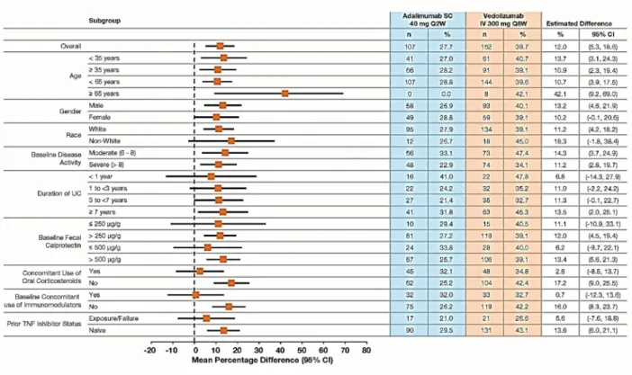
Figure S2. Clinical Remission at Week 52 in Subgroups by Patient Demographic and Baseline Characteristics (FAS).
Plot of treatment differences (dots) and 95% confidence intervals (CIs; horizontal bars) for comparing the proportion of patients in clinical remission between vedolizumab and adalimumab at Week 52.
CI, confidence interval; FAS, full-analysis set; IV, intravenous; SC, subcutaneous; TNF, tumor necrosis factor; UC, ulcerative colitis.
Clinical remission is defined as a total score on the Mayo scale of ≤2 points (or a partial score on the Mayo scale of ≤2 points, if the total score on the Mayo scale was not assessed at the visit) and no individual subscore >1 point.
Patients with missing clinical remission status are considered as non-responders.
The 95% CI for the treatment difference is based on crude estimates using the normal approximation method (the Fisher’s exact method used if the numerator is ≤5).
Figure S3. Clinical Remission and Endoscopic Improvement at Week 52 by Baseline Concomitant Medication Use.
CI, confidence interval; eCRF, electronic Case Report Form; IV, intravenous; IWRS, interactive Web response system; SC, subcutaneous
Absence of baseline steroid use reflects data reported by IWRS. Absence of baseline immunomodulator use reflects data reported on the eCRF.
Figure S4. Clinical Remission at Week 14 in the Overall population and Subgroups by Prior TNF Inhibitor Therapy (FAS).
CI, confidence interval; FAS, full-analysis set; IV, intravenous; SC, subcutaneous; TNF, tumor necrosis factor.
Point estimate and 95% CI for the overall analysis were obtained from Cochran-Mantel-Haenszel method adjusted by randomization stratification factors: concomitant use of oral corticosteroids (Yes/No) and prior use of TNF inhibitor (Yes/No).
Point estimates and the 95% CIs for TNF inhibitor subpopulations were obtained from Cochran- Mantel-Haenszel method adjusted by randomization stratification factor: concomitant use of oral corticosteroids (Yes/No) or the Fisher’s exact method if the numerator is ≤5.
Patients with missing data to determine outcome status were considered as treatment failures;
i.e., non-responders.
Figure S5. Endoscopic Improvement at Week 52 by Patient Demographic and Baseline Characteristics (FAS).
Plot of treatment differences (dots) and 95% confidence intervals (CIs; horizontal bars) for comparing the proportions of patients with endoscopic improvement between vedolizumab and adalimumab at Week 52.
CI, confidence interval; FAS, full-analysis set; IV, intravenous; SC, subcutaneous; TNF, tumor necrosis factor; UC, ulcerative colitis.
Endoscopic improvement is defined as a Mayo score endoscopic subscore of ≤1 point.
Patients with missing mucosal healing status are considered as non-responders (i.e., No Mucosal Healing).
The 95% CI of the treatment difference is based on crude estimates using the normal approximation method (the Fisher’s exact method used if the numerator is ≤5).
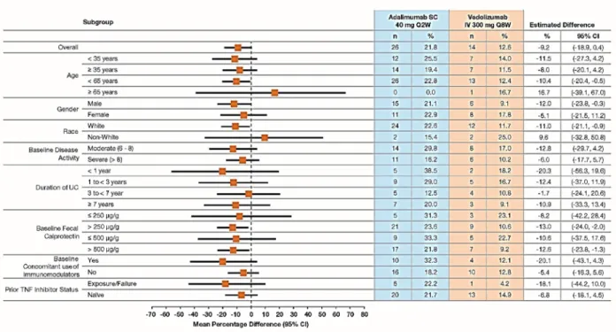
Figure S6. Corticosteroid-free Clinical Remission at Week 52 by Patient Demographic and Baseline Characteristics (FAS).
Plot of treatment differences (dots) and 95% confidence intervals (CIs; horizontal bars) for comparing the proportions of patients with corticosteroid-free clinical remission between vedolizumab and adalimumab at Week 52.
CI, confidence interval; FAS, full-analysis set; IV, intravenous; SC, subcutaneous; TNF, tumor necrosis factor; UC, ulcerative colitis.
The 95% CI for the treatment difference is based on crude estimates using the normal approximation method (the Fisher’s exact method used if the numerator is ≤5).
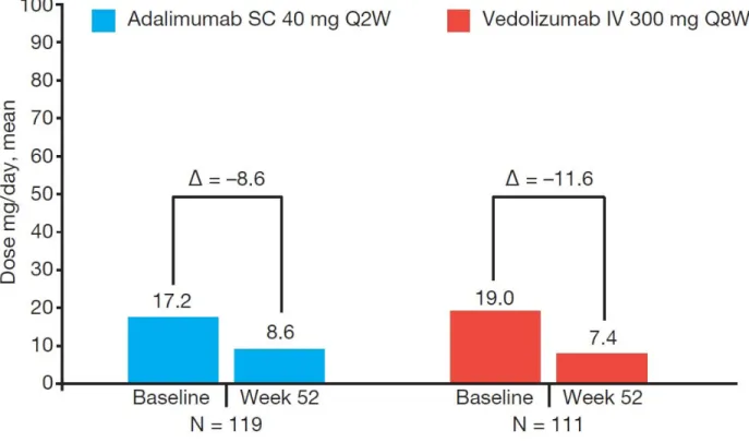
Figure S7. Oral Corticosteroid Use.
Mean change in median oral corticosteroid use at Week 52.
eCRF, electronic Case Report Form; IV, intravenous; SC, subcutaneous.
Only patients with concomitant oral corticosteroid (excludes budesonide) use at Baseline in the eCRF are included.
Patients receiving oral corticosteroids at screening who achieved clinical response at the week 6 and at any subsequent visits underwent a corticosteroid tapering regimen.
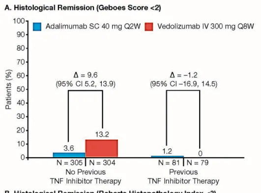
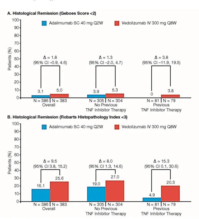
Figure S8. Histological Remission at Week 52 in Subgroups by Prior TNF Inhibitor Therapy.
A: Geboes Grading System.
B: Robarts Histopathology Index.
CI, confidence interval; IV, intravenous; SC, subcutaneous; TNF, tumor necrosis factor.
Shown are the percentages of patients who had histologic remission as indicated by a Geboes score lower than 2.0 (on a scale from 0 to 5.4, with higher scores indicating more severe disease activity) (Panel A) or by a Robarts Histopathology Index score lower than 3 (on a scale from 0 to 33, with higher scores indicating more severe disease activity) (Panel B).
Point estimates and the 95% CIs were obtained from Cochran-Mantel-Haenszel (CMH) method stratified by randomization stratification factor: concomitant use of oral corticosteroids (Yes/No) or the Fisher’s exact method if the numerator is ≤5.
Patients with missing data to determine outcome status were considered as treatment failures;
i.e., non-responders.
Figure S9. Histological Remission at Week 14.
A: Geboes Grading System.
B: Robarts Histopathology Index.
CI, confidence interval; IV, intravenous; SC, subcutaneous; TNF, tumor necrosis factor.
Shown are the percentages of patients who had histologic remission as indicated by a Geboes score lower than 2.0 (on a scale from 0 to 5.4, with higher scores indicating more severe disease activity) (Panel A) or by a Robarts Histopathology Index score lower than 3 (on a scale from 0 to 33, with higher scores indicating more severe disease activity) (Panel B).
Point estimate and 95% CI for the overall analysis were obtained from Cochran-Mantel-Haenszel method adjusted by randomization stratification factors: concomitant use of oral corticosteroids (Yes/No) and prior use of TNF inhibitor (Yes/No).
Point estimates and the 95% CIs for TNF inhibitor subpopulations were obtained from Cochran- Mantel-Haenszel method adjusted by randomization stratification factor: concomitant use of oral corticosteroids (Yes/No) or the Fisher’s exact method if the numerator is ≤5.
Patients with missing data to determine outcome status were considered as treatment failures;
i.e., non-responders.
Figure S10. Minimal Histologic Disease Activity at Week 52.
A: Geboes Grading System.
B: Robarts Histopathology Index.
CI, confidence interval; IV, intravenous; SC, subcutaneous.
Shown are the percentages of patients who had minimal histologic disease activity as indicated by a Geboes score lower than 3.2 (on a scale from 0 to 5.4, with higher scores indicating more severe disease activity) (Panel A) or by a Robarts Histopathology Index score lower than 5 (on a scale from 0 to 33, with higher scores indicating more severe disease activity) (Panel B).
Point estimates and the 95% CIs were obtained from Cochran-Mantel-Haenszel (CMH) method stratified by randomization stratification factor: concomitant use of oral corticosteroids (Yes/No) and prior use of TNF inhibitor (Yes/No).
Patients with missing data to determine outcome status were considered as treatment failures;
i.e., non-responders.
Figure S11. Clinical Response based on Total Score on the Mayo Scale at Week 14 in the Overall Population and Subgroups by Prior TNF Inhibitor Therapy.
IV, intravenous; SC, subcutaneous; TNF, tumor necrosis factor.
Point estimates and the 95% CIs for the overall analysis were obtained from Cochran-Mantel- Haenszel (CMH) method stratified by randomization stratification factor: concomitant use of oral corticosteroids (Yes/No) and prior use of TNF inhibitor (Yes/No).
Point estimates and the 95% CIs for TNF inhibitor subpopulations were obtained from Cochran- Mantel-Haenszel method adjusted by randomization stratification factor: concomitant use of oral corticosteroids (Yes/No) or the Fisher’s exact method if the numerator is ≤5.
Clinical response based on total score on the Mayo scale is defined as a reduction in total score on the Mayo scale of ≥3 points and ≥30% from baseline with an accompanying decrease in rectal bleeding subscore of ≥1point or absolute rectal bleeding subscore of ≤1 point. Patients with missing clinical response status were considered non-responders.
Early Termination Visit Week 52
(b)
Follow- up Visit
Week 68 (b) Study Day/Week: Days -28 to
-1 Day 1 (a)
2 4 6 8, 10,
12 14 16, 18,
20 22 24, 26, 28 30
32, 34,
36 38 40, 42,
44 46 48, 50 Visit Windows
(Days): ±3 ±3 ±3 ±3 ±3 ±3 ±3 ±3 ±3 ±3 ±3 ±3 ±3 ±3 ±7 ±7
Visit Number:
1 2 3 4 5 6-8
(c) 9 10-
12 (c) 13
14- 16 (c) 17
18- 20 (c) 21
22- 24 (c) 25
26- 27
(c) 28 29
Informed consent X Inclusion/exclusion
criteria X X (a)
Demographics and
medical history X
Concurrent medical
conditions X
Medication history X Physical
examination X X (a) X X X X X X X X X X
Vital signs (d) X X (a) X X X X X X X X X X
Weight and height X Flexible
sigmoidoscopy (e) X X X
Total score on the
Mayo scale X X X
Partial score on the
Mayo scale X X X X X X X X
Patient diary X X (a) X X X X X X X X X X X X X X X
Clinical laboratory
testing (f) X X (a) X X X X X X
Urinalysis X X
ADA testing (g) X (a) X X X X X X X
PK testing (g) X (a) X X X X X X X
HIV/Hepatitis panel X Tuberculosis
screening (h) X X
(h) X (h)
Pharmacogenomic DNA and RNA
sample collection X (a)
Serum pregnancy
test (i) X
Urine pregnancy test
(i) X (a) X X X X
(i) X X
(i) X X
(i) X X
(i) X X
(i) X X
(i) X X
IBDQ X (a) X X
ECG X X
Stool sample for C.
difficile X
Stool sample for
calprotectin X X X X
PML checklist X X (a) X X X X X X X X X
PML wallet card X X
SC dosing (adalimumab or
placebo) X X X X X X X X X X X X X X X
IV dosing (vedolizumab or
placebo) X X X X X X X X
PTE assessment (j) X X
Adverse events (k) X X X X X X X X X X X X X X X X X
Concomitant medications and
procedures (l) X X X X X X X X X X X X X X X X X
ADA, antidrug antibody; AE, adverse event; ECG, electrocardiogram; HIV, human immunodeficiency virus; IBDQ, Inflammatory Bowel Disease Questionnaire; IV, intravenous; PK, pharmacokinetics;
PML, progressive multifocal leukoencephalopathy; PTE, pretreatment event; SC, subcutaneous; TB, tuberculosis.
(a) Assessments to be completed predose.
(b) Patients discontinued from the study for any reason will complete the Early Termination Visit and the Follow-up Visit Week 68.
(c) Non-clinic visits. Patient will self-inject at home. On other visits SC dosing will be performed by site staff or patient under supervision of site staff.
(d) Vital signs will include body temperature, respiratory rate, blood pressure, and pulse (bpm). On dosing days, vital signs are taken predose.
(e) Biopsies to be collected at Screening and Weeks 14 and 52. For patients without cancer surveillance endoscopy performed in last 12 months, the investigator can perform a colonoscopy at Screening.
Evaluation of endoscopy results will be performed by the central reader.
(f) Blood sample obtained during Screening should be in fasted state.
(g) Blood samples for the ADA (against vedolizumab or adalimumab) assessments will be collected from all patients at Day 1 and Weeks 6, 14, 22, 30, 38, 52, and 68. On dosing days, blood samples must be taken predose. An aliquot of this sample will be used to determine the serum concentration of vedolizumab.
(h) Assessed by QuantiFERON test or TB skin test reaction. For sites in Taiwan, QuantiFERON test will also need to be performed at Week 22 and Week 52/Early Termination visit.
(i) Women of childbearing potential only. Urine pregnancy test should be done before every IV infusion and at Weeks 10, 18, 26, 34, 42, and 50.
(j) PTEs will be captured immediately following the signing of the informed consent at the Screening Visit, up until the first dose of trial drug. Collection of AEs will begin following first dose of trial drug
Characteristic
Adalimumab (N = 386)
Vedolizumab (N = 385) Total score on the Mayo scale — no. (%)*
Mild (score <6)† 5 (1.3) 9 (2.3)
Moderate (score = 6 to 8)‡ 169 (43.8) 154 (40.0)
Severe (score = 9 to 12)‡ 210 (54.4) 217 (56.4)
Partial score on the Mayo scale (mean ± SD)¶
6.0±1.3 6.1±1.5
*Mayo scores range from 0 to 12, with higher scores indicating more active disease; sub- components: stool frequency, rectal bleeding, endoscopy (sigmoidoscopy), physician global assessment. Scores were available for 384 patients in the adalimumab group and 380 patients in the vedolizumab group.
†Patients with mild disease represented significant protocol deviations.
‡In total, 98.2% of patients in adalimumab group and 96.4% of patients in the vedolizumab group had moderately to severely active ulcerative colitis.
¶The partial score on the Mayo scale consists of the total score on the Mayo scale minus the sigmoidoscopy subscore; range, 0 to 9, with higher scores indicating more active disease. Scores were available for 384 patients in the adalimumab group and 381 patients in the vedolizumab group.
Table S3. Post Hoc Analysis of Clinical Remission and Endoscopic Improvement at Week 52, Using the Weighted Cochran–Mantel–Haenszel Method.
Week 52 Outcomes
Stage 1 (N = 93)
Stage 2 (N = 676)
CHW Method (N = 769) Adalimumab,
n (%) (N = 45)
Vedolizumab, n (%) (N = 48)
Adalimumab, n (%) (N = 341)
Vedolizumab, n (%) (N = 335)
Adjusted P value Clinical remission 11 (24.4) 14 (29.2) 76 (22.3) 106 (31.6) 0.0062 Endoscopic
improvement 13 (28.9) 18 (37.5) 94 (27.6) 134 (40.0) 0.0005
The P values from the Cochran–Mantel–Haenszel (CHW) method were obtained from a weighted combination of test statistics from Stage 1 (Interim Analysis 1 data) and Stage 2 (post Interim Analysis 1 data) weighted by actual interim sample size and planned total sample size.
The results were consistent with the planned efficacy analysis using the Cochran–Mantel–Haenszel method.
Week 52 Outcomes
Adalimumab SC 40 mg Q2Wa
Vedolizumab IV 300 mg Q8Wa
Treatment Differenceb
(95% CI) P value Clinical remission, % (95%
CI)c
(N = 386) 25.9 (21.3, 30.4)
(N = 383)
37.2 (32.2, 42.2) 11.3 (4.6, 18.0) 0.002 Endoscopic improvement,
% (95% CI)c
(N = 386) 33.6 (28.5, 38.7)
(N = 383)
46.8 (41.5, 52.1) 13.2 (6.0, 20.3) <0.001 Corticosteroid-free
remission, % (95% CI)c
(N = 119) 24.7 (16.6, 32.7)
(N = 111)
16.9 (9.3, 24.4) –7.8 (–18.8, 3.1) NS CI, confidence interval; Q2W, once every two weeks; Q8W, once every 8 weeks; NS, not significant.
aThe 95% CI for the proportions was based on the Clopper-Pearson method.
bThe treatment difference in proportions and the corresponding 95% CI and P value were based on the Cochran- Mantel-Haenszel method, stratified by concomitant use of oral corticosteroids (Yes/No) and prior use of a TNF inhibitor (Yes/No). The proportions, treatment difference, and P value were combined using Rubin’s rules from 50 imputed datasets using PROC MIANALYZE. The Wilson-Hilferty transformation was applied to test statistics before PROC MIANALYZE.
cSensitivity Analysis: The impact of dropouts was explored by using a hybrid imputation approach where patients with missing efficacy data by discontinuation due to an adverse event or lack of efficacy were imputed as non- responder (under Missing Not At Random assumption) and missing efficacy data due to other reasons were imputed using multiple imputation (under Missing At Random assumption). For the multiple imputation, each component of the total score on the Mayo scale was imputed by treatment group via a multivariate step-wise approach using fully conditional specification (FCS ordinal logistic) methods,1 respectively. Missing baseline total score on the Mayo scale, if any, was imputed using the relevant demographic and baseline disease characteristic data (namely, age, duration of UC, baseline disease severity). Subsequent visits were imputed using all the previous visits in a stepwise fashion. Fifty imputation datasets were computed for each component of total score on the Mayo scale. The total and/or partial scores on the Mayo scale were derived subsequently. The efficacy outcome status was then determined from the total score on the Mayo scale derived using the observed and the imputed Mayo subscores.
1. Ratitch B, Lipkovich I, O’Kelly M. Combining analysis results from multiply imputed categorical data.Paper presented at: PharmaSUG 2013; May 12-15, 2013; Chicago, IL. Abstract SP03. Available at:
https://pharmasug.org/proceedings/2013/SP/PharmaSUG-2013-SP03.pdf
Outcomea
40 mg Q2W (N=386)
300 mg Q8W (N=383)
Treatment Difference, (95% CI)p
Clinical remissionb at Week 52, n % 87 (22.5) 120 (31.3) 8.8 (2.5, 15.0)
Endoscopic improvement (mucosal healing)c at Week 52, n (%)
107 (27.7) 152 (39.7) 11.9 (5.3, 18.5)
CS-free clinical remissiond at Week 52, n (%) 26 (21.8) 14 (12.6) -9.3 (-18.9, 0.4)
Clinical responsee at Week 52, n (%) 166 (43.0) 211 (55.1) 12.0 (5.1, 19.0)
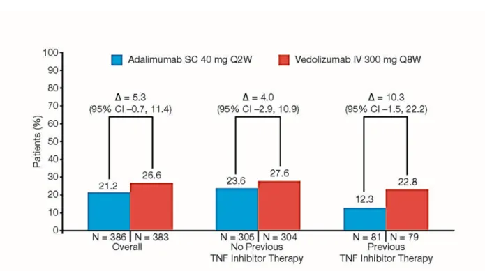
Clinical remissionb at Week 14, n (%) 82 (21.2) 102 (26.6) 5.3 (-0.7, 11.4)
Rectal bleeding subscore indicative of mild disease (≤1) at Week 52, n (%)
211 (54.7) 252 (65.8) 11.1 (4.2, 17.9)
Physician’s Global Assessment (PGA) subscore indicative of mild disease (≤1) at Week 52, n (%)
189 (49.0) 234 (61.1) 12.1 (5.1, 19.0)
Stool frequency subscore indicative of mild disease (≤1) at Week 52, n (%)
173 (44.8) 223 (58.2) 13.3 (6.4, 20.3)
Clinical remissionb where rectal bleeding subscore of 0 and endoscopy subscore of 0 at Week 52, n (%)
54 (14.0) 85 (22.2) 8.2 (2.8, 13.5)
Endoscopy subscore of 0, rectal bleeding subscore of 0, and stool frequency subscore decreases or no change from Baseline at Week 52, n (%)
55 (14.2) 89 (23.2) 8.9 (3.5, 14.4)
Endoscopy subscore ≤1, rectal bleeding subscore of 0, and stool frequency subscore of 0 at Week 52, n (%)
75 (19.4) 91 (23.8) 4.3 (-1.5, 10.1)
Endoscopy subscore ≤1, rectal bleeding subscore of 0, and 91 (23.6) 127 (33.2) 9.5 (3.2, 15.9)
Table S5. Results from Prespecified Analyses of Efficacy Outcomes.
Outcomea
Adalimumab SC 40 mg Q2W
(N=386)
Vedolizumab IV 300 mg Q8W
(N=383)
Treatment Difference, (95% CI)p
Endoscopy subscore ≤1, rectal bleeding subscore of 0, stool frequency subscore decreases or no change from Baseline, and total score (sum of these 3) ≤1 at Week 52, n (%)
79 (20.5) 112 (29.2) 8.7 (2.7, 14.8)
IBDQ score change of ≥16 points from Baseline to Week 52, n (%)
163 (42.2) 199 (52.0) 9.7 (2.7, 16.7)
Clinical remission based on IBDQ score >170 at Week 52, n (%)
156 (40.4) 192 (50.1) 9.6 (2.8, 16.5)
*Change in oral corticosteroid use from Baseline to Week 52, mean (SD):
Cumulative CS exposure (in mg of prednisone equivalent)f
3321.8 (3373.9) 3764.9 (3647.5) –
Duration of CS use (days)g 194.6 (138.4) 219.1 (167.0) –
Change in median oral CS dose from baseline up to Week 52 (in mg of prednisone equivalent)
–8.55 (10.77) –11.62 (11.12) –
CS-free clinical remissionc at Week 14, n (%) 8 (6.7) 7 (6.3) -0.5 (-6.8, 5.9)
Major UC-related events,h Hazard ratio, 95% CI:i
UC-related hospitalizations, n (%)
UC-related bowel resections, n (%)
UC-related procedures, n (%)
20 (5.2) 3 (0.8) 8 (2.1)
15 (3.9) 0 7 (1.8)
0.061 (0.035, 0.105)
Table S5. Results from Prespecified Analyses of Efficacy Outcomes.
Outcomea
Adalimumab SC 40 mg Q2W
(N=386)
Vedolizumab IV 300 mg Q8W
(N=383)
Treatment Difference, (95% CI)p
*Change in fecal calprotectin concentrations (µg/g), mean (SD) LOCF:
Baseline to Week 14
Baseline to Week 30
Baseline to Week 52
–1004.4 (4579.12) –1043.3 (4575.62) –1160.3 (4508.21)
–1393.2 (6052.67) –1601.5 (6463.41) –1631.5 (6424.48)
–409.9 (-824.9, 5.1) –399.4 (-838.2, 39.5) –315.7 (–764.2, 132.8) Change in histology from Baseline to Week 52 per Geboes
(≥ 3 at baseline), n
Improving (change ≤1)
No change (-1< change <1)
Worsening (change ≥1)
Missing
150 12 41 146
225 4 29 104
– – – – Histological remission, per Geboes, n (%)
Week 14
Week 52
12 (3.1) 12 (3.1)
19 (5.0) 40 (10.4)
1.8 (-0.9, 4.6) 7.3 (3.8, 10.8) Histological remission, per RHI, n (%)
Week 14
Week 52
62 (16.1) 77 (19.9)
98 (25.6) 144 (37.6)
9.5 (3.8, 15.2) 17.6 (11.3, 23.8)
Table S5. Results from Prespecified Analyses of Efficacy Outcomes.
Outcomea
Adalimumab SC 40 mg Q2W
(N=386)
Vedolizumab IV 300 mg Q8W
(N=383)
Treatment Difference, (95% CI)p
Exploratory Efficacy Outcomes Defined in the Statistical Analysis Plan but not in the Protocol:
Clinical responsee at Week 14, n (%) 177 (45.9) 257 (67.1) 21.2 (14.4, 28.0)
Total score on the Mayo scale ≤1 and rectal bleeding subscore = 0 at Week 52, n (%)
70 (18.1) 101 (26.4) 8.2 (2.4, 14.0)
Durable clinical remission at Week 52j , n (%) 46 (11.9) 70 (18.3) 6.3 (1.3, 11.3)
Clinical remissionk at Week 52 and clinical remission for
≥14 weeks leading up to Week 52, n (%)
74 (19.2) 105 (27.4) 8.2 (2.3, 14.2)
Disease controll at Week 52, n (%) 23 (6.0) 41 (10.7) 4.7 (0.8, 8.6)
Rectal bleeding subscore = 0 at Week 52, n (%) 170 (44.0) 214 (55.9) 11.8 (4.8, 18.8)
Major UC-related events (eg, hospitalizations, bowel resection, and procedures) throughout the trial up to Week 52, n (%)
25 (6.5) 17 (4.4) -2.0 (-5.2, 1.2)
FCP ≤250 µg/g at Week 14, 30, 52 (among those in the FAS with FCP >250 µg/g at baseline), n (%)
Week 14
Week 30
Week 52
80 (26.8) 73 (24.5) 86 (28.9)
97 (31.9) 103 (33.9) 107 (35.2)
5.3 (-1.9, 12.6) 9.6 (2.4, 16.9) 6.6 (-0.7, 14.0)
Still on adalimumab or vedolizumab at Week 68, n (%) 21 (5.4) 15 (3.9) -1.5 (-4.5, 1.5)
Table S5. Results from Prespecified Analyses of Efficacy Outcomes.
Outcomea
Adalimumab SC 40 mg Q2W
(N=386)
Vedolizumab IV 300 mg Q8W
(N=383)
Treatment Difference, (95% CI)p
*Change from baseline in IBDQ-specific bowel symptoms domain, mean (SD) LOCF:
Week 30
Week 52
13.8 (14.61) 13.9 (15.77)
17.3 (14.51) 17.4 (15.74)
3.1 (1.1, 5.2) 3.0 (0.8, 5.2)
*Time to first clinical remission (day)k, median 154 100 –
*Time to first clinical response (day)m, median 28 29 –
Clinical remissionk by visit, n (%):
Week 2
Week 4
Week 6
Week 14
Week 22
Week 30
Week 38
Week 46
Week 52
66 (17.1) 109 (28.2) 124 (32.1) 140 (36.3) 162 (42.0) 162 (42.0) 152 (39.4) 154 (39.9) 143 (37.0)
52 (13.6) 109 (28.5) 154 (40.2) 194 (50.7) 213 (55.6) 202 (52.7) 212 (55.4) 206 (53.8) 191 (49.9)
– – – – – – – – –