High serum Hsp70 level predicts poor
survival in colorectal cancer: Results obtained in an independent validation cohort
Laszló Gráfa, Lóránd Barabásb, Balázs Madarasc, Nóra Garama, Éva Malátid, Laura Horvátha, Zoltán Prohászkaa, Zsolt Horvátheand Judit Kocsisa,∗
a3rdDepartment of Internal Medicine, Semmelweis University, Budapest 1125, Hungary
b2ndDepartment of Surgery, Semmelweis University, Budapest 1125, Hungary
cNational Institute of Oncology, Budapest 1122, Hungary
dJános Balassa Hospital, County Hospital Tolna, Szekszárd 7100, Hungary
eDepartment of Oncoradiology, Bács-Kiskun County Hospital, Kecskemét H6000, Hungary
Abstract.
BACKGROUND:Hsp70 plays important role in the development and progression of cancer. Previously we described the asso- ciation between serum Hsp70 levels and mortality of colorectal cancer.
OBJECTIVE:In this new prospective study we aimed to confirm and extend our previous findings in a larger cohort of patients, based on a longer follow-up period.
METHODS:Two hundred and thirty-two patients diagnosed with colorectal cancer were enrolled in the study. Baseline serum Hsp70 level and classical biomarker levels were measured. Patients were treated according to stage of the tumor and follow-up lasted for a median 46.4 months.
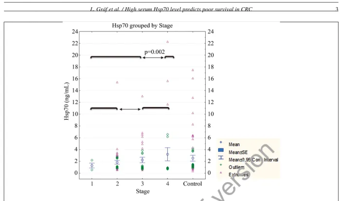
RESULTS:We found that serum Hsp70 concentrations increase significantly with stage of the disease (1.79; 2.23 and 3.21 ng/ml in stage I+II, III and IV respectively,p=0.012 and 0.002, Mann-Whitney test) and with other known biomarkers of the disease.
We managed to confirm our previous findings that high baseline serum Hsp70 level (>1.64 ng/ml) predicted poor 5-year sur- vival (risk of death HR: 1.94 CI: 1.294–2.909; univariate; HR: 2.418 CI: 1.373–4.258; multivariate Cox regression analysis) in the whole patient population and also in subgroups of stage IV and stage III disease. The strongest association was observed in women under age of 70 (HR: 8.12, CI: 2.02–35.84;p=0.004; multivariate Cox regression). The power of this colorectal cancer prognostic model could be amplified by combining Hsp70 levels and inflammatory markers. Patients with high Hsp70, CRP and high baseline WBC or platelet count had 5-times higher risk of death (HR: 5.07 CI: 2.74–9.39,p <0.0001; and HR: 4.98 CI:
3.08–8.06,p <0.0001 respectively).
CONCLUSIONS:These results confirm and validate our previous findings that serum Hsp70 is a useful biomarker of colorectal cancer.
Keywords: Hsp70, colorectal cancer, prognostic model, CRP, survival
1. Introduction
1
Heat shock proteins (Hsp) are a family of evolution-
2
ary conserved proteins. Hsps are molecular chaperones
3
∗Corresponding author: Judit Kocsis, 3rd Department of Internal Medicine, Semmelweis University, Kútvölgyi út 4, Budapest 1125, Hungary. E-mail: kocsisjucidr@gmail.com.
with a wide array of functions, including protein fold- 4
ing, transport, and also the repair and degradation of 5
damaged proteins. Hsps have a regulatory role in pro- 6
grammed cell death and apoptosis [1]. A prominent 7
member of the family is Hsp70, probably the most ex- 8
tensively investigated heat shock protein. Hsp70 plays 9
a key role in carcinogenesis. It is overexpressed in most 10
human cancers to promote cancer cell survival, prolif- 11
ISSN 1574-0153/18/$35.00 c2018 – IOS Press and the authors. All rights reserved
uncorrected
proof
version
eration and to evade apoptosis and other forms of can-
12
cer cell death [2]. In the absence of Hsp70, tumor cells
13
become senescent, a state of irreversible growth arrest
14
with specific cell morphology. Senescent cells are un-
15
able to proliferate and are eventually eliminated by the
16
innate immune system (in [3,4]). On the other hand,
17
high levels of intracellular Hsp70-1 correlate with tu-
18
mor burden, advanced stage and worse prognosis in
19
non-small cell lung cancer [5]; breast, endometrial, and
20
uterine cervical carcinoma [6]. In a study of 81 primary
21
human colorectal tissues the expression of Hsp70 and
22
Hsp110 highly correlated with advanced clinical stages
23
and lymph node involvement [7]. Hsp70 expression
24
was associated with poor prognosis, decreased over-
25
all survival in patients suffering from rectal cancer and
26
squamous cell lung cancer [8] and resistance to on-
27
cotherapy in some cancer patient groups [9].
28
Beyond its intracellular occurrence Hsp70 can also
29
be found in the plasma membrane of many solid tu-
30
mors, while this is not true for normal tissues [10,11].
31
Membrane-bound Hsp70 is not only a biomarker in ag-
32
gressive tumors, but can serve as a potential target of
33
antitumor therapies [12]. Moreover it can be released
34
from the cell (the mechanism of this process is still not
35
exactly clarified) and appear in the circulation in the
36
form of soluble Hsp70 (sHSP70), both in healthy in-
37
dividuals [13,14] and in various pathologic conditions.
38
Circulating Hsp70 has been extensively investigated
39
in a multitude of physiologic (pregnancy, aging) and
40
non-physiologic (hearth failure, diabetes, liver disease,
41
asthma, obesity) conditions (reviewed in [2]), on the
42
other hand, it has been studied to a lesser extent in ma-
43
lignancies. According to Gehrmann and co-workers,
44
Hsp70 serum levels were significantly increased in pa-
45
tients with hepatocellular carcinoma (HCC) compared
46
to healthy controls and subjects with chronic hepati-
47
tis [15]. Another group found a significant correlation
48
between sHsp70 and gross tumor volume in adeno-
49
and squamous cell carcinoma of the lung [16]. Previ-
50
ously we reported on strong association between serum
51
Hsp70 levels and stage, as well as unfavourable prog-
52
nosis of small cell lung cancer [17].
53
In 2010 we published preliminary data on the corre-
54
lation between elevated serum Hsp70 levels and high
55
mortality in a cohort of early stage colorectal cancer
56
patients [18]. The present investigation is a confirma-
57
tory work, aimed to reproduce previous findings on a
58
larger cohort of prospectively followed CRC patients,
59
with a longer follow-up period. We intended to prove
60
that high serum Hsp70 level is a poor prognostic factor
61
and propose a powerful prognostic model combining
62
Hsp70 with easily accessible traditional biomarkers.
63
Table 1
Baseline demographic and tumor characteristics of patients with col- orectal cancer
Variable Number (percent)
Gender
Male 138
Female 94
Age at diagnosis (year, mean, SD) 66.82 (11.41) TNM stage
I 9 (3.9)
II 101 (43.5)
III 73 (31.5)
IV 49 (21.1)
Tumor localization
Right colon 43 (18.5)
Colon transversum 16 (6.9)
Left colon 89 (38.4)
Rectum 83 (35.8)
Unknown 1 (0.5)
Tumor grade
1 51 (22.0)
2 113 (48.7)
3 44 (18.9)
Unknown 24 (10.4)
Surgery
Definitive or palliative surgery 210 (90.5)
No surgery 22 (9.5)
2. Methods 64
2.1. Patients, controls and sample collection 65
Two hundred and thirty two patients diagnosed with 66
colorectal cancer and 110 controls were involved in the 67
study between January 2011 and June 2013 in the on- 68
cology ward of 3rd Department of Internal Medicine, 69 Semmelweis University. After confirmation of inva- 70
sive colorectal cancer with any stage, patients were 71
consented consecutively and clinical data and blood 72 samples were collected before starting anticancer ther- 73
apy. Baseline demographic and clinical characteris- 74
tics of patients are summarized in Table 1. Mean age 75
of patients was 66.8 years, with a male/female ra- 76
tio of 138/94. After diagnosis and adequate surgery 77
patients were treated and followed by the oncology 78
ward according to the stage of their disease and 79 to the actual national and European [19] guidelines. 80
Patients with rectal cancer received radiochemother- 81
apy before definitive surgery from cT3 or N+ dis- 82
ease. Twenty-two patients who had unresectable and/or 83
metastatic disease received upfront primary systemic 84
treatment without definitive surgery. During a follow- 85
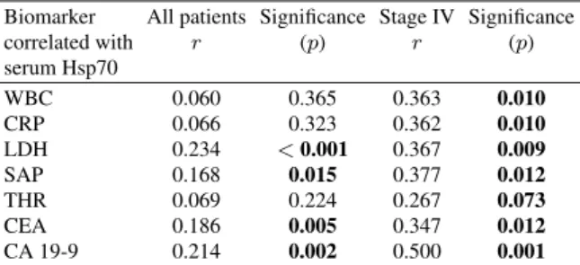
up period that lasted for maximum 5 years (median 86 46.42 months), progression free survival and overall 87
survival data were collected. 88
The control group consisted of 110 healthy individ- 89 uals (mean age 64.5 years, male/female ratio 43/67), 90
uncorrected
proof
version
Fig. 1. Baseline serum Hsp70 level of healthy subjects (N =110) and colorectal cancer patients according to stage of the disease. Differences between groups were calculated with Mann-Whitney test. Significant differences are shown between stages (Stage I+II vs III+IV and Stage I–III vs IV). Non significant difference was observed between controls and patients with advanced (Stage IV) disease (not shown in the figure).
Explanation for stages: Stage I: T1-2 N0; Stage II: T3-4b N0; Stage III (IIIa-b): any T N1-2; Stage IV: any T any N M1 (according to 7thedition of TNM staging system).
who underwent screening colonoscopy in the preced-
91
ing 2 months and were free of colorectal cancer or
92
premalignant lesions and whose history was negative
93
for colorectal cancer or other malignancies. The study
94
was approved by the Medical Research Council Sci-
95
entific and Research Committee. Serum samples were
96
aliquoted and stored at −70 degrees of Celsius for
97
Hsp70 analysis.
98
2.2. Serum Hsp70 analysis
99
Soluble Hsp70 level was measured by using R&D
100
Systems (Minneapolis, MN, USA, Catalogue No.
101
DYC1663E) enzyme-linked immunosorbent assay kit.
102
Ninety-six-well microtitre plates were coated with
103
mouse anti-human Hsp70 capture antibody (100 µl;
104
2µg/ml) in carbonate buffer (pH 9.5) overnight at 4◦C.
105
Plates were washed with phosphate-buffered saline
106
(PBS) containing 0.1% Tween 20 three times and
107
nonspecific binding sites blocked by incubation with
108
200µl of PBS containing 0.5% gelatinec and Tween 20
109
for 1 h at room temperature. After washing, 100µl of
110
the reference preparation (recombinant human Hsp70, 111
0–10 µg/ml) or samples (1:1) were added and the 112
plates were incubated for 2 h at room temperature. 113
Plates were subsequently washed and Hsp70 bind- 114 ing was determined using biotinylated rabbit anti- 115
human antibody (100 µl; 0.5 µg/ml) in PBS gela- 116
tine. After 1.5 h at room temperature, plates were 117
washed and incubated with streptavidin-horseradish- 118
peroxidase (1:200) in PBS gelatine for 20 min at room 119
temperature. Plates were washed and 100 µl of o- 120
phenylene-diamine (Sigma, St Louis, MO, USA) in 121 citrate buffer was added. The optical density was mea- 122
sured atλ=490 nm (reference atλ=620 nm). The 123
detection range of the assay was 0.05–10 ng/ml, the 124 intra/inter-assay variability<10/<16%, respectively. 125
2.3. Tumor marker and other prognostic biomarker 126
analysis 127
Determination of the additional laboratory parame- 128
ters including complete blood counts, clinical chem- 129
istry and tumor markers were performed by Roche In- 130
tegra 800 analyzer, by Cell-Dyn 3500 hematology an- 131 alyzer at the time of study entry of each patient. 132
2.4. Statistical analysis 133
For descriptive purposes data are given as mean 134
±standard deviation (SD) or median and interquar- 135
uncorrected
proof
version
Table 2
Correlation between serum Hsp70 level and the known biomarkers of colorectal cancer. Spearman’s rank correlation test was used
Biomarker All patients Significance Stage IV Significance
correlated with r (p) r (p)
serum Hsp70
WBC 0.060 0.365 0.363 0.010
CRP 0.066 0.323 0.362 0.010
LDH 0.234 <0.001 0.367 0.009
SAP 0.168 0.015 0.377 0.012
THR 0.069 0.224 0.267 0.073
CEA 0.186 0.005 0.347 0.012
CA 19-9 0.214 0.002 0.500 0.001
tile range (IQR) if data were not Gaussian distributed. 136
The differences between groups were evaluated with 137
the Mann-Whitney test. Correlations between the vari- 138
ables were expressed using the non-parametric Spear- 139
man’s correlation coefficients. The association of ser- 140
um protein levels on survival was analysed with Cox 141 regression. Survival was calculated according to the 142 Kaplan-Meier method. The curves were compared for 143 statistical significance using long-rank testing. Re- 144 ceiver operating characteristic (ROC) curve analysis 145 was used to determine the optimal cut-off value of 146 Hsp70. Cut-off value of other biomarkers and tumour 147 markers were selected according to the upper level of 148 normal range used by the local laboratory. All tests 149 were two-tailed,pvalues of<0.05 were accepted as 150
statistically significant. 151
Statistical analysis was performed using the Graph- 152
Pad Prism v6.01 (GraphPad Software Inc, San Diego, 153
CA, USA, www.graphpad.com) and SPSS v22 (SPSS 154
Inc., Chicago, IL, USA) software. 155
3. Results
156
3.1. Serum Hsp70 concentration in patients with
157
colorectal cancer according to stage of the
158
disease and in healthy subjects
159
We studied whether baseline serum concentration of
160
soluble Hsp70 is different between patients with CRC
161
and controls (Fig. 1). Circulating Hsp70 level was in
162
the same range in the whole patient population and
163
controls (2.21 (SD: 2.36) versus 2.55 (SD: 2.66) ng/ml,
164
NS). However, within colorectal cancer patients Hsp70
165
levels increased along with the stage of the disease. In
166
early stage CRC (Stage I and II) mean Hsp70 level was
167
1.79 ng/ml (SD: 1.53), in stage III it was 2.23 (SD:
168
1.93) and in metastatic, stage IV disease we measured
169
3.21 ng/ml (SD: 3.87). The difference was statistically
170
Fig. 2. Survival (Kaplan-Meier) of colorectal cancer patients ac- cording to high (black curves) or low (grey curves) serum Hsp70 level. A: all patients (n=232); B: patients with metastatic stage IV disease (n=49); C: patients with stage III disease (n=73).
Log Rank overall comparison showed significant difference in sur- vival between patients with high (>1.64 ng/ml) versus low (61.64 ng/ml) baseline serum Hsp70 level: 10.66;p =0.001; 6.84;p = 0.009 and 3.53;p=0.06.
uncorrected
proof
version
Table 3
Univariate and multivariate Cox-regression analysis: Association between baseline clinical parameters and serum biomarker levels and colorectal cancer patient’s 5 year survival
Univariate Cox regression HR Significance Multivariate Cox regression HR Significance
(95% CI) (p) (95% CI) (p)
Age at diagnosis (>68 year) 2.118 (1.394–3.216) <0.001 2.223 (1.231–4.014) 0.008
Gender (male versus female) 1.070 (0.712–1.608) 0.745 NA
TNM Stage (stage IV versus stage I–III) 6.615 (4.312–10.150) <0.001 6.516 (3.689–11.510) <0.001
Tumor grade (grade 2/3 versus grade 1) NA NA
Grade 2 1.638 (0.885–3.033) 0.116
Grade 3 2.038 (1.006–4.131) 0.048
Tumor localization (right versus left colon) 1.430 (0.930–2.201) 0.103 NA NA
Hsp70 (>1.64 ng/ml) 1.940 (1.294–2.909) 0.001 2.418 (1.373–4.258) 0.002
WBC (>10 800 /ul) 2.368 (1.477–3.796) <0.001 2.123 (1.076–4.186) 0.030
CRP (>5 mg/l) 2.569 (1.634–4.040) <0.001 NA NA
LDH (>248 U/l) 1.750 (1.146–2.671) 0.010 NA NA
SAP (>120 U/l) 3.175 (1.993–5.040) <0.001 NA NA
THR (>300 /ul) 1.611 (1.078–2.407) 0.020 NA NA
CEA (>4 ng/ml) 3.141 (2.093–4.714) <0.001 NA NA
CA 19-9 (>39 ng/ml) 4.077 (2.559–6.509) <0.001 NA NA
The cut-off value for serum biomarkers were the upper limit of their normal range (shown in column (1)). Cut-off value of serum Hsp70 level (>
1.64 ng/ml) was calculated by ROC curve analysis. The same variables were included in multivariate analyses (column (4)) as in the univariate analysis (column (2)), and the best adjusted set of significant variables were highlighted.
significant between early and advanced stage disease
171
(stage I+II versus III+IV,p=0.012) or between non
172
metastatic and metastatic (stage I–III versus stage IV,
173
p=0.002) disease.
174
Presence or absence of a primary tumor at sample
175
collection (i.e. sample collection before or after op-
176
eration) was not associated with altered serum Hsp70
177
levels. Similarly, we did not find a significant differ-
178
ence in Hsp70 levels between right (n =59, HSP70
179
= 2.03 ng/ml) and left-sided (n = 173, Hsp70 =
180
2.27 ng/ml) colorectal tumors.
181
3.2. Correlation of soluble Hsp70 level with other
182
biomarkers
183
Hsp70 levels showed significant but weak positive
184
correlation with tumor markers and other biomarkers
185
that are known prognostic factors of CRC. These cor-
186
relations were more pronounced (however also weak)
187
in metastatic disease. In this subgroup we found posi-
188
tive association of Hsp70 level with LDH, SAP, CRP,
189
baseline platelet and white blood cell count as well as
190
CA19.9 and CEA (Table 2).
191
3.3. The relationship of Hsp70 and other biomarkers 192
with survival 193
Using the ROC curve analysis the cut-off value of 194
Hsp70 was 1.64 ng/ml. Values61.64 ng/ml were re- 195
garded to be favorable and values>1.64 unfavorable 196
prognostic markers. According to the Kaplan-Meier 197
survival estimate it is clearly demonstrated that high 198
Hsp70 levels correlate with poor survival in the whole 199
patient population, as well as in the subgroups of stage 200
IV (metastatic) and stage III disease (Fig. 2;pvalues 201
are 0.001; 0.009 and 0.06 respectively). The risk of 202
death within 5 years was two-fold higher with high ini- 203
tial Hsp70 level, according to univariate (HR: 1.94 CI: 204
1.29–2.91) and multivariate (HR: 2.42 CI: 1.37–4.26) 205
Cox regression analysis. In addition to Hsp70 level 206
age, tumor stage, grade, high WBC and platelet count, 207
high CRP, LDH, SAP and tumor marker proved to be 208
predictive factors of 5-year survival (Table 3). With the 209
multiple Cox regression analysis age, Hsp70, tumor 210
stage and high baseline white blood cell count were in- 211
dependent factors of death in the entire patient popula- 212
tion. As in our pivotal publication [18] we observed the 213
strongest relationship in the subgroup of women un- 214
der the age of 70. Using the same set of variables be- 215
side advanced stage of the disease (HR: 6.6, CI: 2.08– 216
21.48;p = 0.001), high Hsp70 level (HR: 8.12, CI: 217
2.02–35.84;p=0.004, white blood cell number (HR: 218
6.8, CI: 1.56–29.79;p=0.011) and high baseline CRP 219
level (HR: 6.6, CI: 1.84–24.22; p = 0.011) proved 220
to be the strongest independent predictors of death by 221
multiple Cox regression analysis. 222
3.4. Combined prognostic model of survival 223
Next we determined whether our earlier model [20] 224
that proposed the aggregate prognostic effect of high 225
Hsp70 levels and high acute phase protein levels like 226
uncorrected
proof
version
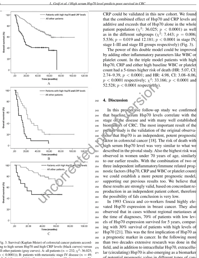
Fig. 3. Survival (Kaplan-Meier) of colorectal cancer patients accord- ing to high serum Hsp70 and high CRP levels (black curves) versus all other patients (gray curves). A: all patients (n=232;χ2: 36.025;
p <0.0001)); B: patients with metastatic stage IV disease (n=49;
χ2: 7.443;p=0.006); C: patients with stage III disease (n=73;
χ2: 12.181;p <0.0001). Cut off value for Hsp70: 1.64 ng/ml, CRP:
5 mg/l.
CRP could be validated in this new cohort. We found 227
that the combined effect of Hsp70 and CRP levels are 228
additive and exceeds that of Hsp70 alone in the whole 229
patient population (χ2: 36.025; p <0.0001) as well 230
as in the different subgroups (χ2: 7.443;p =0.006; 231
5.536;p=0.019 and 12.181;p <0.0001 in stage IV, 232 stage I–III and stage III groups respectively) (Fig. 3). 233
The power of this double model could be improved 234
by adding other inflammatory parameters like WBC or 235 platelet count. In the triple model patients with high 236
Hsp70, CRP and either high baseline WBC or platelet 237
count had a 5-times higher risk of death (HR: 5.07, CI: 238 2.74–9.39,p <0.0001; and HR: 4.98, CI: 3.08–8.06, 239
p <0.0001 respectively;χ2: 33.166;p <0.0001 and 240
52.528;p <0.0001 respectively). 241
4. Discussion
242
In this prospective follow-up study we confirmed
243
that baseline serum Hsp70 levels correlate with the
244
stage of the disease and with many well established
245
biomarkers of CRC. The most important result of the
246
present study is the validation of the original observa-
247
tions that Hsp70 is an independent, potent prognostic
248
factor in colorectal cancer [18]. The risk of death with
249
high serum Hsp70 level was very similar to what we
250
described in the pivotal study. Also the highest risk was
251
observed in women under 70 years of age, similarly
252
to our earlier results. With the combination of two or
253
three independent inflammatory/immune related prog-
254
nostic factors (Hsp70, CRP and WBC or platelet count)
255
we could establish a more potent prognostic model,
256
supporting our previous results too. We believe that
257
these results are strongly valid, based on concordant re-
258
production in an independent patient cohort, therefore
259
the possibility of fals conclusion is very low.
260
In 1993 Ciocca and co-workers found highly ele-
261
vated Hsp70 expression in breast cancer. They also
262
observed that in cases without regional metastases at
263
the time of diagnoses, 70% of patients with low lev-
264
els of Hsp70 expression survived for 5 years, compar-
265
ing with 30% survival of patients with high levels of
266
Hsp70 [21]. This was the first implication of Hsp70 as
267
a prognostic marker in cancer. In the following more
268
than two decades extensive research was done in the
269
field, and in addition to intracellular Hsp70, extracellu-
270
lar (circulating) Hsp70 is also emerging as a biomarker
271
of potential prognostic value in different types of can- 272
cer. 273
Our recent results are in line with our previous ob- 274
servations [17,22] that high serum Hsp70 levels sig- 275
uncorrected
proof
version
nificantly correlate with poor outcome and predict a 276
shorter than expected overall survival. 277
Hsp70 is a versatile protein, crucial in maintain- 278
ing cellular integrity and homeostasis. Cancer cells 279
heavily depend on Hsp70 overexpression, since it pro- 280
tects them from exogenous (chemotherapy, irradia- 281
tion, hypoxia) and endogenous (oncogene accumula- 282
tion) stress. Oncogene accumulation engages senes- 283
cence (OIS=oncogene induced senescence) however, 284
cancer cells can bypass through the up-regulation of 285
Hsp70 [23]. Membrane-bound and extracellular Hsp70 286
is known to interact with the innate and adaptive im- 287
mune system, although this interaction is paradoxical 288
and not fully understood yet. On one hand, Hsp70 can 289
elicit an anti-tumor immune response, mainly by pre- 290
senting antigenic peptides to APCs, which in turn ac- 291
tivate cytotoxic T lymphocytes [24–27]. Natural killer 292
(NK) cells were found to kill mHsp70-positive tumor 293
cells after activation with a naturally occurring Hsp70 294
peptide (TKD) plus low dose IL-2 (TKD/IL-2). In their 295
ongoing proof-of-concept study Multhoff and her team 296
examine whether adjuvant treatment of NSCLC pa- 297
tients after platinum-based radiochemotherapy (RCTx) 298
with TKD/IL-2 activated, autologous NK cells is clin- 299
ically effective [11]. On the other hand there are data 300
supporting that Hsp70 can also play a role in sup- 301
pressing immune-mediated tumor-killing. Jaattela and 302
Wissing found that Hsp70 can protect cells from
303
monocyte cytotoxicity [28], moreover; another group
304
reported that membrane bound Hsp70, located in
305
exosomes, can activate myeloid-derived suppressor
306
cells, thereby counter-regulating anti-tumor immune
307
responses [29].
308
Knowing it’s multitude of housekeeping functions
309
in cancer cells, it is no wonder Hsp70 is an important
310
target of anti-cancer drug development [30,31]. More
311
than a dozen Hsp70 inhibitors have been reported,
312
some of these molecules reaching early phase clini-
313
cal trials. Of note is 15-deoxyspergualin, ver-155008,
314
PES and others ([32], review in [33]). Even though the
315
primary target of these agents is intracellular Hsp70,
316
high concentrations of circulating Hsp70 could influ-
317
ence their efficacy and probably would have to be taken
318
into account, in a future clinical scenario.
319
The era of immuno-oncology is on the doorstep,
320
with novel drugs (antibodies) targeting the immune
321
system to enhance anti-tumor immunity, mainly by
322
inhibiting cancer immune tolerance [34]. Knowing
323
Hsp70’s interplay with the immune system it is an in-
324
teresting question whether the concentration of serum
325
Hsp70 influences the efficacy of immune-oncology
326
treatments (i.e. PD-1 inhibitors); data are lacking in
327
this field yet. On the other hand it is also a question,
328
whether high circulating Hsp70 could influence pre-
329
existing tumor-specific immune response. According
330
to our present results it should be a negative effect,
331
shifting the immune response toward immune toler-
332
ance.
333
Colo-rectal cancer is the second leading cause of
334
cancer mortality worldwide, in 2017 more than 50000
335
patients are estimated to die of the disease just in
336
the US [35]. Apart from disease stage at diagno-
337
sis, there are other prognostic factors that influence
338
mortality in early CRC. Standard prognostic factors
339
are grade of cancer, presence or absence of lym-
340
phatic/venous/perineural invasion and the involvement
341
of resection margins. High serum concentrations of
342
CEA, and to a lesser extent CA19-9, indicate a negative
343
prognosis. Bowel obstruction and perforation are clin-
344
ical traits associated with poor prognosis [36]. From
345
an array of molecular markers some have established
346
prognostic value (18q deletion – negative for progno-
347
sis; microsatellite instability/mismatch repair – posi-
348
tive for prognosis), others are still under investigation
349
(TP53, bcl-2 expression, TGF-alpha etc.) [37]. Recent
350
research is focusing on the immune status and immune
351
environment of colorectal cancer. According to Gal-
352
lon and co-workers it seems that immunoscore, that
353
reflects the amount of memory and cytotoxic T cells 354
in the tumor and tumor microenvironment is a strong 355
prognostic factor of survival [38]. 356
In summary, according to our recent and former very 357
concordant results, we propose that circulating Hsp70 358 levels could be considered in the staging and risk as- 359 sessment of colorectal cancer, either alone or in combi- 360
nation with CRP, platelet or WBC levels. Moreover, as 361
Hsp70 can modulate antitumor immunity, it is possible 362
that these findings will have relevance in the develop- 363
ment of new immunooncology therapy modalities. Re- 364
producibility of results hold considerable value in the 365
era of many unreproducible observations. 366
Abbreviations 367
CEA: carcinoembryonic antigen CA: 19-9 cancer antigen 19-9 CRC: colorectal cancer CRP: C-reactive protein Hsp: heat shock protein
SAP: serum alkaline phosphatase HCC: hepatocellular carcinoma
368
uncorrected
proof
version
Conflict of interest
369
The authors declare that they have no conflict of in-
370
terest.
371
References
372
[1] K. Richter, M. Haslbeck and J. Buchner, The heat shock re-
373
sponse: Life on the verge of death,Mol Cell40(2010), 253–
374
66.
375
[2] J. Radons, The human HSP70 family of chaperones: Where
376
do we stand?Cell Stress Chaperones21(2016), 379–404.
377
[3] M. Braig and C.A. Schmitt, Oncogene-induced senescence:
378
Putting the brakes on tumor development,Cancer Res66
379
(2006), 2881–4.
380
[4] M.Y. Sherman and V.L. Gabai, Hsp70 in cancer: Back to the
381
future,Oncogene34(2015), 4153–61.
382
[5] E. Malusecka, A. Zborek, S. Krzyzowska-Gruca and Z.
383
Krawczyk, Expression of heat shock proteins HSP70 and
384
HSP27 in primary non-small cell lung carcinomas. An im-
385
munohistochemical study,Anticancer Res21(2001), 1015–
386
21.
387
[6] D.R. Ciocca and S.K. Calderwood, Heat shock proteins in
388
cancer: Diagnostic, prognostic, predictive, and treatment im-
389
plications,Cell Stress Chaperones10(2005), 86–103.
390
[7] T.S. Hwang, H.S. Han, H.K. Choi, Y.J. Lee, Y.J. Kim, M.Y.
391
Han and Y.M. Park, Differential, stage-dependent expression
392
of Hsp70, Hsp110 and Bcl-2 in colorectal cancer,J Gastroen-
393
terol Hepatol18(2003), 690–700.
394
[8] K. Pfister, J. Radons, R. Busch, J.G. Tidball, M. Pfeifer, L.
395
Freitag, H.J. Feldmann, V. Milani, R. Issels and G. Multhoff,
396
Patient survival by Hsp70 membrane phenotype: Association
397
with different routes of metastasis,Cancer110(2007), 926–
398
35.
399
[9] V.L. Gabai, K.R. Budagova and M.Y. Sherman, Increased ex-
400
pression of the major heat shock protein Hsp72 in human
401
prostate carcinoma cells is dispensable for their viability but
402
confers resistance to a variety of anticancer agents,Oncogene
403
24(2005), 3328–38.
404
[10] G. Multhoff, Heat shock protein 70 (Hsp70): Membrane
405
location, export and immunological relevance,Methods43
406
(2007), 229–37.
407
[11] H.M. Specht, N. Ahrens, C. Blankenstein, T. Duell, R. Fi-
408
etkau, U.S. Gaipl, C. Gunther, S. Gunther, G. Habl, H. Haut-
409
mann, M. Hautmann, R.M. Huber, M. Molls, R. Offner, C.
410
Rodel, F. Rodel, M. Schutz, S.E. Combs and G. Multhoff,
411
Heat shock protein 70 (Hsp70) peptide activated natural killer
412
(NK) cells for the treatment of patients with non-small cell
413
lung cancer (NSCLC) after radiochemotherapy (RCTx) –
414
from preclinical studies to a clinical phase II trial,Front Im-
415
munol6(2015), 162.
416
[12] L. Friedrich, P. Kornberger, C.T. Mendler, G. Multhoff, M.
417
Schwaiger and A. Skerra, Selection of an Anticalin(R) against
418
the membrane form of Hsp70 via bacterial surface display
419
and its theranostic application in tumour models,Biol Chem
420
(2017).
421
[13] A.G. Pockley, J. Shepherd and J.M. Corton, Detection of heat
422
shock protein 70 (Hsp70) and anti-Hsp70 antibodies in the
423
serum of normal individuals,Immunol Invest27(1998), 367–
424
77.
425
[14] J. Thorsteinsdottir, S. Stangl, P. Fu, K. Guo, V. Albrecht, S.
426
Eigenbrod, J. Erl, M. Gehrmann, J.C. Tonn, G. Multhoff and
427
C. Schichor, Overexpression of cytosolic, plasma membrane 428
bound and extracellular heat shock protein 70 (Hsp70) in pri- 429
mary glioblastomas,J Neurooncol135(2017), 443–452. 430 [15] M. Gehrmann, M. Cervello, G. Montalto, F. Cappello, A. 431
Gulino, C. Knape, H.M. Specht and G. Multhoff, Heat shock 432
protein 70 serum levels differ significantly in patients with 433
chronic hepatitis, liver cirrhosis, and hepatocellular carci- 434
noma,Front Immunol5(2014), 307. 435
[16] S. Gunther, C. Ostheimer, S. Stangl, H.M. Specht, P. Mozes, 436
M. Jesinghaus, D. Vordermark, S.E. Combs, F. Peltz, M.P. 437
Jung and G. Multhoff, Correlation of Hsp70 serum levels with 438
gross tumor volume and composition of lymphocyte subpop- 439
ulations in patients with squamous cell and adeno non-small 440 cell lung cancer,Front Immunol6(2015), 556. 441
[17] M. Balazs, H. Zsolt, G. Laszlo, G. Gabriella, T. Lilla, O. 442
Gyula, D. Balazs, M. Eva, B. Zoltan, P. Zoltan and K. Ju- 443
dit, Serum heat shock protein 70, as a potential biomarker for 444
small cell lung cancer,Pathol Oncol Res23(2017), 377–383. 445 [18] J. Kocsis, B. Madaras, E.K. Toth, G. Fust and Z. Prohaszka, 446 Serum level of soluble 70-kD heat shock protein is associated 447
with high mortality in patients with colorectal cancer without 448
distant metastasis,Cell Stress Chaperones15(2010), 143–51. 449
[19] H.J. Schmoll, E. Van Cutsem, A. Stein, V. Valentini, B. 450
Glimelius, K. Haustermans, B. Nordlinger, C.J. van de Velde, 451 J. Balmana, J. Regula, I.D. Nagtegaal, R.G. Beets-Tan, D. 452
Arnold, F. Ciardiello, P. Hoff, D. Kerr, C.H. Kohne, R. Labi- 453
anca, T. Price, W. Scheithauer, A. Sobrero, J. Tabernero, D. 454
Aderka, S. Barroso, G. Bodoky, J.Y. Douillard, H. El Ghaz- 455
aly, J. Gallardo, A. Garin, R. Glynne-Jones, K. Jordan, A. 456 Meshcheryakov, D. Papamichail, P. Pfeiffer, I. Souglakos, S. 457
Turhal and A. Cervantes, ESMO Consensus Guidelines for 458
management of patients with colon and rectal cancer. A per- 459
sonalized approach to clinical decision making,Ann Oncol23 460
(2012), 2479–516. 461
[20] J. Kocsis, T. Meszaros, B. Madaras, E.K. Toth, S. Kamondi, P. 462
Gal, L. Varga, Z. Prohaszka and G. Fust, High levels of acute 463
phase proteins and soluble 70 kDa heat shock proteins are in- 464
dependent and additive risk factors for mortality in colorectal 465
cancer,Cell Stress Chaperones16(2011), 49–55. 466 [21] D.R. Ciocca, G.M. Clark, A.K. Tandon, S.A. Fuqua, W.J. 467
Welch and W.L. McGuire, Heat shock protein hsp70 in pa- 468
tients with axillary lymph node-negative breast cancer: Prog- 469
nostic implications,J Natl Cancer Inst85(1993), 570–4. 470
[22] P. Rozenberg, J. Kocsis, M. Saar, Z. Prohaszka, G. Fust and 471 Z. Fishelson, Elevated levels of mitochondrial mortalin and 472
cytosolic HSP70 in blood as risk factors in patients with col- 473
orectal cancer,Int J Cancer133(2013), 514–8. 474
[23] V.L. Gabai, J.A. Yaglom, T. Waldman and M.Y. Sherman, 475
Heat shock protein Hsp72 controls oncogene-induced senes- 476 cence pathways in cancer cells,Mol Cell Biol29(2009), 559– 477
69. 478
[24] D. Arnold-Schild, D. Hanau, D. Spehner, C. Schmid, H.G. 479
Rammensee, H. de la Salle and H. Schild, Cutting edge: 480
Receptor-mediated endocytosis of heat shock proteins by pro- 481 fessional antigen-presenting cells, J Immunol 162 (1999), 482
3757–60. 483
[25] H. Singh-Jasuja, R.E. Toes, P. Spee, C. Munz, N. Hilf, S.P. 484
Schoenberger, P. Ricciardi-Castagnoli, J. Neefjes, H.G. Ram- 485
mensee, D. Arnold-Schild and H. Schild, Cross-presentation 486 of glycoprotein 96-associated antigens on major histocompat- 487
ibility complex class I molecules requires receptor-mediated 488
endocytosis,J Exp Med191(2000), 1965–74. 489
uncorrected
proof
version
[26] J. Radons and G. Multhoff, Immunostimulatory functions of
490
membrane-bound and exported heat shock protein 70,Exerc
491
Immunol Rev11(2005), 17–33.
492
[27] G. Multhoff, A.G. Pockley, T.E. Schmid and D. Schilling, The
493
role of heat shock protein 70 (Hsp70) in radiation-induced im-
494
munomodulation,Cancer Lett368(2015), 179–84.
495
[28] M. Jaattela and D. Wissing, Heat-shock proteins protect cells
496
from monocyte cytotoxicity: Possible mechanism of self-
497
protection,J Exp Med177(1993), 231–6.
498
[29] F. Chalmin, S. Ladoire, G. Mignot, J. Vincent, M. Bruchard,
499
J.P. Remy-Martin, W. Boireau, A. Rouleau, B. Simon, D. Lan-
500
neau, A. De Thonel, G. Multhoff, A. Hamman, F. Martin, B.
501
Chauffert, E. Solary, L. Zitvogel, C. Garrido, B. Ryffel, C.
502
Borg, L. Apetoh, C. Rebe and F. Ghiringhelli, Membrane-
503
associated Hsp72 from tumor-derived exosomes mediates
504
STAT3-dependent immunosuppressive function of mouse and
505
human myeloid-derived suppressor cells,J Clin Invest120
506
(2010), 457–71.
507
[30] I.V. Guzhova, M.A. Shevtsov, S.V. Abkin, K.M. Pankra-
508
tova and B.A. Margulis, Intracellular and extracellular Hsp70
509
chaperone as a target for cancer therapy,Int J Hyperthermia
510
29(2013), 399–408.
511
[31] M. Shevtsov and G. Multhoff, Heat shock protein-peptide
512
and HSP-based immunotherapies for the treatment of cancer,
513
Front Immunol7(2016), 171.
514
[32] G. Jego, A. Hazoume, R. Seigneuric and C. Garrido, Targeting
515
heat shock proteins in cancer,Cancer Lett332(2013), 275–
516
85.
517
[33] M.E. Murphy, The HSP70 family and cancer,Carcinogenesis 518
34(2013), 1181–8. 519
[34] A.K. Salama and S.J. Moschos, Next steps in immuno- 520 oncology: Enhancing antitumor effects through appropriate 521
patient selection and rationally designed combination strate- 522
gies,Ann Oncol28(2017), 57–74. 523
[35] National Cancer Institute, Epidemiology, and End Results 524
Program, Cancer Stat Facts: Colon and Rectum Cancer, 525
2017. 526
[36] H.S. Chen and S.M. Sheen-Chen, Obstruction and perforation 527
in colorectal adenocarcinoma: An analysis of prognosis and 528
current trends,Surgery127(2000), 370–6. 529
[37] R. Labianca, B. Nordlinger, G.D. Beretta, S. Mosconi, M. 530 Mandala, A. Cervantes, D. Arnold and E.G.W. Group, Early 531
colon cancer: ESMO Clinical Practice Guidelines for diagno- 532
sis, treatment and follow-up,Ann Oncol24(Suppl 6) (2013), 533
vi64–72. 534
[38] B. Mlecnik, G. Bindea, H.K. Angell, P. Maby, M. Angelova, 535 D. Tougeron, S.E. Church, L. Lafontaine, M. Fischer, T. 536 Fredriksen, M. Sasso, A.M. Bilocq, A. Kirilovsky, A.C. Obe- 537
nauf, M. Hamieh, A. Berger, P. Bruneval, J.J. Tuech, J.C. 538
Sabourin, F. Le Pessot, J. Mauillon, A. Rafii, P. Laurent-Puig, 539
M.R. Speicher, Z. Trajanoski, P. Michel, R. Sesboue, T. Fre- 540
bourg, F. Pages, V. Valge-Archer, J.B. Latouche and J. Galon, 541 Integrative analyses of colorectal cancer show immunoscore 542
is a stronger predictor of patient survival than microsatellite 543
instability,Immunity44(2016), 698–711. 544