Full Terms & Conditions of access and use can be found at
http://www.tandfonline.com/action/journalInformation?journalCode=ytsr20
Topics in Stroke Rehabilitation
ISSN: 1074-9357 (Print) 1945-5119 (Online) Journal homepage: http://www.tandfonline.com/loi/ytsr20
Trunk alignment in different standing positions in healthy subjects and stroke patients -a
comparative study with a simple method for the everyday practice.: Trunk alignment in healthy and stroke subjects
Anna Fehér-Kiss, Edit Nagy, Andrea Domján, Katalin Jakab, János Kránicz &
Gyöngyi Horváth
To cite this article: Anna Fehér-Kiss, Edit Nagy, Andrea Domján, Katalin Jakab, János Kránicz & Gyöngyi Horváth (2018): Trunk alignment in different standing positions in healthy subjects and stroke patients -a comparative study with a simple method for the everyday practice.: Trunk alignment in healthy and stroke subjects, Topics in Stroke Rehabilitation, DOI:
10.1080/10749357.2018.1517490
To link to this article: https://doi.org/10.1080/10749357.2018.1517490
© 2018 The Author(s). Published by Informa UK Limited, trading as Taylor & Francis Group
Published online: 03 Oct 2018.
Submit your article to this journal
Article views: 30
View Crossmark data
ARTICLE
Trunk alignment in different standing positions in healthy subjects and stroke patients -a comparative study with a simple method for the everyday practice.:
Trunk alignment in healthy and stroke subjects
Anna Fehér-Kissa, Edit Nagyb, Andrea Domjánb, Katalin Jakab c, János Krániczdand Gyöngyi Horváth e
aFaculty of Medicine, Department of Orthopaedics, Physiotherapeutic Center, University of Szeged, Szeged, Hungary;bFaculty of Health Sciences and Social Studies, Department of Physiotherapy, University of Szeged, Szeged, Hungary;cFaculty of Medicine, Department of Neurology, University of Szeged, Szeged, Hungary;dFaculty of Health Science, Department of Physiotherapy and Sport Science, University of Pecs, Pecs, Hungary;eFaculty of Medicine, Department of Physiology, University of Szeged, Szeged, Hungary
ABSTRACT
Objectives: Weight-bearing (WB) on the lower extremities is an important outcome parameter in the rehabilitation of poststroke hemiparesis. However, the patients often regain this ability by compensatory movement patterns.
Methods: Our goal was to characterize with a simple method the trunk alignment of healthy subjects and stroke patients (n= 17 for both groups) during standing and following lateral weight shift (WS). To describe trunk alignment, five markers were placed on the subjects’ back, and the angles of the trunk at both sides were defined by the lines drawn from the posterior angle of the acromion and the iliac crest on the same body side to the seventh thoracic spinal process. Weight distributions on the lower extremities during standing and lateral WS were determined with a force platform.
Results: The patients had significantly limited WB capacity on their paretic limb, which was accompanied with significant asymmetry in the trunk alignment during standing and following WS to the paretic side.
Discussion: Our results show that this patient population tends to use abnormal compensatory movement patterns to optimize weight shifting, and changes of trunk alignment play a key role in this. This should be taken into consideration during rehabilitation.
ARTICLE HISTORY Received 9 March 2018 Accepted 25 August 2018 KEYWORDS
Trunk alignment;
hemiparesis; weight distribution; rehabilitation;
stroke; asymmetry
1. Introduction
Controlled trunk stability and mobility including weight shifting are essential components of balance for everyday activities.1,2 Even slight impairments of these functions may lead to func- tional asymmetry.3The optimal alignment of the trunk during quiet standing (as starting position: SP) and following lateral weight shift (WS) requires normal sensory and motor proces- sing and proper interaction between the musculoskeletal and nervous systems.4,5Postural impairments are common in stroke patients, and there is a correlation between asymmetry in weight-bearing (WB) capacity and the severity of stroke.1,2,6,7 The majority of individuals having undergone stroke bear less weight on the paretic limb in standing position,8–11which is due to the alterations in muscle tone and strength,12the impaired sense of trunk position,13poor postural control,14and/or per- ceptual problems.15,16These impairments significantly interfere with balance and everyday activities;17therefore, physiothera- pists focus not only on improving postural control but also on avoiding inappropriate compensatory strategies.13,18Most stu- dies determine functional mobility or investigate asymmetry in relation to the weight distribution on the lower extremities in stroke patients.8,9,19–22Sophisticated methods have been applied to underscore the importance of trunk alignment during SP and
WS in healthy subjects. It has been shown that trunk displace- ment has a very important role in postural control.23–26 However, according to our knowledge, there isn’t a sound rationale regarding a standardized postural compensation that occurs during weight shifting in the healthy controls, although therapists are often emphasizing that there should be an elonga- tion on the weight-bearing side (WBS) of the trunk while practi- cing WS. Furthermore, the exact angles and their changes during these body positions have not been investigated either in healthy subjects or in patients with movement disorders.
Therefore, our aim was to characterize trunk alignment with a simple and reproducible method during quiet standing and following voluntary lateral WS in the frontal plane in healthy subjects and patients with stroke. The result of this study might provide a clearer picture about the postural compensation to widen the knowledge how the trunk alignment is changing following lateral, voluntary WS in order to specify the treatment interventions. Our hypothesis was that in healthy subjects fol- lowing lateral WS in standing, the trunk alignment would change, i.e. the WBS would be elongated, and there would be a proportional shortening on the non-weight-bearing side (NWBS) compared to the SP and that this would be altered in stroke patients.
CONTACTAnna Fehér-Kiss feherne.kiss.anna@med.u-szeged.hu;fehernekiss@gmail.com Semmelweis u. 6, Szeged H-6725 Color versions of one or more of the figures in the article can be found online atwww.tandfonline.com/ytsr.
© 2018 The Author(s). Published by Informa UK Limited, trading as Taylor & Francis Group
This is an Open Access article distributed under the terms of the Creative Commons Attribution-NonCommercial-NoDerivatives License (http://creativecommons.org/licenses/by-nc-nd/4.0/), which permits non-commercial re-use, distribution, and reproduction in any medium, provided the original work is properly cited, and is not altered, transformed, or built upon in any way.
2. Methods 2.1. Participants
Seventeen subjects with hemiparesis due to a single cerebrovas- cular accident between 3 and 45 months previously (right/left:
10/7, male/female: 9/8, age: 59 [SEM: 2.9] years, weight: 78 [2.7] kg, height: 167 [1.8] cm) and 17 healthy controls (male/
female: 7/10, age: 60 [2.5] years, weight: 75 [4.0] kg, height: 168 [2.5] cm) participated in the study. The anthropometric para- meters of the two groups were comparable. Regarding the patients’physical function, it was not analyzed in a large detail, but the patients were eligible for the study if they were able to meet the following conditions: (1) at least 3 months had passed since their cerebrovascular event, (2) if they had hemiparesis, (3) if they were able to stand independently, and (4) if they were free of any musculoskeletal or neurological disorder other than the cerebrovascular event. The control subjects were selected (1) according to their age (age-matched participants) and (2) if they were free of any musculoskeletal or neurological disorder.
Participation was voluntary. All of the subjects gave their informed consent prior to participation in the study, which was approved by the local Institutional Ethics Committee and conformed to the Declaration of Helsinki in all respects. The manuscript conforms to the STROBE guidelines.
2.2. Markers and angles determination
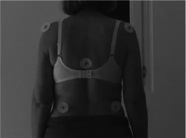
To enable the description of the trunk alignment during the tasks, five markers (circular patches of 4 cm diameter, white with black point in the middle) were placed on the subjects’ back (Figure 1). The placement was partially based on earlier studies,25,27,28as follows:
Marker 1 and 2: bilaterally and symmetrically on the posterior angle of the acromion.
Marker 3: on the spinal process of the seventh thoracic vertebra (the apex of kyphosis).
Marker 4 and 5: bilaterally and symmetrically at the level of the iliac crests.
The angles of the trunk (acromion–vertebra–iliac crest angles; AVIAs) on both sides in the different positions were determined with the use of angular dimension tool of Corel Draw (Corel Corp., Canada).
2.3. Ground contact force determination
All subjects were tested on a force platform (ZWE-PII Stabilometer; 50 × 50 cm; Elektro-Bionika LTD, Budapest, Hungary) to determine the contact forces (as a measure of WB capability) of both legs. The measurements were pre- ceded by verbal instruction and demonstration of the actual task. Initially, the subjects were instructed to stand on the platform with their feet 10 cm apart with their arms by their side and to gaze at the wall 3 m in front of them.21,29 Then, the subjects were asked to stand putting equal body weight on their lower extremities (SP). Next, the subjects were instructed to shift as much weight as possible onto the right leg without lifting off the left foot (WS position), then return to SP. The next task was to shift as much weight as possible onto the left leg without lifting off the right foot (WS position). This sequence was repeated 3 times, and every position was held for 3 s. All of the subjects could perform the tasks.
In the WS position, the two sides were designated as WBS and NWBS. The WBS was the loaded side while the NWBS was the unloaded side. Since no significant differences were found between the three trials, the means of the WS values were analyzed. Regarding SP, the mean of the first three measurements was used for further analyses. Ground contact forces were determined for each leg in all positions and expressed as percentage of body weight (relative ground force: RGF). The movement of the patients was also recorded with a video camera, and the positions of the markers were analyzed offline.
2.4. Data and statistical analyses
Asymmetry indices (AIs) were introduced based on a formula of symmetry index.30 These indices provide measures of RGFs (Asymmetry Indices of Weight bearing: AIW) and AVIAs (Asymmetry Indices of Angles: AIA) on both sides (see below). The data underlying the study are available at the corresponding author.
2.4.1. Data collection and analysis for SP
Two categories were defined according to RGF: the more loaded (MS) and the less loaded (LS) sides for both groups.
In the control group, all the subjects had more weight on MS than on the LS. However, since all of the patients had more load on their paretic side compared to the non-paretic side, therefore the MS was the non-paretic side, while the LS was the paretic side in all cases.
The asymmetry index of WB in starting position (AIW- SP) was determined as follows:
AIWSP¼ð½RGFMSRGFLS 100Þ=
0:5½RGFMSþRGFLS
ð Þ
Figure 1.Presentation of marker setup.
2 A. FEHÉR-KISS ET AL.
An asymmetry index for AVIAs in starting position (AIA-SP) was also calculated:
AIASP¼ð½AVIAMSAVIALS 100Þ=
0:5½AVIAMSþAVIALS
ð Þ
For both indices, 0 indicates perfect symmetry; a positive score indicates a higher value on the MS side, while a negative score indicates a lower value on the MS side.
2.4.2. Data collection and analysis for WS
Four categories were introduced for WS. Two are based on the side of WB (WBS vs. NWBS) and the categories deter- mined during SP (LS vs. MS):
In the case of WS to the LS: WBS-LS and NWBS-MS In the case of WS to the MS: WBS-MS and NWBS-LS Regarding the WB ability, AIs of RGF were introduced for both WS positions (WS to the LS and MS):
AIWWSLS¼ð½RGFWBSLSRGFNWBSMS 100Þ=
0:5½RGFWBSLSþRGFNWBSMS
ð Þ;and
AIWWSMS¼ð½RGFWBSMSRGFNWBSLS 100Þ=
0:5½RGFWBSMSþRGFNWBSLS
ð Þ;
Since RGF was always higher on the WBS compared to the NWBS, these values were positive. Regarding the analysis of the trunk angles in WS position, AIs were introduced as follows1:
AIA WS LS¼ð½AVIAWBSLSAVIANWBSMS 100Þ=
0:5½AVIAWBSLSþAVIANWBSMS
ð Þ;and
AIAWSMS¼ð½AVIAWBSMSAVIANWBSLS 100Þ=
0:5½AVIAWBSMSþAVIANWBSLS
ð Þ;
where positive scores represent greater angle on the WBS, while“0”represents perfect symmetry.
To determine the degree of trunk elongation and short- ening on both the WBS and NWBS and using SP as the baseline, the elongation index (EI) was introduced2:
EIWBS=NWBS¼ AVIAWBS=NWBSAVIASP
A positive value means elongation, and a negative value means shortening on the given side.
Data are presented as mean ± SEM. Since the data sets were not normally distributed, the nonparametric Mann–WhitneyU test was used to compare the two groups, and the Wilcoxon matched pairs signed-rank test was applied to perform the within-subject comparisons. To characterize correlations between the different parameters, Spearman’s rho was calcu- lated. Statistical analyses were performed in STATISTICA for Windows 12.0 (Statistica Inc., Tulsa, Oklahoma, USA). A P value lower than 0.05 was considered significant.
3. Results
3.1. Analysis of the SP
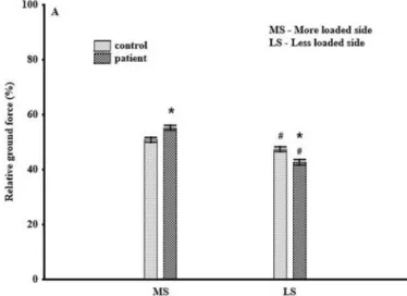
Regarding WB, both groups exhibited significant asymmetry between the two sides, but it was more pronounced in the patient group (Figure 2A); thus, the AIW-SP value also indi- cated significant difference between the two groups (signed with * inTable 1).
As for the AVIAs, no significant differences were found between the two groups on either side; however, the patient group was characterized by significantly smaller AVIAs on the LS than on the MS (P < 0.05) (signed with# in Figure 3A).
Therefore, the AIA-SP also showed significant difference between the two groups (signed with * inTable 1).
3.2. Analysis of WS position
Regarding the WS position, while the control subjects shifted slightly less weight on their LS than on the MS, the patients shifted significantly less weight on the LS than on the MS.
(Figure 2B)Thus, both groups were characterized by less asymmetry in the WS position when shifting the weight to LS than to MS, but the degree of the asymmetry was
Figure 2A.Relative ground force (RGF)in starting position (SP) in patients and controls.
*P< 0.05 compared to control group.#P< 0.05 compared to contralateral side.
Table 1.The mean (95% confidence intervals) of asymmetry indices of weight distribution (AIW) and trunk angles (AIA) in healthy subjects and stroke patients.
AIW AIA
SP
Control 7.1 ± (4.5–9.6) −2.4 ± (−6.6–1.9)
Patients 25.6 ± (4.2–16.7)* 4.5 ± (0.2–8.7)*
WS MS LS MS LS
Control 141 ± (125.5–157.3) 125 ± (105.8–144.4)# −1.7 ± (−5.32–1.83) 4.7 ± (−1.63–10.93)
Patients 125 ± (106.8–143.2) 82 ± (62.7–103.2)*# −0.5 ± (−6.17–5.21) −7.6 ± (−13.31 to−1.99)*
SP: Starting position; WS: (lateral) weight shift; MS: more loaded side; LS: less loaded side.
* and#denote significant differences (p< 0.05) compared to control group and MS, respectively.
Figure 2B.Relative ground force (RGF) in weight shift position in patients and controls.
*P< 0.05 compared to control group.#P< 0.05 compared to contralateral side.
Figure 3.Trunk angles (AVIAs) on both sides in starting and weight shift positions in patients and controls. *P< 0.05 compared to control group.#P< 0.05 compared to contralateral side (MS: more loaded side; LS: less loaded side WS: weight shift).
4 A. FEHÉR-KISS ET AL.
significantly lower in the patients when shifting weight to LS than in the control subjects (Table 1).
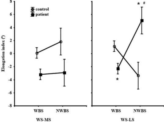
Regarding the changes of AVIAs in WS position on the MS, there were no significant differences between the WBS and NWBS and between the two groups (Figure 3B). On the contrary, in the case of WS on the LS, the stroke patients showed a perfectly opposite trend in the angles at the two sides compared to the control group, i.e. the AVIA values were significantly higher on the NWBS than the WBS (Figure 3C). Therefore, the AI for the angles in WS to the MS did not reveal significant asymmetries in either group, while in WS to the LS, showed an opposite pattern between the patients (decrease) and controls (increase); therefore, sig- nificant differences were observed between the two groups (Table 1). Regarding the EIs of the trunk only, patients showed significant shortening on the WBS and elongation on the NWBS in WS to the LS (Figure 4).
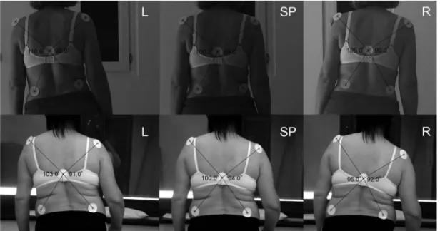
As shown inFigure 5, the difference between a patient and a control subject is well observable.
3.3. Correlation analyses
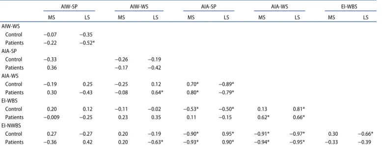
No significant correlations were found between the different RGFs in either group. As for the correlation between AVIAs and RGFs, only a few significant correlations were found (Table 2). The correlation analysis of AIs and EIs, however, revealed significant negative correlation between the WB asymmetry in SP and in WS, and significant positive correla- tion between WB asymmetry in WS and trunk angles asym- metry in WS in the patient group when shifting the weight to
the LS (Table 3). Furthermore, significant correlations were observed between trunk angles asymmetry in SP and in WS in both groups and on both sides. Since the EIs and AIs were calculated from the AVIAs, in most cases, there were signifi- cant correlations between these indices.
4. Discussion
Our study revealed major differences in trunk alignment between healthy subjects and stroke patients. It was sym- metrical in healthy subjects during quiet standing, while, in contrast with our hypothesis, only nonsignificant shorten- ing was detected in this group on the NWBS in WS posi- tion to the LS. In patients, the larger trunk angles on the NWBS compared to the WBS in WS to the LS revealed an altered (i.e. compensatory) strategy of trunk movement.
The few correlations between trunk angles and RGFs sug- gest that trunk angles do not necessarily reflect WS ability only but also the compensatory strategy. The significant correlations between angles asymmetry in SP and WS in both groups and on both sides show that subjects with a higher degree of asymmetry during SP will have a higher degree of asymmetry also in WS. The strong correlations between EIs and AIAs suggest that EIs might be used as parameters for the characterization of trunk alignment in different WS positions.
During SP, the expected body weight on a single limb would be 50% of the total body weight, and the control group showed only slight RGF asymmetry. Regarding the WS ability in our healthy group (82–86%), it was lower than in an earlier study
Figure 4.Elongation indices on both sides in starting and weight shift positions in patients and controls.*P< 0.05 compared to control group.#P< 0.05 compared to contralateral side (MS: more loaded side/LS: less loaded side; WBS: weight-bearing side; non-weight-bearing side).
(95% in individuals between 58 and 70 years of age),31which might be due to the involvement of older subjects in our study.
The significant inverse correlation (R=−0.75) between the age and WS percentages in the healthy group suggests slightly impaired postural stability in the older subjects.3,8,32Regarding the patient group, in agreement with earlier studies,1,8,9,32,33the non-paretic side had greater WB ability compared to the paretic one during SP; thus, the degree of asymmetry in the RGF was significantly higher than in the control group (Table 1).
The alterations of trunk alignment in SP and in different WS positions indicate that the stroke patients were not able to elongate the affected side; therefore, they performed WS in a compensatory manner. Surprisingly, asymmetry in WB
and trunk angles (AIW and AIA) correlated significantly (R = 0.64) only in patients shifting weight to the paretic side (LS), suggesting that asymmetry in WB is not neces- sarily linked to the asymmetries of trunk alignment. Since WS to the paretic side is a challenging task for these patients, it is assumed that the abnormal elongation/short- ening strategy might compensate for the impaired WB ability and may be applied as a protective strategy against falling.
Stroke patients are unable to perform satisfactory exten- sion of the lower extremity to support the body weight leading to passive standing.1 This passive WB position together with lateral flexion of the trunk to the WBS can
Figure 5.Changes of trunk alignment during weight shift in a healthy control subject (top) and a stroke patient (bottom). SP: The starting position (quiet standing);
L: shift to the left; R: shift to the right.
Table 2.Correlations between relative ground force (RGF) and acromion–vertebra–iliac crest angle (AVIA) values.
RGF-SP RGF-WS AIW-SP
MS LS MS LS MS LS
RGF-WS
Control 0.14 0.30
Patients −0.36 0.43
AVIA-SP
Control −0.10 −0.16 −0.03 0.44
Patients 0.53* −0.04 −0.32 0.09
AVIA-WS-WBS
Control 0.02 −0.21 −0.10 0.32
Patients 0.45 0.11 −0.17 0.29
AVIA-WS-NWBS
Control −0.09 0.55* 0.20 0.11
Patients −0.17 −0.44 −0.09 −069*
AIW-WS
Control −0.07 −0.35
Patients −0.22 −0.52*
AIW: Asymmetry Indices of Weight bearing; SP: starting position; WS: (lateral) weight shift; MS: more loaded side; LS: less loaded side; WBS: weight-bearing side;
NWBS: non-weight-bearing side.
*Significant degree of correlation.
6 A. FEHÉR-KISS ET AL.
help the patients to perform the required task. The weak abductors and adductors on the affected side and/or the deficient interaction or coordination between these muscles can also contribute to the observed impairments.34 Furthermore, the overactivity of the unaffected side and the stiffness of the paretic side can constrain WS toward either side, which the patients may try to compensate by trunk movements.21,34,35
A major focus of rehabilitation in hemiparesis is to increase the patients’ ability to bear weight on the paretic limb.5,7,9,21,22 However, so far, no particular attention has been paid to such patients’ trunk alignment in different standing positions. Our results suggest that a quantitative analysis of WB in itself is not enough to build up a correct treatment plan, since trunk alignment may also contribute to the movement performance. With more information about the pathological postural alignment associated with stroke, the therapist is better equipped to determine the state of the patients and to establish appropriate interventions. For instance, in patients with shortening on the WB side, atten- tion must be paid to the correction of this alignment, that is elongation should be facilitated on the loaded side.
The study had several limitations. One important limita- tion might be that the results are based on a relatively small sample of stroke patients. This fact has to be taken into consideration when interpreting such findings as moderate correlations between WB and the trunk angles. Another lim- itation is that we focused only on the description of WB and trunk alignment in these groups, but in this study, we did not evaluate the subjects’physical functions (motor control, coor- dination, strength, walking speed, and stroke severity). Thus, these results cannot be generalized to the entire population of stroke patients with hemiparesis. Furthermore, we did not evaluate the risk of falling and its correlation to trunk align- ment and weight distribution in this study; e.g. if the hemi- paretic limb utilization leads to increased or decreased fall
and if patients with moderate impairments can have full WB on the affected side. The amount of WB can be different in stroke patients with severe, moderate, or slight impairment.
Future studies should include the measurements of physical functions and the risk of falling. Clearly, studies with larger samples are needed with the abovementioned, extended mea- surements and training paradigms.
5. Conclusions
To our knowledge, this study is the first attempt to give an exact characterization of trunk alignment during quiet stand- ing and lateral WS in healthy and stroke patients. Obviously, this study was not designed to substantiate ultimate conclu- sions; its aim was rather to point out the practical importance of trunk alignment. The results revealed that the stroke patients’ WB ability is deficient and that they tend to com- pensate for this deficiency with abnormal trunk alignment.
Therefore, the measurement of WB ability in itself is an unsatisfactory proxy of therapeutic success, and attention must also be paid to how the individual patient performs WS, and incorporate this information in the therapeutic plan. Our simple method can help to reach that end by allowing the therapist to numerically describe patients’ trunk alignment in an easier way.
Acknowledgments
The authors wish to thank Csilla Keresztes and Janos Wodala for the linguistic correction of the manuscript.
Disclosure of interest
The authors report no conflicts of interest.
Table 3.Correlations between the asymmetry/elongation indices.
AIW-SP AIW-WS AIA-SP AIA-WS EI-WBS
MS LS MS LS MS LS MS LS MS LS
AIW-WS
Control −0.07 −0.35
Patients −0.22 −0.52*
AIA-SP
Control −0.33 −0.26 −0.19
Patients 0.36 −0.17 −0.42
AIA-WS
Control −0.19 0.25 −0.25 0.12 0.70* −0.89*
Patients 0.30 −0.43 −0.08 0.64* 0.80* −0.79*
EI-WBS
Control 0.20 0.12 −0.11 −0.02 −0.53* −0.50* 0.13 0.81*
Patients −0.009 −0.25 0.23 0.35 0.11 −0.15 0.62* 0.66*
EI-NWBS
Control 0.27 −0.27 0.20 −0.19 −0.90* 0.95* −0.91* −0.97* 0.30 −0.66*
Patients −0.36 0.42 0.20 −0.63* −0.93* 0.90* −0.94* −0.95* −0.33 −0.39
AIW: Asymmetry Indices of Weight bearing; AIA: Asymmetry Indices of Angles; EI: Elongation Indices; SP: starting position; WS: weight shift; WBS: weight-bearing side; NWBS: non-weight-bearing side.
*Significant degree of correlation.
ORCID
Katalin Jakab http://orcid.org/0000-0002-5157-8625 Gyöngyi Horváth http://orcid.org/0000-0002-6025-4577
References
1. Eng JJ, Chu KS. Reliability and comparison of weight-bearing ability during standing tasks for individuals with chronic stroke.
Arch Phys Med Rehabil.2002;83:1138 Phys.
2. de Haart M, Geurts AC, Dault MC, et al. Restoration of weight shifting capacity in patients with postacute stroke: a rehabilitation cohort study. Arch Phys Med Rehabil. 2005;86:7555 Ph.
doi:10.1016/j.apmr.2004.10.010
3. Blaszczyk JW, Prince F, Raiche M, et al. Effect of ageing and vision on limb load asymmetry during quiet stance. J Biomech. 2000;33:
1243omech.
4. Ryerson S, Levit K. Functional Movement Re-Education: A Contemporary Model for Stroke Rehabilitation. Philadelphia (PA):
Churchill Livingstone Inc;1997.
5. Mudie MH, Winzeler-Mercay U, Radwan S, et al. Training symmetry of weight distribution after stroke: a randomized controlled pilot study comparing task-related reach, Bobath and feedback training approaches. Clin Rehabil. 2002;16:5822 Re. doi:10.1191/0269215502 cr527oa
6. Dettman MA, Linder MT, Sepic SB. Relationship among walking performance, postural stability, and functional assessments of the hemiparetic patient.Am J Phys Med.1987;66:7787 .
7. Dickstein R, Dvir Z, Jehosua EB, et al. Automatic and voluntary lateral weight shifts in rehabilitation of hemiparetic patients.Clin Rehabil.1994;8:9194 . doi:10.1177/026921559400800201
8. Marigold DS, Eng JJ. The relationship of asymmetric weight-bear- ing with postural sway and visual reliance in stroke.Gait Posture.
2006;23:2496 Po. doi:10.1016/j.gaitpost.2005.03.001
9. Sackley CM, Baguley BI, Gent S, et al. The use of a balance performance monitor in the treatment of weight-bearing and weight-transference problems after stroke. Physiother.
1992;78:9072iot. doi:10.1016/S0031-9406(10)60498-1
10. Bohannon RW, Larkin PA. Lower extremity weight bearing under various standing conditions in independently ambulatory patients with hemiparesis.Phys Ther.1985;65:1323 Ther.
11. Laufer Y. The effect of walking aids on balance and weight-bearing patterns of patients with hemiparesis in various stance positions.
Phys Ther.2003;83:1123 Th.
12. Bohannon RW, Andrew AW. Limb muscle strength is impaired bilaterally after stroke. J Phys Ther Sci. 1995;7:199. doi:10.1589/
jpts.7.1
13. Ryerson S, Byl NN, Brown DA, et al. Altered trunk position sense and its relation to balance functions in people post-stroke.J Neurol Phys Ther.2008;32:1408u. doi:10.1097/NPT.0b013e3181660f0c 14. Dickstein R, Shefi S, Marcovitz E, et al. Anticipatory postural
adjustment in selected trunk muscles in post-stroke hemiparetic patients.Arch Phys Med Rehabil.2004;85:2614 Ph.
15. Barra J, Oujamaa L, Chauvineau V, et al. Asymmetric standing pos- ture after stroke is related to a biased egocentric coordinate system.
Neurology.2009;72:1582ology. doi:10.1212/WNL.0b013e3181a4123a 16. Spinazzola L, Cubelli R, Della Sala S. Impairments of trunk move-
ments following left or right hemisphere lesions: dissociation between apraxic errors and postural instability. Brain.
2003;126:2656nrmen. doi:10.1093/brain/awg266
17. Di Fabio RP, Badke MB. Relationship of sensory organization to balance function in patients with hemiplegia. Phys Ther.
1990;70:5420 Th.
18. Levin MF, Kleim JA, Wolf SA. What do motor f sensory organ“- compensation” mean in patients following stroke? Neurorehabil Neural Repair.2009;23:3139ore. doi:10.1177/1545968308328727 19. Pai YC, Rogers MW, Hedman LD, et al. Alterations in weight
transfer capabilities in adults with hemiparesis. Phys Ther.
1994;74:6474 Th.
20. Turnbull GI, Charteris J, Wall JC. Deficiencies in standing weight shifts by ambulant hemiplegic subjects. Arch Phys Med Rehabil.
1996;77:3566 Ph.
21. Goldie P, Evans O, Matyas T. Performance in the stability limits test during rehabilitation following stroke. Gait Posture.
1996;4:3156 Po. doi:10.1016/0966-6362(95)01059-9
22. Garland SJ, Willems DA, Ivanova TD, et al. Recovery of standing balance and functional mobility after stroke. Arch Phys Med Rehabil.2003;84:1753 Phys.
23. Day BL, Steiger MJ, Thompson PD, et al. Effect of vision and stance width on human body motion when standing: implications for afferent control of lateral sway.J Physiol.1993;469:4793ysi.
24. Blackburn JT, Riemann BL, Myers JB, et al. Kinematic analysis of the hip and trunk during bilateral stance on firm, foam and multi- axial support surfaces. Clin Biomech. 2003;18(7):6553 Bi.
doi:10.1016/S0268-0033(03)00091-3
25. Horak FB, Nashner LM. Central programming of postural move- ments: adaptation to altered support-surface configurations. J Neurophysiol.1986;55:1369uroph. doi:10.1152/jn.1986.55.6.1369 26. Rietdyk S, Patla AE, Winter DA, et al. Balance recovery from
medio-lateral perturbations of the upper body during standing.J Biomech.1999;32:1149omech.
27. Frigo C, Carabalona R, Dalla Mura M, et al. The upper body segmental movements during walking by young females. Clin Biomech.2003;18:4193 Bi. doi:10.1016/S0268-0033(03)00028-7 28. Vismara L, Menegoni F, Zaina F, et al. Effect of obesity and low
back pain on spinal mobility: a cross sectional study in women.J Neuroeng Rehabil.2010;7(3):1:1. doi:10.1186/1743-0003-7-1 29. Kim JW, Kwon Y, Jeon HM, et al. Feet distance and static postural
balance: implication on the role of natural stance.Biomed Mater Eng.2014;24(6):2681ed Ma. doi:10.3233/BME-141085
30. Robinson RO, Herzog W, Nigg BM. Use of force platform variables to quantify the effects of chiropractic manipulation on gait sym- metry.J Manipulative Physiol Ther.1987;4:1727nip.
31. Goldie PA, Matyas TA, Evans OM, et al. Maximum voluntary weight-bearing by the affected and unaffected legs in standing following stroke. Clin Biomech. 1996;11:3336 Bi. doi:10.1016/
0268-0033(96)00014-9
32. Cheng PT, Wang CM, Chung CY, et al. Effects of visual feedback rhytmic weight-shift training on hemiparetic stroke patients.Clin Rehabil.2004;18:7474 Re. doi:10.1191/0269215504cr778oa 33. Shumway-Cook A, Anson D, Haller S. Postural sway biofeedback:
its effect on reestablishing stance stability in hemiparetic patients.
Arch Phys Med Rehabil.1988;69:3958 Ph.
34. Kirker SG, Simpson DS, Jenner JR, et al. Stepping before standing:
hip muscle function in stepping and standing balance after stroke.J Neurol Neurosurg Psychiatry.2000;68:4580uro.
35. Dickstein R, Heffes Y, Laufer Y, et al. Activation of selected trunk muscles during symmetric functional activities in post stroke hemi- paretic and hemiplegic patients. J Neurol Neurosurg Psychiatry.
1999;66:2189uro.
8 A. FEHÉR-KISS ET AL.
