Review
Monitoring the Redox Status in Multiple Sclerosis
Masaru Tanaka1,2 and LászlóVécsei1,2,*
1 MTA-SZTE, Neuroscience Research Group, Semmelweis u. 6, H-6725 Szeged, Hungary;
tanaka.masaru.1@med.u-szeged.hu
2 Department of Neurology, Interdisciplinary Excellence Centre, Faculty of Medicine, University of Szeged, Semmelweis u. 6, H-6725 Szeged, Hungary
* Correspondence: vecsei.laszlo@med.u-szeged.hu; Tel.:+36-62-545-351
Received: 21 September 2020; Accepted: 9 October 2020; Published: 12 October 2020 Abstract: Worldwide, over 2.2 million people suffer from multiple sclerosis (MS), a multifactorial demyelinating disease of the central nervous system. MS is characterized by a wide range of motor, autonomic, and psychobehavioral symptoms, including depression, anxiety, and dementia.
The blood, cerebrospinal fluid, and postmortem brain samples of MS patients provide evidence on the disturbance of reduction-oxidation (redox) homeostasis, such as the alterations of oxidative and antioxidative enzyme activities and the presence of degradation products. This review article discusses the components of redox homeostasis, including reactive chemical species, oxidative enzymes, antioxidative enzymes, and degradation products. The reactive chemical species cover frequently discussed reactive oxygen/nitrogen species, infrequently featured reactive chemicals such as sulfur, carbonyl, halogen, selenium, and nucleophilic species that potentially act as reductive, as well as pro-oxidative stressors. The antioxidative enzyme systems cover the nuclear factor erythroid-2-related factor 2 (NRF2)-Kelch-like ECH-associated protein 1 (KEAP1) signaling pathway.
The NRF2 and other transcriptional factors potentially become a biomarker sensitive to the initial phase of oxidative stress. Altered components of the redox homeostasis in MS were discussed in search of a diagnostic, prognostic, predictive, and/or therapeutic biomarker. Finally, monitoring the battery of reactive chemical species, oxidative enzymes, antioxidative enzymes, and degradation products helps to evaluate the redox status of MS patients to expedite the building of personalized treatment plans for the sake of a better quality of life.
Keywords: oxidative stress; redox; antioxidant; multiple sclerosis; biomarker; neurodegenerative disease; personalized medicine
1. Introduction
Multiple sclerosis (MS) is an immune-mediated demyelinating disease of the brain and spinal cord, which over 2.2 million people worldwide suffer from; it affects primarily young adults from 20 to 40 years of age. After one to two decades, many MS patients enter a progressive phase of the disease. As survival rates have improved, MS patients suffer throughout the adult life. Years lived with disability begin to increase steeply early in the second decade of life and disability-adjusted life years peak in the sixth decade of life [1]. MS encompasses a wide range of symptoms from motor and autonomic dysfunctions to psychobehavioral disturbances, including gait difficulties, paresthesia, spasticity, vision problems, dizziness and vertigo, incontinence, constipation, sexual disturbances, pain, cognitive and emotional changes, anxiety, and depression [2–5]. Increased risk of depression and painful conditions in chronic illnesses are likely to be mediated by the kynurenine pathway of tryptophan metabolism [6,7].
Several genetic susceptibilities, environmental factors, and the aging process have been proposed to alter the risk of developing MS, but the underlying cause of the disease remains
Biomedicines2020,8, 406; doi:10.3390/biomedicines8100406 www.mdpi.com/journal/biomedicines
unknown [8,9]. The most typical pathomechanisms involved in MS are simultaneous inflammatory and neurodegenerative processes [10,11]. Main pathological findings of MS include the blood-brain barrier disruption, multifocal inflammation, demyelination, oligodendrocyte loss, reactive gliosis, and axonal degeneration [12]. Multifocal immune-mediated destruction of myelin and oligodendrocytes leading to progressive axonal loss is a main cause of neurological deficits in MS [13–15].
The diagnosis of MS is confirmed by the presence of two or more multifocal inflammatory or demyelinating attacks in the central nervous system (CNS) with objective clinical evidence, a single attack with magnetic resonance imaging (MRI)-detected lesions, and positive cerebrospinal fluid (CSF) analysis, or insidious neurological progression with positive brain MRI or CSF analysis [16].
The symptomatic course classifies MS into four subtypes. Approximately 85% of MS patients have alternating episodes of neurological disability and recovery that last for many years, termed relapsing-remitting MS (RRMS). Almost 90% of RRMS patients progress to steady neurological decline within 25 years, termed secondary progressive MS (SPMS). Nearly 10% of MS patients suffer from steady deterioration of neurological functions without recovery, termed primary progressive MS (PPMS). As few as 5% of MS patients present progressive neurological deficits with acute attacks with or without recovery, termed progressive-relapsing MS (PRMS) [17]. However, PRMS is no longer considered a subtype of MS and is now grouped into PPMS with active disease of new symptoms or changes in the MRI scan [18]. In addition, clinically isolated syndrome (CIS) is a single episode of monofocal or multifocal neurological deficits that lasts at least 24 h. CIS is one of the courses of MS disease [12].
As in other neurodegenerative diseases, MS is a clinically classified disease of CNS in which multifactorial factors, including genetic, environmental, socioeconomic, cultural, personal lifestyle, and aging play an initial role to form a causative complex, eventually converging into similar pathognomonic clinical pictures [4]. Inflammatory and demyelinating attacks are unique manifestations in MS, but different pathomechanisms govern the distinguished clinical courses in each subtype of MS.
Currently there is no cure for MS. Disease-modifying therapy is the mainstay of MS treatment.
Immunomodulators, immunosuppressors, and cytotoxic agents are main groups of medicine.
Immunomodulators, such as interferon (IFN)-beta (β), glatiramer acetate (GA), and siponimod are used for CIS. Short courses of high-dose corticosteroid methylprednisolone alleviate acute flare-ups of RRMS, indicating that an inflammatory process predominates in RRMS and relapse prevention of RRMS [19,20].
Siponimod is indicated for RRMS [20]. For active RRMS monoclonal antibodies alemtuzumab and ocrelizumab, and immunomodulator dimethyl fumarate, fingolimod, and teriflunomide are prescribed besides IFN-βand GA. For highly active RRMS, cytotoxic agents cladribine and mitoxantrone are indicated, besides immunomodulatory fingolimod, and monoclonal antibodies, natalizumab and ocrelizumab. PPMS and SPMS are characterized by a neurodegenerative process leading to neural death [18]. An immunosuppressive monoclonal antibody ocrelizumab is indicated for treatment of PPMS [21] and diroximel fumarate, siponimod, and ofatumumab were recently licensed for treatment of SPMS [20,22,23] (Table1).
Table 1.Licensed disease-modifying drugs in multiple sclerosis [19–26].
Class Drugs Indications
Immunomodulators
Interferon beta (IFN-β) CIS
Active RRMS Methylprednisolone Acute flare-ups RRMS
Relapse prevention
Glatiramer acetate (GA) CIS
Active RRMS
Dimethyl fumarate Active RRMS
Diroximel fumarate
CIS RRMS Active SPMS
Fingolimod Active RRMS
High-active RRMS
Teriflunomide Active RRMS
Siponimod
CIS RRMS SPMS
Immunosuppressors
Alemtuzumab Active RRMS
Natalizumab High-active RRMS
Ocrelizumab
Active RRMS High-active RRMS
PPMS Ofatumumab
CIS RRMS Active SPMS
Cytotoxic Agents Cladribine High-active RRMS
Mitoxantrone High-active RRMS
Either causative, accompanying, or resultant events of inflammation and neurodegeneration, disturbance of reduction-oxidation (redox) metabolism have been observed and play a crucial role in pathogenesis of MS [27–29]. The serum proteomics revealed that ceruloplasmin, clusterin, apolipoprotein E, and complement C3 were up-regulated in RRMS patients, compared to healthy controls. Vitamin D-binding protein showed a progressive trend of oxidation and the increased oxidation of apolipoprotein A-IV in progression from remission to relapse of MS [30]. CSF samples of patients in the remission stage of RRMS showed higher purine oxidation product uric acid, reduced antioxidant, and increased intrathecal synthesis of IgG [31]. The observations suggest the presence of redox metabolism disturbance and involvement of inflammatory process in RRMS. Furthermore, higher serum alpha (α)-tocopherol levels were associated with reduced T1 gadolinium (Gd+)-enhancing lesions and subsequent T2 lesions in MRI of RRMS patients on IFN-β. Antioxidant glutathione (GSH) mapping showed lower GSH concentrations in the frontoparietal region of patients suffering from PPMS and SPMS than RRMS and no significant difference between those of RRMS and controls. Thus, the oxidative stress in CNS was linked to neurodegeneration in progressive types of MS [32].
This review article reviewed participants of redox homeostasis, including reactive chemical species, oxidative and antioxidative enzymes, and resulting degradation products, all of which serve as evidence of biochemical assaults and tissue injuries under redox disequilibrium. Literature research was carried out using PubMed/MEDLINE and Google Scholar, using appropriate search terms, such as:
“reactive oxygen species”, “oxidative stress”, “redox”, “biomarker”, “neurodegenerative disease”,
“inflammation”, “neurodegeneration”, and/or “multiple sclerosis”. Meta-analyses, systematic reviews,
expert reviews, case-control studies, and cohort studies were included in this review. Selection criteria and a risk of bias assessment are described in TablesA1andA2of AppendixA. Alterations of redox components in MS in general, phase-, and treatment-specific components were reviewed in search of a potential diagnostic, prognostic, predictive, and/or therapeutic redox biomarker.
Finally, monitoring different redox components, including oxidative enzymes, antioxidative enzymes, and degradation products during the disease progression helps to evaluate the redox status of MS patients, thus expediting the building of the most appropriate personalized treatment plans for MS patients [33].
2. Oxidative Stress
A redox reaction is a type of chemical reaction that involves the transfer of electrons between two molecules. A pair of electrons transfers from a nucleophile to an electrophile, forming a new covalent bond. The redox reaction is common and vital to the basic function of life such as cellular respiration, in which sugar is oxidized to release energy, which is stored in ATP. Redox metabolisms constitute multiple metabolic pathways involved in the series of redox chemical reactions indispensable for sustaining life and, at same time, engaged in removal of electrophilic oxidative species and other harmful nucleophiles. The dynamic activities range from a single electron transfer, enzyme reaction, chemical reaction cascade, to signaling in cells, tissues, organ systems, and whole organismal levels [34]. Oxidative stress is a state caused by an imbalance between the relative levels of production of reactive oxidizing metabolites and their elimination by the enzymatic or non-enzymatic antioxidant system. The oxidative state is induced by oxidative stressors either derived from xenobiotics or produced from the activities of oxidative enzymes and essential cellular constituents [35] (Figure1a).
Oxidative stressors are linked to the aging process, neurologic disease, and psychiatric disorders including Alzheimer’s disease (AD), Parkinson’s disease (PD), MS, amyotrophic lateral sclerosis (ALS), and depression [36–40].
Biomedicines 2020, 8, x FOR PEER REVIEW 4 of 34
species and other harmful nucleophiles. The dynamic activities range from a single electron transfer, enzyme reaction, chemical reaction cascade, to signaling in cells, tissues, organ systems, and whole organismal levels [34]. Oxidative stress is a state caused by an imbalance between the relative levels of production of reactive oxidizing metabolites and their elimination by the enzymatic or non- enzymatic antioxidant system. The oxidative state is induced by oxidative stressors either derived from xenobiotics or produced from the activities of oxidative enzymes and essential cellular constituents [35] (Figure 1a). Oxidative stressors are linked to the aging process, neurologic disease, and psychiatric disorders including Alzheimer’s disease (AD), Parkinson’s disease (PD), MS, amyotrophic lateral sclerosis (ALS), and depression [36–40].
Figure 1. Redox homeostasis and antioxidant supplementation. (a) Oxidative stressors comprise of external and internal stressors, exerting oxidative chemical reactions in organism. Oxidation is an indispensable bioenergetic process to sustain life. (b) Reductive stressors are products of antioxidative enzymes that generate antioxidants in response to regular oxidation activity and increased oxidative stress. (c) Antioxidant supplementation attempts to shift the biphasic response from adverse to beneficial phase to maintain nucleophilic tone. This mechanism is called parahormesis.
2.1. Endogenous Oxidative Stressors: Oxidative Enzymes and Reactive Species
Endogenous oxidative stressors are produced from cellular activities of cytosolic xanthine dehydrogenase (XDH), membrane-bound nicotinamide adenine dinucleotide phosphate (NADPH) oxidase, inflammatory lipoxygenase (LOX), and cyclooxygenase (COX), phagocytic myeloperoxidase (MPO) in respiratory burst, a second messenger nitrogen oxide (NO)-producing nitric oxide synthetase (NOS), mitochondrial and lysosomal electron transfer chain (ETC) enzymes, among others. Transition metals such as iron (Fe2+) and copper (Cu+) also play a crucial role in the formation of oxidative stressors via the Fenton reaction [41].
2.1.1. Oxidative Enzymes Generating Reactive Oxygen Species
Reactive oxygen species (ROS) include several free radicals, such as superoxide (O2−•) and hydroxyl radical (OH•) and nonradical molecules such as hydrogen peroxide (H2O2) and organic hydroperoxide (ROOH) (Table 2). ROS are produced in endogenously in the cytosol, the plasma membrane, the membranes of mitochondria and endoplasmic reticulum, peroxisomes, and
Figure 1.Redox homeostasis and antioxidant supplementation. (a) Oxidative stressors comprise of external and internal stressors, exerting oxidative chemical reactions in organism. Oxidation is an indispensable bioenergetic process to sustain life. (b) Reductive stressors are products of antioxidative enzymes that generate antioxidants in response to regular oxidation activity and increased oxidative stress. (c) Antioxidant supplementation attempts to shift the biphasic response from adverse to beneficial phase to maintain nucleophilic tone. This mechanism is called parahormesis.
2.1. Endogenous Oxidative Stressors: Oxidative Enzymes and Reactive Species
Endogenous oxidative stressors are produced from cellular activities of cytosolic xanthine dehydrogenase (XDH), membrane-bound nicotinamide adenine dinucleotide phosphate (NADPH) oxidase, inflammatory lipoxygenase (LOX), and cyclooxygenase (COX), phagocytic myeloperoxidase (MPO) in respiratory burst, a second messenger nitrogen oxide (NO)-producing nitric oxide synthetase (NOS), mitochondrial and lysosomal electron transfer chain (ETC) enzymes, among others. Transition metals such as iron (Fe2+) and copper (Cu+) also play a crucial role in the formation of oxidative stressors via the Fenton reaction [41].
2.1.1. Oxidative Enzymes Generating Reactive Oxygen Species
Reactive oxygen species (ROS) include several free radicals, such as superoxide (O2−•
) and hydroxyl radical (OH•) and nonradical molecules such as hydrogen peroxide (H2O2) and organic hydroperoxide (ROOH) (Table2). ROS are produced in endogenously in the cytosol, the plasma membrane, the membranes of mitochondria and endoplasmic reticulum, peroxisomes, and phagocytic cells [42,43] (Figure2). ROS is difficult for direct measurement in biological tissues due to its highly reactivity and short life.Biomedicines 2020, 8, x FOR PEER REVIEW 6 of 34
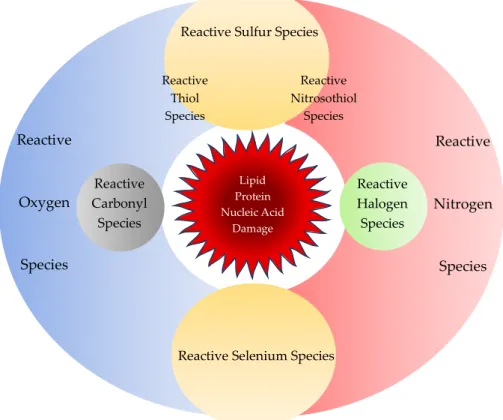
Figure 2. Reactive chemical species. Reactive chemical species comprise of not only reactive oxygen species and reactive nitrogen species, but also reactive sulfur, carbonyl, halogen, and selenium species. Sulfur reacts with oxygen or nitrogen to form reactive thiol or nitrosothiol species, respectively. All reactive chemical species react in concert during regular cellular activity but may cause oxidative stress to damage cellular components such as proteins, lipids, and nucleic acids.
In the cytosol, ROS can be generated by soluble intercellular components, such as catecholamines, hydroquinones, flavins, and thiols (RSHs) that undergo reduction reactions [44]. The cytosolic enzyme XDH normally catalyzes xanthine, NAD+, and water (H2O) to urate, reduced form of NAD+, NADH, and hydrogen ion (H+). Reversible oxidation of cysteine residues or irreversible Ca2+-stimulated proteolysis converts XDH to xanthine oxidase (XO) that transfers electrons to molecular oxygen (O2), producing superoxide (O2−•) during xanthine or hypoxanthine oxidation [45].
The serum levels of uric acid, a major endogenous antioxidant was measured in patients with PPMM, RRMM, and SPMM. The uric acid levels were significantly lower in active MS than inactive MS, and the uric acid levels were independently correlated with gender, disease activity and duration of the disease [46] (Table 3).
Table 3. Oxidative stress biomarkers of multiple sclerosis. The redox status can be monitored by the activities of oxidative and antioxidative enzymes and the presence of degradation products derived from cellular components. ↑: increase, ↓: decrease, -: unknown.
Classes Types Human Samples
Reference Blood CSF
Reactive Species Reactive Nitrogen Species ↑ ↑ [47]
Oxidative Enzymes
Xanthine Dehydrogenase
(XDH) ↓ - [46]
Nicotinamide Adenine Dinucleotide Phosphate
(NADPH) Oxidase
mixed - [48]
Superoxide Dismutase (SOD) ↑ ↓ [49–52]
Reactive Sulfur Species
Reactive Selenium Species Lipid
Protein Nucleic Acid
Damage
Reactive
Oxygen
Species
Reactive
Nitrogen Species Reactive
Halogen Species Reactive
Thiol Species
Reactive Nitrosothiol
Species
Reactive Carbonyl
Species
Figure 2.Reactive chemical species. Reactive chemical species comprise of not only reactive oxygen species and reactive nitrogen species, but also reactive sulfur, carbonyl, halogen, and selenium species.
Sulfur reacts with oxygen or nitrogen to form reactive thiol or nitrosothiol species, respectively.
All reactive chemical species react in concert during regular cellular activity but may cause oxidative stress to damage cellular components such as proteins, lipids, and nucleic acids.
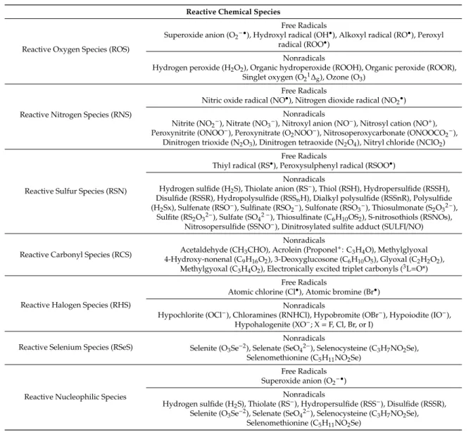
Table 2. Reactive chemical species. During regular cellular activity living cells generate numerous reactive chemical species containing oxygen, nitrogen, sulfur, carbonyl, halogen, or selenium. Reactive sulfur species can contain thiols or nitrothiols. Free radicals possess at least one unpaired electron that makes highly reactive and short-lived. Nonradicals are oxidizing chemicals or easily converted to free radicals. Superoxide, reactive sulfur species, or reactive selenium species can be reducing agents as reactive nucleophilic species.
Reactive Chemical Species
Reactive Oxygen Species (ROS)
Free Radicals
Superoxide anion (O2−•), Hydroxyl radical (OH•), Alkoxyl radical (RO•), Peroxyl radical (ROO•)
Nonradicals
Hydrogen peroxide (H2O2), Organic hydroperoxide (ROOH), Organic peroxide (ROOR), Singlet oxygen (O21∆g), Ozone (O3)
Reactive Nitrogen Species (RNS)
Free Radicals
Nitric oxide radical (NO•), Nitrogen dioxide radical (NO2•) Nonradicals
Nitrite (NO2−), Nitrate (NO3−), Nitroxyl anion (NO−), Nitrosyl cation (NO+), Peroxynitrite (ONOO−), Peroxynitrate (O2NOO−), Nitrosoperoxycarbonate (ONOOCO2−),
Dinitrogen trioxide (N2O3), Dinitrogen tetraoxide (N2O4), Nitryl chloride (NClO2)
Reactive Sulfur Species (RSN)
Free Radicals
Thiyl radical (RS•), Peroxysulphenyl radical (RSOO•) Nonradicals
Hydrogen sulfide (H2S), Thiolate anion (RS−), Thiol (RSH), Hydropersulfide (RSSH), Disulfide (RSSR), Hydropolysulfide (RSSnH), Dialkyl polysulfide (RSSnR), Polysulfide (H2Sx), Sulfenate (RSO−), Sulfinate (RSO2−), Sulfonate (RSO3−), Thiosulmonate (S2O32−),
Sulfite (RS2O32−), Sulfate (SO42−), Thiosulfinate (C6H10OS2), S-nitrosothiols (RSNOs), Nitrosopersulfide (SSNO−), Dinitrosylated sulfite adduct (SULFI/NO)
Reactive Carbonyl Species (RCS)
Nonradicals
Acetaldehyde (CH3CHO), Acrolein (Proponel+: C3H4O), Methylglyoxal 4-Hydroxy-nonenal (C9H16O2), 3-Deoxyglucosone (C6H10O5), Glyoxal (C2H2O2),
Methylgyoxal (C3H4O2), Electronically excited triplet carbonyls (3L=O*)
Reactive Halogen Species (RHS)
Free Radicals
Atomic chlorine (Cl•), Atomic bromine (Br•) Nonradicals
Hypochlorite (OCl−), Chloramines (RNHCl), Hypobromite (OBr−), Hypoiodite (IO−), Hypohalogenite (XO−; X=F, Cl, Br, or I)
Reactive Selenium Species (RSeS)
Nonradicals
Selenite (O3Se−2), Selenate (SeO42−), Selenocysteine (C3H7NO2Se), Selenomethionine (C5H11NO2Se)
Reactive Nucleophilic Species
Free Radicals Superoxide anion (O2−•)
Nonradicals
Hydrogen sulfide (H2S), Thiolate (RS−), Hydropersulfide (RSS−), Disulfide (RSSR), Selenite (O3Se−2), Selenate (SeO42−), Selenocysteine (C3H7NO2Se),
Selenomethionine (C5H11NO2Se)
In the cytosol, ROS can be generated by soluble intercellular components, such as catecholamines, hydroquinones, flavins, and thiols (RSHs) that undergo reduction reactions [44]. The cytosolic enzyme XDH normally catalyzes xanthine, NAD+, and water (H2O) to urate, reduced form of NAD+, NADH, and hydrogen ion (H+). Reversible oxidation of cysteine residues or irreversible Ca2+-stimulated proteolysis converts XDH to xanthine oxidase (XO) that transfers electrons to molecular oxygen (O2), producing superoxide (O2−•
) during xanthine or hypoxanthine oxidation [45]. The serum levels of uric acid, a major endogenous antioxidant was measured in patients with PPMM, RRMM, and SPMM.
The uric acid levels were significantly lower in active MS than inactive MS, and the uric acid levels were independently correlated with gender, disease activity and duration of the disease [46] (Table3).
Table 3.Oxidative stress biomarkers of multiple sclerosis. The redox status can be monitored by the activities of oxidative and antioxidative enzymes and the presence of degradation products derived from cellular components.↑: increase,↓: decrease,-: unknown.
Classes Types Human Samples
Reference
Blood CSF
Reactive Species Reactive Nitrogen Species ↑ ↑ [47]
Oxidative Enzymes
Xanthine Dehydrogenase (XDH) ↓ - [46]
Nicotinamide Adenine Dinucleotide Phosphate
(NADPH) Oxidase mixed - [48]
Superoxide Dismutase (SOD) ↑ ↓ [49–52]
Inducible Nitric Oxide Synthase (iNOS) ↑ ↑ [18,53–55]
Myeloperoxidase (MPO) mixed - [48]
Antioxidative Enzymes and Transcriptional Factors
Glutathione Peroxidase (GPx) ↑(relapse)
↓(remission) ↓ [56–61]
Glutathione Reductase (GSR) - ↑ [61,62]
Catalase ↓ - [61,62]
Xanthine oxidase (XO)-Uric Acid ↓ - [46]
Nuclear Factor Erythroid 2-Related Factor (Nrf2) ↑ - [52]
Peroxisome proliferator-activated receptors (PPARs) - ↑ [63]
Peroxisome proliferator-activated receptor gamma
coactivator 1-alpha (PGC-1α) ↓ - [64]
Degradation Products and End Products
Protein
Protein carbonyls ↑ - [65–71]
3-nitrotyrosin (3-NO-Tyr) ↑ - [68–70,72]
Protein glutathionylation - ↑ [73]
Dityrosine ↑ - [74]
Advanced oxidation protein products (AOPPs) ↑ - [49,74,75]
Advanced glycation end products (AGEs) ↑ ↑ [74,75]
Amino acids Asymmetric dimethylarginine (ADMA) ↓ - [76]
Lipid
F2-isoprostane (F2-isoP) ↑ ↑ [77–81]
Malondialdehyde (MDA) mixed ↑ [61,82–85]
4-hydroxynonenal (4-HNE) - ↑ [86]
Hydroxyoctadecadienoic acid (HODE) ↑ ↑ [87]
Oxysterol mixed ↑ [88]
DNA 8-dihydro-20deoxyguanosine (8-oxodG) ↑ - [56]
The plasma membrane is a network of phospholipid bilayer and integral proteins which protects the cellular organelles from the outer environment and is responsible for several cellular functions, such as cell adhesion, ion transport, cell signaling, and phagocytosis. The main ROS of the plasma membrane is a superoxide (O2−•) produced by the membrane-bound enzyme nicotinamide adenine dinucleotide phosphate oxidase (NOX), which is composed of two membrane proteins, three cytosolic proteins, and a small GTP-binding protein [89,90]. The expression of NOX isoform NOX5 was significantly increased, but the expression NOX4 was significantly decreased in serum of RRMS patients, suggesting that differential NOX isoform expression contributes to OS-associated vascular changes in MS [48]. ROS is also produced by COX and LOX, which convert arachidonic acid to prostaglandins, thromboxanes, and leukotrienes. Phospholipase A2generates ROS during arachidonic acid oxidation [91]. In the presence of transition metal ions, such as Fe2+and Cu+, hydrogen peroxide (H2O2), organic hydroperoxide (ROOH), and organic peroxide (ROOR) produce hydroxyl (OH•), alkoxyl (RO•), and peroxyl radical (ROO•), respectively [92] (Table3).
Superoxide dismutase (SOD) catalyzes the disproportionation of two superoxide (O2−•
) into molecular oxygen (O2) and hydrogen peroxide (H2O2). These enzymes are present in almost all aerobic cells and in extracellular fluids. SODs contain metal ion cofactors that, depending on the isozyme, can be copper, zinc, manganese, or iron [93,94]. There are three isozymes in humans. Dimeric copper- and zinc-coordinated SOD1 is in the cytoplasm; tetrameric manganese-coordinated SOD2 is confined to the mitochondria; tetrameric copper- and zinc-coordinated SOD3 is extracellular [95].
The mitochondrial ROS production takes place at four protein complexes, ubiquinone, and cytochrome c of the ETC, embedded in the inner membrane of the mitochondria [96]. The Complexes I/III/IV utilize NADH as the substrate, while Complexes II/III/IV use succinic acid. The complex II is also glycerol 3-phosphate dependent. The primary mitochondrial ROS is superoxide (O2−•) that is converted by
mitochondrial SOD into hydrogen peroxide (H2O2), which can be turned into hydroxyl (OH•) via the Fenton reaction [97]. The Complex I and III release superoxide (O2−•) into the mitochondrial matrix where it can damage the mitochondrial DNA, while the Complex III also releases superoxide (O2−•
) into the intermembrane space where it is accessible to the cytosol [98]. Reduced levels of antioxidant α-tocopherol was observed in the blood of patients with Leber’s hereditary optic neuropathy, which is caused by mitochondrial mutation of the Complex I, suggesting that oxidative load was elevated and antioxidant capacity was compromised [99]. Other mitochondrial enzymes that contribute to hydrogen peroxide (H2O2) production are monoamine oxidases, dihydroorotate dehydrogenase,α-glycerophosphate dehydrogenase, andα-ketoglutarate dehydrogenase (α-KGDH) complex. Succinate dehydrogenase also generates ROS [100].
Significantly higher mean activity of SOD in erythrocyte lysates was reported in RRMS than controls. Interestingly, the SOD activity of CIS was higher than that of RRMS [101]. The erythrocyte SOD activity can be a diagnostic biomarker of CIS and RRMS. The SOD activity was significantly lower in the erythrocyte lysates of RRSM patients upon relapse than controls but increased following the intravenous administration of corticosteroid methylprednisolone and remained higher during remission period than controls. The SOD is a potent predictive and therapeutic biomarker. The serum/plasma samples also showed significantly higher mean SOD activity in RRMS compared to control groups [82]. However, platelet SOD1 and SOD2 activity was unchanged in MS patients [49]. Intriguingly, SOD activity was significantly low in CSF of CIS and RRMS patients, despite the significantly high activity of plasma SOD. Furthermore, there were negative correlations between the erythrocyte SOD activity and disease duration and expanded disability status scale (EDSS) in CIS and RRMS, between the erythrocyte SOD activity and Gd+enhancement lesion volume in CIS patients [50]. These findings suggest the SOD activity as a possible diagnostic and prognostic marker (Tables3and4).
The peripheral blood mononuclear cells (PBMCs) SOD1 proteins and mRNA expression were significantly lower in RRMS patients than controls and became significantly more elevated following IFN-β1b treatment than the baseline [51]. These studies suggest SOD as a potential therapeutic biomarker (Table4). However, the erythrocyte SOD activity remained unchanged following the treatment of natalizumab, a humanized monoclonal antibody against the cell adhesion molecule α4-integrin. But levels of carbonylated protein and oxidized guanosine were reduced [52].
In the inner membrane of mitochondria and the endoplasmic reticulum, a heme-containing monooxygenase cytochrome P450 (CYP) enzymes are responsible for oxidizing steroids, cholesterols, and fatty acids. The CYPs forms ROS superoxide (O2−•) and hydrogen peroxide (ROOH) by substrate cycling [102]. Protonation of hydrogen peroxide (ROOH) forms hydrogen peroxide (H2O2) which, furthermore, cleaves into hydroxy radicals (OH•). The redox cycling produces free radical semiquinone from quinoid substrates [103]. In the mitochondrial transport chain, flavoprotein reductase forms ROS by direct reduction of O2and via the mediation of quinones. [104]. Superoxide (O2−•) is produced by XO in the reperfusion phase of ischemia, LOX, COX, and NADPH-dependent oxidase [105]. In the endoplasmic reticulum NADH cytochrome b5 reductase can leak electrons to molecular oxygen (O2) to generate superoxide (O2−•
) during the NADPH-dependent oxidation of xenobiotics [106].
Most enzymes in the peroxisomes produce ROS during the catalysis of fatty acidα- andβ-oxidation, amino acid and glyoxylate metabolism, and synthesis of lipidic compounds. A large fraction of hydrogen peroxide (H2O2) generated inside peroxisomes was observed to penetrate the peroxisomal membrane and diffuse to the surrounding media [107]. The peroxide can diffuse through the channel formed by the peroxisomal membrane protein Pxmp2 and hydrogen peroxide (H2O2) generated by the peroxisomal urate oxidase can release through crystalloid core tubules into the cytosol [108].
Meanwhile, peroxisomes also possess protective mechanisms to counteract oxidative stress and maintain redox balance. Reduction in peroxisomal gene and protein expression was observed in MS gray matter [109].
Table 4.Possible redox biomarkers in multiple sclerosis. Reactive chemical species, oxidative enzymes, antioxidants, antioxidative enzymes, degradation products, and end products are potential biomarkers for multiple sclerosis (MS). Diagnostic biomarkers allow early detection and secondary prevention;
prognostic biomarkers suggest the likely clinical course; predictive biomarkers predict the response of MS patients to a specific therapy; and therapeutic biomarkers indicate a target for therapy. CIS:
clinically isolated syndrome, PPMS: primary progressive MS; RRMS: relapsing-remitting MS, SPMS:
secondary progressive MS, mixedMM: mixed population of MS.
Class Components Biomarkers
Diagnostic Prognostic Predictive Therapeutic
Reactive Chemical Species
Total nitrite (NO2−
)/nitrite (NO3− ) value (tNOx)
PPMS, RRMS,
Relapse, SPMS RRMS - -
S-nitrosothiol RRMS, SPMS Spinal injury - -
Oxidative Enzymes
Superoxide dismutase (SOD) CIS, RRMS CIS, RRMS RRMS RRMS
Myeloperoxidase (MPO) RRMS RRMS - -
Inducible nitric oxide synthase (iNOS) RRMS - - -
Antioxidants and Antioxidative
Enzymes
Xanthine oxidase (XO)-Uric acid PPMM, RRMM,
SPMM - - -
Selenium RRMS - - -
Glutathione reductase (GSR) mixedMM MixedMM - -
Catalase CIS, RRMS RRMS - -
Thioredoxin-Peroxiredoxin (TRX-PRDX) MS - - -
Nuclear factor erythroid 2-related
factor (Nrf2) RRMS - RRMS -
Peroxisome proliferator-activated
receptors (PPARs) RRMS - - -
Peroxisome proliferator-activated receptor gamma coactivator
1-alpha (PGC-1α)
PPMS, SPMS - - -
Degradation Products and End Products
Protein carbonyls RRMS, SPMS RRMS, SPMS RRMS -
3-nitrotyrosine
(3-NO-Tyr) RRMS, SPMS - RRMS -
Glutathionylation Acute attack - - -
Dityrosine RRSM - - -
Advanced oxidation protein
products (AOPPs) CIS, RRMS RRMS RRMS -
Advanced glycation end products (AGEs) RRMS - - -
Asymmetric dimethylarginine
(ADMA) RRMS, SPMS - - -
F2-isoprostane
(F2-isoP) RRMS, SPMS SPMS - -
Malondialdehyde (MDA) RRMS RRMS RRMS -
4-hydroxynonenal
(4-HNE) PPMS, RRMS, SPMS - - -
Hydroxyoctadecadienoic acid (HODE) CIS, RRMS - - -
Oxocholesterols MixedMS SPMS - -
Oxidized low-density
lipoprotein (oxLDL) RRMS, SPMS - - -
8-OH2dG RRMS - - -
The lysosomal ETC plays a central role to support the positive proton gradient to maintain an optimal pH of the acid hydrolases [110]. The ETC is made up of a flavin-adenine dinucleotide, a b-type cytochrome and ubiquinone with the donor NADH and ending to acceptor molecular oxygen (O2), transferring three electrons. Superoxide (O2−•
) is possibly produced in the acidic environment which favors dismutation of hydrogen peroxide (H2O2) into hydroxy radical (OH•) by ferrous iron [111].
Furthermore, ozone (O3) and ozone-like oxidants are generated from singlet oxygen (O21∆g) catalyzed by antibody or amino acid. Ozone (O3) reacts with superoxide anion (O2−•) to form hydrogen peroxide (H2O2) in the presence of Fe+2[112].
2.1.2. Oxidative Enzymes Generating Reactive Nitrogen Species
Reactive nitrogen species (RNS) are a group of nitrogen-congaing molecules including free radicals, nitric oxide (NO), and nitrogen dioxide (NO2). Free radicals are nitric oxide (NO•) and nitrogen dioxide (NO2•) radicals, while nonradicals are nitrite (NO2−) and nitrate (NO3−), among others (Table2). RNS are derived from nitric oxide (NO) and superoxide anion (O2−•
) produced by nitric oxide
synthetase 2 (NOS2), NADPH oxidase, XO, LOX, and COX, among others [113,114]. At physiological concentrations, a gaseous molecule nitric oxide (NO) is a second messenger involved in blood pressure regulation, smooth muscle relaxation, defense mechanisms, immune regulation, and neurotransmission contributing the function of memory and learning [47].
Cross-sectional studies show that the levels of nitric oxide metabolites nitrite (NO2−
) and nitrite (NO3−), measured as a total value (tNOx), are significantly higher in plasma or serum of patients with RRMS [83,115]. A longitudinal study revealed that higher serum tNOx is significantly correlated with relapsing rate, suggesting prognostic biomarker of NOS [116]. Many studies of CSF samples reported significantly higher levels of tNOx in RRMS and PPMS, compared to healthy controls [117,118]. A study observed significantly higher levels of CSF tNOx in RRMS than SPMS, suggesting an inflammatory role of RNS [119]. Significantly higher levels of CSF tNOx were reported in patients with acute relapsing phase of RRMS than those with stable remitting phase of RRMS [120,121] (Tables3and4).
In cGMP-dependent pathways, nitric oxide radical (NO•) generated by endothelial NOS in endothelium, brain, and heart relaxes blood vessels and maintains normal blood pressure, while nitric oxide radicals (NO•) produced by neuronal NOS serve as a neurotransmitter to regulate blood pressure in the brain. Inducible NOS (iNOS) in macrophages and smooth muscle cells gives rise to nitric oxide radicals (NO•) as in reaction to bacterial lipopolysaccharides and/or cytokines [122].
Nitric oxide radical (NO•) is produced from the metabolism of L-arginine by NOS that converts L-arginine into L-citrulline and nitric oxide radical (NO•) by a 5-electron oxidation of a guanidine nitrogen of L-arginine [123]. In mitochondria nitric oxide radicals (NO•) react with respiratory Complex III to inhibit electron transfer and facilitate superoxide anion (O2•−) production. The nitric oxide radicals (NO•) also compete with molecular oxygen (O2) for the binding site at the binuclear center of cytochrome coxidoreductase, inducing a reversible inhibition of cytochrome c oxidase.
Nitric oxide (NO) neutralizes ROS [45]. However, RNS react with oxygen molecules (O2) and ROS, giving rise to a variety of nitrogen oxides (NOs), such as nitrogen dioxide radical (NO2•), nitrogen dioxide (NO2), dinitrogen trioxide (N2O3), peroxynitrite (ONOO−), nitrite (NO2−
), and nitrate (NO3−
). Higher concentrations of nitric oxide (NO) become toxic by forming nitrosothiols which oxidize tyrosine, cysteine, methionine, and GSH. In mitochondria, nitric oxide radicals (NO•) inhibit Complex I byS-nitrosation [124]. Together with other RNS, this contributes the damage of cell membranes, proteins, and lipid membrane leading to the degradation of mitochondria, lysosomes, and DNA. The chain of events culminates in the inhibition of immune response and production of carcinogenic nitrosamines [125]. Nitric oxide (NO) is also involved in metal homeostasis including Fe, Cu, and Zn [126].
Highly toxic peroxynitrite (ONOO−) is formed by the reaction of nitric oxide (NO) and superoxide anions (O2−•
), leading to the production of more reactive compounds that oxidize methionine and tyrosine residues of proteins, lipids, and DNA. Reacting with superoxide anion (O2−•), nitric oxide radicals (NO•) form peroxynitrite which causes reversible inhibition of cellular respiration in the mitochondria [127]. In peroxisomes nitric oxide radicals (NO•) react with superoxide anions (O2−•
) produced by XO to form peroxynitrite and hydrogen peroxides [111]. In addition, insulin resistance favors peroxintrite formation [128].
In response to bacterial lipopolysaccharides and inflammatory stimuli,iNOS generates nitric oxide (NO) that protects tissue hypoxia and serves as a neurotransmitter. However, overexpression iNOS, increase of nitric oxide (NO), and subsequent inflammation have been implicated in pathophysiology of neurodegenerative diseases including MS [47]. Calmodulin mediates the oxidative stress and inflammasome activation involving calmodulin-binding proteins, calcineurin, and calcium/calmodulin-dependent kinase II. Calmodulin inhibitors improved cognitive functions in an animal model of vascular dementia [129]. TheiNOS activity is upregulated in acute MS plaques [19,53].
Increased activity and expression ofiNOS in lymphocytes were found in active relapsing phase of RRMS [54]. CSFiNOS expression was shown in MS patients and mean CSF NOS activity was significantly higher, compared to controls [55] (Table3).
2.1.3. Reactive Sulfur Species
Reactive sulfur species (RSS) are sulfur-based redox-active compounds able to oxidize or reduce biomolecules under physiological conditions, often formed by thiols (RSHs) and disulfides (RSSHs).
RSS include cysteine and methionine, GSH, trypanothione, and mycothiol [130] (Table2).
Thiyl radicals (RS•), very reactive oxidants produced in the active site of enzymes such as the ribonucleotide reductase can react with nitric oxide radicals (NO•) [131]. Thiolate ions (RS−) are better nucleophiles than alkoxides because sulfur is more polarizable than oxygen [132]. Thiol (RSH) is a metal ligand [133]. Hydrogen sulfide (H2S) is produced from from L-cysteine by cistationine-γ-lyase.
Hydrogen sulfide (H2S) increases the activity ofN-methyl-D-aspartate receptor and β-adrenergic receptors through a cAMP-dependent protein kinase and activates NOS and the hemoxygenase favoring the formation of Nitric oxide (NO) and carbon monoxide (CO) from heme metabolism [134]. Hydrogen sulfide (H2S) is a metal ligand that reacts with other biological electrophilic sulfur species such as hydropersulfide (RSSR) and sulfenic acid (RSOH) [135]. Cysteine residues in GSH were found to be readily oxidized by superoxide anions (O2−•) to form singlet oxygen (O21∆g), glutathione disulfide (GSSG), and glutathione sulfonate (GSO3−
) in a reaction involved with peroxysulphenyl radical (RSOO•).
This mechanism may apply to cysteine residues in proteins [136]. Hydroxyl radicals (OH•) may also initiate the conversion of amino acids to peroxyl radicals [92]. Another reaction catalyzed by XO is the decomposition of S-nitrosothiols (RSNO), a RNS, to nitric oxide (NO), which reacts with a superoxide (O2−•
) anion to form peroxynitrite (ONOO−) under aerobic conditions [137]. Hydropersulfide (RSSH) is a nucleophile, as well as electrophilic molecule that is readily reduced to extremely potent reductant thiol (RSH). Disulfide (RSSR) is electrophilic RSS that can be reduced to thiol (RSH). Hydropolysulfide (RSSnH) and dialkyl polysulfide (RSSnR) are like hydropersulfide (RSSH) [135]. RSS can interact with ROS, generating sulfur oxides such as peroxysulphenyl radical (RSOO•), sulfenate (RSO−), sulfinate (RSO2−
), sulfonate (RSO3−
), thiosulmonate (S2O32−
), and SO42−
[138]. Sulfenate (RSO−) reacts with other thiols to give disulfides, RSSR. RSS can also interact with RNS, leading to the formation of S-N hybrid molecules, such as thiazate (NSO−), thionitrite (SNO−) isomers, S-nitrosothiols (RSNOs), nitrosopersulfide (SSNO−), and the dinitrosylated sulfite adduct, SULFI/NO. S-nitrosothiols (RSNOs) can be reduced to thiol (RSH) and nitroxyl (HNO) [139]. XO catalyzes S-nitrosothiols (RSNOs) to nitric oxide (NO), which reacts with a superoxide (O2−•) anion to form peroxynitrite (ONOO−) under aerobic conditions [123]. The properties of thiazate (NSO−), thionitrite (SNO−) isomers, nitrosopersulfide (SSNO−) and polysulfides dinitrososulfites (Sulfi/NO), are to be determined and they appear to be a source of nitric oxide (NO) and nitroxyl (HNO) [140–142] (Table2).
An antioxidantN-acetyl cysteine administration was reported to improve cognitive functions in patients in MS andN-acetyl cysteine supplement is under clinical trial for the treatment of fatigue in MS patients [143,144]. The levels of methionine reported mixed results. The plasma methionine levels were significantly reduced in RRMS and dietary methionine supplement was proposed for the treatment [145]. The level of methionine sulfoxide was elevated more than two-fold in CSF of MM patients and the reduction of dietary methionine was reported to slow the onset and progression of MM [146,147]. The levels of GSH in MS have not reached a consensus [147–149]. Trypanothione and mycothiol have not been investigated in MS. The serum S-nitrosothiol levels were increased in RRMS and SPMS, and selectively correlated with spinal cord injury and, thus, a high level of S-nitrosothiol is proposed to be a potential prognostic biomarker for spinal cord injury in MS [150] (Tables3and4).
2.1.4. Reactive Carbonyl/Halogen/Selenium Species
Reactive carbonyl species (RCS) are metabolically generated highly reactive molecules with aldehydes and electronically excited (3L=O*) triplet carbonyls, known for their harmful reactions to nucleic acids, proteins, and lipids [92]. In addition, RCS are considered to participate in electrophilic signaling of adaptive cell response and post-transcriptional protein modification [151]. RCS are classified intoα,β-unsaturated aldehydes; keto-aldehyde and di-aldehydes. In the presence of catalase and bicarbonate, XO was found to produce the strong one-electron oxidant carbonate radical anion
from oxidation with acetaldehyde. The carbonate radical was likely produced in one of the enzyme’s redox centers with a peroxymonocarbonate intermediate [45]. Self-reaction of lipid peroxyl radical (LOO•) produced by the oxidation of polyunsaturated fatty acids (PUFAs) by hydroxyl radical (HO•) generates electronically excited triplet carbonyls (3L=O*) yielding to singlet oxygen (O21∆g) [89]
(Table2). RCS react with amines and thiols leading to advanced glycation end products (AGEs), biomarkers of ageing and degenerative diseases [152].
MPO, a lysosomal heme-containing enzyme present in granulocytes and monocytes catalyzes the conversion of hydrogen peroxide (H2O2) and chloride anion (Cl−) to hypochlorous acid (HClO) during the respiratory burst. An adipocyte producing hormone leptin stimulates the oxidative burst [153].
MPO also oxidizes tyrosine to tyrosyl radical [154]. MPO mediates protein nitrosylation, forming 3-chlorotyrosine (3-Cl-Tyr) and dityrosine crosslinks [155,156] (Table2).
Studies on MPO activity reported mixed results. The mean MPO activity of peripheral leukocyte was observed reduced in a mixed population of MS patients, compared to controls [157]. Significantly higher serum MPO activity was measured in opticospinal phenotype (OSMS) of RRMS at relapse and remission and in conventional phenotype of RRMS at remission, compared to controls. A positive correlation was associated between Kurtzke’s EDSS and MPO activity at remission of OSMS [158]
(Table4). The mean MPO activity of peripheral leukocytes was found higher, but statistically not significant in RRMS, compared to controls [56] (Table3). No study regarding CSF MPO activity in MS was found. Selenium is an essential micronutrient with similar chemical and physical properties to sulfur, but more easily oxidized and kinetically more labile than sulfur. Selenium is a component of proteinogenic selenocysteine, naturally occurring selenomethionine and selenoproteins, such as glutathione peroxidase (GPx), thioredoxin reductase (TRXR), and selenoprotein P [57]. Reactive selenium species (RSeS) are selenium-containing inorganic and organic compounds including selenite (O3Se−2), selenocysteine (C3H7NO2Se), and selenomethionine (C5H11NO2Se) [58] (Table2). Selenium have both beneficial and harmful actions. At low concentration it works as an antioxidant, inhibiting lipid peroxidation and detoxifying ROS as a component of GPx and TRXR, while at high concentration it becomes a toxic pro-oxidant, generating ROS, inducing lipid oxidation and forming cross-linking in thioproteins [159] (Table2). The serum selenium levels were measured significantly lower in MS patients, compared to controls, suggesting antioxidant capacity is impaired in MS [160] (Table3).
2.1.5. Exogenous Oxidative Factors
Oxidative stressors are generated in reaction to exogenous stimuli such as pollutants, food and alcohol, cigarette smoke, heavy metals, chemotherapy, drug and xenobiotics, or radiation. Aging becomes more susceptible to their insults. Organic solvents, organic compounds such as quinone, pesticides and heavy metals including lead, arsenic, mercury, chromium, and cadmium are common sources of oxidative stressors [41]. Ultraviolet and infrared-B radiations generate oxygen radicals endogenously [161] (Figure1a). The levels of serum arsenic, malondialdehyde (MDA), and lactate were elevated and ferric-reducing activity of plasma was reduced RRMS patients and the levels of serum lithium were significantly lower and the levels of nitric oxide (NO) were higher in RRMS patients, compared to healthy controls, suggesting environmental factors seem to play a role in pathogenesis of MS [162,163].
3. Reductive Stress
3.1. Reactive Nucleophilic Species
Endogenous reductive stressors include nucleophilic free radical, inorganic, and organic molecules and antioxidative enzyme. (Figure1b). Superoxide (O2−•
) anion is one of the reactive nucleophilic species and a powerful reducing agent under physiological conditions, which initiates reaction cascades generating another ROS, such as hydrogen peroxide (H2O2) and sulfur dioxide (SO2) derivatives.
Hydrogen sulfide (H2S), thiolate (RS−), hydropersulfide (RSS−), and disulfide (RSSR) are reactive
nucleophilic species that can participate in nucleophilic substitution in vivo [133]. Selenium is more nucleophilic than sulfur due to its greater electron density. The selenol (RSeH) portion of selenocysteine (C3H7NO2Se) is ionized at physiological pH, making it more nucleophilic against oxidative species [164,165] (Table2).
3.2. Antioxidative Enzymes
Reductive stress is induced by excessive levels of reductive stressors that results from an elevation in GSH/GSSG ratio, NAD+/NADH, NADP+/NADPH and/or or overexpression of antioxidative enzymatic systems such as the GSH system, catalase, thioredoxin-peroxiredoxin (TRX-PRDX) system,α-ketoglutarate dehydrogenase (GPDH), and glycerol phosphate dehydrogenase [166,167].
The reductive stressors deplete reactive oxidative species and are harmful as oxidative stressors and implicated in pathological processes in AD, PD, and sporadic motor neuron disease, among others [168].
The GSH system consists of GSH, the enzymes for synthesis and recycling including gamma-glutamate cysteine ligase, glutathione synthetase, glutathione reductase (GSR), and gamma glutamyl transpeptidase, and the enzymes for metabolism and antioxidation including glutathione S-transferase and GPx [169]. The GPx is an enzyme containing four selenium-cofactors that catalyzes the reduction of hydrogen peroxide (H2O2) to water molecule (H2O) and organic hydroperoxide (ROOH) to alcohol (ROH) by converting reduced monomeric GSH to GSSG. Glutathione s-transferases show high activity with lipid peroxides [170]. Eight isozymes are in the cytosol, membrane, and plasma, protecting the organisms from oxidative stress [171].
Most studies on peripheral blood GPx activity reported nonsignificant results in a mixed population of MS [56,59,172–174]. However, lower mean GPx activity of erythrocyte lysates in remission and higher mean GPx were reported in acute relapse of RRMS [60]. GPx activity in CSF was found lower in MS patients [61]. The GSR activity of lymphocyte and granulocyte lysates were not significantly different in MS, compared to controls. However, a significant correlation of GPx and GRx was observed in controls, but not in MS [62]. Mean GRx activity of CSF was found significantly higher in MS patients [61] (Tables3and4).
Catalases a tetrameric heme- or manganese-containing dismutase that catalyzes the conversion of two hydrogen peroxide (H2O2) molecules to water (H2O) in the presence of small amount of hydrogen peroxide. The cofactor is oxidized by one molecule of hydrogen peroxide and then regenerated by transferring the bound oxygen to a second molecule of substrate. The enzyme is located in the peroxisomes, the cytosol of erythrocytes, and the mitochondria, removing harmful hydrogen peroxides to prevent cellular and tissue damage [175].
Studies on the catalase activity of peripheral blood samples reported equivocal results in MS.
The catalase activity of granulocyte lysates was found lower in MS patients, compared to controls [176].
The activities of CSF and plasma catalase were found increased in CIS and RRMS patients, compared to healthy controls, and MS patients with lower EDSS had higher plasma and CSF catalase activities [84]
(Tables3and4).
In TRX-PRDX system PRDXs catalyze the reduction of H2O2to H2O. H2O2oxidizes the peroxidatic cysteine of PRDXs to protein sulfenic acid (PSOH), which can react with the thiol (SH) group of the resolving cysteine to yield the formation of an inter-(typical) or intramolecular (atypical) disulfide bond.
TRX/TRXR system mediates the reduction of the PRDX disulfide bond. TRX reduced state is maintained by the flavoenzyme TRXR in the presence of NADPH. When H2O2exceeds the normal levels, PRDXs are overoxidized from PSOH to protein sulfinic acids (PSO2H). The latter can be reduced back to the native form of the enzyme by sulfiredoxin (SRX) in the presence of ATP. However, further oxidation of PRDXs to PSO3H is irreversible [177].
Serum Trx1 was significantly increased in the newly diagnosed MS patients, compared to controls.
TRX1andAPEX1mRNA expressions were significantly higher in the newly diagnosed MS patients, patients under INF-βtreatment, and patients who received immunosuppressant azathioprine or betamethasone, compared to healthy controls [178]. PRDX2mRNA is upregulated and PRDX2
expression is higher in MS lesions white matter of autopsy tissue of patients its expression level is positively correlated with the degree of inflammation and oxidative stress [179] (Tables3and4).
α-KGDH is a mitochondrial enzyme in Krebs cycle, which catalyzesα-ketoglutarate, coenzyme A and NAD+to succinyl-CoA, NADH and CO2,transferring an electron to the respiratory chain [180].
KGNH activity is sensitive to redox status. H2O2reversibly inhibits KGNH by glutathionylation of lipoic acid cofactor, resulting reducing electron supply to the respiratory chain. A lipid peroxidation product 4-hydroxy-2-nonenal (4-HNE) reacts with lipoic acid cofactor, inhibitingα-KGDH activity [181].
The pyruvate tolerance test showed higher activity ofα-KGDH in serum of MS patients [182]. However, reduced expression and activity of mitochondrialα-KGDH was observed in demyelinated axons that correlated with signs of axonal dysfunction (Table3) [183].
α-GPDH catalyzes the reversible redox conversion of dihydroxyacetone phosphate to sn-glycerol 3-phosphate, linking carbohydrate and lipid metabolism. A loss ofα-GPDH in oligodendrocytes were observed in chronic plaques of MS patients, suggesting the presence of antioxidant capacity impairment [63] (Table3).
Nrf2 is a transcriptional factor of the antioxidative enzyme genes including catalase, GPx, GRx, glutathione S-transferase, and SOD. In response to oxidative stress, the Kelch-like ECH-associated protein 1 (KEAP1) inhibits the ubiquitin-proteasome system in the cytosol and facilitates the translocation of Nrf2 into the nucleus to bind to thecis-acting enhancer sequence of the promotor region, the antioxidant response elements [184,185]. Activation of the Nrf2-Keap1 pathway has been observed in various types of cancers, accompanied with reduced antioxidant capacity and elevated oxidative stress and inflammation [186]. The cytoplasmic and nucleic Nrf2 protein expression of PBMC was increased and correlated with clinical improvement in MS patients on 14-month course of natalizumab, anα4 integrin receptor blocker [110] (Tables3and4).
Other transcriptional factors involved in energy metabolism have been investigated. Peroxisome proliferator-activated receptors (PPARs) are a transcriptional factor of the gene regulating energy metabolism including glucose metabolism, fatty acid oxidation, thermogenesis, lipid metabolism, and anti-inflammatory response [187]. PPARs have attracted growing attention as promising targets of many diseases such as diabetes and hyperlipidemia [188]. An isoform PPAR-gamma (PPAR-γ) was elevated in CSF samples of MS, compared to controls [64]. Peroxisome proliferator-activated receptor gamma coactivator 1-α(PGC-1α) 4 integrin receptor blocker is a transcriptional coactivator that regulates the genes involved in energy metabolism. Reduced PGC-1αexpression was associated with mitochondria changes and correlated with neural loss in MS [189] (Tables3and4).
3.3. Exogenous and Endogenous Antioxidants
A daily diet rich in naturally occurring polyphenolic antioxidants, such as flavonoids and phenolic acids are regularly recommended for disease prevention and antioxidant supplements, such as vitamin C, vitamin E, N-acetyl cysteine, L-carnitine and folic acid are frequently employed as a complementary therapy for various diseases [190,191]. Those preventive and therapeutic measures are based on the pathogenesis of diseases which are induced and developed under oxidative cellular environment. Furthermore, plant polyphenols were reported to alleviate oxidative stress and enhance neuroprotection [192]. Olive leaf-derived polyphenols are strong antioxidants and their therapeutic use are of particular interest against oxidant-induced diseases including cancer and neurodegenerative diseases [193–195]. The Mediterranean diet can present a biphasic dose-response curve toward hormetic stimuli, preventing low-grade inflammation and inflammageing [196,197]. There is a growing interest in supplementation of nonessential compounds that trigger the redox feedback loop activating the nucleophilic response, parahormesis (Figure1c) [198].
N-Methyl-D-aspartic acid receptor antagonist memantine and memantine-ferulic acid conjugate improved oxidative stress in patients with AD [199]. A meta-analysis of randomized controlled trials showed that an exogenous antioxidantN-acetylcysteine supplement improved cognitive function in patients with schizophrenia [200]. Traditional Chinese medicine curcumin is an antioxidant and
![Table 1. Licensed disease-modifying drugs in multiple sclerosis [19–26].](https://thumb-eu.123doks.com/thumbv2/9dokorg/1088187.74060/3.892.176.716.160.715/table-licensed-disease-modifying-drugs-multiple-sclerosis.webp)