Elevated levels of mitochondrial mortalin and cytosolic HSP70 in blood as risk factors in patients with colorectal cancer
Perri Rozenberg1*, Judit Kocsis2*, Moran Saar1, Zoltan Prohaszka2, George F€ust2†and Zvi Fishelson1
1Department of Cell and Developmental Biology, Sackler School of Medicine, Tel Aviv University, Tel Aviv, 69978, Israel
2Research Laboratory, 3rd Department of Internal Medicine, Semmelweis University, Kutv€olgyiut 4, Budapest, 1125, Hungary
Mortalin/GRP75 is a ubiquitous mitochondrial chaperone related to the cytosolic heat shock protein 70 (HSP70). It protects cells from senescence and apoptosis and is overexpressed in cancer cells. Cell resistance to complement-dependent cytotoxic- ity depends on mortalin and during complement attack mortalin is released from cells. Our goal was to determine whether cancer patients have circulating mortalin in blood. The significance of mortalin in blood to survival prospects of colorectal cancer patients was evaluated. Occurrence of extracellular soluble HSP70 (sHSP70) is documented. We developed a sensitive ELISA for mortalin. The association between mortalin level and survival was subjected to the Cox proportional hazards analy- sis (univariate and multivariate analyses). Mortalin concentration in serum of colorectal cancer patients was 10–214 ng/ml.
Survival data of the patients were known from an earlier study of sHSP70 in these samples. Cox regression analysis indicated that high mortalin (>60 ng/ml) is a risk factor for shorter survival. Serum levels of sHSP70 and mortalin in patients were inde- pendent variables. Concurrence of high sHSP70 and mortalin was associated with rapid disease progression (HR54, 2.04–
8.45,p<0.001). Addition of high sHSP70 and mortalin to a baseline model of age, sex and TNM stage, significantly
(p<0.001) enhanced the risk score to 8 (3.26–20.46). This is the first demonstration of circulating mortalin in cancer
patients. Analysis of mortalin in blood, and even more so of mortalin and sHSP70, adds a high prognostic value to the TNM stage and will identify colorectal cancer patients at high risk of poor survival.
Mortalin (mthsp70/grp75), the mitochondrial heat shock protein 70, plays a major role in import and refolding of mi- tochondrial proteins.1–3Mortalin is ubiquitously and constitu- tively expressed in all eukaryotic cells and its expression is not heat-induced, yet may be affected following ionizing radiation and glucose deprivation.4,5 Overexpression of mortalin protects cells from glucose deprivation and reactive oxygen species accu- mulation6and from serum deprivation,7reduced oxidative stress, antagonized ischemic damage,8resulted in lifespan extension of fibroblasts and promoted tumorigenesis.9 In contrast, knock down of mortalin by RNA interference caused senescence-like growth arrest in immortalized cells.10 Synthesized with a mito-
chondrial-targeting sequence, mortalin is mainly located in mitochondria, yet few reports identified it in other cellular com- partments such as the cytosol and plasma membrane.11
Several human transformed and tumor cells have been shown to express elevated levels of mortalin.9,12 In human colorectal adenocarcinoma, higher mortalin expression corre- lated with poor patient survival.13 Mortalin plays a role in the protection of cancer cells from complement-dependent cytotoxicity.14,15 It facilitates elimination of the complement membrane attack complex (MAC) from the cell surface by exo-vesiculation.14 Mortalin inhibitors sensitized the cells to complement-dependent cytotoxicity and inhibited the shed- ding of mortalin with the MAC.15
In vitro studies showed that during complement MAC attack, mortalin is shed from attacked cells.14,16This was the first demonstration of extracellular mortalin,14 yet the release of cytosolic HSP70 from cells is a well-known phenom- enon.17 Extracellular HSP70 was also identified in blood plasma of patients with colorectal cancer,18and was found to be a useful, stage-independent prognostic marker in colo- rectal cancer, especially in patients without distant metastasis.
Plasma levels of HSP70 and acute phase proteins could inde- pendently predict survival in patients with colorectal cancer but their combined measurements gave even a higher predic- tion value in specific subgroups of patients.19 To determine the amount of mortalin in the plasma of the same cancer patients, we developed a sensitive ELISA for mortalin. By using this ELISA we identified mortalin in blood serum of Key words:colorectal cancer, mortalin, GRP75, cytosolic HSP70,
prognosis
Additional Supporting Information may be found in the online version of this article.
*P.R. and J.K. contributed equally to this work
†George F€ust passed away on July 5, 2012
Grant sponsor:US–Israel Binational Science Foundation;Grant sponsor:Israel Science Foundation
History:Received 13 Nov 2012; Revised 19 Dec 2012; Accepted 28 Dec 2012; Online 15 Jan 2013
DOI: 10.1002/ijc.28029
Correspondence to:Zvi Fishelson, Department of Cell and Developmental Biology, Sackler School of Medicine, Tel Aviv University, Tel Aviv 69978, Israel, Tel.:197-23-6409620, Fax:197-23-6407432, E-mail: lifish@post.tau.ac.il
Short Report
International Journal of Cancer
colorectal patients. As shown here, higher serum mortalin level correlated with faster disease progression. The hazard index was found to be very high when levels of both mortalin and HSP70 in serum were elevated and even higher when combined with the TNM cancer stage.
Material and Methods Serum samples
Serum samples of colorectal cancer patients were collected at the outpatient oncology clinic of the 3rd Department of Internal Medicine, Semmelweis University, Budapest, between October 2000 and March 2005. A number of 175 consecutive patients, diagnosed with colorectal cancer, who were willing to give informed consent for the study, were enrolled regard- less of tumor stage.18Serum samples of 121 of these patients were available for this study and the median follow-up time was increased from 33 to 42 months. This patient cohort was already tested for the association between sHSP70 levels and survival18and between acute phase proteins plus sHSP70 and survival,19 except for four patients with no available serum samples for the present study. In the majority of cases, the primary tumor was removed surgically, according to relevant international guidelines, and patients were enrolled 4–6 weeks after surgery when blood was collected for the current study. In 16 cases, the primary tumor could not be removed before inclusion; these patients had advanced, metastatic tumors and were referred for primary chemotherapy. The patients were followed during and after chemotherapy in a protocol-based manner and health status and disease out- come were registered. More information on the patients is available in our earlier publications.18,19 The study received approvals by the ethical committees of Semmelweis Univer- sity, Budapest and Tel Aviv University, Tel Aviv. One hun- dred sixty-three of the 175 patients were followed up until May 2011 and their survival recorded.
Mortalin ELISA
To be able to measure serum levels of mortalin, we developed the following capture ELISA. Wells of 96-well MaxiSorp Nunc-Immuno plate (Thermo, Rochester, NY) were coated overnight at 4C with 1 mg/ml mouse monoclonal antibody directed to human mortalin (StressMarq, Victoria, Canada) in Tris buffer (TB) (50 mM Tris Base pH 7.0, 100 mM NaCl). The wells were blocked with bovine serum albumin 15 mg/ml (Sigma-Aldrich, Israel) in Tris-buffered saline for 1
hr at 37C and then serum samples diluted 1:3 in TB were added to the wells for 2 h at 37C. Next, the wells were washed with TBT (TB with 0.05% Tween-20) and treated with a goat anti-mortalin polyclonal antibody (Santa Cruz Bio- technology, Santa Cruz, CA) diluted in TB for 1 hr at 37C.
Finally, after a wash with TBT, peroxidase-conjugated donkey anti-goat IgG (Jackson ImmunoResearch Laboratories, West Grove, PA) diluted in TB was added for 1 hr at 25C. Anti- body binding was quantified by using TMB One Component Microwell Substrate (SouthernBiotech, Birmingham, AL) and absorbance was read at 450 nm in a Microplate Reader (Spec- trafluor plus, Tecan, Austria). Calibration of the quantity of mortalin was included in each ELISA plate with 0–2.5 ng puri- fied recombinant mortalin (Saar et al., in preparation) mixed with normal human serum (same dilution as the tested sam- ples). Specificity of the anti-mortalin antibodies used was con- firmed in Western blots showing a single protein band in whole cell lysates (similar to the bands shown in Ref. 15). Pu- rity of the recombinant mortalin was also confirmed.15 Ab- sorbance of normal serum without recombinant mortalin was subtracted from all samples in the plate. A typical calibration curve is shown in Supporting Information Figure S1.
Statistical analysis
Statistical analysis was performed using the SPSS 15.0 soft- ware (SPSS Inc., Chicago, IL). Patients’ survival was tested by the Kaplan–Meier survival analysis, using the Log Rank test.
The power of the study to identify the observed difference in mortality rate (0.18) between low- and high-mortalin groups was 0.78. The impact of soluble mortalin and other factors on patients’ survival was tested by the Cox proportional hazards analysis (univariate and multivariate analyses). The results of the Cox regression models are presented as hazard ratios, the corresponding 95% confidence intervals (CI) and the Wald chi-square andpvalues of likelihood ratio tests.
Results
Concentration of mortalin in sera of patients with colorectal cancer
Mortalin is shed in vitro from complement attacked viable human erythroleukemia cells.14 Release of mortalin in vitro was also demonstrated from human colon carcinoma HCT116 cells (Supporting Information Fig. S2), mouse colo- rectal CT26 cells, mouse lymphoma EL4 cells, mouse bladder carcinoma MBT2 cells and human B lymphoma Raji cells (Masarwa and Fishelson, in preparation). In order to measure What’s new?
Mortalin is a ubiquitous mitochondrial chaperone protein that protects cells from apoptosis and is overexpressed in cancer cells. Here, a novel ELISA developed for the detection and quantification of circulating mortalin in sera of colorectal cancer patients is described. The assay revealed that high circulating levels of both mortalin and the closely related cytosolic protein HSP70 are indicative of short patient survival. The results suggest that mortalin, HSP70, and TNM staging are independent survival determinants for colorectal cancer.
Short Report
mortalin level in patients’ blood, we set to develop a sensitive mortalin ELISA with a low background reading. Several alter- native protocols were tested and finally the captured ELISA described in details under Methods was found to be satisfac- tory. This mortalin ELISA gave a reproducible and sensitive detection of 0.25 ng of mortalin in 33diluted human se- rum. Each ELISA plate had wells with normal human serum alone or human serum with increasing amounts of recombi- nant mortalin. Normal human serum gave a low background readout that was subtracted from all samples. The recombi- nant mortalin samples gave a linear dose-dependent calibra- tion curve (Supporting Information Fig. S1) that was used to convert the optical density readout of the clinical samples into ng mortalin.
By using this ELISA, significant amounts of soluble mor- talin were detected in serum samples of all colorectal cancer patients (14–215 ng/ml). Patients were divided into two groups, according to their serum mortalin level. The low mortalin group (n588) had serum mortalin concentration below 60 ng/ml, whereas the high mortalin group (n533) had levels equal to or higher than 60 ng/ml. This concentra- tion of mortalin was chosen based on receiver operating characteristic (ROC) calculation to yield optimal stratification of patients regarding mortalin levels and survival.
Mortalin concentrations were analyzed in relation to the tumor stage of the patients determined in earlier studies.18,19 There was no difference in the mortalin levels among the patients with different size of tumors (TNM-T) (p50.151, Kruskal–Wallis test), among patients with different extent of lymph node involvement (TNM-N) (p50.346, Kruskal–
Wallis test) or between patients with or without distant
metastases (TNM-M) (p50.205, Mann–Whitney test). Like- wise, there was no significant association between mortalin levels and different Duke’s grade categories and grade of pri- mary tumor (both p>0.050, data not shown). Similarly, data were available on the serum levels of sHSP70, C-reactive pro- tein (CRP) and C1-Inhibitor in these patients,18,19 Mortalin concentrations did not show correlation to the sHSP70 con- centrations (Spearman correlation coefficient R50.051, p50.580) or CRP levels (R50.018,p50.878) whereas a sig- nificant correlation (R50.335,p<0.001) was found between serum mortalin and C1-Inhibitor concentrations.
Univariate survival analysis
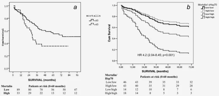
Kaplan–Meier survival analysis of patients with low (<60 ng/ml) vs. high (>60 ng/ml) mortalin level in serum during a 10-year follow-up gave a Log Rank overall comparison of p50.042 between the two groups, with 113 months esti- mated median survival in the low mortalin group and 29 months median survival in the high mortalin group (Fig. 1a).
Univariate analysis by Cox regression of the variables: age and sex of the patients, TNM-N and TNM-M stage of the disease as well as serum mortalin and sHSP70 concentrations are presented in Supporting Information Table S1. Only patients without missing data were included in this analysis (n599). Age was found to be a highly significant factor with a hazard of 1.04 per year (p50.002). Females had a Hazard of 1.62 (p50.029) relative to males. Both TNM-N (metasta- ses in the regional lymph node) and TNM-M (distant metas- tases) were highly significant predictors of mortality while TNM-T was not (data not shown). High sHSP70 serum con- centration (>1.65 ng/ml) was associated with mortality with
Figure 1.Survival (Kaplan-Meier) of colorectal cancer patients. (A) Patients divided according to mortalin level in their serum; high (60 ng=ml) or low (<60 ng=ml). Log Rank overall comparison showed significant equality of survival distribution (p50.042). (B) Patients di- vided according to their mortalin (low:<60 ng=ml, high:>60 ng=ml) and sHSP70 (low:<1.65 ng=ml, high:>1.65 ng=ml) levels. Analysis by Cox regression HR (with 95% CI) of patients with both high mortalin and high sHSP70 vs. both low mortalin and low sHSP70 is indi- cated. Number of patients in each category, on months: 0, 12, 24, 36, 48 and 60, is shown below the figures.
Short Report
a HR of 1.86 (p50.006). High mortalin (>60 ng/ml) level is a significant factor (p50.048) with a hazard ratio of 1.73. In this analysis, patients with high and low serum mortalin lev- els had a mean survival time of 43.19 (32.39–53.99) months and 72.52 months (62.78–82.76), respectively (p50.046).
Multivariate analysis: Independent and additive effects of risk factors
The association of the same variables with mortality of the can- cer patients was evaluated by multivariate Cox regression anal- ysis (Table 1). The effects of age and sex were found to be insignificant, while the hazard of TNM-N11TNM-N2 stages vs. TNM-N0 stage (lymph nodes involved, yesvs. no) remained highly significant (HR 3.34 (p50.001). Similarly, patients with distant metastasis had significantly higher risk (HR: 4.04;
p<0.001) than those with no distant metastases. In the multi- variable model, the hazard ratio associated with high sHSP70 was 3.508 (p<0.001) while high mortalin concentrations exhibited a weak but still significant association with mortality of patients, independently of the sHSP70 levels (Table 1).
Thereafter, we analyzed the survival of the patients strati- fied according to sHSP70 and mortalin levels (Fig. 1b). In this type of multivariate analysis, high mortalin level with low sHSP70 or low mortalin with high sHSP70 did not affect the mortality of patients. In contrast, patients with concomi- tant high mortalin and high sHSP70 concentrations had a 4.2 times higher hazard of mortality than patients with both low sHSP70 and low mortalin levels, indicating the additive na- ture of these two biomarkers (Fig. 1b). Median survival time of the patients with high mortalin/high sHSP70 and low mortalin/low sHSP70 levels were 27.5 (13.3–34.3) months and 57.0 (31.0–81.0) months, respectively.
Finally, we examined the additive impact of serum con- centrations of mortalin and sHSP70 on prediction of patients’
survival, as compared to the baseline model that includes
the basic demographic variables as well as the tumor stage (Table 2). According to the results of the likelihood-ratio test (v2526.846. p<0.0001), addition of the biomarkers sHSP70 and mortalin to the baseline model significantly increased the survival-predicting value of the model. In the subgroup of patients with concomitantly high sHSP70 and mortlain levels, a hazard rate of 8.176 (95% CI 3.267–20.463,p<0.001) was observed, indicating that the baseline model (clinical stage, age, and sex) and the biomarkers are independent and addi- tive predictors of mortality in colorectal cancer.
Discussion
As shown here, colorectal cancer patients have elevated mor- talin concentrations in their blood serum. Development of a novel and sensitive mortalin ELISA permitted us to quantify the soluble mortalin level in blood serum of patients with colorectal cancer. The amount of circulating soluble mortalin was found to be variable (10–214 ng/ml) and independent on stage of the disease.
Table 1.Multivariate analysis by Cox-regression of the association between age and sex of the patients as well as high baseline sHSP70 levels and high baseline mortalin levels on survival of the patients with colorectal cancer
95.0%
confidence interval Variable1
Significance (p)
Hazard
ratio Lower Upper
Age at diagnosis 0.488 1.013 0.977 1.050
Sex 0.900 0.959 0.497 1.850
TNM-N 0.001 3.337 1.661 6.704
TNM-M <0.001 4.044 2.014 8.118
Mortalin 0.044 1.976 1.019 3.831
sHSP70 <0.001 3.508 1.753 7.019
1Age (years); Sex (female/male); TMN-N (1 or 2 metastasis in the regional lymph nodesvs. 0, yes/no); TNM-M (distant metastasis, yes/
no); sHSP70 (high:>median 1.67 ng/ml, low:<1.67 ng/ml); mortalin (high:>median, 60 ng/ml, low:<60 ng/ml).
Table 2.Additive effect of the high mortalin and high sHSP70 levels in serum on the age and tumor TNM-dependent mortality risk of patients with colorectal cancer
95.0%
confidence interval Variable1
Significance (p)
Hazard
ratio Lower Upper Model 1
Age at diagnosis 0.098 1.030 0.995 1.066
Sex 0.865 0.947 0.508 1.768
TNM-N 0.003 1.853 1.234 2.784
TNM-M 0.001 3.130 1.630 6.013
Model 2
Age 0.323 1.017 0.983 1.052
Sex 0.571 1.218 0.617 2.404
TNM-N 0.003 1.879 1.244 2.840
TNM-M <0.0001 5.000 2.423 10.318
Mortalin low & sHSP70 low
– – – –-
Mortalin high & sHSP70 low
0.223 0.390 0.086 1.772
Mortalin low & sHSP70 high
0.123 1.836 0.849 3.974
Mortalin high & sHSP70 high
<0.001 8.176 3.267 20.463
Likelihood-ratio-test as compared to model 1:v2526.846.
p<0.0001
1Age (years); Sex (female/male); TMN-N (1 or 2 metastasis in the re- gional lymph nodesvs. 0, yes/no); TNM-M (distant metastasis, yes/
no); sHSP70 (high:>median 1.67 ng/ml, low:<1.67 ng/ml); Mortalin (high:>median, 60 ng/ml, low:<60 ng/ml). Number of patients in the different categories (mortalin/sHsp70): low/low-46; high/ low-43; low/
high-14; high/high-18).
Analysis by model-building tool of Cox regression analysis.
Short Report
The cohort of cancer patients studied here was part of a larger group of 175 patients with colorectal cancer studied earlier in which the level of soluble cytosolic HSP70 was ana- lyzed. Correlation was found between high sHSP70 and faster disease progression in colorectal cancer.18 A follow-up study examined in the same patients several other potential prog- nostic bio-markers.19 High (above median) levels of CRP, C1-INH and sHSP70 were found to be independently associ- ated with poor patient survival. The additive effect of high sHSP70, CRP and C1-INH levels on the survival of patients exceeded that of high sHSP70 alone.19 Updated survival in- formation since the previous study was available on 163 patients. Serum sample of 121 patients were tested for soluble mortalin levels. Patients that have a higher mortalin level (>60 ng/ml) also have a significantly shorter median survival time (29 months) than patients that have low mortalin level (113 months, p50.042). The hazard value of high mortalin is 1.730. Therefore, high circulating mortalin level is pro- posed as a risk factor in colorectal carcinoma. Multivariate analysis of survival data of 99 patients, for which age, sex, cancer stage, soluble HSP70 and mortalin information was available, indicated that patients with both high soluble HSP70 and high mortalin had more than eight-times higher risk of mortality (8.176, 3.267–20.463, p<0.001) as com- pared to those with low concentrations of both biomarkers.
We conclude that having concomitant high circulating HSP70 and mortalin levels indicates bad prognosis in patients with colorectal cancer, thus measurement of these two biomarkers may be informative in this setting.
Immunohistochemical analysis demonstrated that mor- talin is overexpressed in several tumor types, including colo- rectal adenocarcinoma and hepatocellular carcinoma.9,12,13,20 Elevated mortalin in liver cancer was associated with metas- tasis and early cancer recurrence.20 Over-expression of mor- talin in colorectal adenocarcinomas is also correlated with poor survival.13 Apparently, mortalin confers an advantage to cancer growth and metastasis, but the mechanism is still poorly characterized. In addition, mortalin promotes cell re- sistance to complement-dependent cytotoxicity (CDC) and may permit escape of cancer cells from antibody-based immunotherapy.14–16 Down-regulation of mortalin, using siRNA, antibodies or MKT-077 enhances cells sensitivity to CDC, whereas overexpression of mortalin enhanced cell re- sistance to CDC. As shown here, serum levels of mortalin and HSP70 and the TNM stage are apparently independent determinants in survival of patients with colorectal cancer.
This suggests that mortalin and HSP70 are secreted into the blood from different tissues and/or under distinct signals.
The trigger that has induced in these patients, mortalin release into the blood and the origin of this mortalin, remains to be identified. Possibly, inflammatory or other stress responses cause secretion of mortalin into the circula- tion from metastatic cells and/or non-cancerous affected tissues.
Acknowledgement
Authors thank Ilana Gelernter for her valuable assistance with the statistical analysis of the results.
References
1. Kang PJ, Ostermann J, Shilling J, et al.
Requirement for hsp70 in the mitochondrial matrix for translocation and folding of precursor proteins.Nature1990;348:137–43.
2. Bhattacharyya T, Karnezis AN, Murphy SP, et al.
Cloning and subcellular localization of human mitochondrial hsp70.J Biol Chem1995;270:1705–
10.
3. Wiedemann N, Frazier AE, Pfanner N. The protein import machinery of mitochondria.J Biol Chem2004;279:14473–6.
4. Merrick BA, Walker VR, He C, et al. Induction of novel Grp75 isoforms by 2-deoxyglucose in human and murine fibroblasts.Cancer Lett 1997;119:185–90.
5. Massa SM, Longo FM, Zuo J, et al. Cloning of rat grp75, an hsp70-family member, and its expression in normal and ischemic brain.J Neurosci Res1995;40:807–19.
6. Liu Y, Liu W, Song XD, et al. Effect of GRP75/
mthsp70/PBP74/mortalin overexpression on intracellular ATP level, mitochondrial membrane potential and ROS accumulation following glucose deprivation in PC12 cells.Mol Cell Biochem2005;268:45–51.
7. Taurin S, Seyrantepe V, Orlov SN, et al.
Proteome analysis and functional expression identify mortalin as an antiapoptotic gene induced by elevation of [Na1]i/[K1]i ratio in cultured vascular smooth muscle cells.Circ Res 2002;91:915–22.
8. Xu L, Voloboueva LA, Ouyang Y, et al.
Overexpression of mitochondrial Hsp70/Hsp75 in rat brain protects mitochondria, reduces oxidative stress, and protects from focal ischemia.J Cereb Blood Flow Metab2009;29:365–74.
9. Wadhwa R, Takano S, Kaur K, et al.
Upregulation of mortalin/mthsp70/Grp75 contributes to human carcinogenesis.Int J Cancer 2006;118:2973–80.
10. Wadhwa R, Takano S, Taira K, et al. Reduction in mortalin level by its antisense expression causes senescence-like growth arrest in human immortalized cells.J Gene Med2004;6:
439–44.
11. Ran Q, Wadhwa R, Kawai R, et al.
Extramitochondrial localization of mortalin/
mthsp70/PBP74/GRP75.Biochem Biophys Res Commun2000;275:174–9.
12. Takano S, Wadhwa R, Yoshii Y, et al. Elevated levels of mortalin expression in human brain tumors.Exp Cell Res1997;237:38–45.
13. Dundas SR, Lawrie LC, Rooney PH, et al.
Mortalin is over-expressed by colorectal adenocarcinomas and correlates with poor survival.J Pathol2005;205:74–81.
14. Pilzer D, Fishelson Z. Mortalin/GRP75 promotes release of membrane vesicles from immune attacked cells and protection from complement- mediated lysis.Int Immunol2005;17:
1239–48.
15. Pilzer D, Saar M, Koya K, et al. Mortalin inhibitors sensitize K562 leukemia cells to complement-dependent cytotoxicity.
Int J Cancer2010;126:1428–35.
16. Pilzer D, Gasser O, Moskovich O, et al. Emission of membrane vesicles: roles in complement resistance, immunity and cancer.Springer Semin Immunopathol2005;27:375–87.
17. De Maio A. Extracellular heat shock proteins, cellular export vesicles, and the Stress Observation System: a form of communication during injury, infection, and cell damage. It is never known how far a controversial finding will go! Dedicated to Ferruccio Ritossa.
Cell Stress Chaperones2011;16:
235–49.
18. Kocsis J, Madaras B, Toth EK, et al. Serum level of soluble 70-kD heat shock protein is associated with high mortality in patients with colorectal cancer without distant metastasis.Cell Stress Chaperones2010;15:143–51.
19. Kocsis J, Meszaros T, Madaras B, et al. High levels of acute phase proteins and soluble 70 kDa heat shock proteins are independent and additive risk factors for mortality in colorectal cancer.Cell Stress Chaperones2011;16:49–55.
20. Yi X, Luk JM, Lee NP, et al. Association of mortalin (HSPA9) with liver cancer metastasis and prediction for early tumor recurrence.Mol Cell Proteomics2008;7:315–25.