Inflammatory Myopathies According to Clinical Features
A Longitudinal Study of 162 Cases
Katalin Danko´, MD, PhD, Andrea Ponyi, MD, Tama´s Constantin, MD, Ga´bor Borgulya, MD, MSc, and Gyula Szegedi, MD, DSc
Abstract:The idiopathic inflammatory myopathies are character- ized by chronic muscle inflammation and involvement of internal organs, which contribute considerably to the morbidity and mor- tality of the disease. We conducted the current study to determine the survival data for patients with idiopathic inflammatory myo- pathies according to the presence of extramuscular clinical manifestations. We also determined the cumulative survival prob- ability and the long-term prognosis and analyzed the causes of death at a single clinical immunology center.
A survival analysis was performed using data for 162 patients diagnosed between 1976 and 1997 according to Bohan and Peter’s criteria. Patients were followed up for a minimum of 5 years (median, 101.5 mo) or to date of death. Cumulative survival prob- ability was calculated by the Kaplan-Meier method. The influence of extraskeletal and extramuscular involvement was analyzed as prognostic factors for death by Cox proportional hazards survival model.
Eighteen disease-specific deaths occurred; pulmonary and cardiac complications were the most frequent causes of death.
Global survival rates were 95%, 92%, and 89% for 1, 5, and 10 years, respectively. Analysis for clinicopathologic subgroups revealed that cancer-associated myositis had the worst prognosis, while juvenile and overlap myositis had the best prognosis. Five- and 10-year survival rates were 94.2% and 89.4% for patients with primary polymyositis and 90.1% and 86.4% for primary dermato- myositis patients, respectively. In the whole group of patients with idiopathic inflammatory myopathy, cardiac (p < 0.01) and res- piratory muscle involvement (p = 0.045) were significant prog- nostic factors for death. In the group of patients with primary
polymyositis/dermatomyositis, cardiac involvement was the main prognostic factor for death (p < 0.01).
Myositis patients described in this study have higher survival rates than reported previously worldwide. We examine the reasons for the differences between the data in the current study and the available survival data in the relevant literature.
(Medicine2004;83:35–42)
INTRODUCTION
T
he idiopathic inflammatory myopathies (IIMs) are systemic autoimmune diseases characterized by chronic muscle inflammation resulting in progressive weakness and frequent involvement of internal organs, mainly the pulmo- nary, gastrointestinal, and cardiac systems, which contribute considerably to the morbidity and mortality of the IIMs. Mor- tality rate was 50% in a year for the most frequent myopathies, polymyositis (PM) and dermatomyositis (DM), before the widespread use of glucocorticoids1. In the past decades, earlier diagnosis and adequate treatment regiments have become the standard of care, so survival of patients with IIMs has improved progressively worldwide10,18.We report here a long-term study of 162 Hungarian patients who were followed at a single clinical immunology center with a particular interest in the disease and which is responsible for the health provision of about 2.5 million people. Our series represents most of the types of IIMs defined by Bohan and Peter. To our knowledge, this is among the largest survival studies on patients with IIMs in the world.
METHODS Patient Selection: Recruitment and Analysis Dates
We identified 162 consecutive inpatients and out- patients who were diagnosed, treated, and followed by the
Abbreviations: DM = dermatomyositis, IIM = idiopathic inflammatory myopathy, PM = polymyositis.
From the 3rd Department of Internal Medicine, Division of Clinical Immunol- ogy, Medical and Health Science Center, University of Debrecen (KD, AP, GS), Debrecen; and the 2nd Department of Pediatrics, Semmelweis University, Faculty of Medicine (TC, GB), Budapest, Hungary.
Address reprint requests to: Katalin Danko´, MD, PhD, 3rd Department of Internal Medicine, Division of Clinical Immunology, Medical and Health Science Center, University of Debrecen 4004 Debrecen, Mo´ricz Zs Krt 22, Hungary. E-mail: danko@iiibel.dote.hu.
Copyrightn2004 by Lippincott Williams & Wilkins ISSN: 0025-7974/04/8301-0035
DOI: 10.1097/01.md.0000109755.65914.5e
3rd Department of Internal Medicine, University of Debre- cen, Hungary. The diagnosis of IIM was made between 1 January 1976 and 31 December 1997. We analyzed the medical records retrospectively and recorded the following data: age, sex, time of diagnosis, duration of symptoms before the diagnosis was made, extraskeletal and extramuscular manifes- tations at any time during the clinical course, and date of death or end of follow-up. All patients were white.
Diagnosis
Diagnosis of IIM was based on the criteria defined by Bohan and Peter3in all cases:
1. Progressive, symmetric weakness of proximal muscles;
2. Elevation of creatinine-kinase, lactate acid dehydrogenase;
3. Characteristic triad of electromyographic alterations;
4. Muscle biopsy evidence;
5. Characteristic dermatologic features in DM.
Patients were classified into clinicopathologic sub- groups according to the Bohan and Peter schema as follows:
I. Primary, adult PM;
II. Primary, adult DM;
III. Juvenile idiopathic inflammatory myositis: below 18 years old at onset of disease, both DM and PM;
IV. Cancer-associated myositis;
V. Overlap myositis: myositis associated with all or some clinical and immunologic features of another systemic autoimmune disease (fulfill partly or completely the diagnostic criteria of another connective tissue disease).
Clinical Evaluation
At the time of diagnosis all patients underwent a standardized clinical evaluation to detect extramuscular (Raynaud phenomenon, interstitial lung disease) and extra- skeletal (dysphagia, cardiac and respiratory muscle involve- ment) manifestations:
1. Chest radiograph, pulmonary function tests, high- resolution computerized tomography (CT) scan of the lungs;
2. Electrocardiogram, echocardiography, Holter monitoring;
3. Dysphagia—clinical history of disordered swallowing, requirement for nasogastric tube;
4. Nailfold capillary microscopy.
During the clinical course of the disease, these tests were usually repeated annually or as required (for example, if a relapse occurred).
Systemic involvement was defined as follows:
1. Interstitial lung disease was considered if chest radio- graph and/or high-resolution CT scan indicated the presence of bibasilar interstitial fibrosis or alveolar infiltrates; with pulmonary function tests abnormalities characterized by a restrictive pattern.
2. Diagnosis of cardiac involvement was based on the ex- clusion of other causes of rhythm disturbances, conduc- tion defects, myocarditis, cardiomyopathy, and congestive heart failure. In some cases (8 from the total of 15 patients with cardiac involvement), a histopathologic evaluation confirmed the presence of cardiac involvement.
3. Respiratory muscle involvement,resulting from the weak- ness of the respiratory musculature, was considered if patients had ventilatory failure with decreased vital- capacity.
Diagnosis of extramuscular and extraskeletal manifes- tations was considered if other causes of interstitial lung dis- ease, dysphagia, cardiac involvement, or respiratory muscle involvement were excluded.
Duration of Follow-Up and Endpoints
The data collection for this study was terminated by 1 December 2002, when the present study was performed.
Duration of follow-up was determined from time 0 cor- responding to the date of diagnosis to either the date of death or the date of the latest appearance at our department (endpoints). The median follow-up was 101.5 months for surviving patients (range, 5.0–312.5 mo; 25th percentile:
65.7 and 75th percentile: 144.1) and 47.5 month for patients who died due to IIM (range, 0.03–234.5 mo; 25th percentile:
2.3 and 75th percentile: 107.3). In the group of patients with primary PM/DM, median duration of follow-up was 96.0 months for surviving patients (range, 5.0–276.0 mo; 25th percentile: 58.7 and 75th percentile: 144.1) and 59 months for patients who died due to IIM (range, 0.03–145.0 mo;
25th percentile: 1.1 and 75th percentile: 96.0).
Of the 162 IIM patients, 119 patients were followed for a minimum of 5 years (except for those who had died earlier), and 32 patients (19.7%) were lost to follow-up.
Among 117 patients with primary PM/DM, 82 were followed for a minimum of 5 years (except for those who had died earlier), and 27 patients (23.1%) were lost to follow-up. Data of these dropout patients were included in the calculation of cumulative survival probability. All of the dropout patients were in remission when we lost contact. We speculate that most of these patients were lost due to a complete recovery (24/32 patients had a monophasic disease). Therefore, the survival probability may be underestimated.
The causes of death were evaluated by autopsy or death certificate: when a patient died in our department or in another hospital, we used the date and cause of death that appeared in the autopsy record (14 patients). When the death occurred outside the hospital, we used the cause of death stated in the death certificate (6 patients).
Prognostic Factors
We selected clinical features characteristic of IIM in addition to skeletal muscle weakness. The following
prognostic factors were considered: gender, age at time of diagnosis, presence of Raynaud phenomenon, interstitial lung disease, dysphagia, respiratory muscle involvement and cardiac involvement at any time in the clinical course. We did not analyze the effect of myositis-specific autoantibodies on survival because the tests for these autoantibodies were not available in some patients at the time of diagnosis. There were no missing values among the prognostic factors in the data set.
Statistical Analysis
Data were analyzed using SPSS for Windows 10.0 statistics software. Differences in frequencies of systemic manifestations among the clinicopathologic subgroups and correlation among the supposed prognostic factors were examined using the chi-square test. The survival curves were drawn using the Kaplan-Meier method. The log-rank test was used to determine the statistical significance of the observed differences in survival rates between patient groups.
Stratified Cox regression analysis with forward stepwise variable selection method was used to assess the variables predicting death4. The number of events was insufficient to analyze the interactions between the variables—this would lead to instability in the Cox model, so we decided to discard interactions. P values equal to or less than 0.05 were con- sidered significant.
RESULTS Clinical Characteristics
Clinical characteristics of our cohort of IIM patients are presented in Table 1. Distribution of clinicopathologic subgroups revealed that the most frequent forms were PM (46.3%) and DM (25.9%). Age distribution exhibited the classical bimodal pattern with peaks at 10–15 and 35–45 years of age. The mean age at the time of diagnosis was different among the subgroups: PM and overlap myositis
patients were younger at the onset of the disease than DM and cancer-associated myositis patients. The female:male ratios were also different among the subgroups: there were larger female:male ratios in DM and overlap myositis patients than in PM patients. However, males were more predominant than females in the groups of juvenile patients and cancer-associated myositis. All cancer-associated myo- sitis patients had DM.
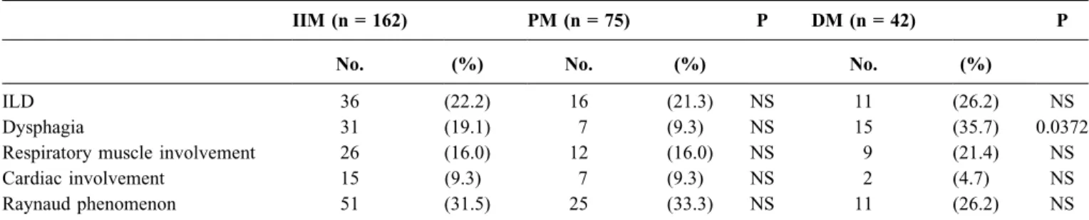
Further analysis of patients with primary PM/DM revealed that most of the PM patients had definite myositis (58 patients), while 17 had probable PM. The median duration of symptoms preceding the diagnosis was 5 months (range, 0.25–120 mo). Among patients with DM, 40 patients were considered to have definite DM and 2 had probable DM. The median onset of muscular and/or cutaneous symptoms was 2 months (range, 0.25–24 mo) before the diagnosis of DM. Proximal muscle weakness was present in all patients; and in the cases of DM, all the patients showed the typical skin manifestations. Frequencies of extraskeletal and extramuscular manifestations present at any time in the clinical course are shown in Table 2. Dysphagia was sig- nificantly more frequent among DM patients (p = 0.0372), while other systemic manifestations (cardiac and respiratory muscle involvement, interstitial lung disease, Raynaud phe- nomenon) were observed to a similar extent among PM and DM patients.
Causes of Death
Twenty patients died in our cohort of patients.
Eighteen disease-specific deaths occurred at a mean age of 49.3 ± 10.1 years (range, 30–65 yr), after a median duration follow-up of 30 months (range, 0.03–234.5 mo). The global survival curve of our patients (Figure 1) shows that the mortality of myositis is higher during the first year after diagnosis than later in the course. Of those patients with disease-related death, 8 died within 12 months after the diagnosis and 7 patients died more than 5 years after the diagnosis of myositis. The female:male ratio of the deceased (9 female:9 male patients) was not similar to the ratio typical of the disease. Causes of death were cardiac (11 patients), pulmonary (4 patients), and gastrointestinal (1 patient) com- plications and cancer (in 2 patients who had cancer- associated myositis). Pulmonary complications occurred mostly within the first 12 months, while cardiovascular complications caused death mainly after 5 years’ disease duration. In the groups of primary PM/DM, 13 disease- specific deaths were observed: 4 deaths were due to pulmonary complications (1 PM patient had severe oropha- ryngeal dysfunction leading to aspiration pneumonia and 3 DM patients had respiratory muscle involvement resulting in pulmonary insufficiency), 8 deaths were due to cardiovas- cular manifestations (2 cases of arrhythmia, 3 of heart failure, 2 of cardiac arrest, and 1 case of myocardial TABLE 1.Characteristics of Patients with Idiopathic
Inflammatory Myopathies: By Clinicopathologic Subgroup
No. of Patients (%)
Female:
Male Ratio
Age at Time of Diagnosis (yr) All Patients 162 (100) 2.1:1 39.2 ± 13.6 Polymyositis 75 (46.3) 1.8:1 39.3 ± 11.1 Dermatomyositis 42 (26.0) 2.8:1 43.7 ± 12.4 Juvenile patients 9 (5.5) 1:2 10.4 ± 4.4 Cancer/associated
myositis
7 (4.3) 1:1.3 48.6 ± 18.7 Overlap myositis 29 (17.9) 4.8:1 39.4 ± 10.9
infarction) and 1 death was due to gastrointestinal compli- cations (1 PM patient who had toxic megacolon).
Difference Among Clinicopathologic Subgroups
Global survival rates were 95%, 92%, and 89% for 1, 5, and 10 years, respectively. Survival analysis for further clinicopathologic subgroups (see Figure 1) revealed that patients with various forms of IIMs have different survival rates. None of the juvenile patients in our study died. Cancer- associated myositis patients had a survival curve signifi- cantly different from the other subgroups (p = 0.03). Further, although overlap myositis patients had the best prognosis, there was actually no significant difference from the other subgroups (p = 0.65). All 3 deaths in the overlap myositis subgroup were due to cardiovascular complications, and both of the 2 deaths in the cancer-associated myositis subgroup were the consequence of the underlying malignant disease.Five- and 10-year survival rates were 94.2% and 89.4% for patients with primary PM and 90.1% and 86.4%
for patients with primary DM. There was no significant difference between PM and DM survival curves (p = 0.5417).
Both the 5- and 10-year survival rates were 96% for overlap myositis patients and 71% for patients with cancer-associated myositis, respectively.
Influence of Clinical Features on Survival
The effects of different extraskeletal and extramuscular manifestations were evaluated in the group of patients with primary PM/DM (Figures 2 and 3). PM patients with dysphagia (p < 0.01) and cardiac involvement (p < 0.01) had significantly worse cumulative survival probability than PM patients without dysphagia or cardiac involvement. In the group of patients with DM, survival was significantly worse for male patients (p = 0.0382), patients who were older than 45 years at time of diagnosis (p = 0.0217), patients with interstitial lung disease (p = 0.0228), and patients with cardiac involvement (p < 0.01).
FIGURE 1. Survival curves of patients with idiopathic inflammatory myopathies (left) and survival curves according to clinicopathologic subgroups (right). JDM indicates juvenile dermatomyositis; OM, overlap myositis; PM, polymyositis;
DM, dermatomyositis; CAM, cancer-associated myositis.
TABLE 2.Frequency of Extraskeletal and Extramuscular Manifestations in Patients with Idiopathic Inflammatory Myopathies
IIM (n = 162) PM (n = 75) P DM (n = 42) P
No. (%) No. (%) No. (%)
ILD 36 (22.2) 16 (21.3) NS 11 (26.2) NS
Dysphagia 31 (19.1) 7 (9.3) NS 15 (35.7) 0.0372
Respiratory muscle involvement 26 (16.0) 12 (16.0) NS 9 (21.4) NS
Cardiac involvement 15 (9.3) 7 (9.3) NS 2 (4.7) NS
Raynaud phenomenon 51 (31.5) 25 (33.3) NS 11 (26.2) NS
Abbreviations: IIM = idiopathic inflammatory myopathies; PM = polymyositis; DM = dermatomyositis; NS = not significant; ILD = interstitial lung disease.
Prognostic Factors
Although the chi-square test of variables revealed a cor- relation between cardiac and respiratory muscle involvement, we analyzed the effect on survival of the presence of different clinical manifestations in a multivariate model, too. Significant prognostic factors for death determined in the whole group of patients with IIM were cardiac (coefficient = 3.182; p < 0.01) and respiratory muscle involvement (coefficient = 1.16;
p = 0.045). In the group of patients with primary PM/DM, cardiac involvement was the main prognostic factor for death (coefficient = 3.553; p < 0.01).
DISCUSSION
We report a long-term survival study on patients with myositis who were diagnosed, treated, and followed in a
single clinical immunology department in Hungary. Our cohort of IIM patients represents most of the clinicopatho- logic forms, and their clinical characteristics (distribution of different types, female:male ratio, age distribution, and frequencies of extraskeletal and extramuscular involvement) were similar to those in other series in the literature10,12,17.
Available data on survival of patients with IIMs, based on studies with sufficient numbers of patients, come from the United States, England, France, and Israel (Table 3). The clinicopathologic subgroups of IIMs included in these studies differ considerably. Therefore, to compare our work with data of the relevant literature, we decided to report the survival probability for the whole group of IIM patients as well as for primary PM/DM patients in our series. However, survival studies are influenced by several factors that modify FIGURE 2. Survival curves of patients with primary polymyositis with or without interstitial lung disease (top left) and respiratory muscle involvement (bottom left); and patients with dermatomyositis with or without interstitial lung disease (top right) and respiratory muscle involvement (bottom right). PM indicates polymyositis; DM, dermatomyositis; ILD, interstitial lung disease;
NS, not significant.
the results considerably, and the observed differences in mortality rates are attributable to 1) the means of selection of patients (diagnostic criteria, recruitment from a single or more than 1 department, which subgroups of IIMs are included in the analysis); 2) involving a sufficient number of patients and inclusion or exclusion of data of cases lost to follow-up; and 3) the duration of follow-up.
Myositis patients in the current study had higher survival rates than previously reported worldwide. This may be attributable to 3 aspects, discussed below: 1) these large series were based on patients who were diagnosed 2–3 decades before2,8,14; 2) the distribution of clinicopathologic subgroups was not similar, especially the frequency of
cancer-associated cases; and 3) improved diagnostic tools and therapeutic modalities have resulted in an increase in survival rates.
First, comparison of survival data is limited by the fact that studies reported by Medsger et al14and Hochberg et al8 had started before Bohan and Peter published their diagnostic criteria in 19753.
Second, all of these long-term large series had different inclusion criteria, especially concerning the subgroups of IIMs they studied. The worst survival rates and prognosis were experienced in studies that involved higher rates of cancer-associated myositis patients2,13. Results of recent long-term studies of IIM patients from France and England, FIGURE 3. Survival curves of patients with primary polymyositis with or without dysphagia (top left) and cardiac involvement (bottom left) and patients with dermatomyositis with or without dysphagia (top right) and cardiac involvement (bottom right).
PM indicates polymyositis; DM, dermatomyositis; NS, not significant.
which are more comparable to our experience, vary considerably depending on whether cancer-associated myo- sitis patients are excluded or not: Sultan et al18 reported a 95% 5-year survival rate, while Marie et al12reported 77%.
Having different etiologic, clinical, and serologic features from the other clinicopathologic forms of IIM, cancer- associated myositis patients should not be included when measuring the cumulative survival probability and prognosis of IIM. Cancer-associated myositis patients having the worst prognosis are hardly comparable with patients with other forms of IIM, because prognosis and life expectancy is determined by the underlying malignant disease. Consider- ing that mainly DM patients have a higher risk for ma- lignancy6, studies including cancer-associated cases among DM patients report worse survival rates for patients with DM than for those with PM.
Finally, improved survival of patients with IIM, and other patients as well, can be attributed to the following:
1. Widespread use of new diagnostic tools and serologic testing, which contribute to early diagnosis and to rec- ognition of milder forms of the disease;
2. Early use of more appropriate and more aggressive immunosuppressive and supportive therapy1;
3. Regular follow-up in departments specializing in the disease;
4. Better general medical care;
5. Better understanding of the natural history of the disease.
Our survival curve was heterogeneous, with an accel- erated mortality during the first year after diagnosis and a slower mortality during the following 10 years, except for those who had cardiac complications later in the clinical course. We found no significant difference between groups for patients with primary PM and primary DM, although most of the studies report better survival rates for PM patients.
Major causes of death in the current study were similar to the findings of the relevant literature9,18. Deaths due to pulmonary complications were observed early in the course (median, 1 mo) in our cohort of patients, and cardiovascular complications occurred late in the course (median, 59 mo).
Myositis patients may have severe extraskeletal and extramuscular manifestations—pulmonary, gastrointestinal, and cardiac involvement—which often affect the prognosis unfavorably. In patients with primary PM, the presence of dysphagia and the presence of cardiac involvement were associated with a significantly worse survival probability. In patients with primary DM, survival rates were worsened significantly by male gender, older age at the time of diagnosis (more than 45 yr), presence of interstitial lung disease, and presence of cardiac involvement. Effect of pulmonary involvement, especially interstitial lung disease, on survival may be controversial5,7,15, however, most authors found higher mortality rates compared to patients without interstitial lung disease2,11.
Classic factors of poor prognosis are older age, male sex, African-American race, interstitial lung disease, pres- ence of anti-Jo-1 and anti-SRP autoantibodies, associated malignancy, delayed or inadequate treatment, dysphagia, dysphonia, cardiac involvement, and pulmonary involve- ment2,9,10,13. In our study, unfavorable prognostic signs were respiratory muscle and cardiac involvement.
ACKNOWLEDGMENT
The authors thank Dr. Lisa Rider for critical reading of the manuscript.
REFERENCES
1. Adams EM, Plotz PH. The treatment of myositis. How to approach resistant disease.Rheum Dis Clin North Am. 1995;21:179–202.
2. Benbassat J, Geffel D, Larholt K, Sukenik S, Morgenstern U, Zlotnick A. Prognostic factors in polymyositis/dermatomyositis: a TABLE 3.Survival of Patients with Idiopathic Inflammatory Myopathies Present and Previous Reports
First Author (Ref.) No. of Clinicopathologic Survival rates (%)
Year Location Follow-Up Patients Subgroup 1-year 5-year >5-year
Medsger (14) 1971 USA 1947–1968 124 PM, DM, JDM, OM, CAM 72% 65% 53% (7 yr)
Hochberg (8) 1986 USA 1970–1981 76 PM, DM, JDM, OM 94.5% 80.4% 72.8% (8 yr)
Benbassat (2) 1985 Israel 1956–1976 92 PM, DM, JDM, OM, CAM 72% 52%
Maugars (13) 1996 France 1973–1984 69 PM, DM, JDM, OM, CAM 82.6% 66.7% 55.4% (9 yr)
Marie (12) 2001 France 1983–1998 77 PM, DM, CAM 83% 77% 61% (15 yr)
Sultan (18) 2002 England 1978–1999 46 PM, DM, JDM, OM 95% 83.8% (10 yr)
Danko (PR) 2003 Hungary 1976–2002 162 PM, DM, JDM, OM, CAM 95% 92% 89% (10 yr)
Abbreviations: PM = polymyositis; DM = dermatomyositis; JDM = juvenile dermatomyositis; OM = overlap myositis; CAM = cancer-associated myositis;
PR = present report.
computer-assisted analysis of 92 cases. Arthritis Rheum. 1985;28:
249–255.
3. Bohan A, Peter BJ. Polymyositis and dermatomyositis. Parts 1 and 2.
N Engl J Med. 1975;292:344–347, 403–407.
4. Cox DR. Regression models and life-tables (with discussion).J R Stat Soc B. 1972;39:86–94.
5. Grau JM, Miro O, Pedrol E, Casademont J, Masanes F, Herrero C, Haussman G, Urbano-Marquez A. Interstitial lung disease related to dermatomyositis. Comparative study with patients without lung in- volvement.J Rheumatol. 1996;23:1921–1926.
6. Hill CL, Zhang Y, Sigurgeirsson B, Pukkala E, Mellemkjaer L, Airio A, Evans SR, Felson DT. Frequency of specific cancer types in der- matomyositis and polymyositis: a population-based study. Lancet.
2001;357:96 –100.
7. Hirakata M, Nagai S. Interstitial lung disease in polymyositis and dermatomyositis.Curr Opin Rheumatol. 2000;12:501–508.
8. Hochberg MC, Feldman D, Stevens MB. Adult onset polymyositis- dermatomyositis: an analysis of clinical and laboratory features and survival in 76 patients, with a review of the literature.Semin Arthritis Rheum. 1986;3:168–178.
9. Hochberg MC. Epidemiology of polymyositis/dermatomyositis. Mt Sinai J Med. 1988;55:447–452.
10. Love LA, Leff RL, Fraser DD, Targoff IN, Dalakas M, Plotz PH, Miller FW. A new approach to the classification of the idiopathic in- flammatory myopathy: myositis-specific autoantibodies define use-
ful homogeneous patient groups. Medicine (Baltimore). 1991;70:
360 –374.
11. Marie I, Hatron P-Y, Hachulla E, Wallaert B, Michon-Pasturel U, Devulder B. Pulmonary involvement in polymyositis and dermatomyo- sitis.J Rheumatol. 1998;25:1336–1343.
12. Marie I, Hachulla E, Hatron P-Y, Hellot M-F, Levesque H, Devulder B, Courtois H. Polymyositis and dermatomyositis: short term and long term outcome, and predictive factors of prognosis.J Rheumatol. 2001;28:
2230 –2237.
13. Maugars YM, Berthelot J-MM, Abbas AA, Mussini J-MB, Nguyen J-MD, Prost AM. Long-term prognosis of 69 patients with dermato- myositis or polymyositis.Clin Exp Rheumatol. 1996;14:263–274.
14. Medsger TA, Robinson H, Masi AT. Factors affecting survivorship in polymyositis. A life-table study of 124 patients. Arthritis Rheum.
1971;14:249–258.
15. Miro O, Laguno M, Grau JM. Survival of patients with polymyositis/
dermatomyositis and pulmonary involvement.J Rheumatol. 1999;26:
1852–1853.
16. Rider LG, Miller FW. Idiopathic inflammatory muscle disease: clinical aspects.Baillieres Clin Rheumatol. 2000;14:37–54.
17. Spiera R, Kagen L. Extramuscular manifestations in idiopathic in- flammatory myopathies.Curr Opin Rheumatol. 1998;10:556 –561.
18. Sultan SM, Ioannou Y, Moss K, Isenberg DA. Outcome in patients with idiopathic inflammatory myositis: morbidity and mortality.Rheumatol- ogy. 2002;41:22–26.