Subclinical epileptiform activity accelerates the progression of Alzheimer’s disease: A long-term EEG study
Andras Attila Horvath
a,b,⇑, Aniko Papp
a,c, Janos Zsuffa
d, Anna Szucs
a, Janos Luckl
e, Ferenc Radai
e, Ferenc Nagy
e, Zoltan Hidasi
f, Gabor Csukly
f, Gabor Barcs
a, Anita Kamondi
a,gaNational Institute of Clinical Neurosciences, Department of Neurology Budapest, Hungary
bSemmelweis University, Department of Anatomy Histology and Embryology, Budapest, Hungary
cSemmelweis University, School of PhD Studies, Budapest, Hungary
dJános Zsuffa, Zsuffa-Med Ltd., Budapest, Hungary
eDepartment of Neurology, Kaposi Mór County Hospital, Kaposvár, Hungary
fSemmelweis University, Department of Psychiatry and Psychotherapy, Budapest, Hungary
gSemmelweis University Department of Neurology, Budapest, Hungary
See Editorial, pages 1961–1963
a r t i c l e i n f o
Article history:
Accepted 29 March 2021 Available online 8 May 2021 Keywords:
Alzheimer’s disease Epilepsy
Epileptiform discharge Electroencephalography Neuropsychology
h i g h l i g h t s
Epileptiform discharges are presented in half of Alzheimer patients without epileptic seizures.
Alzheimer patients with epileptiform discharges have significantly lower memory performance.
Epileptiform discharges associate with 1.5-times faster cognitive decline in a prospective follow-up.
a b s t r a c t
Objective: While many studies suggest that patients with Alzheimer’s disease have a higher chance for developing epileptic seizures, only a few studies are available examining independent epileptic dis- charges. The major aims of our study was to determine the prevalence of subclinical epileptiform activity (SEA) in AD compared to healthy elderly controls with the hypothesis that SEA is more frequent in AD than in cognitively normal individuals. Another aim was to analyze the effect of baseline SEA captured with electroencephalography on the progression of the disease with longitudinal cognitive testing.
Methods: We investigated 52 Alzheimer patients with no history of epileptic seizures and 20 healthy individuals. All participants underwent a 24-hour electroencephalography, neurology, neuroimaging and neuropsychology examination. Two independent raters analyzed visually the electroencephalograms and both raters were blind to the diagnoses. Thirty-eight Alzheimer patients were enrolled in a 3-year long prospective follow-up study with yearly repeated cognitive evaluation.
Results: Subclinical epileptiform discharges were recorded significantly (p:0.018) more frequently in Alzheimer patients (54%) than in healthy elderly (25%). Epileptiform discharges were associated with lower performance scores in memory. Alzheimer patients with spikes showed 1.5-times faster decline in global cognitive scores than patients without (p < 0.001). The decline in cognitive performance scores showed a significant positive correlation with spike frequency (r:+0.664; p < 0.001).
Conclusions: Subclinical epileptiform activity occurs in half of Alzheimer patients who have never suf- fered epileptic seizures. Alzheimer patients with subclinical epileptiform activity showed accelerated cognitive decline with a strong relation to the frequency and spatial distribution (left temporal) of spikes.
Significance:Our findings suggest the prominent role of epileptiform discharges in the pathomechanism of Alzheimer’s disease which might serve as potential therapeutic target.
Ó2021 International Federation of Clinical Neurophysiology. Published by Elsevier B.V. This is an open access article under the CC BY license (http://creativecommons.org/licenses/by/4.0/).
https://doi.org/10.1016/j.clinph.2021.03.050
1388-2457/Ó2021 International Federation of Clinical Neurophysiology. Published by Elsevier B.V.
This is an open access article under the CC BY license (http://creativecommons.org/licenses/by/4.0/).
⇑Corresponding author at: 57 Amerikai út, 1145-Budapest, Hungary.
E-mail address:horvath.andras1@med.semmelweis-univ.hu(A.A. Horvath).
Contents lists available atScienceDirect
Clinical Neurophysiology
j o u r n a l h o m e p a g e : w w w . e l s e v i e r . c o m / l o c a t e / c l i n p h
1. Introduction
Alzheimer’s disease (AD) is the major cause of cognitive decline creating ponderous economic burden on the aging societies. It is well known that some factors modify the progression, e.g. the onset of cognitive decline, an advanced disease stage at the time of diagnosis, educational background, female gender, and comor- bid cardiovascular disease (Ito et al., 2011; Devanand et al., 2013; Doody et al., 2010).
A recently recognized concomitant condition is epilepsy. In ani- mal models of AD, epileptic seizures are common (Palop et al., 2008; Palop and Mucke, 2010). Also, neuropathology studies have identified many similarities between AD and temporal lobe epi- lepsy such as the presence of amyloid plaques, tau neurofibrils, hippocampal sclerosis and hippocampal hyperexcitability (Puvenna et al., 2006; Riascos et al., 2014; Tai et al., 2016). Growing body of evidence suggests that AD patients have an increased risk to develop epileptic seizures and seizures associate with worse cognitive functioning (Horváth et al., 2016; Palop and Mucke, 2009). In addition to taking into account the cognitive harm of clin- ical seizures, modern epileptology has recognized the negative impact of epileptiform discharges on the cognitive performance of epilepsy patientsUng et al., 2017).
Considering the harm of epileptic discharges on cognitive func- tions in epilepsy, it is intriguing to investigate the effect of epilep- tiform activity in AD patients without clinical seizures (subclinical epileptiform activity- SEA) (Fisher et al., 2014). SEA could be observed among healthy individuals (Santoshkumar et al., 2009);
however, the physiological background of the phenomenon (as a potential marker of cortical hyperexcitability or an indicator of an undiagnosed neurological disorder) is unclarified (McLachlan and Luba, 2002). Many early studies demonstrated that SEA might serve as a predictive marker for the further development of epilep- tic seizures (Saito et al., 1987), however, it was not clarified with sensitive neurophysiological techniques or meta-analysis (So 2010). Recent studies demonstrated that SEA might predict poor outcome in acute vascular events (Tabaeizadeh et al., 2020), asso- ciate with more severe symptoms in autism spectrum disorders (Mulligan and Trauner, 2014) and predict epileptic seizures in chil- dren with attention-deficit hyperactivity disorders (Richer et al., 2002). There is clearly a scientific trend suggesting that SEA might have a role in the development of many disorders affecting cogni- tive functions (Horvath et al., 2020). It is confirmed by a recent human study that SEA might impair the cognitive functions of AD patients (Vossel et al., 2016); however, these results have not been replicated yet.
The aim of our prospective study was to determine the preva- lence of SEA in AD compared to healthy elderly controls with the hypotheses that 1, SEA is more frequent in AD than in cognitively normal individuals, and 2, baseline SEA captured with electroen- cephalography (EEG) associate with faster progression of the dis- ease. We also aimed to establish the link between the temporal and spatial distribution of epileptiform spiking and cognitive decline.
2. Materials and methods 2.1. Participants
We studied 80 AD patients with clinically typical, predomi- nantly memory-associated symptoms who met the diagnostic cri- teria of the National Institute of Aging- Alzheimer’s Association (NIA-AA) for probable AD (McKhann et al., 2011) and 20 cogni- tively healthy controls (HC) in the Department of Neurology of National Institute of Clinical Neurosciences in Budapest and in
the Department of Neurology of Kaposi Mór County Hospital in Kaposvár, Hungary between 2015 and 2019. All subjects were native Hungarians.
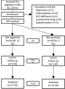
We collected medical history including collateral data and med- ical records for excluding those patients with epileptic seizures or seizure like events. Participants who had epileptic seizures or ele- vated risk for epileptic seizures including prior infection of central nervous system, clinically significant brain lesions (stroke, severe periventricular white matter disease, white matter infarcts), head trauma with loss of consciousness, demyelinating conditions, hydrocephalus, untreated vitamin B12 deficiency or hypothy- roidism, syphilis or HIV infection, major depression, schizophrenia, electroconvulsive therapy, renal insufficiency, liver disease, signif- icant systemic medical illness, alcohol or substance dependency, psychoactive drugs affecting cognitive functions except antide- mentia medication were excluded from the current analysis (n:21). Furthermore, 7 patients with major depression measured with neuropsychological tests were also excluded (Fig. 1).
Data of 52 AD patients were analyzed in the prevalence analysis (studies at year 0). Data from 32 patients were used in a previous publication of our group (Horvath et al., 2018). In that study, only the demographic characteristics were analyzed to compare AD patients with SEA to AD patients with epileptic seizures.
We followed our patients for 3 years and repeated the same neuropsychology test battery each year. During the 3-year follow-up, we excluded 5 AD patients from the prospective analy- sis who in addition to AD suffered significant physical or mental ill- ness potentially affecting cognitive functioning during the follow- up period. Four AD patients developed seizures during the follow-up period, therefore their data were excluded from the sta- tistical analysis. Five more patients were lost to follow-up because of being unreachable. Finally, we analyzed the prospective data of the remaining 38 AD patients at the end of the 3rd year in the lon- gitudinal analysis (Fig. 1).
2.2. Standard protocol approvals, registration, and phenotyping
The Hungarian Medical Research Council authorized our research (reference number: 024505/2015). We obtained informed
Fig. 1.FLOW-CHART OF PARTICIPANT RECRUITMENT AND ENROLLMENT. AD:
Alzheimer’s disease; HC: healthy control; EEG: electroencephalography; SEA:
subclinical epileptiform activity.
written consent from each participant. Surrogate consents were not required since all cases were mild so that all patients con- sented for themselves.
2.3. Clinical testing
The participants underwent detailed physical, neurological, and epileptological examination, as well as routine blood checks including thyroid functions and vitamin B12 level. All subjects had structural brain magnetic resonance imaging (MRI).
The controls had normal neurology status and neuropsychology scores, normal brain MRI and blood results, and they had no cogni- tive complaints.
2.4. Neuropsychological examination
The neuropsychological tests were carried out by trained neu- rologists or neuropsychologists in Hungarian language at the beginning of the study (year 0) and at the beginning of each upcoming year (year 1, year 2, year 3) within 335–395 days follow- ing the previous examination. We used the Hungarian version of Addenbrooke Cognitive Examination (ACE) (Stacho et al., 2002) as primary test battery because of its high sensitivity and speci- ficity in the diagnosis of major neurocognitive disorders. ACE scores range from 0 to 100, higher score represents better cognitive performance. ACE measures six cognitive domains including orien- tation (spatial and temporal- e.g. identification of home city of cur- rent year), attention (e.g. counting), memory (anterograde, retrograde and delayed recall- e.g. memorization of an address or words), verbal fluency (category and letter- e.g. listing of animals), language (e.g. execution of written or oral orders) and visuo-spatial skills (e.g. clock drawing and copying of a cube or intersecting pen- tagons). ACE is an accurate tool in differentiating frontotemporal dementia and AD too, and it serves properly in the assessment of dementia severity since Mini-Mental State Examination Score (MMSE) can be extracted from the test data (Dudas et al., 2005).
Scores in the MMSE range from 0 to 30, higher scores denote better cognitive performance. ACE also includes the ratio of verbal fluency and language skills divided by the scores of delayed recall memory and orientation (VLOM ratio). The normal value for VLOM ratio is 2.2–3.2, scores < 2.2 indicate frontotemporal type deficit while scores > 3.2 denote Alzheimer-type impairment.
Since anxiety and depression might compromise cognitive func- tions, we also recorded the Hungarian version of Spielberger State and Trait Anxiety Inventory (STAI) (Sipos and Sipos, 1983) and Beck Depression Inventory II (BDI-II) (Miklosi et al., 2011). Based on large samples, <45 value on STAI represents low level of anxiety in both the trait and state category (Horvath et al., 2016).
Scores < 13 indicate minimal depression, the 14–19 range repre- sents mild, the range 20–28 moderate and a score higher than 29 signalizes severe depression. To increase our diagnostic accuracy, patients with a STAI > 45 and a BDI II > 13 were not included in our analysis (possible indicators of major depression).
Controls had MMSE > 26 (Janka et al., 1989) Addenbrooke Cog- nitive Examination (ACE) score > 84 (Stacho et al., 2002), STAI < 45 (Sipos and Sipos, 1983), BDI II < 13 (Miklosi et al., 2011).
2.5. EEG examination
We performed 34-channel 24-hour long inpatient EEG record- ing (Micromed Morpheus, 10–20 electrode placement system) in all participants within 5 days following the neuropsychological and clinical testing at the beginning of the study (year 0). EEGs dur- ing the follow-up period were not repeated. Peri-orbital leads were applied to aid sleep staging. We used the following EEG settings:
bipolar longitudinal montage, 10 microvolts/mm sensitivity,
30 mm/sec speed, 70 Hz low pass and 0.5 Hz high pass filter with 50 Hz notch filter on.
Subclinical epileptiform discharges were defined as paroxysmal EEG graphoelements (spikes or sharp waves) with 20–200 ms duration, with the disruption of background EEG activity, followed by slow waves (Noachtar and Rémi, 2009). Two independent raters analyzed visually the EEGs; a graphoelement was identified as epileptiform if both raters marked it so. Both raters were blind to the diagnoses. For avoiding misinterpretation of epileptic tran- sients, we identified and excluded from the calculation the follow- ing variants: wicket spikes, occipital sharp transients of sleep, benign epileptiform transients in sleep, and rhythmic temporal theta series in superficial sleep. Based on these evaluations, partic- ipants were categorized into EEG positive (AD + SEA) and negative (AD-SEA) subgroups.
The number of spikes was visually counted. The average num- ber of spikes/hours was calculated as the total number of spikes divided by the hours of recordings. The scalp distribution of SEA was analyzed both visually and with the application of automatic EEG software (Micromed SystemPLUS 98, Compumedics NeuroS- can Curry 7). Recognition of spatial distribution of SEA was based on the largest electronegativity corresponding to scalp electrodes in the 10–20 electrode placement system as follows: frontal (Fp1, Fp2, F3, F4), frontocentral (Fz), central (C3, Cz, C4), centroparietal (Pz), frontotemporal (F7, F8), temporal (T3, T4, T5, T6), parietal (P3, P4) and occipital (O1, O2) electrodes.
2.6. Statistical analysis
IBM SPSS 20 software (https://www.ibm.com/support/pages/
ibm-spss-statistics-20-documentation) was applied for statistical analysis. A recent study of Vossel et al. (Vossel et al., 2016) re- ported difference in MMSE change /year in AD + SEA patients (mean = 3.9, SD = 1.6) compared to AD-SEA patients (mean = 1.6, SD = 2.4). Based on their results and our power calculations the probability was equal or greater than 80% to find a significant (al- pha = 0.05) difference between study groups in MMSE deteriora- tion (in delta MMSE / year) with a sample size of 50. Drop of rate (27%) was higher than calculated. In pairwise comparisons, t- tests were used for continuous data with parametric distribution, while Mann-Whitney U-test was applied for data with non- parametric distribution. Distribution was analyzed with Shapiro- Wilk test. For pairwise comparisons of categorial variables, chi- square test was applied. Holm-Bonferroni method was applied for correction of multiple comparisons. Logistic regression with the age as a covariant was applied for comparison of SEA preva- lence across AD patients and controls, since AD patients repre- sented an older cohort.
Longitudinal changes in the neuropsychological data repre- sented by ACE score and MMSE score (at the beginning, after 1- year, after 2-year and after 3-year follow up) across AD + SEA and AD-SEA patients were compared using repeated measure gen- eral linear model (r-GLM). Linear model was selected because Shapiro-Wilk test indicated normal distribution of ACE and MMSE data (p > 0.05). Between subject factor was the presence (AD + SEA) or absence of epileptiform activity (AD-SEA), while the measured ACE and MMSE scores at 0 time point, at 1-year, at 2-year and at 3-year represented the within subject variable (dependent factor).
Greenhouse-Geisser correction was applied to report p- and F- values for pairwise comparisons since Mauchly’s test reported that sphericity could not be assumed (p < 0.05). Tukey-test was applied for post-hoc analysis with Bonferroni correction to analyze changes between different time points. We report adjusted p- values for the multiple comparisons of 4 time points (0-, 1-, 2-, 3-year) and indicate significance where p < 0.0125.
Known progression modifying factors such as age at onset of cognitive decline, gender, education level (represented in years of education) and disease severity (represented in 0 timepoint MMSE score) were added as covariates to the model. Effect of these factors derived from the linear model was reported. P < 0.05 represented statistically significant effect. Spearman-correlation was applied to measure the association between progression of cognitive decline and spike frequency.
All statistical analyses concerning EEG data were performed using the data derived from the long-term EEG recording per- formed at year 0.
2.7. Data availability
The data that support the findings of this study and not pre- sented in this article are available on request from the correspond- ing author.
3. Results
3.1. Demographics and clinical characteristics at year 0
The AD group (n: 52) was older than the control group (n: 20) (p < 0.001). Patients were in the mild or moderate phase of the dis- ease. Neuropsychology indicated typical AD representation of cog- nitive impairment (deficit dominantly in orientation and episodic memory) in all patients (Table 1). MRIs were analyzed with visual inspection and in all patients showed characteristic bifrontal- bitemporal atrophy as well as hippocampal atrophy typical of AD.
3.2. Prevalence, spatial distribution and spike frequency of SEA in AD at year 0
We detected SEA in 54% of AD patients (28/52) and in 25% of HCs (5/20), the difference is significant (p:0.018). SEA was detected predominantly over the temporal electrodes (23/28 patients = 82%) (Fig. 2). Temporal SEA lateralized predominantly to the left side (12/23 = 52%), while bitemporal (6/23 = 26%) and right temporal occurrence (5/23 = 22%) were less common (Fig. 2). In patients with SEA, spike frequency was 0.29–6.68 spikes/hour (in average 2.02 spikes/hour). The vast majority of spikes (92%) occurred dur-
ing sleep. Spikes appeared most frequently during stage 2 (31%) and stage 3 (34%) sleep, while 23% of spikes were detected in stage 1 sleep. Only 4% of spikes occurred during rapid eye movement (REM) sleep (Fig. 2).
3.3. Characteristics of AD patients with and without subclinical epileptiform activity at year 0
Based on the presence or absence of SEA in the EEG recordings we divided our patients into two subgroups: AD + SEA (n: 28) and AD-SEA (n: 24) respectively. Presence of SEA was not associated with significantly different clinical or epidemiologic features of AD patients. They did not show differences in therapeutic regime, handedness, or in the course of dementia nor in global neuropsy- chology scores. Furthermore, patients with SEA showed higher (non-significant) VLOM ratios (Table 2). Analysis of various ACE subscores related to different cognitive domains revealed that patients with SEA had reduced performance in memory (Md:3.84; p:0.007) and visuo-spatial scores (Md:1.05; p:0.03) (Fig. 3). Difference in memory remained significant after Holm- Bonferroni correction (p < 0.008).
3.4. Prospective analysis of the effect of subclinical epileptiform activity on the progression of Alzheimer’s disease at year 3
In our prospective study, we analyzed the data of 38 AD patients who completed the 3-year long follow-up (Table 3).
AD + SEA patients (n = 21) differed only in the VLOM ratios from AD-SEA patients (n = 17) (Md:-0.57; p:0.039). Spatial distribution of SEA at year 0 in the AD + SEA group was the following: left tem- poral (7/21 = 33%), right temporal (5/21 = 24%), bitemporal (4/21 = 19%), right frontal (1/21 = 5%), bifrontal (3/21 = 14%), biparietal (1/21 = 5%).
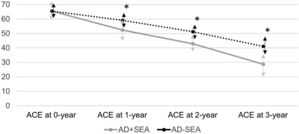
In the longitudinal analysis, AD + SEA patients showed signifi- cantly faster cognitive decline represented by average yearly decrease of total ACE scores (12.15 points/year in AD + SEA patients and 8.17 points/year in AD-SEA patients, F:15.891; p:<0.001) (Fig. 4) and by average yearly decrease in MMSE scores (2.71 points/year in AD + SEA patients and 2.22 points/year in AD-SEA patients, F:9.64; p:0.01). Cohen’s d effect size for 3-year ACE decline was 1.53 and 0.86 for 3-year MMSE decline. Significant dif- ferences were found with Tukey post-hoc analysis across the mea- sured time points (1st, 2nd and 3rd years) in ACE and MMSE scores (all p’s < 0.001). The 1.5 times greater decrease in ACE and 20%
greater decrease in MMSE in the presence of SEA remained signif- icant in the covariance weighted analysis applying the onset of dementia (years), sex (% of females), educational level (total years) and 0 timepoint disease severity (MMSE score). In our cohort, only the 0 timepoint disease severity factor (MMSE score) showed sig- nificant progression-modifying effect (F:9.661; p:<0.001 for ACE;
F:8.212, p:0.01 for MMSE). Decline in ACE score showed a signifi- cant positive correlation with the measured baseline spike fre- quency (year 0) (r:+0.664; p:0.001) (Fig. 5). We demonstrated smaller but significant correlation between spike frequency and reduction of MMSE score (r:+0.48; p:<0.01) as well. Since we found higher prevalence of spikes in the temporal regions with promi- nent left sided occurrence, we also measured the potential effect of spatial distribution of spikes on the progression of cognitive decline with ANOVA analysis comparing left, right and bitemporal appearances (N: 16). We found non-significant trend for differ- ences (F:3.775; p:0.051) across the 3 groups, where left did not dif- fer from bitemporal occurrence (p:1), while right occurrence was associated with lower non-significant decrease in ACE than left appearance (Md: 11.85; p:0.058) (Fig. 5).
Table 1
Demographic and clinical parameters of participants.
Characteristic Controls AD
patients p-value
Number 20 52 -
Female sex (n; %) 9 (45%) 31
(59,6%) 0.346 Age (years, mean ± SD) 67.8 ± 4.8 75.5 ± 8 *0.01
Right handedness (%) 18 (90%) 48
(92.3%) 0.772 Number of years of education in median
score (interquartile range)
12 (12–
17)
12 (12–
17)
0.142 MMSE median score (interquartile range) 28.5
(27.3–29)
20 (16–
23)
*<0.001 ACE median score (interquartile range) 92.5
(89.3–
94.8)
66 (56–
74)
*<0.001
VLOM median ratio (interquartile range) 2.7 (2.5–
3)
3.4 (3.2–
4.1)
*<0.001
Statistical tests were Chi-square for sex and handedness, t-test for age and Mann- Whitney U-test for MMSE score, ACE score and VLOM ratio.
AD: Alzheimer’s disease, MMSE: Mini-Mental Score Examination, ACE: Adden- brooke Cognitive Examination, VLOM ratio: sum of verbal fluency and language scores divided by sum of orientation and delayed memory recall scores, SD: stan- dard deviation. *indicates significant differences (p < 0.05).
4. Discussion
In the current study, we analyzed 24-hour EEGs of 52 AD patients and 20 healthy elderly controls. We demonstrated that AD patients show significantly higher prevalence of SEA than the control subjects (54% vs 20% respectively). Temporal distribution of SEA showed that 65% of the discharges occur in stage 2 or deeper sleep, while analysis of spatial occurrence revealed strong left side dominance. In the comparison of AD patients with and without SEA, we showed that patients with positive EEG had significantly lower performance in memory and visuo-spatial domains. In a 3- year prospective analysis of AD patients with repeated neuropsy-
chological test battery, we revealed that patients with SEA showed significantly faster progression of cognitive decline.
There are only a few studies focusing on SEA in AD with ambiguous prevalence data. Liedorp and his colleagues found epileptiform discharges in only 2 % of 1674 AD patients (Liedorp et al., 2010). However, they performed only 30-min long daytime EEGs, therefore they could not detect most of the SEA events which, as we show in the present study, appear mostly during slow wave sleep. Also, Vossel et al found SEA in just 6% of 42 AD patients, that can be explained by evaluating only daytime routine EEGs in 91% of their patients (Vossel et al., 2013). Our remarkably higher 54% - SEA rate may be due to the fact that we analyzed 24 hours EEGs including whole night sleep. It has been clearly shown in several epilepsy studies that interictal epileptiform discharges accumulate in sleep (Sammaritano et al., 1991; Steriade, 2003) and we had similar findings in AD patients in our previous study (Horvath et al., 2017) and in the current one as well (92% of spikes occurred in sleep). In the study of Vossel, the routine EEG was nor- mal in 60% of AD patients who had manifest epileptic seizures, while sleep deprived serial EEGs showed epileptiform discharges only in 16% (Vossel et al., 2013). In our previous study we also showed that 1-hour sleep recording is twenty-times more sensitive in detecting epileptiform activity than 1-hour awake EEG (Horvath et al., 2017). Thus, the possible explanation for our finding of ele- vated SEA rate is the higher sensitivity of long-term EEG containing sleep recordings. This is supported by another study about the temporal distribution of SEA in AD showing that epileptic activity occurs almost exclusively (90%) during sleep (Vossel et al, 2016).
Congruently with our data, these authors found SEA in 42% of AD patients.
In our healthy control sample, the relatively high incidence of SEA (25%) is curious; however, some studies also found increased risk for epileptiform event by non-epileptic elderly. In the study of McBride et al, epileptiform discharges were reported in 26% of 94 patients by patients aged 60 years and older using long-term EEG monitoring (McBride et al., 2002). Others found intermittent rhythmic delta activity in 17% of patients at the age 90 + applying 1-hour EEG recordings (Peltz et al., 2002). A study from Chochoi et al identified epileptiform discharges in 28% of 43 elderly patients Fig. 2.NEUROPHYSIOLOGIC FEATURES OF SUBCLINICAL EPILEPTIFORM ACTIVITY (SEA) IN AD PATIENTS AT YEAR 0. A: Spike detected in the left temporal region with phase inversion at temporal (T3) electrode in patient 005; B: Spike detected in the right frontotemporal region with maximum electronegativity at F8 electrode in patient 008; C:
Spatial distribution of SEA showing the dominant occurrence of spikes in the temporal regions with left sided predisposition.; D: Temporal distribution of SEA demonstrating that spikes occur almost exclusively in sleep, dominantly in deep sleep.
Table 2
Epidemiologic and clinical data of the AD + SEA and AD-SEA subgroups.
Parameter AD-SEA AD + SEA p-
value
Number of patients 24 28 -
Female sex (%) 14 (58%) 17 (61%) 0.579
Memantine therapy (%) 5 (20%) 6 (21%) 0.51
Cholinesterase inhibitor therapy (%) 24 (100%) 28 (100%) 1 Age (years, mean ± SD) 73.5 ± 7.8 71.9 ± 7.5 0.441
Right handedness (%) 21 (88%) 27 (96%) 0.321
Age at the onset of dementia (years, mean ± SD)
70.7 ± 7.5 69 ± 7.4 0.434 Duration of dementia in years with median
(interquartile range)
3 (1–4) 3 (2–3) 0.76 Number of years of education in median
(interquartile range)
12 (12–
12)
12 (12–
17)
0.26 MMSE median score (interquartile range) 19.5 (16–
24.8)
20 (16–
21.8)
0.665 ACE median score (interquartile range) 66 (58.5–
69)
65 (55.3–
77)
0.919 VLOM median ratio (interquartile range) 3.4 (3.2–
3.6)
3.6 (3.3–
4.6)
0.07
Statistical tests were Chi-square for sex, antidementia medication and handedness, t-test for age and for the onset of disease and Mann-Whitney U-test for MMSE score, ACE score and VLOM ratio.
AD: Alzheimer’s disease; SEA: subclinical epileptiform activity; ACE: Addenbrooke Cognitive Examination; MMSE: Mini-Mental Score Examination; VLOM ratio: sum of verbal fluency and language scores divided by sum of orientation and delayed memory recall scores, SD: standard deviation.
using long-term EEG (Chochoi et al., 2017). A guideline report from Mayo Clinic proposes that SEA is presented in 12% of the healthy elderly (Davidson and Davidson, 2012). The incidence of SEA reaches 50% by non-epileptic elderly with syncope (Hughes and Zialcita, 2000). The significance of SEA in healthy elderly needs fur- ther studies in larger cohorts. As it has been reported by Vossel (Vossel et al., 2013) and was confirmed by our current findings too, SEA occurs in AD significantly more frequently than in healthy elderly. Interestingly, at the time of diagnosis (first neuropsychol- ogy testing) higher SEA did not associate with reduced global cog- nitive scores, however the more sensitive subscores of cognitive domains revealed associations to more severe impairment of
memory and visuo-spatial skills in our cohort. These findings sug- gest that SEA might serve as an indicator of faster progressing AD and become a more important contributor of disease progression in the later course of AD. These results need further investigations.
The spatial distribution of epileptiform discharges was scruti- nized in only five studies; four of them analyzed AD patients with epileptic seizures (interictal epileptiform discharges) (Vossel et al., 2013; Cretin et al., 2016; Rao et al., 2009; Sarkis et al., 2016) and one of them investigated AD patients with SEA only (Vossel et al., 2016). In the summary of these reports, eighty percent of the discharges appeared in the frontotemporal regions, and 60%
of them on the left side. In our study, SEA appeared almost exclu- sively in the temporal/fronto-temporal areas, involving the left side in 52%, the bitemporal areas in 26% and the right temporal electrodes in 22%. In view of the known temporal and frontal involvement of AD-related morphology changes, the frontotempo- ral dominance of the epileptiform activity is not surprising. How- ever, the overwhelming left dominance of epileptiform activity in a neurodegenerative condition considered symmetric or leastwise bilateral, is curious. Noticeably, in frontotemporal dementia, a more severe left sided atrophy is a frequent imaging finding;
nowadays we even consider this phenomenon a diagnostic hall- mark (Frisoni et al., 1999). Interestingly, the strong left side dom- inance of interictal epileptiform discharges has been demonstrated in epilepsy studies as well (Doherty et al., 2003). If we accept that SEA or interictal activity are more frequent in the left temporal area and that higher spike frequency associate with faster cognitive decline, the clinical significance of this ‘‘left side phenomenon” warrants further investigations both in epilepsy and in neurodegenerative research.
The high prevalence of SEA is important if we consider the 1.5- times faster progression of cognitive deterioration of AD patients with the presence of SEA. The only available study in the literature focusing on the impact of SEA in AD had similar results (Vossel et al., 2016). In that study of Vossel et al, AD + SEA patients suffered 2.5-times faster decline in their yearly measured MMSE scores than patients with no SEA (Vossel et al., 2016). This finding remained significant even after correcting for the effects of age, gender and educational differences, similarly to our findings. In our cohort, we found smaller, 1.22-times faster decline in MMSE Fig. 3.DOMAIN SPECIFIC CHARACTERISTICS OF PATIENTS WITH (AD + SEA) AND WITHOUT SUBCLINICAL EPILEPTIFORM ACTIVITY (AD-SEA) AT YEAR 0. Mann-Whitney U-test was applied for pairwise comparisons. * indicates significant differences (p < 0.008, after Holm-Bonferroni correction). AD: Alzheimer’s disease; SEA: subclinical epileptiform activity.
Table 3
Baseline epidemiologic and clinical data of the AD-SEA and AD + SEA patient groups that completed the 3-year prospective follow-up.
Parameter AD-SEA AD + SEA p-
value
Number of patients 17 21 -
Female sex (n; %) 11 (65%) 11 (52%) 0.33
Memantine therapy (n; %) 4 (24%) 5 (24%) 0.94
Cholinesterase inhibitor therapy (n; %) 17 (100%) 21 (100%) 1 Age (years, mean ± SD) 74.2 ± 7.3 71.5 ± 5.9 0.22
Right handedness (%) 17 (100%) 21 (100%) 1
Age at the onset of dementia (years, mean ± SD)
70.8 ± 7.2 68.6 ± 5.5 0.304 Duration of dementia in years with
median score (median; interquartile range)
3 (3–4) 3 (3–3) 0.055
Number of years of education in median score (interquartile range)
12 (12–
14.5)
12 (12–17) 0.857 MMSE median score (interquartile range) 18 (15.5–
22.5)
20 (16–21) 0.37 ACE score (mean ± SD) 65.5 ± 9.1 65.5 ± 12.5 0.98 VLOM median ratio (interquartile range) 3.3 (3.2–
3.5)
3.6 (3.3–
4.5)
*0.039
Statistical tests were Chi-square for sex, antidementia medication and handedness, t-test for age, for the onset of disease, for ACE score and Mann-Whitney U-test for MMSE score and VLOM ratio. * indicates significant differences (p < 0.05).
AD: Alzheimer’s disease; SEA: subclinical epileptiform activity; ACE: Addenbrooke Cognitive Examination; MMSE: Mini-Mental Score Examination; VLOM ratio: sum of verbal fluency and language scores divided by sum of orientation and delayed memory recall scores, SD: standard deviation.
scores in patients with SEA. The slower progression rate in MMSE scores might be explained by the differences among the subjects of the two study samples in both the AD + SEA and AD-SEA groups (2.71 points/year in AD + SEA patients and 2.22 points/year in AD-SEA patients in our report vs 3.9 points/year in patients with epileptiform activity vs. 1.6 points/year in patients without in Vos- sel’s experiment). In our cohort, we analyzed older patients (aver- age age for prospective study was 73 in our cohort and ~61 in Vossel’s study), with shorter duration of dementia (~3 years in our study vs~5 years in Vossel’s experiment) and with higher pro- portion of patients taking antidementia medication (24% for Memantine and 100% for cholinesterase inhibitor therapy in our study vs 2.5% for Memantine and 56% for cholinesterase inhibitor therapy in Vossel’s report). All of these factors might influence the progression of AD. However, we could replicate the major strik- ing results with a more sensitive neuropsychological test battery (ACE versus MMSE) and larger sample size (38 vs 26 patients).
We also revealed a direct link between spike activity and cognitive decline with the demonstration of strong correlation across spike frequency and decline in cognitive scores.
There are limitations to our study. Firstly, we did not perform EEG examination at the end of the follow-up, so there is a possibil- ity that spike frequency and spatial distribution have changed dur- ing the follow-up period. Another important limitation is that positron emission tomography, cerebrospinal fluid analysis or genetic testing were not applied in the current experiment. Fur- thermore, AD group was significantly older than the control group (75.5 ± 8 vs 67.8 ± 4.8 years respectively), however, statistical anal- ysis was corrected for age. The strengths of the current report are the rigorous patient selection, the long follow-up period, and the application and analysis of long-term EEG containing sleep.
To conclude, 24-hour EEG recording detects subclinical epilepti- form activity in more than half of the AD patients. SEA appears typ- ically in the frontotemporal areas, predominantly in the left hemisphere and it seems to accelerate the progression of AD. Based on our findings it seems reasonable that development of new antiepileptic drugs targeting SEA control might represent a promis- ing a novel strategy to slow cognitive decline in Alzheimer’s disease. More studies are required to further elucidate the impor- tance of epileptiform activity in neurodegenerative disorders.
Fig. 5.RESULTS OF LONGITUDINAL PROSPECTIVE FOLLOW-UP AT YEAR-3 IN RELATION TO BASELINE (YEAR-0) FREQUENCY AND SPATIAL DISTRIBUTION OF EPILEPTIFORM SPIKES. A: the decline in ACE scores shows strong positive (r:+0.664) and significant (p < 0.001) correlation with spike frequency. B: left temporal (n:7) and bitemporal (n:4) spikes associate with faster cognitive decline than right spikes (n:5) with marginal significance (p:0.051). ACE: Addenbrooke Cognitive Examination.
Fig. 4.RESULTS OF LONGITUDINAL PROSPECTIVE FOLLOW-UP. AD + SEA patients show significant (p < 0.001), 1.5-times higher decline in total ACE scores than AD-SEA patients using repeated general linear model. * indicates significant differences (p < 0.001). AD: Alzheimer’s disease; SEA: subclinical epileptiform activity; ACE: Addenbrooke Cognitive Examination.
Funding
Our study was supported by National Brain Research Program I, II (KTIA_NAP_13-1-2013-0001; 2017-1.2.1-NKP-2017-00002), Hungarian Scientific Research Fund 2019 of the National Research, Development and Innovation Office (PD- 132652), Janos Bolyai Research Scholarship of the Hungarian Academy of Sciences (bo_78_20_2020). This is an EU Joint Programme- Neurodegenera- tive Disease Research (JPND) project. The project is supported through the following funding organization under the aegis of JPND- www.jpnd.eu (National Research,Development and Innova- tion, Hungary, 2019-2.1.7-ERA-NET-2020-00006).
Declaration of Competing Interest
The authors declare that they have no known competing finan- cial interests or personal relationships that could have appeared to influence the work reported in this paper.
Acknowledgements
We thank all the patients who participated in this study. Special thanks to Zita Csepella for analyzing the EEG recordings.
References
Chochoi M, Tyvaert L, Derambure P, Szurhaj W. Is long-term electroencephalogram more appropriate than standard electroencephalogram in the elderly?. Clin Neurophysiol 2017;128:270–4.
Cretin B, Sellal F, Philippi N, Bousiges O, Di Bitonto L, Martin-Hunyadi C. Epileptic prodromal Alzheimer’s disease, a retrospective study of 13 new cases:
expanding the spectrum of Alzheimer’s disease to an epileptic variant?. J Alz Dis 2016;52:1125–33.
Davidson PN, Davidson KA. Electroencephalography in the elderly. Neurodiag J 2012;52:3–19.
Devanand D, Lee J, Luchsinger J, Manyl J, Marder K, Mayeux R, Stern Y. Lessons from epidemiologic research about risk factors, modifiers, and progression of late onset Alzheimer’s disease in New York City at Columbia University Medical Center. J Alz Dis 2013;33:S447–55.
Doherty MJ, Simon E, De Menezes MS, Kuratani JD, Saneto RP, Holmes MD, et al.
When might hemispheric favouring of epileptiform discharges begin?. Seizure 2003;12:595–8.
Doody RS, Pavlik V, Massman P, Rountree S, Darby E, Chan W. Predicting progression of Alzheimer’s disease. Alzheimers Res Ther 2010;2:2.
Dudas RB, Berrios GE, Hodges JR. The Addenbrooke’s cognitive examination (ACE) in the differential diagnosis of early dementias versus affective disorder. Am J Geriatr Psy 2005;13:218–26.
Fisher RS, Acevedo C, Arzimanoglou A, Bogacz A, Cross JH, Elger CE, et al. Official report: a practical clinical definition of epilepsy. Epilepsia 2014;55:475–82.
Frisoni G, Laakso M, Beltramello A, Geroldi C, Bianchetti A, Soininen H, et al.
Hippocampal and entorhinal cortex atrophy in frontotemporal dementia and Alzheimer’s disease. Neurology 1999;52:91-91.
Horváth A, Montana X, Lanquart J-P, Hubain P, Sz}ucs A, Linkowski P, et al. Effects of state and trait anxiety on sleep structure: A polysomnographic study in 1083 subjects. Psychiatry Res 2016;244:279–83.
Horvath A, Szucs A, Barcs G, Kamondi A. Sleep EEG detects epileptiform activity in Alzheimer’s Disease with high sensitivity. J Alz Dis 2017;56:1175–83.
Horvath A, Szucs A, Barcs G, Noebels JL, Kamondi A. Epileptic seizures in Alzheimer disease: a review. Alzheimer Dis Assoc Disord 2016;30:186–92.
Horvath A, Szucs A, Hidasi Z, Csukly G, Barcs G, Kamondi A. Prevalence, semiology, and risk factors of epilepsy in Alzheimer’s disease: an ambulatory EEG study. J Alz Dis 2018;63:1045–54.
Horvath AA, Csernus EA, Lality S, Kaminski RM, Kamondi A. Inhibiting epileptiform activity in cognitive disorders: possibilities for a novel therapeutic approach.
Front Neurosci 2020;14 557416.
Hughes JR, & Zialcita ML. EEG in the elderly: seizures vs. syncope. Clin Electroencephalogr 2000;31:131-137.
Ito K, Corrigan B, Zhao Q, French J, Miller R, Soares H, et al. Disease progression model for cognitive deterioration from Alzheimer’s Disease Neuroimaging Initiative database. Alzheimers Dement 2011;7:151–60.
Janka Z, Somogyi A, Magloczki E. Dementia screening by a short cognitive test. Orv Hetil 1989;129:2797–800.
Liedorp M, Stam CJ, Van Der Flier WM, Pijnenburg YA, Scheltens P. Prevalence and clinical significance of epileptiform EEG discharges in a large memory clinic cohort. Dement Geriatr Cogn Disord 2010;29:432–7.
McBride AE, Shih TT, Hirsch LJ. Video-EEG monitoring in the elderly: a review of 94 patients. Epilepsia 2002;43:165–9.
McKhann GM, Knopman DS, Chertkow H, Hyman BT, Jack CR, Kawas CH, et al.
diagnosis of dementia due to Alzheimer’s disease: Recommendations from the National Institute on Aging-Alzheimer’s Association workgroups on diagnostic guidelines for Alzheimer’s disease. Alzheimer Dement 2011;7:263–9.
McLachlan RS, Luba N. Cortical location of benign paroxysmal rhythms in the electrocorticogram. Can J Neurol Sci 2002;29:154–8.
Miklosi M, Martos T, Kocsis-Bogar K, Perczel DF. Psychometric properties of the Hungarian version of the Cognitive Emotion Regulation Questionnaire.
Psychiatr Hung 2011;2:102–11.
Mulligan CK, Trauner DA. Incidence and Behavioral Correlates of Epileptiform Abnormalities in Autism Spectrum Disorders. J Autism Dev Disord 2014;44:452–8.
Noachtar S, Rémi J. The role of EEG in epilepsy: a critical review. Epilepsy Behav 2009;15:22–33.
Palop JJ, Mucke L. Epilepsy and cognitive impairments in Alzheimer disease. Arch Neurol 2009;66:435–40.
Palop JJ, Mucke L. Amyloid-b–induced neuronal dysfunction in Alzheimer’s disease:
from synapses toward neural networks. Nature Neurosci 2010;13:812–8.
Palop JJ, Chin J, Roberson ED, Wang J, Thwin MT, Bien-Ly N, et al. Aberrant excitatory neuronal activity and compensatory remodeling of inhibitory hippocampal circuits in mouse models of Alzheimer’s disease. Neuron 2008;55:697–711.
Peltz CB, Kim HL, Kawas CH. Abnormal EEGs in Cognitively and Physically Healthy Oldest-Old: Findings from The 90+ Study. J Clin Neurophysiol 2002;4:292.
Puvenna V, Engeler M, Banjara M, Brennan C, Schreiber P, Dadas A, et al. Is phosphorylated tau unique chronic traumatic encephalopathy. Brain Res 2006;1630:225–40.
Rao SC, Dove G, Cascino GD, Petersen RC. Recurrent seizures in patients with dementia: frequency, seizure types, and treatment outcome. Epilepsy Behav 2009;14:118–20.
Riascos D, Nicholas A, Samaeekia R, Yukhananov R, Mesulam MM, Bigio EH, et al.
Alterations of Ca(2)(+)-responsive proteins within cholinergic neurons in aging and Alzheimer’s disease. Neurobiol Aging 2014;35:1325–33.
Richer LP, Shevell MI, Rosenblatt BR. Epileptiform abnormalities in children with attention- deficit–hyperactivity disorder. Pediatr Neurol 2002;26:125–9.
Saito F, Fukushima Y, Kubota S. Small sharp spikes: possible relationship to epilepsy. Clin Electroencephalogr 1987;18:114–9.
Sammaritano M, Gigli GL, Gotman J. Interictal spiking during wakefulness and sleep and the localization of foci in temporal lobe epilepsy. Neurology 1991;41:290- 290.
Santoshkumar B, Chong JJ, Blume WT, McLachlan RS, Young GB, Diosy DC, et al.
Prevalence of benign epileptiform variants. Clin Neurophysiol 2009;120:856–61.
Sarkis RA, Dickerson BC, Cole AJ, Chemali ZN. Clinical and neurophysiologic characteristics of unprovoked seizures in patients diagnosed with dementia. J Neuropsychiatry Clin Neurosci 2016;28:56–61.
Sipos K, Sipos M. The development and validation of the Hungarian Form of the State-Trait Anxiety Inventory. Ser Clin Community Psychol 1983;2:27–39.
So EL. Interictal epileptiform discharges in persons without a history of seizures:
what do they mean?. J Clin Neurophysiol 2010;27:229–38.
Stacho L, Dudas R, Ivady R, Kothencz G, Janka Z. Addenbrooke’s Kognitív Vizsgálat: a magyar változat kifejlesztése. Psychiatr Hung 2002;18:226–40.
Steriade M. Neuronal substrates of sleep and epilepsy. Cambridge University Press;
.
Tabaeizadeh M, Nour HA, Shoukat M, Sun H, Jin J, Javed F, et al. Burden of Epileptiform Activity Predicts Discharge Neurologic Outcomes in Severe Acute Ischemic Stroke. Neurocrit Care 2020;32:697–706.
Tai XY, Koepp M, Duncan JS, Fox N, Thompson P, Baxendale S, et al.
Hyperphosphorylated tau in patients with refractory epilepsy correlates with cognitive decline: a study of temporal lobe resections. Brain 2016;139:2441–55.
Ung H, Cazares C, Nanivadekar A, Kini L, Wagenaar J, Becker D, et al. Interictal epileptiform activity outside the seizure onset zone impacts cognition. Brain 2017;140:2157–68.
Vossel KA, Beagle AJ, Rabinovici GD, Shu H, Lee SE, Naasan G, et al. Seizures and epileptiform activity in the early stages of Alzheimer disease. JAMA Neurol 2013;70:1158–66.
Vossel KA, Ranasinghe KG, Beagle AJ, Mizuiri D, Honma SM, Dowling AF, et al.
Incidence and impact of subclinical epileptiform activity in Alzheimer’s disease.
Ann Neurol 2016;80:858–70.