Neurocognitive Signatures of Parkinson’s and Alzheimer’s Disease
Ph.D. Dissertation
Dr. Nikoletta Bódi
Semmelweis University Mental Health Sciences
Supervisor: Prof. Szabolcs Kéri, M.D., Ph.D., D.Sc.
Reviewers: Prof. Csaba Pléh, M.D., Ph.D., D.Sc.
Dr. Tibor Kovács, M.D., Ph.D., D.Sc.
Chairman of final examination committee: Prof. Mária Kopp, M.D., Ph.D., D.Sc.
Members of final examination committee: Prof. István Czigler, Ph.D., D.Sc.
Dr. György Purebl, M.D., Ph.D.
Budapest
2012
TABLE OF CONTENTS
Table of contents 1.
Abbreviations 4.
1. Introduction 6.
1.1. The importance of common neurodegenerative diseases 6.
1.2. Beyond the motor symptoms: reinforcement learning in PD 8.
1.2.1. Basic pathology: from molecules to cognition 8.
1.2.2. Reinforcement learning in PD 11.
1.2.3. Is altered sensitivity to reinforcers related to personality changes in PD? 14.
1.3. The mirror image of parkinsonian cognition in AD 16.
1.3.1. The medial temporal lobe and beyond 16.
1.3.2. Novel functions of the medial temporal lobe and AD: the acquired equivalence
paradigm 19.
1.3.3. Visuospatial abilities in AD and other dementias 21.
2. Aims 24.
3. Methods 25.
3.1. Participants 25.
3.1.1. Participants in the reinforcement learning experiment 25.
3.1.2. Participants in the stimulus-learning experiment 26.
3.1.3.Participants in the visuospatial assessment 27.
3.2. Tasks 29.
3.2.1. Tasks in the reinforcement learning experiment 29.
3.2.2. Tasks in the stimulus-learning experiment 34.
3.2.3. Tasks in the visuospatial assessment 38.
4. Results 40.
4.1. Results in the reinforcement learning experiment 40.
4.1.1. Differences between never-medicated and recently-medicated PD patients in sensitivity to positive and negative feedback 40.
4.1.2. Personality measures in never-medicated and recently-medicated PD patients 42.
4.1.3. Correlation between performance on the feedback-based task and personality
measures 43.
4.1.4. Longitudinal result from the feedback-based task: retesting the never-medicated PD patients after the initialization of dopamine agonist therapy 48.
4.1.5. Longitudinal data from personality measures 51.
4.1.6. Effect of different dopamine agonists, illness duration, and symptoms 51.
4.2. Results in the stimulus-learning experiment 52.
4.2.1. Training phase 52.
4.2.2. Transfer phase 52.
4.2.3. Card pairing test 52.
4.3. Results in the visuospatial assessment 54.
4.3.1. Demographic and cognitive test results for groups 54.
4.3.2. Overall and error analysis of CDT comparing control, AD, and FTD groups 55.
5. Discussion 58.
5.1. Outline of the results 58.
5.2. Reinforcement learning in PD 59.
5.2.1. The role of dopamine in salience, motivation, and reward 59.
5.2.2. The possible clinical relevance of reward-learning and personality changes
associated with dopamine agonists in PD 62.
5.3. Transfer and flexibility of stimulus-response associations in AD 65.
5.3.1. Habit learning in AD, striatal automaticity, and hippocampal pattern
completion 65.
5.3.2. Is feedback-guided stimulus-response learning an implicit memory process? 67.
5.4. Visuospatial functions discriminate between AD and FTD 70.
6. Conclusions 74.
7. Summary 76.
8. References 80.
9. List of own publications related to the thesis 98.
10. List of own publications independent of the thesis 99.
Acknowledgement 101.
ABBREVIATIONS
AD – Alzheimer’s disease
ANCOVA – analyses of covariance ANOVA – analyses of variance
CBDS – corticobasal degeneration syndrome CDT – Clock Drawing Test
DDS – dopamine dysregulation syndrome DRS – Dementia Rating Scale
FTD – frontotemporal dementia
FTD-bv – frontotemporal dementia behavioral variant GDS – Global Deterioration Scale
HAM-A – Hamilton Anxiety Rating Scale HAM-D – Hamilton Depression Rating Scale HSD – Honestly Significant Difference test MANOVA – multivariate analyses of variance MMSE – Mini - Mental State Examination MRI – Magnetic Resonance Imaging
NINCDS-ADRDA – National Institute of Neurological and Communicative Disorders and Stroke and the Alzheimer’s Disease and Related Disorders Association
PA – progressive aphasia PD – Parkinson’s disease
PET – positron emission tomography PNFA – primer non-fluent aphasia PSP – progressive supranuclear palsy SD – semantic dementia
TCI - Temperament and Character Inventory TPQ – Tridimensional Personality Questionnaire UPDRS – Unified Parkinson’s Disease Rating Scale WAIS-R – Wechsler Adult Intelligence Scale
1. INTRODUCTION
1.1. The importance of common neurodegenerative diseases
Neurodegenerative diseases comprise one of the major public health concerns in our aging population. According to the data from the EURODEM study, the incidence of dementia ranges between 0.6-8.2/100 individuals in the age range of 70-90 years, whereas the prevalence is 2.9-30.8% (Launer et al., 1999). The exact etiology and pathophysiology remain elusive, but large-scale studies identified several risk factors that may contribute to the diseases. These general risk factors include various genetic polymorphisms, gene copy number variations, increasing age, poor education, endocrine diseases, oxidative stress, inflammation and infection, hypertension, diabetes, smoking, head trauma, depression, vitamin deficiencies, nutritional problems, and toxic environmental exposure (Launer et al., 1999).
The proper clinical diagnosis of neurodegenerative diseases is often difficult given the large overlap in signs and symptoms, although in more than 75% of cases the clinical diagnosis is confirmed by postmortem examination (Mok et al., 2004). The situation is even more complex, because neurodegenerative diseases may overlap and co-exist at the molecular pathological level (Armstrong et al., 2005). Some experts suggest a neuropathological classification of diseases instead of clinical symptoms, such as alpha-synucleinopathies (e.g., Parkinson’s disease), amyloidopathies (e.g., Alzheimer’s disease), and taupathies (e.g., fronto-temporal dementia) (Jellinger, 2008).
In the clinical practice, physicians focus on signs and symptoms and their progression; the classification systems of diseases is based on expert consensus bearing the sign of subjectivity, and we have to face with the fact that definitive biomarkers are still missing from the everyday practice. One possible solution is the standardized application of neuropsychological tests measuring the functional integrity of neuronal circuits, possibly with markers from peripheral blood and brain imaging.
Here, we focus on the neurocognitive bases of two common disorders, Parkinson’s and Alzheimer’s disease (PD and AD). PD is traditionally considered as a motor disorder characterized by intact or mildly affected cognitive functions until the latest stages of the disease (Lees et al., 2009). The cognitive deficit is predominantly
described as an anomaly of processing speed, attention, and executive functions, establishing the concept of subcortical dementia that occurs in approximately 20% of cases. PD dementia must be differentiated from Dementia with Lewy Bodies in which we can observe severe fluctuations in alertness and attention, recurrent visual hallucinations, and parkinsonian motor symptoms. In this disorder, cognitive problems related to cortical functions occur earlier, and therefore easier to confuse with AD (Korczyn and Reichmann, 2006; Selikhova et al., 2009). When memory, language, and visuo-spatial functions are predominantly affected, we can define the condition as a cortical dementia. This classification is nevertheless oversimplified and can hardly be considered valid today. Both AD and PD are common in the elderly, and patients with PD who develop dementia may have AD as well (Bonelli and Cummings, 2008).
In the forthcoming experiments, we will present data regarding three novel aspects of PD and AD
1. We explore specific cognitive deficits and personality changes in the early- stage of PD, with a special reference to reinforcement learning guided by positive and negative feedback. The effect of dopamine receptor agonists on these early non-motor changes will be investigated, showing how these drugs shift the balance of reward and punishment sensitivity.
2. We show that in early AD the cognitive deficit is not generalized. However, even successful feedback-guided stimulus-outcome learning results in rather context- specific memory traces that can not be applied in new retrieval situation in a flexible way.
3. Finally, we explore how more traditional clinical neuropsychological tests of visuo-spatial orientation and constructive abilities may serve to differentiate AD from other types of dementia.
1.2. Beyond the motor symptoms: reinforcement learning in PD
1.2.1. Basic pathology: from molecules to cognition
PD was described by James Parkinson in 1817, although a similar symptomatic delineation can be found in the work of Ferenc Pápai Páriz from 1690 with all four cardinal symptoms: resting tremor, rigidity, bradykinesia, and gait disturbances (Bereczki, 2010). The classic neuroanatomical definition of PD is based on the loss of neuromelanine-positive dopaminergic cells in substantia nigra pars compacta, leading to the disturbance of the nigrostriatal pathway (Lees et al., 2009). Neuronal loss may be induced by accumulated alpha-synuclein in these cells that comprises the core of the Lewy-bodies. Damaging alpha-synuclein may occur via phosphorylation by intracellular kinases, truncation by proteases, modification by free radicals, reactive nitrogen species, toxins, overproduction (gene duplication), and decreased elimination by parkin and synphilin (Venda et al., 2010). However, alpha-synuclein is not the sole player in mitochondrial damage and abnormal ubiquitine-proteosomal functions; other relevant factors are Parkin (PARK2), PINK1 [phosphatase and tensin homolog-induced putative kinase 1] (PARK6), DJ-1 (PARK7), LRRK2 [leucine-rich repeat kinase 2] and dardarin (PARK8) (Mellick et al., 2010). From our point of view, alpha-synuclein and its interactive proteins are especially important because this molecule, acting as a presynaptic regulator of dopamine release, may bridge neurodegeneration and cognition (Kéri et al., 2010).
Regarding the functional localization of PD, the hypothesis of selective nigrostriatal dysfunction seems to be problematic given that it can hardly explain several non-motor symptoms of PD: olfactory, sleep, and vegetative abnormalities, blunted affect, changes in personality traits (e.g., decreased novelty seeking and increased neuroticism), and atypical responses to dopaminergic medications in some patients (e.g., impulse control disorders and psychotic symptoms) (Bassetti, 2011).
Consistent with post-mortem studies showing that the loss of dopaminergic neurons in the substantia nigra is just an intermediate step in neurodegeneration, Jubault et al.
(2009) demonstrated in vivo imaging evidence for an early volume loss in the medulla
oblongata/pontine tegmentum, which is followed by neurodegeneration in substantia nigra/amygdala, and then in the cortex.
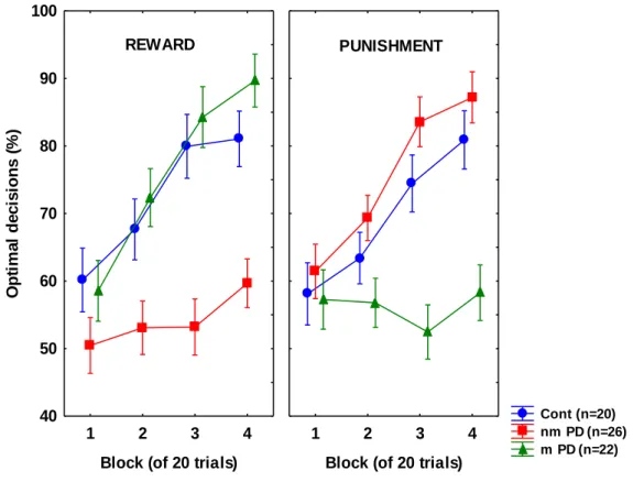
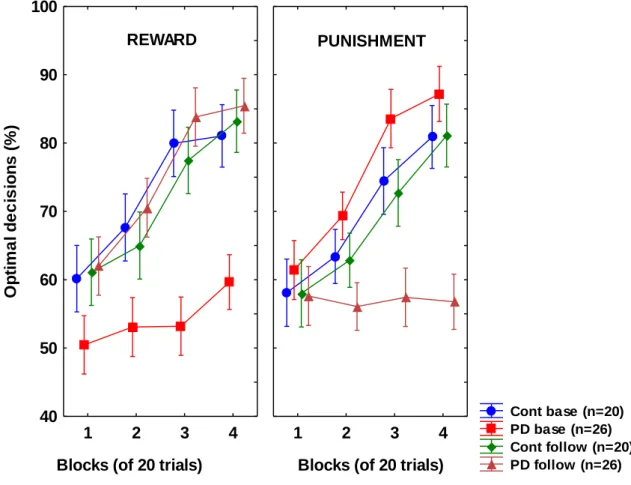
Although dopaminergic deficit is a hallmark of PD, the loss of this neurotransmitter is not evenly distributed in the striatum. Specifically, there is a more pronounced reduction of dopamine in the dorsal than in the ventral striatum (Kish et al., 1988). Therefore, therapies that restore dopamine level in the dorsal striatum result in dopamine “overdose” in the ventral striatum, which may lead to impaired performance on some cognitive tasks (Gotham et al., 1988; Cools et al., 2001, 2003; Shohamy et al., 2006; Jahanshahi et al., 2010; MacDonald et al., 2011) and in some cases to psychotic symptoms, including hallucinations and delusions (Mehler-Wex et al., 2006; Maia and Frank, 2011). At the same time, there is evidence that dopaminergic therapy enhances learning from reward signals and decreases learning from punishment signals in PD (Frank et al., 2004, 2007; Cools et al., 2006; Graef et al., 2010; Kobayakawa et al., 2010), and the ventral striatum plays a crucial role in reinforcement learning (Yin and Knowlton, 2006). Figure 1 shows a renewed scheme of the cortico-striatal system that clearly indicates the non-motors functions of this complex system. The effect of dopamine receptor agonists on non-motor functions in PD was explored in first series of our studies.
Figure 1.
Based on Bradshaw and Shepard, 2000
Pathways from the frontal cortex project to different regions of the striatum that in turn give output to the putamen and pallidum, which send afferents to the thalamus and cortex. Frontal regions are responsible for the processing of plans (dorsolateral region), complex motor programs (premotor and supplementary motor area), and affective/social representations (ventral frontal and cingular region). In the striatum neuronal activation patterns are selected and modified according to the current motivational state and reinforcers. This process is regulated by dopamine in the archistriatum (ventral striatum or nucleus accumbens). Striosomes, which are rich in acetylcholinesterase activity, are important in affective functions, whereas the matrix is more closely related to motor functions.
1.2.2. Reinforcement learning in PD Fronto – striato - pallido-
thalamic circuits:
-Cognitive(plan, dorsal PFC – dorsal neostriatum) -Motor (movement,
PMC/SMA – putamen, matrix) -Affective(emotion-
motivation, ventral cingulum – archistriatum, striosome)
Plans, motor programs,
affective impulses
Pattern selection, motivation and
reinforcement (archistriatum,
dopamine)
Primary motor area
Supplementary motor area (SMA) Premotor area (PMC)
Prefrontal cortex (PFC) Cingulum
Fine structure of striatum:
Matrix Striosome
Execution of movement (convergence
to motor cortex)
The cortico – striato – thalamo - cortical system
/plus: sensory cortex/
There are multiple lines of evidence that the striatum and its dopaminergic projections from the midbrain are important for learning to predict rewarding outcomes (Schultz, 2007). This reward prediction signal is critical for tasks during which responses to salient stimuli are modified by feedback. Given the well-known loss of dopaminergic signals in the striatum of PD, it is expected that PD patients show impairments on tasks requiring feedback-based reinforcement learning (Yin and Knowlton, 2006).
Animal studies indicate that neurons in the mesencephalic dopaminergic centers show phasic excitatory responses following primary rewards and after intensive and unexpected sensory stimuli. There are three groups of these neurons: A9 (pars compacta of substantia nigra), A10 (ventral tegmental area) and A8 (dorsolateral substantia nigra).
A8-A10 cells project their axons to the dorsal and ventral striatum, as well as to the dorsolateral and ventromedial frontal cortex. Interestingly, these neurons are transiently inhibited when reward is omitted or when conditioned stimuli are presented that predict the absence of reward (Schultz, 2007).
Dopamine, a classic neurotransmitter related to PD, psychosis, and mood disorders, seems to regulate the functional connectivity of striatal, limbic, and prefrontal areas during conditioning and reinforcement learning, and enhances responses to salient stimuli regardless of their hedonic value, possibly acting in the ventral striatum (Berridge, 2007). The most likely scenario is that some mesencephalic dopamine neurons encode motivational valence (reward), whereas others encode motivational salience. Both valence and salience encoding neuronal populations are facilitated by novelty and alerting signals in order to detect important environmental objects and events (Bromberg-Martin et al., 2010). Therefore, phasic dopamine responses may serve as a “teaching signal” during reinforcement learning. Interestingly, similar mechanisms have been revealed in humans using high-resolution functional neuroimaging techniques during various laboratory tasks, which provide unique insight into the organization of human dopaminergic cell groups in the mesencephalon (Düzel et al., 2009).
In a seminal study, Knowlton et al. (1996) compared amnesic patients with hippocampal damage and nondemented patients with PD on a probabilistic classification task in which participant acquired which of two outcomes would occur on
each trial (a pattern of figures predict rain or sunshine). Surprisingly, amnesic patients performed well on this feedback-guided task despite the fact that they had severe declarative memory impairment and hardly were able to consciously recall the stimuli.
Lesions to the prefrontal cortex did not affect learning, too. However, PD patients failed on the feedback learning phase, but showed intact declarative memory for the figures (Knowlton et al., 1996).
It is especially intriguing how PD patients exhibit compensatory brain activation during feedback-based reinforcement learning tasks. Patients with PD show less activation in the striatum and greater activation in the prefrontal cortex and medial temporal lobe, which may indicate that they use an explicit memory strategy during a reinforcement learning procedure (Moody et al., 2004). Overall, it is concluded that the neostriatum is essential for the gradual and incremental learning of associations. These findings were clearly pioneered by Saint-Cyr et al. (1988) who demonstrated a similar double dissociation in PD and amnesia with more traditional test procedures.
However, this type of gradual associative learning depends on L-dopa medication, which enhances global dopamine level and impairs certain types of feedback-based learning, presumably because of the “overdosing” of dopamine in brain areas less affected in PD, i.e.. the ventral striatum (Cools et al., 2001, 2003; Shohamy et al., 2006). The biased effect of dopamine replacement may improve motor functions and cognitive flexibility, but, at the same time, may induce impulsivity and decreased performance on tasks involving negative feedback-based learning (Cools et al., 2001, 2003; Shohamy et al., 2006).
Current models suggest that the ventral striatum underlies learning of stimulus associations, whereas the dorsal striatum is responsible for the processing of competing patterns during response selection. In decision making tasks, dopamine replacement therapy impairs stimulus-stimulus relation learning, but facilitates the selection of appropriate responses when conflicting informations must be resolved. This is consistent with activation of the ventral vs. dorsal striatal regions during stimulus- stimulus association learning and response selection, respectively (MacDonald et al., 2011).
Frank et al. (2004, 2007) demonstrated that PD patients off L-dopa are better at learning from punishment (negative outcome, e.g. losing points in a game) than from
reward (positive outcome, e.g. winning points); L-dopa reverses this pattern of task performance, enhancing learning from reward at the expense of punishment. Cools et al.
(2006) confirmed these findings using a feedback-based reversal learning task. In the study of Cools et al. (2006), medication-induced deficit for punishment was particularly pronounced in patients who received the dopamine D3 receptor agonist pramipexole.
D3 dopamine receptors are densely expressed in the ventral striatum, and it is possible that the stimulation of these receptors leads to decreased punishment sensitivity. Alpha- synuclein is a presynaptic negative regulator of dopamine release (Venda et al., 2010), and individuals with copy number variations of the alpha-synuclein gene, leading to the over-expression of the protein, display pronounced reward learning deficits well before the emergence of motor symptoms (Kéri et al., 2010).
Overall, models describing the effect of dopaminergic medication on reinforcement learning in PD do not take into consideration a couple of key factors.
First, in PD it is possible that tonic increase of dopamine after L-dopa administration over-rides phasic release of dopamine, which is the key „teaching signal” for learning.
Second, it is important that the selective stimulation of dopamine receptors may have a substantially different effect compared to L-dopa. Such kind of receptor-specific stimulation occurs in the case of dopamine agonist administration.
The first class of these drugs includes ergolinic dopamine agonists, such as bromocriptine or pergolide, whereas later non-ergolinic drugs, such as pramipexole and ropinirole, were marketed. Ergoline agents bind with high affinity to D2 receptors but also bind to D1, adrenergic, and serotonin receptors. Non-ergolines have a higher selectivity to D2 and D3 receptors; pramipexole shows an especially strong affinity to D3 receptors (Kvernmo et al., 2006). D3 receptors are prominently expressed in the ventral striatum, and therefore drugs stimulating this receptor may modulate reward- related learning processes (Cools et al., 2006). Drugs binding to D1 receptors stimulate the direct cortico-striato-thalamic pathways, which are considered to act as a stimulatory pathway for reinforcement learning providing “go” signals (Frank et al., 2004).
However, the exact receptor-specific effect on learning has not been studied and its clinical relevance is not clear, too.
In addition to the classic side effects of dopaminergic medications, such as nausea, psychotic symptoms, and orthostatic hypotension, dopamine agonists also
induce daytime somnolence, impulse-control disorders (drug addiction, gambling, overactive sexual behavior), and heart valve fibrosis in some patients. It is not clear whether dopamine agonists have a disease-modifying, i.e. neuroprotective effect and whether these drugs are more effective in the treatment on non-motor symptoms such as depression (Perez-Lloret and Rascol, 2010). This latter symptom of PD may be directly related to reinforcement learning.
1.2.3. Is altered sensitivity to reinforcers related to personality changes in PD?
In the previous section, we delineated a mechanistic model for dopaminergic functions in the mesencephalic-striatal system for reinforcement learning. However, dopamine may play a crucial role in higher-level human functions, such as patterns of emotional reactions, attitudes, and cognitive schemas comprising the multidimensionality of human personality. The most deeply investigated dopamine- related personality trait, in which norepinephrine could also be important, is novelty seeking including exploratory excitability, impulsiveness, extravagance, and disorderliness, as measured by the Temperament and Character Inventory (TCI) (Cloninger, 1994). There are numerous case reports suggesting rigid, punctual, and introverted personality in PD, but the results are mixed and non-conclusive (Menza, 2000; Jacobs et al., 2001; Tomer and Aharon-Peretz, 2004). In accordance with decreased dopaminergic transmission, some reports suggest decreased novelty seeking in PD (Menza, 2000), but other authors emphasize increased scores on harm avoidance (anticipatory worry, fear of uncertainty, shyness, and fatigability), which may be related to PD-associated depression (Jacobs et al., 2001). Premorbid personality may be especially important, because it may affect vulnerability to dopamine dysregulation syndrome induced by medications.
Krebs et al. (2009) investigated the neuronal correlates of novelty seeking and reward dependent personality traits. Using a reinforcement learning task with monetary incentives, functional magnetic resonance imaging hemodynamic responses were registered from the substantia nigra/ventral tegmental area, nucleus accumbens, and hippocampus. Novelty seeking was positively correlated with activation in the substantia nigra/ventral tegmental area elicited by new stimuli that did not predict
reward, whereas reward-dependence was related to activations elicited by novel cues that predicted reward. The responses of these mesencephalic regions to novelty and reward are differentially associated with novelty seeking and reward-dependence.
Persons with higher novelty seeking scores tend to respond to new stimuli even when it is not associated with reward, and in their case less reward is necessary to enhance hippocampal activation and to boost their memory for new stimuli. It is notable that midbrain dopamine autoreceptor density is negatively correlated with novelty seeking (Zald et al., 2008).
An important limitation of the literature regarding both reinforcement learning tasks and personality assessment is that studies include chronic elderly patients with PD receiving multiple medications, and many of these patients exhibit multiple neuropsychiatric co-morbidities from mood disorder to psychotic symptoms and generalized cognitive decline. Longitudinal follow-up studies are missing from the literature. Finally, it is unknown how sensitivity to reward and punishment in laboratory tasks is related to complex personality traits. Young-onset PD may be an especially important condition, because it is associated with slower progression of motor symptoms, longer disease course with spared cognitive function, but sometimes an earlier appearance of motor fluctuations, dyskinesias, and psychiatric symptoms (Schrag and Scott, 2006). The pathology is more circumscribed than in late-onset PD, but, paradoxically, in some cases cell loss in the substantia nigra is very definitive (Gibb and Lees, 1988).
1.3. The mirror image of parkinsonian cognition in AD
1.3.1. The medial temporal lobe and beyond
The pathological process resulting in AD and first hitting the medial temporal lobe is still not clear, but it is at least partly different from that observed in PD. AD is also considered as a protein misfolding disease (proteopathy); in this case the key protein is not alpha-synuclein, as for PD (although Lewy bodies can occur in AD), but the basic alteration may be the accumulation of abnormally folded amyloid-beta and tau
proteins. Amyloid plaques consist of 39–43 amino acids peptides. In AD hyperphosphorylation of microtubule-associated protein tau causes abnormal changes in cellular transport (Finder, 2010). From the amyloid small toxic fragments may be released (amyloid-derived diffusible ligands) that bind to prion protein in the cell membrane and disrupt synapses (Laurén et al., 2009). This mechanism may explain how neurodegenerative process is spreading in the brain.
What are the neuropsychological consequences of this degenerative process? If we reconcile the Saint-Cyr et al. (1988) and Knowlton et al. (1996) experiments, it is not difficult to make inferences from amnesia to AD, given that the earliest stage of AD is dominated by declarative memory dysfunctions, although the attentional system may be affected even earlier (Parasuraman et al., 2000). Nevertheless, patients with early AD show superior performances on feedback-based stimulus-outcome learning tasks in which PD patients are impaired (Klimkowicz-Mrowiec et al., 2008). Declarative memory impairment in AD is linked to the atrophy of the medial temporal lobe, with a special reference to the entorhinal cortex and later the hippocampus proper. However, the scenario seems to be more complex. In a systematic quantitative meta-analysis of structural and functional neuroimaging studies including 1351 patients and 1097 healthy controls, Schroeter et al. (2009) found that in early AD the trans-entorhinal and hippocampal regions are decreased in volume, whereas metabolism and blood flow are reduced in the inferior parietal lobules and precuneus. Atrophy in the trans-entorhinal cortex and hippocampus, as well as hypometabolism/hypoperfusion in the inferior parietal lobules predict progression from amnestic mild cognitive impairment to AD. In fully developed AD, the medial frontal cortex and its connection with the thalamus is also disrupted.
In 917 patients with mild cognitive impairment, a prodromal form of AD, and in 809 healthy controls, Nickl-Jockschat et al. (2011) showed grey matter reduction in bilateral amygdala and hippocampus, extending to the left medial temporal pole, thalamus, and bilateral precuneus. A voxel-wise analysis revealed a correlation between grey matter loss and progressive cognitive deficits in the right hippocampus and amygdala, and in the left thalamus. Figure 2 shows the structural organization of the medial temporal lobe and related brain regions.
How do these structural alterations affect cognition? Using functional magnetic resonance imaging, de Rover et al. (2011) demonstrated differential hippocampal activation in mild cognitive impairment and control groups during the paired associates learning task: patients displayed higher activation than controls at low memory loads and less at higher loads. Importantly, this functional impairment was confined to the hippocampal region, consistently with the structural alteration of it (grey matter reduction). Hanseeuw et al. (2011) showed that declarative associative memory correlates with hippocampal volume.
Impaired reversal of stimulus context, which inversely correlates with right hippocampal volume, is also a characteristic alteration in mild cognitive impairment and early AD (Levy-Gigi et al., 2011). However, it does not depend on the valence of the feedback used in the task: it is observed both when previously negative outcomes became positive and when previously positive outcomes became negative. The valence- independence of context reversal learning dysfunction may indicate normal affective processing and intact ventromedial frontal cortex, as well as intact ventral striatal functions (Levy-Gigi et al., 2011) in contrast to PD where the balance between positive and negative outcome is disturbed during reversal learning (Cools et al., 2006).
Figure 2. The structural organization of the medial temporal lobe
The proper functional and structural delineation of the medial temporal lobe is still a matter of debate. The analysis of 210 neuropsychologically characterized individuals demonstrated that delayed retention is associated with the basal metabolism of the entorhinal cortex, whereas recognition performance is associated with metabolism of the dentate gyrus (Brickman et al, 2010). Regarding episodic memory, it has been proposed that distinct items (e.g., objects and persons) are represented in the perirhinal and lateral entorhinal cortex, whereas spatial context of the item is processed in the parahippocampal and medial entorhinal cortex (Eichenbaum et al., 2007). These latter structures may be especially vulnerable in early AD. Mueller et al. (2010) provided evidence that the CA1-2 zone is better than the total hippocampal volume for discrimination between healthy controls and persons with early memory disturbances.
The hippocampus may be responsible for placing items in spatial context (Eichenbaum et al., 2007), which is impaired in early AD.
1.3.2. Novel functions of the medial temporal lobe and AD: the acquired equivalence paradigm
1. CA3/dentate gyrus 2. CA1
3. Subiculum
4. Entorhinal cortex 5. Perirhinal cortex
CA = Cornu Ammonis
Hippocampus
Parahippocampal area Ento-/perirhinal cortex
Based on Eichenbaum et al., 2007
AD is traditionally characterized by a severe dysfunction of declarative memory, which refers to the conscious recollection of facts and events. This deficit is associated with the pathology of the medial temporal lobe, including the hippocampus, in the early stage of the disease (deToledo-Morrell et al., 2007). Although traditional associative learning, such as remembering face-name or object-place pairs, is related to the hippocampal region as a sensitive marker of AD (Sperling, 2007), recent evidence suggests that not all types of associative learning require medial temporal lobe involvement. This is especially true when associations are acquired via feedback during extended and gradual training sessions. Previous studies assessed non-demented elderly individuals with and without hippocampal atrophy on stimulus-response associative learning tasks (Myers et al., 2002, 2003). Surprisingly, individuals with atrophy of the medial temporal lobe were able to learn stimulus-response associations and were able to perform a series of object discriminations when acquisition was gradual and guided by feedback after each decision. However, they were impaired on a generalization task in which familiar features and objects were recombined. PD patients showed the opposite pattern of performance: they required more trials to learn the associations but showed intact generalization (Myers et al., 2002, 2003).
Nagy et al. (2007) tested never-medicated PD patients and individuals with amnestic mild cognitive impairment on a chaining stimulus-response associative learning task. In the training phase of the task, each link in a sequence of stimuli leading to reward is trained step-by-step using feedback after each decision until the complete sequence is learned. The computer-assisted task was to find a way out from a chain of four rooms in which only one door was open. In the probe phase of the chaining task, the context of stimulus-response associations must be used (the position of the associations in the sequence). Patients with PD showed impaired learning during the feedback-guided training phase of the task, but their performance was spared in the context-dependent probe phase. The opposite was observed in mild cognitive impairment in which medial temporal lobe functions are disturbed: these patients showed intact learning during the feedback-based training phase, but their performance was impaired in the probe phase when context (which door is open in each room) must be used in a flexible manner (Nagy et al., 2007).
The results of these studies suggest that the hippocampal region may not be critical for simple feedback-guided stimulus-response associative learning, but it is indispensable for the generalization and flexible application of this knowledge. This raises the possibility that generalization deficit may be a sensitive marker of medial temporal lobe pathology and may be an early indicator of risk for cognitive decline in AD.
One particularly intriguing form of generalization is acquired equivalence, which is a well-known paradigm in traditional animal behavior research (Ellis et al., 1964). There are two basic forms of acquired equivalence. In the first case, rats are trained with two visual stimuli (X and Y) followed by the same outcome (food) or by different outcomes (food and no food). After this conditioning procedure, X is paired with an electric shock and the generalization of conditioned suppression to X is determined. Suppression to stimulus X is larger when X and Y were both followed by food than when they had different outcomes in the conditioning phase (Honey and Hall, 1989). In the second from of acquired equivalence, three stimuli (X, Y, Z) are used. In one group of animals, two stimuli are paired with food and one (Z) is not followed by any reinforcement. In the control group, Z is reinforced by food and the other two stimuli are not reinforced. In this paradigm, animals respond equivalently to stimuli if their reinforcement history is similar, that is, they were followed by food or not (Honey and Hall, 1989).
Coutureau et al. (2002) investigated the neuronal basis of acquired equivalence. Excitotoxic lesion was used to impair the entorhinal cortex or the hippocampus proper in rats before training in a reinforcement schedule. In two environmental contexts (A and B), stimulus X was followed by food, whereas stimulus Y was not reinforced. In two other contexts (C and D), Y was followed by food and X was non-reinforced, reversing the contingencies. After this conditioning, rats received food ad librium in context A but not in context C. As expected, control rats without brain lesion showed more activity in context B than in context D, which is a manifestation of acquired equivalence. A similar behavior was observed after the lesion of the hippocampus proper, but not after the lesion of the entorhinal cortex when acquired equivalence was severely disrupted.
In summary, acquired equivalence is a phenomenon in which prior training to treat two stimuli as equivalent with similar outcomes increases generalization between them, even if the stimuli are perceptually very dissimilar. Acquired equivalence learning is markedly impaired in individuals with the atrophy of the hippocampal region (Myers et al., 2003). The question is open how patients with early AD are able to learn stimulus-response associations and how they are able to generalize this knowledge in an acquired equivalence situation and in other conditions that require conscious access and flexibility in stimulus-response associations.
1.3.3. Visuospatial abilities in AD and other dementias
The clock drawing test (CDT) is a widely used clinical measure to screen for dementia, especially AD (Pinto et al., 2009). The CDT has several advantages in the clinical practice: the procedure is quick, easy to administer, well tolerated by patients, straightforward to score providing results independent of culture (Shulman, 2000). The CDT taps several cognitive functions including planning, visual memory, graphomotor ability, and visuospatial and constructive skills (Freedman et al., 1994).
Despite the widespread application of the CDT in clinical populations, its neuronal correlates are less clarified. Tranel et al. (2008) investigated 133 patients whose lesions were localized in various parts of the neocortex and white matter tracts.
Impairments on the CDT were associated with damage to the right supramarginal gyrus and left inferior frontal-parietal opercular cortices. Visuospatial errors were more frequent in patients with right hemisphere damage, whereas time setting errors were observed in patients with left hemisphere lesions.
However, correlation with lesion localization may be affected by the scoring system used during the assessment of the patients. Regardless of the scoring method, CDT scores were positively correlated with grey matter volume in the right parietal lobe. Shulman’s CDT scores were positively correlated with grey matter volume in the bilateral posterior temporal lobes, and Rouleau’s scores were correlated with the right posterior superior temporal lobe (Matsuoka et al., 2011).
The CDT is suitable for the discrimination of AD from dementia with Lewy bodies and PD (Cahn-Weiner et al., 2003). According to some authors, CDT is one of
the best methods to make a differentiation between AD as a cortical dementia and other dementias with subcortical pathology. However, data are sometimes controversial in the literature. Fukui et al. (2009) directly tested the hypothesis that subcortical cognitive impairment and AD can be differentiated by using visuospatial tasks. These authors assessed 60 patients with AD and 63 patients with extrapyramidal diseases with cognitive impairment with the clock drawing/reading/matching tests and Frontal Assessment Battery. In the mild stage of the disease, results from all measures were similar in AD and patients with subcortical pathology. However, in the moderate-severe stage, clock drawing scores were lower in the case of subcortical pathology as compared with AD. The results raise the possibility that in the mild stage of the disease, visuospatial functions are not sufficiently sensitive to differentiate patients with different types of dementia. The fact that subcortical pathology was also associated with frontal symptoms, which can be interpreted by the disruption of fronto-striatal loops, further complicates the situation raising the possibility that executive dysfunction may also contribute to abnormal CDT performance. This controversy should be elucidated by the investigation of dementia types with direct frontal involvement.
Cosentino et al. (2004) demonstrated that in the command version of the CDT, errors on the Time subscale correlated with impaired executive functions, and in the copy condition errors on the Perseveration/Pull to Stimulus subscale showed a similar correlation. In the command condition, when white matter tracts are disrupted by ischemic lesions, performance is similarly impaired to that observed in PD, which is even more prominent than that found in AD. Therefore, large-scale interconnected fronto-striatal network may be crucial, providing evidence for the hypothesis that executive functions are important for the successful completion of the CDT.
To elucidate the proper neuronal correlates of the CDT, it is essential to choose the right control condition. For example, in the test condition subjects are asked to draw the hands of a clock according to the time presented acoustically. In the control task, they draw horizontal and vertical lines and recite the numbers presented acoustically.
Functional magnetic resonance imaging revealed activation in the test condition relative to the control condition in the bilateral posterior parietal cortices with right-sided dominance, bilateral dorsal premotor areas, left pre-supplementary motor area, left ventral prefrontal cortex, left precentral gyrus, and bilateral cerebellum. The posterior
parietal cortex and the dorsal premotor area yielded the strongest activation, and it was consistent across each participant (Ino et al., 2003).
Frontotemporal dementia (FTD) has an estimated prevalence of 12.5% of autopsied cases (Brun, 1987), but it is not a homogeneous group. At least three subtypes are differentiated: behavioral variant, progressive non-fluent aphasia, and semantic dementia. Similarly, FTD is not a homogeneous group at the neuropathological level. At least three main subtypes can be differentiated: (1) classic neuropathology of Pick's disease with 3-repeat tau protein inclusions, (2) tau-positive pathology other than the 3- repeat variant (e.g., FTDP-17, corticobasal degeneration, progressive supranuclear palsy), (3) TDP-43/ubiquitin positive and tau-negative frontal degeneration with or without motor neuron loss (Galaritois et al., 2005; Kertesz et al., 2005; Frank et al., 2008).
Executive dysfunctions are most prominent in the behavioral variant of FTD, which is characterized by attentional dysfunction, behavioral inhibition or release, perseveration, utilization behavior, and various social cognitive impairments. It is important that patients with FTD show relatively preserved medial temporal lobe functions, which is the traditional discriminative sign from AD. Behavioral and language impairments appear earlier in FTD than in AD (Kertész and Munoz, 1998;
Bozoki and Farooq, 2009).
Although the CDT is widely used in populations with different types of dementia, it is less clear how it is related to FTD vs. AD. Moretti et al. (2002) showed that FTD patients had higher scores than AD patients on a 10-point scoring system.
Rating was confined to overall CDT scores rather than detailed error analysis among the groups. Rascovsky et al. (2002) arrived at the same conclusion and found that pathologically confirmed FTD patients scored higher than AD patients, but only overall differences were examined. Given the complex nature of the CDT, a detailed qualitative error analysis may reveal more substantial differences across groups.
2. SPECIFIC AIMS
The following series of experiments had the following specific aims and hypotheses:
1. We investigated young, never-medicated patients with PD and a matched sample of PD patients who were on dopamine agonist therapy and did not receive any other drugs (e.g. L-dopa, antidepressants). Second, we followed-up the never-medicated sample after the initiation of dopamine agonists pramipexole or ropinirole (longitudinal, within-subject part of the study). We used a feedback-based probabilistic classification learning task that enabled us to investigate stimulus-response learning guided by positive and negative feedback (winning and losing virtual money). Results from this feedback-based task were compared with personality traits as measured by the TCI.
Specifically, we were interested in novelty seeking and harm avoidance in unmedicated and medicated PD patients and their relationship with reward and punishment learning.
2. We investigated feedback-guided stimulus-response learning in early AD and tested the generalization and flexibility of these associations. The data analysis was focused on acquired equivalence and on the retrieval of associations in a free task context (non-directed card pairing) instead of instrumental responding.
3. The third specific aim was to test the discriminative power of the CDT regarding AD vs. FTD. The aim of our study was to examine both overall and specific error differences. We only examined the command condition of the CDT, because it is a more sensitive and cognitively demanding measure compared to the copy condition.
The data analysis was focused on errors related to visuospatial difficulties and conceptual problems, as visuospatial skill can be relatively preserved in FTD patients, and AD patients are expected to display more conceptual errors.
3. METHODS
3.1. Participants
3.1.1. Participants in the reinforcement learning experiment
Participants were patients with idiopathic PD who had never received dopaminergic medications or who had recently begun medication with dopamine
receptor agonists. These patients were compared with healthy volunteers without a history of neurological or psychiatric disorders. The clinical and demographic data are shown in Table 1. The mean dose of pramipexole (n=12) was 4.5 mg/day (range: 2.5- 6.0 mg/day), the mean dose of ropinirole (n=10) was 5.5 mg/day (range: 2.0-7.0 mg/day). After baseline testing, never-medicated PD patients started dopamine agonist therapy and were followed-up for 12 weeks (pramipexole: n=14, mean dose at follow- up: 4.0 mg/day, range 2.0-6.0 mg/day; ropinirole: n=12, mean dose at follow-up: 5.5 mg/day, range: 2.0-7.5 mg/day). After this period, participants were re-evaluated.
The symptoms of PD were evaluated by the Hoehn-Yahr Scale (Hoehn and Yahr, 1967) and the Unified Parkinson’s Disease Rating Scale (UPDRS) (Lang and Fahn, 1989). The Hamilton Depression Rating Scale (HAM-D) and the Hamilton Anxiety Rating Scale (HAM-A) were used to evaluate mood and anxiety symptoms, respectively (Mountjoy and Roth, 1982). The socioeconomic status was evaluated by the Hollingshead Four-Factor Index (Cirino et al., 2002). General intellectual abilities were determined using the revised version of the Wechsler Adult Intelligence Scale (WAIS-R) (Wechsler, 1981). All scales were administered by trained experts who were blind to personality measures, test performances, and medication status. All participants gave written informed consent and the study was approved by the institutional ethics board.
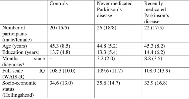
Table 1. Clinical and demographic characteristics of the participants Controls Never medicated
Parkinson’s disease
Recently medicated Parkinson’s disease Number of
participants (male/female)
20 (15/5) 26 (18/8) 22 (17/5)
Age (years) 45.3 (8.5) 44.8 (5.2) 45.3 (8.2)
Education (years) 13.7 (4.8) 13.3 (5.4) 14.4 (6.2) Months since
diagnosis*
– 3.2 (2.0) 8.8 (3.5)
Full-scale IQ (WAIS-R)
108.3 (10.0) 109.6 (11.7) 108.0 (13.9) Socio-economic
status
(Hollingshead)
34.6 (13.0) 35.6 (14.7) 33.9 (16.8)
Novelty seeking* 20.8 (3.2) 17.0 (4.2) 25.0 (7.4) Harm avoidance 15.8 (4.0) 15.5 (3.1) 15.5 (3.3) Reward dependence 16.1 (4.4) 17.3 (4.2) 17.4 (4.1)
Persistence 4.2 (0.8) 4.0 (1.0) 4.1 (1.1)
No. of patients in Hoehn–Yahr Stage
– 1.0:4
1.5:2 2:18 2.5:1 3:1
1.0:2 1.5:2 2:15 2.5:2 3:1
UPDRS – 30.8 (6.4) 27.5 (6.1)
HAM-D – 4.2 (1.4) 4.6 (2.0)
HAM-A – 3.1 (1.8) 3.3 (1.5)
Data are mean (standard deviation).
*Significant difference across group, P<0.05 (for details, see text)
3.1.2. Participants in the stimulus-learning experiment
Twenty-five patients with mild AD and 20 healthy elderly controls participated in the study. Patients and controls were matched for age, gender, and education. The diagnosis of probable AD was made according to the NINCDS-ADRDA criteria (McKhann, Drachman, Folstein, Katzman, Price & Stadlan, 1984). Participants were evaluated with the Mini-Mental State Examination (MMSE) (Folstein, Folstein &
McHugh, 1975) and Global Deterioration Scale (GDS) (Reisberg, Ferris, de Leon &
Crook, 1982). Clinical information included medical history, laboratory tests, brain imaging findings (head magnetic resonance imaging [MRI]), neurological examination, and neuropsychological test results. Exclusion criteria consisted of vascular lesions on MRI scans and prior neurological and psychiatric disorders. The clinical and demographical data are shown in Table 2.
Table 2. Clinical and demographical characteristics of the participants
Controls (n=20) Alzheimer’s patients (n=22)
Age (years) 70.1 (4.8) 69.8 (6.9)
Male/female 12/8 15/7
Education (years) 13.7 (3.2) 13.6 (3.8)
MMSE 29.4 (0.7) 24.0 (1.3)
GDS - 3.7 (0.5)
There were no significant differences between controls and Alzheimer’s patients with the exception of the Mini-Mental State Examination (MMSE) scores (p<0.0001).
3.1.3. Participants in the visuospatial assessment
We analyzed clocks drawn by older individuals without dementia (n = 25) and patients diagnosed with FTD (n =36) and AD (n =25). The FTD group was composed of frontotemporal dementia behavioral variant (FTD-bv) (n=18), primer non-fluent aphasia (PNFA) (n=13), and semantic dementia (SD) (n=5) patients. All study participants were seen between 2003, when CDT according to the Rouleau et al. (1992) system became a regular part of clinical assessment, and early 2005 (Table 3). The AD patients all met the criteria for probable Alzheimer’s disease according to the National Institute of Neurological and Communicative Disorders and Stroke and the Alzheimer’s Disease and Related Disorders Association (NINCDS-ADRDA; McKhann et al., 1984). The FTD sample fulfilled the Neary et al. (1998) and McKhann et al. (2001) criteria. The positive predictive value on autopsy based on the McKhann et al. (1984) and Neary et al. (1998) criteria have been shown to be greater than 80% (Bowler et al., 1998; Kertesz et al., 2005). The PNFA group included patients who were anomic, logopenic, and nonfluent. The SD group was diagnosed by the presence of a prominent comprehension deficit, naming difficulty, and asking the meaning of nouns and objects. All FTD patients were placed into FTD-bv, SD, and PNFA groups based on syndromes at the onset of illness. From the history provided at consultation 15 patients in the FTD-bv group had a behavioral syndrome at onset, 2 had dysexecutive problems observed around the home and at work, and 1 had a combination of both behavioral and dysexecutive symptoms at onset. At the time of the CDT, 3 FTD-bv patients had began to develop symptoms of progressive aphasia (PA), 2 had features of SD, and 1 had signs of motor neuron disease. Of the 5 SD patients, 3 had developed behavioral changes by the time of testing.
Table 3. Demographic characteristics, cognitive test results, and overall CDT scores of participants
FTD (n=36) M (SD)
AD (n=25) M (SD)
Controls (n=25) M (SD)
Total Population (N=86) M (SD)
p value
Age (yrs) 65.14 (7.66) 78.76 (6.04) 65.36 (3.96) 69.16 (8.78) b<.001 Education
(yrs)
13.64 (3.74) 11.88 (4.77) 12.12 (2.93) 12.69 (3.9) .17
Duration of 3.83 (2.02) 3.42 (2.12) .45
illness (yrs) Gender (F:M)
18:18 10:15 13:12 41:45 .65
MMSE (maximum 30)
24.22 (3.98) 22.12 (1.92) 28.84 (1.07) 24.95 (3.86) c<.001
DRS-2 (maximum 144)
113.21 (20.23)
113.25 (11.55)
139.44 (3.59)
121.63 (18.94)
a<.001
CDT (maximum 10)
7.74 (1.99) 5.48 (2.36) 9.54 (.58) 7.6 (2.4) c<.001
Note. AD Alzheimer’s disease, FTD Frontotemporal dementia.
a controls versus FTD, controls versus AD; b AD versus controls, AD versus FTD; c FTD versus controls, FTD versus AD, controls versus AD.
FTD patients with extrapyramidal disorders, such as corticobasal degeneration syndrome (CBDS) and progressive supranuclear palsy (PSP) that would interfere with their ability to perform the CDT, were excluded. This only resulted in the exclusion of two patients. One patient had FTD-bv as a primary syndrome with secondary and tertiary syndromes of CBDS and PA, respectively. The other patient had PNFA as a primary syndrome and PSP as the secondary syndrome.
The exclusion criteria for all patients included metabolic causes of dementia, history of drug abuse, alcohol dependence, serious psychiatric condition, neurological disorder such as stroke or closed head injury, a current major depressive episode, psychosis, acute mania, and bipolar disorder. Imaging was conducted on all patients to exclude other causes of dementia such as stroke or tumor. However, imaging was not used as a confirmatory diagnostic measure; diagnosis was based on the prior mentioned clinical criteria. Control data was obtained from the accompanying caregivers of patients.
The control group was selected to match the FTD groups in age and education.
The inclusion criteria for controls consisted of no history of memory problems, age and education-adjusted scale score of nine or higher (normal range) on the second edition of the dementia rating scale (DRS-2; Jurica et al., 2001); and Mini-Mental State Examination (MMSE; Folstein et al., 1975) scores above age and education adjusted cut-off scores (Crum et al., 1993).
3.2.Tasks
3.2.1. Tasks in the reinfrocement learning experiment
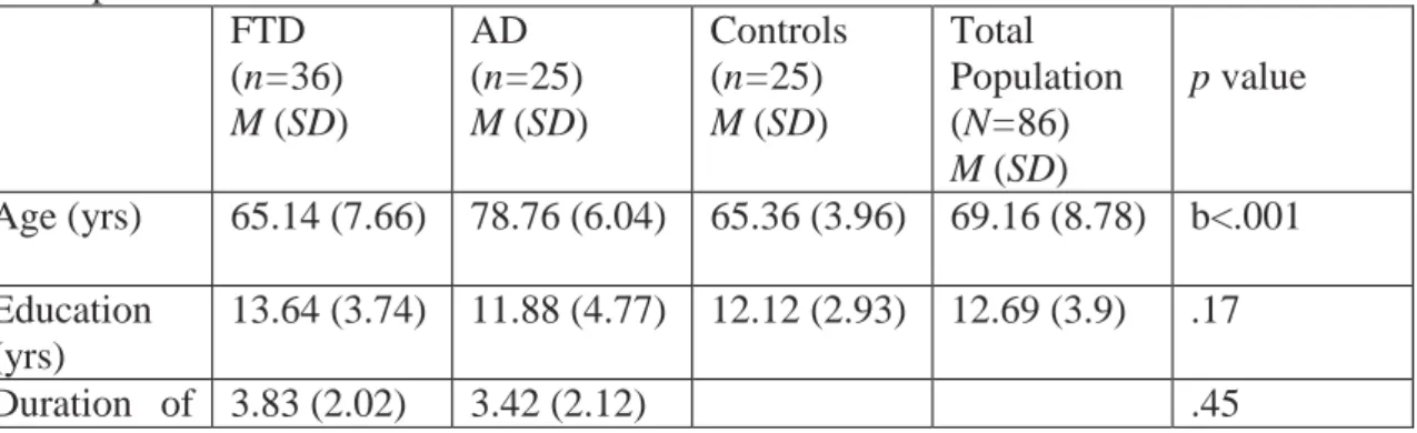
Feedback-based probabilistic classification task
All participants were administered a computer-based probabilistic classification task (Bolikal et al., 2007). On each trial, participants viewed one of four images (Figure 3), and were asked to guess whether it belonged to category A or category B. For each participant, the four images were randomly assigned to be stimuli S1, S2, S3, and S4. A second set of similar images (S5-S8) were used for repeated testing (test-retest reliability based on the repeated testing of controls: r=0.76). On any given trial, stimuli S1 and S3 belonged to category A with 80% probability and to category B with 20%
probability, while stimuli S2 and S4 belonged to category B with 80% probability and to category A with 20% probability (Table 4). Stimuli S1 and S2 were used in the reward-learning task. Two stimuli per valence were employed in order to balance category outcome frequencies, so that one stimulus in each task would be associated with each outcome. Thus, if the participant correctly guessed category membership on a trial with either of these stimuli, a reward of +25 points was received; if the participant guessed incorrectly, no feedback appeared. Stimuli S3 and S4 were used in the punishment-learning task. Thus, if the participant guessed incorrectly on a trial with either of these stimuli, a punishment of –25 was received; correct guesses received no feedback.
Figure 3. The feedback-based probabilistic classification task. (A) On each trial, the participant saw one of four stimuli and was asked whether this stimulus belonged to category A or B. (B) For some stimuli, correct responses were rewarded with visual feedback and 25 points winnings, whereas for others, incorrect responses were punished with visual feedback and loss of 25 points.
Table 4. Category and feedback structure of the probabilistic classification task
Stimulus Probability
Class A (%)
Probability Class B (%)
Feedback
S1 80 20 If correct: +25
S2 20 80 If incorrect: ø
S3 80 20 If correct: ø
S4 20 80 If incorrect: –25
The experiment was conducted on a Macintosh i-book, programmed in the SuperCard language. The participant was seated in a quiet testing room at a comfortable viewing distance from the screen. The keyboard was masked except for two keys, labelled “A” and “B” which the participant could use to enter responses. At the start of
the experiment, the participant read the following instructions: “In this experiment, you will be shown pictures, and you will guess whether those pictures belong to category
“A” or category “B”. A picture does not always belong to the same category each time you see it. If you guess correctly, you may win points. If you guess wrong, you may lose points. You will see a running total of your points as you play. We will start you off with a few points now. Press the mouse button to begin practice.”
The practice phase then walked the participant through an example of a correct and an incorrect response to a sample trial in the punishment-learning task and an example of a correct and incorrect response to a sample trial in the reward- learning task. These examples used images other than those assigned to S1-S4. The participant saw a practice image, with a prompt to choose category A or B, and a running tally of points at the lower right corner of the screen. The tally is initialized to 500 points at the start of practice. The participant was first instructed to press the “A” key, which resulted in a punishment of –25 and updated point tally and then the “B” key, which resulted in no feedback. The participant then saw a second practice figure and was instructed first to press the “B” key which resulted in a reward of +25 and updated point tally and then the “A” key, which resulted in no feedback.
After these two practice trials, a summary of instructions appeared: “So… For some pictures, if you guess CORRECTLY, you WIN points (but, if you guess incorrectly, you win nothing). For other pictures, if you guess INCORRECTLY, you LOSE points (but, if you guess correctly, you lose nothing). Your job is to win all the points you can – and lose as few as you can. Remember that the same picture does not always belong to the same category. Press the mouse button to begin the experiment.”
From here, the experiment began. On each trial, the participant saw one of the four stimuli (S1, S2, S3, S4) and was prompted to guess whether it was an “A” or a “B”. On trials in the reward-learning task (with stimuli S1 or S2), correct answers were rewarded with positive feedback and gain of 25 points; incorrect answers received no feedback.
On trials in the punishment-learning task (with stimuli S3 or S4), incorrect answers were punished with negative feedback and loss of 25 points; correct answers received no feedback. The task contained 160 trials. Within a block, trial order was randomized.
Trials were separated by a 2 second interval, during which time the screen was blank.
Within each block, each stimulus appeared 10 times, 8 times with the more common
outcome (e.g. category “A” for S1 and S3 and “B” for S2 and S4) and 2 times with the less common outcome. Thus, training on the reward-learning task (S1 and S2) and punishment-learning task (S3 and S4) were intermixed. The no-feedback outcome, when it arrived, was ambiguous, as it could signal lack of reward (if received during a trial with S1 or S2) or lack of punishment (if received during a trial with S3 or S4). At the end of the 160 trials, if the participant’s running tally of points was less than 525 (i.e. no more than the points awarded at the start of the experiment), additional trials were added on which the participant’s response was always taken as correct, until the tally is at least 525. This was done in an attempt to minimize frustration in participants by ensuring that all participants terminated the experiment with more points than they had started with. Data from any such additional trials were not analyzed. On each trial, the computer recorded whether the participant made the optimal response (i.e. category A for S1 and S3, and category B for S2 and S4) regardless of actual outcome.
Personality measures
Following the probabilistic classification task, all participants were administered the Hungarian version of the TCI questionnaire, which has a good test-retest reliability (Rózsa et al. 2005). The TCI is suitable for the assessment of temperament and character traits. In this study, we focused on the temperament traits of novelty seeking (exploratory excitability, impulsiveness, extravagance, disorderliness), harm avoidance (anticipatory worry, fear of uncertainty, shyness, fatigability), and reward dependence (sentimentality, openness to warm communication, attachment, dependence), and persistence (eagerness of effort, work hardened, ambitious, perfectionist) (Cloninger, 1994). Thus, in addition to the main focus on novelty seeking and harm avoidance, data also were collected on reward dependence and persistence in order to test the specificity of possible alterations in personality traits.
Data analysis
The normality of data distribution was checked using Kolmogorov-Smirnov tests. All data were normally distributed (p>0.1). Analyses of variance (ANOVAs)
using the general linear model panel of the STATISTICA 7.0 software (StatSoft, Inc., Tulsa) were used to compare controls, never-medicated, and recently-medicated PD patients, and to compare the performance of patients at baseline (no medication) and at follow-up (dopamine agonists). ANOVAs were followed by planned F tests and Tukey Honestly Significant Difference (HSD) tests. Two-tailed t tests were used for the analysis of demographic data and personality measures. Pearson’s product-moment correlation coefficients were calculated between test performance and personality measures. The Williams test was used to compare the correlation coefficients. The level of significance was set at alpha<0.05.
3.2.2. Tasks in the stimulus-learning experiment
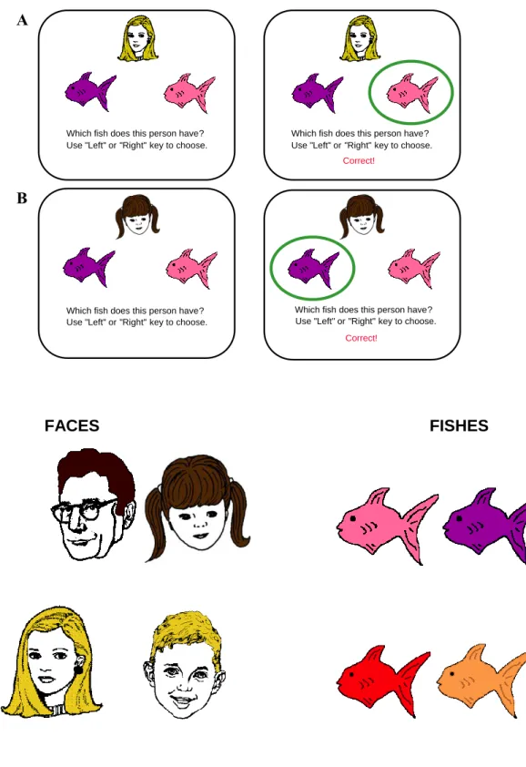
Associative learning test
Stimuli were presented and responses were collected using a Macintosh Power- Book laptop. The antecedent stimuli were four drawings of faces (man, woman, girl, boy). The consequents were drawings of fish colored red, orange, purple, and pink. For each participant, stimuli were randomly assigned as antecedent and consequent stimuli.
At the start of the experiment, the following instruction appeared on the screen:
“Welcome to the experiment. You will see drawings of people who each have some pet fish. Different people have different kinds of fish. Your job is to learn which kinds of fish each person has. At first, you will have to guess.” The experimenter read the instruction aloud to the participant and then clicked the mouse button to begin the acquisition phase. On each trial, a face and two fish drawings were displayed on the computer screen along with the prompt: “Which fish does this person have? Use the Left or Right key to choose”. The participant responded with pressing one of two separate keys labeled as “LEFT” and “RIGHT” to indicate whether the fish on the left or the fish on the right was associated with the face. The selected fish drawing was circled and corrective feedback was given (Figure 4). In the case of an incorrect response, an alert beep sounded. The left-right ordering of the fish drawings was randomized across subjects. There were three stages in the acquisition phase (Table 5).
Stages 1 and 2 terminated after 8 consecutive correct responses, whereas stage 3 terminated after 12 consecutive correct responses. The participant was not informed on the beginning of a new stage. After the termination of the acquisition phase, a new instruction appeared on the screen, informing the participant that the task would remain the same but feedback would no longer be provided. The participant was not informed of the presence of new associations. The transfer phase consisted of 48 trials of which 12 trials were new associations for the testing of learned equivalence and 36 trials were old associations trained during the acquisition phase. The dependent measures were the mean number of errors in the acquisition phase and the proportion of incorrect responses in the transfer phase (for methodological details, see Myers et al., 2003).
After the computer-administered testing phase, participants received cards (size:
5 x 5 cm) depicting the faces and fishes. The task was to pair fishes and faces as learned during the test. The dependent measure was the percentage of correctly retrieved face- fish associations. After the card sorting test, participants were asked to read a newspaper article for 5-min. After this, the original computer-administered testing phase was repeated.
Figure 4. Example screen events during one trial. (A) Stimuli appear. (B) Participant
responds and corrective feedback is given.
FACES FISHES
Table 5. Acquired equivalence learning
Which fish does this person have?
Use "Left" or "Right" key to choose.
Which fish does this person have?
Use "Left" or "Right" key to choose.
Correct!
Which fish does this person have?
Use "Left" or "Right" key to choose.
Which fish does this person have?
Use "Left" or "Right" key to choose.
A
B
Correct!
Acquisition Stage 1:
Shaping
Acquisition Stage 2:
Equivalence Training
Acquisition Stage 3:
New Consequents
Transfer Phase:
Equivalence Testing
A1 → X1 A1 → X1
A2 → X1
A1 → X1 A2 → X2?
A2 → X1 A1 → X2
B1 → Y1 B1 → Y1
B2 → Y1
B1 → Y1 B2 → Y2?
B2 → Y1 B1 → Y2
Data analysis
The number of errors in the training phase of the associative learning test and the clinical parameters were analyzed with two-tailed t tests and Mann-Whitney U test (this non-parametric analysis was used for MMSE values which showed non-Gaussian distribution). Errors from the testing phase were analyzed with a three-way repeated measures analysis of variance (ANOVA) which had the following design: 2 (group) by 2 (immediate vs. delayed testing) by 2 (old vs. new associations). A two-way ANOVA was used for the analysis of errors from the card pairing test with a 2 (group) by 2 (old vs. new associations) design. Tukey Honestly Significant Difference Test (HSD) was used for post hoc analysis. The level of significance was alpha<0.05.