SZÉKFOGLALÓ ELŐADÁSOK A MAGYAR TUDOMÁNYOS AKADÉMIÁN
Michael Berridge
cALcIUM, MEMORY AND
ALZhEIMER’S DISEASE
Michael J. Berridge
CALCIUM, MEMORY AND ALZHEIMER’S DISEASE
SZÉKFOGLALÓK
A MAGYAR TUDOMÁNYOS AKADÉMIÁN INAUGURAL LECTURES
AT THE HUNGARIAN ACADEMY OF SCIENCES A 2013. május 6-án megválasztott
akadémikusok székfoglalói Inaugural lectures by new members
elected on 6 May, 2013
Michael J. Berridge
CALCIUM, MEMORY AND ALZHEIMER’S DISEASE
Magyar Tudományos Akadémia • 2014
Az előadás elhangzott 2014. május 21-én Delivered on 21 May, 2014
Sorozatszerkesztő • Series editor: Bertók, Krisztina
Angol nyelvi lektor • English reader: Torkos, Béla
Borító és tipográfi a • Cover and typography: Auri Grafi ka
ISSN 1419-8959 ISBN 978-963-508-766-2
© Michael J. Berridge
Kiadja a Magyar Tudományos Akadémia • Published by the Hungarian Academy of Sciences Kiadásért felel • Person in charge of publication: Lovász, László, az MTA elnöke • President of HAS
Felelős szerkesztő • Editor-in-chief: Kindert, Judit Nyomdai munkálatok • Printed by: Kódex Könyvgyártó Kft.
INTRODUCTION
When I retired, my American colleagues (Jim Putney and Gary Bird) presented me with a coat of arms they had designed. The motto read “Apis genua est Calcium”, which translates to “Calcium is the bee’s knees” and nicely summarises my lifelong fascination with calcium (Ca2+). The small icons on the shield illustrate various aspects of my research: the fl y represents the model organism that I used to discover the cellular messenger inositol trisphosphate (InsP3 top right) (Berridge 2012a). I discovered that InsP3 acts to generate the Ca2+ signals that often appear in cells in the form of regular oscillations (bottom right). This InsP3/Ca2+ signalling pathway will feature signifi cantly in the following discussion of how Ca2+ functions in memory formation and how such memories are rapidly lost in Alzheimer’s disease (AD).
Figure 1. A coat of arms depicting highlights of the author’s scientifi c work.
The motto, which is translated as “Calcium is the bee’s knees”, refl ects the central role of Ca2+ in regulating the activity of multiple cellular processes
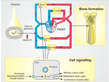
When discussing Ca2+, it is important to establish that this ion has two major functions (Figure 2). Firstly, it has a structural role as part of the skeleton. The Ca2+ is absorbed from the intestine and enters the blood stream to be carried around the body where it is used by osteoblasts to form bone.
Secondly, it also has a vital signalling role operating within the cell to regulate a large number of cellular processes such as fertilization, muscle contraction, secretion of hormones such as insulin and adrenalin, secretion of fl uid by the salivary glands and memory formation. Before discussing the role of Ca2+ in memory in more detail, I shall explore some of the primary features of this Ca2+ signalling system.
Figure 2. Calcium (Ca2+) has two important functions. Firstly, it is taken up from the lumen of the intestine and can then be used for bone formation. Secondly, Ca2+ functions in cell signalling where it acts to regulate
multiple cellular processes
CELL COMMUNICATION MECHANISMS
Cells communicate with each other through two main mechanisms (Figure 3).
Firstly, there is electrical communication that occurs when cells are connected to their neighbours through gap junctions. These low resistance pathways allow free communication between the cells for ions and small messenger molecules.
Secondly, cells are also linked together through chemical communication whereby one cell releases a chemical stimulus (e.g. hormones, neurotransmitters and growth factors) that is then detected by receptors on the target cell. These receptors then activate various internal cell signalling pathways to activate effectors in the cytoplasm (Ecyto) or nucleus (Enucl) to stimulate multiple cellular processes.
Figure 3. Cells communicate through either electrical or chemical signalling mechanisms. Cells that are connected through gap junctions (shown on the left) can communicate rapidly with each other by passing electrical current or through the diffusion of internal messengers. In the case of chemical communication (shown on the right), one cell releases a chemical stimulus, which diffuses to a target cell that has receptors to detect the stimulus and to relay information along various cell signalling pathways to activate effectors either in
the nucleus (Enucl) or cytoplasm (Ecyto)
We now know that there is a large number of such cell signalling pathways (Figure 4). During development, each specifi c cell type selects out and expresses those particular pathways that are suited to carry out their particular function. Of all of these pathways the Ca2+ signalling pathway is expressed most widely and plays a central role in many different cellular processes. As is evident from Figure 4, the Ca2+ signal can be generated by a number of different mechanisms (i.e. Steps 2-5 in Figure 4) with very different properties and this greatly enhances the versatility of the Ca2+ signalling system that is capable of generating Ca2+ signals with very different temporal and spatial properties (Berridge et al 2000). For example, localized Ca2+ signals in nerve ending operate in microseconds to release neurotransmitters, whereas much slower signals lasting for many seconds trigger the process of fertilization.
In order to achieve this versatility, there is a very large Ca2+ signalling toolkit (Figure 5) from which components can be mixed and matched to create these widely different Ca2+ signals (Berridge 2012b). There are a large number of channels (shown in green) responsible for introducing Ca2+ into the cell, Ca2+ pumps and exchangers (shown in red) remove Ca2+. Then there are a large number of internal sensors (shown in orange) that detect the Ca2+ in order to activate different cellular processes (shown in yellow). As will be described later, brief pulses of Ca2+ are responsible for forming memories, whereas slower lower concentrations erase memories. The next aspect to consider, therefore, is how components of the toolkit are assembled to produce such pulses of Ca2+.
Ca
2+signalling mechanism
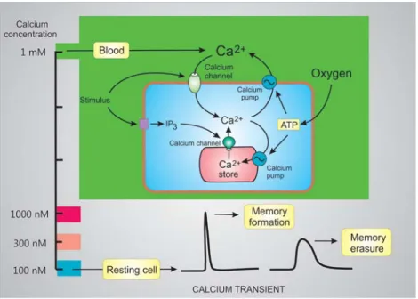
A key aspect of Ca2+ signalling concerns the huge concentration gradient of Ca2+
that exists between the inside and outside of the cell. The concentration of Ca2+
in the blood surrounding the cell is 1-2 mM, whereas that inside the cell is 10,000 times lower at 100 nM (Figure 6). Despite this enormous concentration gradient, very little Ca2+ enters the cell under resting conditions because the
Figure 4. Summary of the major signalling pathways used by cells to regulate cellular processes. Cells have a number of signalling systems that are capable of responding either to external stimuli or to internal stimuli.
The external stimuli acting on cell-surface receptors are coupled to transducers to relay information into the cell using a number of different signalling pathways (Pathways 1–18). Internal stimuli activate signalling
pathways independently of external signals (Pathways 19 and 20). All of these pathways generate internal messengers that then act through internal sensors to stimulate the effectors that bring about different cellular
responses. In this article, attention will be focused on the pathways 3-5 that function to generate the Ca2+
signals responsible for memory formation
Figure 5. Summary of the large Ca2+ signalling toolkit. The green boxes illustrate the membrane Ca2+ channels, whereas the red boxes are the pumps and exchangers that remove Ca2+ either out of the cell or back into the endoplasmic reticulum (ER). The purple boxes represent the buffers located in the cytoplasm or in the endoplasmic reticulum (ER). To carry out its signalling function, Ca2+ binds to sensors (brown boxes) that then
employ a range of effectors to stimulate many different cellular processes shown in the yellow boxes
plasma membrane that surrounds the cell is very impermeable. However, cells have channels in this plasma membrane that open to allow Ca2+ to enter in response to the appropriate stimulus. In addition to this external source of Ca2+, cells also have an internal store of signal Ca2+ contained within the endoplasmic reticulum (ER). Like the plasma membrane, the ER also has channels capable of releasing Ca2+ from this internal store in response to external stimuli (Figure 6).
Many of these stimuli are incapable of entering the cell so they act by binding to receptors in the plasma membrane that then act to generate an internal messenger, such as the InsP3 described earlier. The InsP3 then diffuses into the cell to bind to the channels on the ER to release Ca2+ into the cytoplasm.
Figure 6. The concentration of Ca2+ in the blood, which is 10,000-fold higher than that in the cytoplasm, is one of the sources of signal Ca2+. Cells also have an internal membrane store of Ca2+ that can also be released into the cytoplasm. In response to a stimulus, Ca2+ enters across the plasma membrane or is released from the internal store. In neurons, a brief transient of Ca2+ from a resting level of 100 nM to 1000 nM is responsible for
memory formation. In contrast, a small transient from 100 nM to 300 nM induces memory erasure
As a result of the opening of the external channels and/or the ER channels, the concentration within the cell increases from the resting level of 100 nM to levels between 300 to 1000 nM. Such opening of these channels is normally transient because they rapidly inactivate and once they close, the signal Ca2+ is rapidly removed by being pumped out of the cell and back into the ER (Figure 6). The channels and pumps that operate consecutively to generate the characteristic Ca2+ transients are then responsible for stimulating cellular processes. In the case of memory, Ca2+ transients can regulate both memory formation and memory erasure depending on the amplitude of the transient.
There is increasing evidence that 1000 nM Ca2+ transients form memories, whereas smaller 300 nM transients can erase these memories.
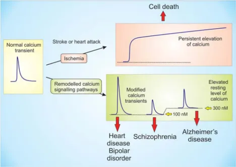
There is increasing evidence that alterations in the properties of these Ca2+
transients are responsible for some of the major diseases in man. An extreme example occurs during stroke or heart attack where blood clots cut off the blood supply to large groups of cells that die because of the persistent elevation of Ca2+ (Figure 7). Since the pumps that remove Ca2+ from the cell depend on a constant supply of energy (ATP), the ischemia and resulting decline in oxygen means that the Ca2+ remains elevated for a prolonged period and this results in cell death. There are many other diseases that result from a more subtle remodelling of the Ca2+ signalling system (Berridge 2012b; Berridge et al 2013) that result in transients that are too large (heart disease) or too low (schizophrenia). In addition, there may be a small elevation in the resting level Ca2+ that may be responsible for inducing Alzheimer’s disease (AD) as will be argued later.
Before considering how Ca2+ dysregulation might explain AD, it is necessary to describe how Ca2+ acts normally to form memories when awake and erase them during sleep. Memory formation is thus intimately connected with the operation of the brain rhythms that characterize the sleep/wake cycle.
BRAIN RHYTHMS AND THE TONIC EXCITATORY DRIVE
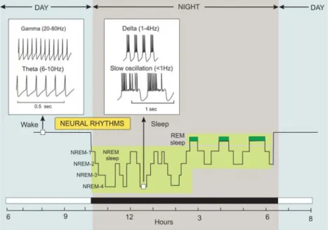
The neural activity of the conscious brain during wakefulness, when we are aware of our surroundings, is very different to that during sleep.
Electroencephalogram (EEG) recordings indicate that when we are awake, our brain cells are oscillating at very high frequencies in the theta and gamma ranges (Figure 8). A rapid decline in neural activity occurs at the onset of sleep
Figure 7. Deregulation of Ca2+ signalling can contribute to multiple human pathologies. During ischemia associated with stroke or a heart attack, a decrease in blood supply and the resulting lack of oxygen prevents the pumping of Ca2+ and the resulting persistent elevation of Ca2+ induces cell death. More subtle remodelling
of Ca2+ signalling pathways results in some of the major diseases in man. Heart disease is caused by Ca2+
transients that are too high, whereas smaller transients result in schizophrenia. An elevation in the resting level of Ca2+ may be responsible for Alzheimer’s disease
as the brain switches from consciousness to a period of unconsciousness. This period of sleep consists of two main phases: rapid eye movement (REM) sleep and non-rapid eye movement (NREM) sleep. The oscillatory rhythms that occur during the sleep/wake cycle are of critical importance for memory in that memories are formed during the fast rhythms that occur while awake, whereas memory erasure occurs during the slow rhythms that occur during NREM sleep. To understand memory, therefore, it is necessary to understand the Ca2+ signals that occur during these different rhythms.
Figure 8. The sleep/wake cycle. Sleep consists of two main phases: non-rapid eye movement (NREM) and rapid eye movement (REM) sleep. The NREM sleep has four different stages. During the wake state, memories are formed when neurons in the brain fi re at the fast theta and gamma neural rhythms. Memories
are erased during the delta and slow oscillations that occur during NREM sleep
One of the master regulators of this sleep/wake cycle is the ascending arousal system, which wakes up the sleeping brain and is responsible for maintaining the period of wakefulness. This arousal system consists of a heterogeneous population of neurons located within the brainstem, midbrain, basal forebrain and hypothalamus that produce sleep/wake regulatory molecules that are released throughout the major regions of the brain such as the cortex and hippocampus (Figure 9).
Figure 9. The human brain has a number of discrete regions that carry out different functions. There is a large cortex that extends around to connect to the hippocampus. The middle of the brain has the corpus callosum, ventricles and the thalamus. Below the thalamus is the hypothalamus that extends down into the pituitary. The cerebellum at the back controls movement. Neurons of the ascending arousal system, which are located in the basal forebrain, midbrain and hindbrain, send their axons out into the rest of the brain where they release transmitters, such as orexin, acetylcholine (ACh), 5-hydroxytryptamine (5-HT) and
norepinephrine (NE) that act to control the sleep/wake cycle
The different oscillatory modes that occur during the sleep/wake cycle are regulated by an interaction between the ascending arousal system and the sleep-inducing system (Figure 10). The ascending arousal system consists of an array of neurons such as the orexinergic neurons, cholinergic neurons, serotonergic neurons, histaminergic neurons, noradrenergic neurons and dopaminergic neurons. The neurotransmitters released by this arousal system act on neural circuits to regulate the sleep/wake cycle (Berridge 2012c; Berridge 2014a). The orexinergic neurons that release orexin are master regulators that act to stimulate the other arousal neurons. The central role of orexinergic neurons is also evident in the way they integrate the action of a number of other sleep regulatory factors such as ghrelin, leptin, glucose and adenosine.
The sleep/wake cycle is also controlled by the circadian clock. The onset of sleep is initiated by neurons located in the preoptic area of the brain such as the ventrolateral preoptic (VLPO) neurons, which act by releasing GABA that inhibits the activity of the ascending arousal neurons. When the arousal neurons are active during the wake period, they release transmitters onto the brain neurons where they induce the tonic excitatory drive that generates and maintains the fast brain rhythms responsible for consciousness.
Tonic excitatory drive
The transmitters that are released from the arousal neurons act on receptors that are coupled to various signalling pathways that provide the depolarization that controls the level of the tonic excitatory drive that regulates the different oscillatory states that characterize the sleep/wake cycle (Berridge 2012c;
Berridge 2014a). Variation in the activity of this tonic drive functions much like a rhythm rheostat in that it controls the hierarchy of rhythms with the lowest frequencies occurring during sleep that are then switched to the higher frequency rhythms of the wake state (Figure 11). The membrane depolarization responsible for the tonic excitatory drive is induced by various signalling
mechanisms that either activate inward Na+ currents or close outward K+ currents (Berridge 2014a). For example, hydrolysis of the phospholipid phosphatidylinositol 4,5-bisphosphate (PtdIns4,5P2) has two effects. Firstly, it closes the KV7.2/KV7.3 channels responsible for the M current. Switching off this M current depolarizes the membrane to increase neuronal activity.
Secondly, hydrolysis of PtdIns4,5P2 to form InsP3 releases Ca2+ that stimulates the Ca2+-activated non-selective cation (CAN) channel. The CAN channel can also be activated by Ca2+ entering through the voltage-operated Ca2+ (VOC)
Figure 10. The sleep/wake cycle is regulated by the ascending arousal and sleep-inducing systems. The ascending arousal system releases transmitters such as orexin, acetylcholine (ACh), 5-hydroxytryptamine (5-HT), norepinephrine (NE) and dopamine (DA) that activate signalling systems responsible for the tonic excitatory drive, which is a rhythm rheostat that controls the neural rhythms that occur during the sleep/wake
cycle. The sleep-inducing system releases the inhibitory transmitter GABA that switches off the ascending arousal neurons to induce sleep
channel. The NE and DA act through the cyclic AMP signalling pathway to enhance the activity of the hyperpolarizing-activated cyclic nucleotide-gated (HCN) channel responsible for the depolarizing Ih current.
An important feature of this tonic excitatory drive is that it is applied equally to both the excitatory and inhibitory neurons and it is essential for proper brain function for this stimulation to be fi nely balanced. There are
Figure 11. The tonic excitatory drive. Neurons of the ascending arousal system release transmitters such as orexin, acetylcholine (ACh), 5-hydroxytryptamine (5-HT), norepinephrine (NE) and dopamine (DA) that activate signalling systems that control the neural rhythms that occur during the sleep/wake cycle. The tonic excitatory drive mechanism depends on membrane depolarization that results from closing the KV7.2/KV7.3 channels responsible for the M current and the opening of the Ca2+-activated non-specifi c cation channel (ICAN) and the hyperpolarizing-activated cyclic nucleotide-gated (HCN) channel responsible for the Ih current.
This tonic excitatory drive functions as a rhythm rheostat to regulate the different neural rhythms that occur during the sleep/wake cycle
indications that alterations in this excitation-inhibition (E-I) balance may occur in psychiatric diseases such as BPD where excessive excitation may be responsible for the manic phase, whereas depression may result from excessive inhibition (Berridge 2014b).
With regard to memory formation, a key feature of these fast brain rhythms is that the excitatory neurons operate in synchrony with each other to create the functional interaction responsible for memory formation. The fast gamma oscillations (20-80 Hz) are generated through the operation of a network oscillator that depends on the feedback interactions between the inhibitory interneurons and the excitatory neurons (Figure 12). A unique feature of these circuits is that each inhibitory interneuron sends out a long axon with multiple terminals that makes contact with and thus controls the activity of many excitatory neurons. The inhibitory interneuron has a primary role in setting up the gamma rhythm in that it fi res an action potential at the crest of each gamma cycle and this induces the hyperpolarization that occurs synchronously in all the excitatory neurons (Figure 12). While all the excitatory neurons participate in each gamma cycle by responding to GABA to produce a hyperpolarization response, they fi re much less frequently and the resulting action potentials occur within a narrow time window towards the end of the pacemaker depolarization. In this way, a single inhibitory interneuron can entrain the inherent rhythmical activity of a large population of pyramidal neurons. When these excitatory neurons are processing information, they communicate with each other (green arrow) and the resulting action potential coincidence (Figure 12) is responsible for memory formation.
MEMORY FORMATION
There are two types of memory: working memory located primarily in the hippocampus (equivalent to the RAM in a computer) and the permanent
memory store located in the cortex (equivalent to the hard drive in a computer) (Figure 9). New memories, which are acquired during the wake period, are stored temporary in the working memory. During sleep, any new information is uploaded into the permanent memory store in the cortex, whereas most of the other memories are erased so that the working memory store is available to acquire new memories during the next wake period. To understand memory,
Figure 12. Many neural circuits (see inset at the bottom) consist of fast spiking inhibitory interneurons (red) and excitatory neurons (green) interacting with each other through a positive/negative feedback loop.
A unique feature of many circuits is that each inhibitory neuron controls the activity of many excitatory neurons (red arrow). The inhibitory neuron fi res an action potential on each gamma cycle and this serves to induce a hyperpolarization that occurs synchronously in all the excitatory neurons. While all the excitatory neurons participate in each gamma cycle, they fi re much less frequently towards the end of the pacemaker depolarization. When two excitatory neurons communicate with each other (green arrow) there is coincidence
in the action potentials and this is critical for memory formation. The ascending arousal system releases transmitters such as acetylcholine (ACh) that excite both the excitatory and inhibitory neurons (blue arrows)
therefore, it is necessary to consider how memories are formed in regions such as the hippocampus when we are conscious and how they are erased when we go to sleep.
The hippocampus, which is one of the regions where the working memory is located, consists of a trisynaptic circuit (Figure 13). At one end, the hippocampus is connected to various cortical regions. The cortical region closest to the hippocampus is the presubiculum and it is through this region that the working memory in the hippocampus can communicate with the permanent memory in the cortex. The hippocampal pyramidal neurons, which are arranged into three regions (dentatae gyrus, CA3 and CA1), are joined together to form a trisynaptic circuit (synaptic connections 1–3 in Figure 13).
The fi rst synapses are on the granule cells of the dentate gyrus that receives in- put from the perforant fi bres. These granule cells send out axons called mossy fi bres that extend into the CA3 region, where they form the second group of synapses by innervating the characteristic pyramidal cells. The axon from the CA3 neuron bifurcates: one part forms the commissural fi bres that are directed down to the septum, whereas the other gives rise to Schaffer collaterals that complete the trisynaptic circuit by innervating the pyramidal neurons in the CA1 region. The axons emanating from the CA1 neurons carry information back to the cortex where the permanent memories are stored.
The dendrites on these three neuronal cell types are encrusted with spines that receive the synaptic inputs, enabling them to communicate with each other. All of these synaptic connections are highly plastic in that they are the sites where memories are formed during the process of action potential coincidence (Figure 14). The action potential in the presynaptic neuron (e.g.
Neuron A in Figure 14) invades the synaptic ending, where it activates a voltage- operated Ca2+ channel (VOC) to produce a local pulse of Ca2+ that triggers the release of the transmitter glutamate. The glutamate then has two important
functions: it activates the AMPA receptors (AMPARs) that gate Na+ to initiate an action potential and the resulting depolarization provides an essential signal to facilitate the opening of the NMDA receptors (NMDARs). The latter are unusual in that they require both glutamate and depolarization before they can open. When both neurons fi re an action potential in synchrony with each other, the NMDAR channel opens to allow Ca2+ to fl ood into the postsynaptic ending to trigger memory formation (Figure 14).
Figure 13. The hippocampus has a trisynaptic local circuit. The fi rst synapses (1) are on the granule cells of the dentate gyrus (orange) that receives input from the entorhinal cortex. These granule cells send out axons
called mossy fi bres that extend into the CA3 region (blue), where they form the second group of synapses (2) by innervating the CA3 pyramidal cells. The axons from the CA3 neurons bifurcate: one part is directed down to the septum, whereas the other gives rise to Schaffer collaterals that complete the trisynaptic circuit by
innervating the pyramidal neurons (3) in the CA1 region (yellow). The CA1 neurons send their axons back to the cortex. This hippocampal circuitry plays an important role in the operation of the temporary
working memory
The resulting burst of Ca2+ is then responsible for inducing three biochemical changes that are responsible for memory formation (Figure 15A).
Firstly, Ca2+ activates CaMKII to phosphorylate the AMPAR resulting in an increase in its sensitivity to glutamate. Secondly, this sensitivity to glutamate is also enhanced by the insertion of more AMPARs through a process of Ca2+- dependent exocytosis. Thirdly, Ca2+ activates actin polymerization resulting in spine elongation and a closer apposition between the pre- and post-synaptic
Figure 14. The action potential (AP) coincidence, which occurs when two neurons communicate with each other, plays a critical role in memory formation. When the AP in neuron A reaches the synaptic terminal, the
depolarization acts to open voltage-operated Ca2+ channels (VOCs) that creates the signal to trigger exocytosis and the release of glutamate. The glutamate has two actions. Firstly, it acts on AMPARs to gate Na+ resulting in depolarization that triggers the action potential. Secondly, glutamate activates the NMDARs to induce the entry of Ca2+ that is responsible for activating the three biochemical processes responsible for memory
formation (See Figure 15 for details)
membranes. These biochemical changes, which constitute a new memory, are then retained in the working memory for the remainder of the wake period.
At the onset of sleep, those memories that represent novel information are then consolidated by being uploaded into the permanent memory store in the cortex before much of the information in the working memory is erased through the low intensity global Ca2+ transients that occur during slow wave sleep.
MEMORY ERASURE
Normal memory erasure, which occurs during the early phase of NREM sleep, is also a Ca2+-dependent process (Figure 15B). Less is known about the signals responsible for these erasure processes, but it is likely to be driven by the slow wave oscillations in Ca2+ that have been recorded during this phase (Errington et al 2012). The remarkable aspect of these oscillations is that each transient is global in that the Ca2+ rises throughout the neuron (Figure 15B) quite unlike the spine-specifi c Ca2+ transients that are responsible for memory formation (Figure 15A).
The global Ca2+ transients that occur during NREM sleep (Figure 15B) result in an elevation of Ca2+ in each spine where they act to erase any memories that were formed during the wake period. This erasure depends on the action of calcineurin (CaN), which is a Ca2+-sensitive enzyme that responds to lower levels of Ca2+ in the 300-500 nM range (Figure 15B). The CaN dephosphorylates the AMPARs thus leading to receptor desensitization.
The AMPARs that were released to the cell surface during memory formation (Figure 15A) are sucked back into the cell by the process of endocytosis. Finally, the CaN acts to depolymerize actin thus reducing its length and restoring the spine to its previous bulbous shape.
In summary, spine-specifi c Ca2+ transients that occur during the wake period are responsible for forming memories, whereas the periodic global Ca2+
Figure 15. The role of Ca2+ in memory formation and memory erasure. A. Memories are formed when the NMDARs open to create the high intensity pulse of Ca2+ that activates CaMKII to phosphorylate and sensitize
the AMPARs. Such sensitization is enhanced further by the exocytosis of vesicles containing AMPARs.
Following the conversion of G to F actin the resulting formation of actin fi laments changes the shape of the spine. B. Memories are erased by lower intensity Ca2+ transients that activate calcineurin (CaN) to reverse the
three biochemical changes that are responsible for memory formation
transients that appear during NREM sleep erase these temporary memories (Figure 16). In the case of Alzheimer’s disease (AD), it is proposed that a permanent elevation of the resting level of Ca2+ into the 300 nM range may act to continuously erase memories shortly after they are formed during the wake period (Berridge 2012c, 2014a).
Figure 16. Calcium-induced memory formation and erasure. During wake periods, high intensity Ca2+ spikes confi ned to specifi c spines are responsible for memory formation. Part of the biochemical change is the formation of actin fi laments that cause spine elongation. The biochemical changes are analogous to a computer digital switch from 0 to 1. During sleep, global elevations of Ca2+ in the 300 nM range result
in memory erasure as the biochemical changes that occur during memory formation are rapidly reversed i.e. the spine is switched back from 1 to 0
CALCIUM HYPOTHESIS OF ALZHEIMER’S DISEASE
The development of Alzheimer’s disease (AD) is driven by the accumulation of amyloid β (Aβ) protein, which is a neuron-derived pathogenic factor that brings about the loss of memory and subsequent neuronal cell death that characterizes the progression of AD. This development of AD is a slow process and attention here will be focused on the initial period of memory loss (Figure 17).
Figure 17. The calcium-induced memory erasure hypothesis of Alzheimer’s disease (AD). AD begins late in life with the loss of memory that then slowly progresses to neuronal cell death and dementia. At the beginning,
the amyloid protein released from neurons begins to activate a gradual elevation in the resting level of Ca2+ into the 300 nM range that erases memories soon after they are formed. As the resting level of Ca2+ rises above
300 nM, it induces the neuronal death that results in dementia
The Ca2+ hypothesis of AD suggests that the deleterious effects of Aβ depend on a dysregulation of Ca2+ signalling (Khachaturian 1989; LaFerla 2002; Stutzmann 2007; Thibault et al 2007; Bezprozvanny and Mattson 2008; Stutzmann and Mattson 2011). The basic idea is that abnormal amyloid metabolism induces an up-regulation of neuronal Ca2+ signalling that is responsible for the initial decline in memory and subsequent apoptosis (Figure 17). When Ca2+ was measured in the spines and dendrites of cortical pyramidal neurons of transgenic mice that express human AD genes, there was a higher than normal resting level in those neurons located close to amyloid deposits (Kuchibhotla et al 2008). Similarly, the resting level of Ca2+ in the cortical neurons of 3xTg-AD animals was 247 nmol/L, which was twice that found in the non-Tg controls (110 nmol/L) (Lopez et al 2008). In addition, there is increasing evidence that Aβ also acts on the neighbouring microglial cells and astrocytes to induce local infl ammatory responses that contribute to Ca2+ signalling deregulation (reviewed in Berridge 2014a).
The deregulation of neuronal Ca2+ signalling may depend on changes in both the entry of external Ca2+ and its release from internal stores. The amyloid β (Aβ) oligomers that accumulate outside diseased neurons (Figure 18) can increase Ca2+ entry following its insertion into the membrane to form channels (Demuro et al 2011) or by activating NMDA receptors. The Aβ can also activate the calcium-sensitive receptor (CaR) to increase the level of InsP3 (Ye et al 1997; Chiarini et al 2009; Armato et al 2012). The CaR is coupled to phospholipase C through the G protein Gq to increase the formation of InsP3. An increase in the formation of InsP3 will enhance the amount of Ca2+ being released from the endoplasmic reticulum (ER) by the InsP3 receptors (InsP3Rs).
Indeed, a feature of AD is an increase in the activity of the InsP3Rs (Cheung et al 2008; Müller et al 2011). Expression of the Cav1.2 L-type Ca2+ channel, which has been implicated in memory formation (Moosmang et al 2005), is induced by Aβ (Webster et al 2006; Dursun et al 2011) and this will enhance
the release of Ca2+ from the RYRs. Such an action would be enhanced further by the amyloid-dependent increase in the expression of the ryanodine receptor (RYR) (Supnet et al 2006). Neuronal levels of the Ca2+ buffer calbindin-28 k (CB) are known to be reduced in AD (Sutherland et al 1992). In addition, Aβ may also reduce Ca2+ extrusion from the cell by inhibiting both the plasma membrane Ca2+-ATPase (PMCA) and the Na+/K+-ATPase that maintains the Na+/Ca2+ exchanger (NCX) (Mark et al 1995). Thus, there are a number of mechanisms that could contribute to the upregulation of Ca2+ signalling to ac- count for the persistent elevation in the resting level of Ca2+ (Kuchibhotla et al 2008; Lopez et al 2008).
There is some evidence, based primarily on AD mouse models, that the symptoms of AD can be reversed by a range of molecules such as Li+, Bcl-2, dantrolene, FK506, MitoQ and vitamin D. All of these treatments impact on the Ca2+ signalling pathways that have been implicated in AD (See the white boxes in Figure 18):
• Lithium. There is evidence that the risk of developing AD disease might be reduced by Li+ (Nunes et al 2007), but how this occurs is not clear. The action of Li+ in bipolar disorder may depend on its ability to reduce the activity of the InsP3/Ca2+ signalling pathway (Berridge 2014b) and exactly the same mechanism could explain its protective effect in AD (Berridge 2014a).
• Bcl-2. The anti-apoptotic factor Bcl-2 reduces the symptoms of AD (Rohn et al 2008). This observation is consistent with the calcium hypothesis of AD because Bcl-2 is known to bind to the InsP3R to reversibly inhibit InsP3-dependent channel opening (Rong and Distelhorst 2008). If such a mechanism operates in neurons, a reduction in the release of Ca2+ from the internal store and the subsequent decline in the level of Ca2+ would support the notion
that the up-regulation of Ca2+ signalling is responsible for driving memory loss in AD.
• FK506. The persistent elevated levels of Ca2+, which are thought to occur in AD, erase memories by stimulating the enzyme CaN (Figure 15B). The level of CaN was found to be elevated in aged rats and in an APP transgenic mouse model of AD that displays defects in cognition. In the case of the transgenic mouse, the defects in cognition could be reversed by FK506, which is an inhibitor of CaN (Dineley et al 2007).
• Dantrolene. AD symptoms in mouse models can also be reduced by dantrolene that acts to inhibit release of Ca2+ through the ryanodine receptors (RYR) (Oules et al 2012).
• MitoQ. An increase in the formation of reactive oxygen species (ROS), which are known to enhance the sensitivity of both the InsP3 and RYRs, contributes to the elevation in intracellular Ca2+ (Figure 18).
One of the sources of ROS is the mitochondria and inhibition of this ROS formation by a mitochondrial-targeted antioxidant MitoQ prevents the cognitive decline in a transgenic mouse model of AD (McManus et al 2012).
• Vitamin D. There are an increasing number of studies indicating that a defi ciency in vitamin D may contribute to the onset of neurodegenerative diseases such as AD and Parkinson’s disease (PD) (Tuohimaa et al 2009; Wang et al 2012). With regard to AD, the decline in cognition that occurs normally in older adults may also be linked to vitamin D defi ciency (Przybelski and Binkley 2007).
Enhanced dietary vitamin D intake lowers the risk of developing AD in a study of older women (Annweiler et al 2012). Since both AD and PD seem to be caused by abnormal elevations in Ca2+, I shall develop
the notion that the deleterious effect of vitamin D defi ciency may be explained by an alteration in its normal role in regulating intracellular Ca2+ homeostasis (Annweiler and Beauchet 2011; Butler et al 2011).
The brain possesses all the enzyme responsible for both vitamin D formation and degradation. Neurons also express the vitamin D re- ceptor (VDR) and VDR polymorphisms have been associated with Parkinson’s disease (Butler et al 2011), age-related decline in cognition and the incidence of depressive symptoms and is also a risk factor for AD (Wang et al 2012; Lehmann et al 2011).
All the evidence outlined above indicates that vitamin D has a signifi cant protective role in the brain by helping to maintain both Ca2+ and ROS homeostasis. Such an action is consistent with the fact that vitamin D can regulate the expression of those Ca2+ signalling toolkit components responsible for reducing Ca2+ levels (Figure 18). For example, vitamin D stimulates the expression of the plasma membrane Ca2+ ATPase (PMCA), the Na+/Ca2+
exchanger (NCX) and Ca2+ buffers such as CB and parvalbumin (de Viragh et al 1989; Wasserman 2004; Pérez et al 2008). Neuronal levels of CB are known to be reduced in AD (Sutherland et al 1992). In addition to enhancing these mechanisms for lowering the level of Ca2+, vitamin Dcan curb the infl ux of external Ca2+ by reducing the expression of L-type voltage-sensitive channels, which are markedly elevated in rat hippocampal neurons (Brewer et al 2006).
In summary, any reduction in vitamin D levels will result in elevated neuronal Ca2+ levels and this could account for a number of neurodegenerative diseases such as AD and PD. A clinical trial is in progress to test whether vitamin D can alleviate some of the degenerative processes associated with AD (Annweiler and Beauchet 2011) and there is every reason to suspect that it might prove effi cacious in other neural diseases such as PD that are driven by a dysregulation of Ca2+ signalling.
Despite this strong evidence for a dysregulation of Ca2+ playing a role in AD, the way in which the elevation of Ca2+ initiates the loss of memory has not been explained. In the calcium-induced memory erasure hypothesis of AD, I have argued that the onset of this disease depends upon a progressive elevation in the resting level of Ca2+ (Berridge 2010, 2011, 2012b, 2012c, 2014a). The prolonged phase of memory loss may be caused by an elevation of the resting level of Ca2+ into the range of 300 nM (Figure 19). In a normal brain, fl uctuations in the level of Ca2+ have a key role to play in regulating both information storage and erasure, which are essential features of normal cognition. During the day,
Figure 18. Reversal of calcium signalling in neurodegeneration. The neurodegeneration in mouse models of Alzheimer’s disease (AD) can be reversed by a variety of agents (shown in the white boxes). Consistent with the calcium hypothesis of AD, many of these agents act to either reduce the abnormal elevation in intracellular calcium that is proposed to be the cause of memory loss and increased apoptosis. FK506 acts to inhibit the Ca2+
activation of calcineurin (CaN) that is responsible for erasing memories
brief high concentration spikes of Ca2+ in the spines on neurons of the working memory are responsible for forming the temporary memories that are then stored until sleep occurs. During slow wave sleep, novel information placed in this temporary memory is then uploaded and consolidated in the more permanent memory store in the cortex. During this phase of sleep, the smaller global elevations in Ca2+ described earlier (Figure 16), erase information from
Figure 19. Calcium-induced memory erasure hypothesis of AD. In normal individuals (left panel), brief high concentration (1000 nM) spikes of Ca2+ that occur during the day are responsible for activating long-term potentiation (LTP) that form memories that are held in a temporary memory store in the hippocampus (white
panel). During sleep, novel memories (red bar) are consolidated following their transfer to the permanent memory store in the cortex. The memories in the temporary store are then erased by a period of intermediate
elevation of Ca2+ (approximately 300 nM). In Alzheimer’s disease (right panel, amyloid metabolism results in a permanent elevation of Ca2+ into this intermediate range that continuously erases memories from the temporary memory store soon after they are formed. Memories can still be formed by brief high-intensity spikes of Ca2+, but the persistent amyloid-dependent elevation of Ca2+ erases these temporary memories before
they can be transferred to the permanent memory store
this working memory. In the case of AD, the basic idea is that the amyloid- dependent elevation of the resting level of Ca2+ acts to erase memories soon after they are formed during wake periods. In effect, the permanent elevation of Ca2+ level quickly erases information from the working memory (Figure 19).
Memories can still be formed by brief spikes of Ca2+, but these are rapidly erased before they can be transferred to the permanent store during sleep.
CONCLUSION
In summary, the buildup of Aβ oligomers during the onset of AD has a profound effect on the activity of the local community of cells in the brain.
The infl ammatory response in both the microglia and astrocytes contribute to this dysregulation of neural Ca2+ signalling that seems to be one of the major factors in the development of AD. It is argued that in the early stages of AD, this alteration in signalling is manifest as a persistent elevation of the resting level of Ca2+ that results in memories acquired during the wake period being rapidly erased before they can be consolidated during sleep.
There are a number of agents that can alleviate the symptoms of AD in mouse models and this not only supports the idea that an up-regulation of Ca2+ may be responsible for the onset of AD but they also provide proof of concept that this debilitating neurodegenerative disease could be alleviated by treatments targeted at neuronal Ca2+ signalling pathways. Vitamin D is of particular interest because it may play a critical role in memory retention by regulating the expression of the Ca2+ components necessary to maintain low resting levels of Ca2+.
REFERENCES
Annweiler C, Rolland Y, Schott AM, Blain H, Vellas B, Herrmann FR. et al. (2012) Higher vita- min D dietary intake is associated with lower risk of Alzheimer’s disease: A 7-year follow- up. J Gerontol A Biol Sci Med Sci 67: 1205-1211.
Annweiler C. and Beauchet O. (2011) Possibility of a new anti-Alzheimer’s disease pharmaceutical composition combining memantine and Vitamin D. Drugs Ageing 29: 1-11.
Armato U, Bonafi ni C, Chakravarthy B, Pacchiana R, Chiarini A, Whitfi eld JF and Dal Prà I (2012) The calcium-sensing receptor: a novel Alzheimer’s disease crucial target? J Neurol Sci 322: 137-140.
Berridge, M.J., Lipp, P. and Bootman, M.D. (2000) The versatility and universality of calcium signalling. Nature Rev. Mol. Cell Biol. 1 11-21.
Berridge, M.J. (2010) Calcium hypothesis of Alzheimer’s disease. Pfl ügers Archiv European Journal of Physiology 459: 441-449.
Berridge, M.J. (2011) Calcium signalling and Alzheimer’s disease. Neurochem. Res. 36: 1149-1156.
Berridge, M.J. (2012a) Discovery of the second messenger inositol trisphosphate, Messenger 1: 3-15.
Berridge, M.J. (2012b) Calcium signalling remodelling and disease. Biochem. Soc.Trans. 40: 297-309.
Berridge, M.J. (2012c) Dysregulation of neural calcium signalling in Alzheimer disease, bipolar disorder and schizophrenia. Prion 6: 1-12.
Berridge, M.J. (2012) Discovery of the second messenger inositol trisphosphate, Messenger 1: 3-15.
Berridge, M.J., Bootman, M.D. and Roderick, H.L. (2003) Calcium signalling: Dynamics, homeostasis and remodelling. Nature Rev. Mol.Cell Biol. 4:517-529.
Berridge, M.J. (2014a) Calcium regulation of neural rhythms, memory and Alzheimer’s disease. J Physiol 592: 281-293.
Berridge, M.J. (2014b) Calcium signalling and psychiatric disease: bipolar disorder and schizophrenia. Cell and Tissue Res. 357: 477-492.)
Bezprozvanny I and Mattson M.P. (2008). Neuronal calcium mishandling and the pathogenesis of Alzheimer’s disease. Trends Neurosci 31, 454-463.
Brewer LD, Porter NM, Kerr DS, Landfi eld PW. and Thibault O. (2006) Chronic 1α,25-(OH)2 vitamin D3 treatment reduces Ca2+-mediated hippocampal biomarkers of aging. Cell Calcium 40: 277-286.
Butler, MW, Burt A, Edwards TL, Zuchner S, Scott WK, Martin ER. et al. (2011) Vitamin D receptor gene as a candidate gene for Parkinson disease. Ann Hum Genet 75: 201–210.
Cheung K-H, Shineman D, Müller M, Cárdenas C, Mei L, Yang J, Tomita T, Iwatsubo T, Lee VM and Foskett JK (2008) Mechanisms of Ca2+ disruption in Alzheimer’s disease by presenilin regulation of InsP3 receptor channel gating. Neuron 58: 871–883.
Chiarini A, Dal Pra I, Marconi M, Chakravarthy B, Whitfi eld JF and Armato U (2009) Calcium- sensing receptor (CaSR) in human brain’s pathophysiology: roles in late-onset Alzheimer’s disease (LOAD). Curr Pharm Biotechnol 10, 317:326.
Demuro A, Smith M and Parker I (2011) Single-channel Ca2+ imaging implicates Abeta1-42 amyloid pores in Alzheimer’s disease pathology. J Cell Biol 195: 515-524.
de Viragh PA, Haglid KG. and Celio MR. (1989) Parvalbumin increases in the caudate putamen of rats with vitamin D hypervitaminosis. Proc Natl Acad Sci USA 86: 3887-3890.
Dineley KT, Hogan D, Zhang WR and Taglialatela G. (2007) Acute inhibition of calcineurin restores associative learning and memory in Tg2576 APP transgenic mice. Neurobiol Learn Mem 88: 217–224.
Dursun E, Gezen-Ak D and Yilmazer S (2011) A novel perspective for Alzheimer’s disease: vitamin D receptor suppression by amyloid-β and preventing the amyloid-β induced alterations by vitamin D in cortical neurons. J Alzheimers Dis 23: 207-219.
Errington AC, Hughes SW and Crunelli V (2012) Rhythmic dendritic Ca2+ oscillations in thalamocortical neurons during slow non-REM sleep-related activity in vitro. J Physiol 590: 3691–3700.
Khachaturian ZS (1989) Calcium, membranes, aging, and Alzheimer’s disease. Introduction and overview. Ann N Y Acad Sci 568, 1-4.
Kuchibhotla KV, Goldman ST, Lattarulo CR, Wu HY, Hyman BT and Bacskai BJ (2008). Abeta plaques lead to aberrant regulation of calcium homeostasis in vivo resulting in structural and functional disruption of neuronal networks. Neuron 59: 214-225.
LaFerla FM (2002) Calcium dyshomeostasis and intracellular signaling in Alzheimer’s disease. Nat Rev Neurosci 3: 862–872.
Lehmann DJ, Refsum H, Warden DR, Medway C, Wilcock GK. and Smith AD. (2011) The vitamin D receptor gene is associated with Alzheimer’s disease. Neur Lett 504:79-82.
Lopez JR, Lyckman A, Oddo S, LaFerla FM, Querfurth HW and Shtifman A (2008) Increased intraneuronal resting [Ca2+] in adult Alzheimer’s disease mice. J Neurochem 105: 262-271.
Mark RJ, Hensley K, Butterfi eld DA and Mattson MP (1995) Amyloid beta-peptide impairs ion- motive ATPase activities: evidence for a role in loss of neuronal Ca2+ homeostasis and cell death. J Neurosci 15: 6239-6249.
McManus, M.J., Murphy M.P. and Franklin J.L. (2011) The mitochondria-targeted MitoQ prevents loss of spatial memory retention and early neuropathology in a transgenic mouse model of Alzheimer’s disease. J Neurosci 31: 15703-15715.
Moosmang S, Haider N, Klugbauer N, Adelsberger H, Langwieser N, Müller J, Stiess M, Marais E, Schulla V, Lacinova L, Goebbels S, Nave KA, Storm DR, Hofmann F and Kleppisch T (2005) Role of hippocampal Cav1.2 Ca2+ channels in NMDA receptor-independent synaptic plasticity and spatial memory. J Neurosci 25: 9883-9892.
Müller M, Cheung KH and Foskett JK (2011) Enhanced ROS generation mediated by Alzheimer’s disease presenilin regulation of InsP3 Ca2+ signaling. Antioxid Redox Signal 14: 1225-1235.
Nunes PV, Forlenza OV. and Gattaz, WF. (2007) Lithium and risk for Alzheimer’s disease in elderly patients with bipolar disorder. British J Psychiatry 190:359-360.
Oules B, Del Prete D, Greco B, Zhang X et al (2012) Ryanodine receptor blockade reduces amyloid-β load and memory impairements in Tg2576 mouse model of Alzheimer’s disease. J Neurosci 32: 11820–11834.
Pérez AV, Picotto G, Carpentieri AR, Rivoira MA, Peralta López ME and Tolosa de Talamoni NG. (2008) Minireview on Regulation of Intestinal Calcium Absorption. Digestion 77:
22-34.
Przybelski, RJ and Binkley NC. (2007) Is vitamin D important for preserving cognition? A positive correlation of serum 25-hydroxyvitamin D concentration with cognitive function. Archiv Biochem Biophys 460: 202-205.
Rohn TT, Vyas V, Hernandez-Estrada T, Nichol KE, Christie L-A. and Head E. (2008) Lack of pathology in a triple transgenic mouse model of Alzheimer’s disease after overexpression of the anti-apoptotic protein Bcl-2. J Neurosci 28: 3051-3059.
Rong YP and Distelhorst CW. (2008) Bcl-2 protein family: Versatile regulators of calcium signaling in cell survival and apoptosis. Ann Rev Physiol 70: 73-91.
Stutzmann GE (2007) The pathogenesis of Alzheimer’s disease is it a lifelong ‘‘calciumopathy’’.
Neuroscientist 13: 546–559.
Stutzmann GE and Mattson MP (2011) Endoplasmic reticulum Ca2+ handling in cells in health and disease. Pharmacol Rev 63: 700-727.
Supnet C, Grant J, Kong H, Westaway D and Mayne, M (2006) Amyloid-β-(1-42) increases ryanodine receptor-3 expression and function in neurons of TgCRND8 mice. J Biol Chem 281: 38440-38447.
Sutherland MK, Somerville MJ, Yoong LK, Bergeron C, Haussler M R. and McLachlan DR.
(1992) Reduction of vitamin D hormone receptor mRNA levels in Alzheimer as compared to Huntington hippocampus: correlation with calbindin-28k mRNA levels. Mol Brain Res 13: 239–250.
Thibault O, Gant JC and Landfi eld PW (2007) Expansion of the calcium hypothesis of brain ageing and Alzheimer’s disease: minding the store. Ageing Cell 6: 307-317.
Tuohimaa P, Keisala T, Minasyan A, Cachat J. and Kalueff A. (2009) Vitamin D, nervous system and aging. Psychoneuroendocrinology 34S: S278—S286.
Wang L, Hara, K, Van Baaren J M. and Price J C. (2012) Vitamin D receptor and Alzheimer’s disease: a genetic and functional study. Neurobiol of Ageing 33: 1844.e1–1844.e9.
Wasserman RH. (2004)Vitamin D and the dual processes of intestinal calcium absorption. J Nutr 134: 3137–3139.
Webster NJ, Ramsden M, Boyle JP, Pearson HA and Peers C (2006) Amyloid peptides mediate hypoxic increase of L-type Ca2+ channels in central neurones. Neurobiol. Aging 27: 439–
445.
Ye C, Ho-Pao CL, Kanazirska M, Quinn S, Rogers K, Seidman CE, Seidman JG, Brown EM and Vassilev PM (1997) Amyloid-beta proteins activate Ca2+-permeable channels through calcium-sensing receptors. J Neurosci Res 47: 547-554.


![neuronalmicroenvironmentcontributetopathologicalpathways[ ].Thesealterationsincludestructuralneuronalchanges,alteredsecretionofneurotransmitters,orlossof vasculature;insmallanimals,itisfoundinonelayer,whileinlargeranimalsandhu- Diabeticpatientsoftensuffer](data:image/gif;base64,R0lGODlhAQABAIAAAP///wAAACH5BAEAAAAALAAAAAABAAEAAAICRAEAOw==)