SYSTEMATIC REVIEW published: 25 May 2020 doi: 10.3389/fnhum.2020.00179
Frontiers in Human Neuroscience | www.frontiersin.org 1 May 2020 | Volume 14 | Article 179
Edited by:
Raffaella Franciotti, Università degli Studi G. d’Annunzio Chieti e Pescara, Italy
Reviewed by:
Ravi L. Hadimani, Virginia Commonwealth University, United States Ulrich Palm, Medical Park Chiemseeblick, Germany
*Correspondence:
Adrienn Holczer holczer.adrienn@med.u-szeged.hu Anita Must must.anita@med.u-szeged.hu
Specialty section:
This article was submitted to Brain Imaging and Stimulation, a section of the journal Frontiers in Human Neuroscience
Received:20 January 2020 Accepted:21 April 2020 Published:25 May 2020
Citation:
Holczer A, Németh VL, Vékony T, Vécsei L, Klivényi P and Must A (2020) Non-invasive Brain Stimulation in Alzheimer’s Disease and Mild Cognitive Impairment—A State-of-the-Art Review on Methodological Characteristics and Stimulation Parameters.
Front. Hum. Neurosci. 14:179.
doi: 10.3389/fnhum.2020.00179
Non-invasive Brain Stimulation in Alzheimer’s Disease and Mild
Cognitive Impairment—A State-of-the-Art Review on
Methodological Characteristics and Stimulation Parameters
Adrienn Holczer1*, Viola Luca Németh1, Teodóra Vékony1, László Vécsei1,2,3, Péter Klivényi1and Anita Must2,4*
1Department of Neurology, Faculty of Medicine, Albert Szent-Györgyi Health Center, University of Szeged, Szeged, Hungary,
2MTA-SZTE Neuroscience Research Group, Szeged, Hungary,3Interdisciplinary Centre of Excellence, University of Szeged, Szeged, Hungary,4Faculty of Arts, Institute of Psychology, University of Szeged, Szeged, Hungary
Background: Transcranial magnetic stimulation (TMS) and transcranial direct current stimulation (tDCS) have been proposed as a new therapeutic way to enhance the cognition of patients with dementia. However, serious methodological limitations appear to affect the estimates of their efficacy. We reviewed the stimulation parameters and methods of studies that used TMS or tDCS to alleviate the cognitive symptoms of patients with Alzheimer’s disease (AD) and mild cognitive impairment (MCI). Moreover, we evaluated the risk of bias in these studies. Our aim was to highlight the current vulnerabilities of the field and to formulate recommendations on how to manage these issues when designing studies.
Methods: Electronic databases and citation searching were used to identify studies administering TMS or tDCS on patients with AD or MCI to enhance cognitive function.
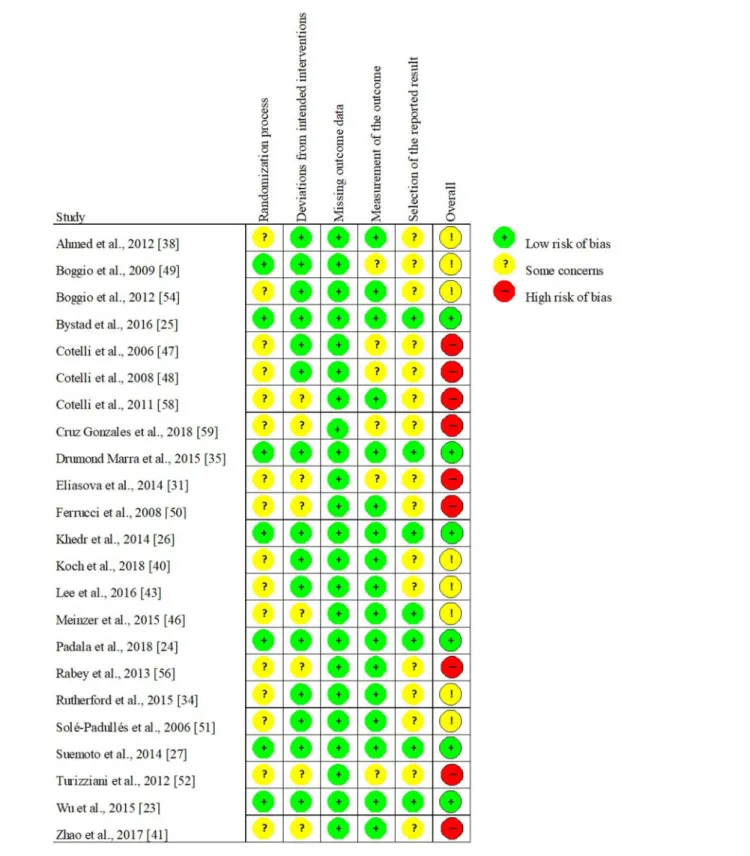
Data were extracted by one review author into summary tables with the supervision of the authors. The risk of bias analysis of randomized-controlled trials was conducted by two independent assessors with version 2 of the Cochrane risk-of-bias tool for randomized trials.
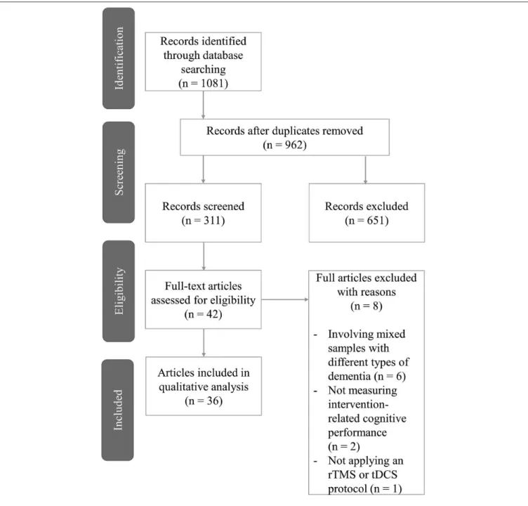
Results: Overall, 36 trials were identified of which 23 randomized-controlled trials underwent a risk of bias assessment. More than 75% of randomized-controlled trials involved some levels of bias in at least one domain. Stimulation parameters were highly variable with some ranges of effectiveness emerging. Studies with low risk of bias indicated TMS to be potentially effective for patients with AD or MCI while questioned the efficacy of tDCS.
Conclusions: The presence and extent of methodical issues affecting TMS and tDCS research involving patients with AD and MCI were examined for the first time. The risk of bias frequently affected the domains of the randomization process and selection of the reported data while missing outcome was rare. Unclear reporting was present involving
randomization, allocation concealment, and blinding. Methodological awareness can potentially reduce the high variability of the estimates regarding the effectiveness of TMS and tDCS. Studies with low risk of bias delineate a range within TMS parameters seem to be effective but question the efficacy of tDCS.
Keywords: Alzheimer’s disease, mild cognitive impairment, research methodology, transcranial magnetic stimulation, transcranial direct current stimulation
INTRODUCTION
Non-invasive brain stimulation (NIBS) has been tested to modify the cognition of healthy participants, as well as to mitigate cognitive symptoms in neurodegenerative disorders (Guse et al., 2010; Vacas et al., 2019). The two most common forms of NIBS, namely transcranial magnetic stimulation (TMS) and transcranial direct current stimulation (tDCS) have both been characterized by a great variability of application and diverse stimulation parameters. Accordingly, the results of NIBS studies are characterized by a large amount of inter- and intra-individual variability. This issue has led to the point that some reviews and meta-analyses have even questioned the efficacy of certain NIBS methods, especially tDCS, in modulating the cognitive performance of either healthy or demented participants (Jacobson et al., 2012; Horvath et al., 2015). Although accumulating evidence supports the efficacy of TMS in modulating cognition, not only the determination of the effectiveness, but also the estimation of the effect size is crucial which likewise needs to be based on reliable data. Reviews indicating positive cognitive effects of NIBS in neurodegenerative disorders have reported serious limitations of the analyzed studies (Freitas et al., 2011; Elder and Taylor, 2014;
Hsu et al., 2015; Vacas et al., 2019). The limitations included high heterogeneity among the applied measurements and stimulation parameters, increased variability due to specific characteristics among demented samples, and low statistical power resulting from small sample sizes. All these factors might contribute to the high variability and hinder the accurate estimation of NIBS efficacy; however, the extent to which each of these factors is present has not been systematically reviewed. Moreover, the reporting of methods is often suboptimal regarding several important design aspects of clinical trials (e.g., allocation concealment, randomization, statistical analyses, and sample characteristics) (Gluud, 2006). Inadequate reporting, as well as the selection of trial design and applied methods, may affect the estimates of NIBS effects (Savovi´c et al., 2012; Weuve et al., 2015;
Polanía et al., 2018) with a more definite influence on subjectively assessed outcomes, such as cognitive status (Savovi´c et al., 2012).
Differences in stimulation parameters may result in the altered efficacy of stimulation. Moreover, some settings of stimulation parameters are designed to achieve different goals e.g., more focal stimulation or the modulation of subcortical structures.
Consequently, clear and detailed reporting of NIBS protocols is crucial to allow the consideration of these differences (Polanía et al., 2018). An overview of the recommended methodological characteristics and stimulation parameters pointing toward fully developed methodology guidelines and consensus regarding the
elements of NIBS is needed (Weuve et al., 2015; Polanía et al., 2018).
The current review aims to examine the presence and extent of methodological issues confounding NIBS studies attempting to alleviate the cognitive symptoms of demented patients. The term cognition covers multiple domains (e.g., attention, memory, language, decision-making, etc.), and each domain can be assessed by numerous types of measurement. However, pooling disparate measures that assess different constructs (i.e., different cognitive subdomains) is generally not recommended, especially in the presence of high heterogeneity of the intervention (Greenfield et al., 2007). By extracting the design characteristics and stimulation parameters of previous studies, we aim to highlight the current vulnerabilities of the field and to formulate recommendations on how to manage these issues when designing studies. We focused on original research articles that applied repetitive transcranial magnetic stimulation (rTMS) or tDCS, i.e., the two most frequent NIBS techniques. We included studies involving patients with mild cognitive impairment (MCI) and Alzheimer’s disease (AD). AD is the most frequent form of dementia that accounts for 50–70% of all dementia cases (Hugo and Ganguli, 2014). Patients with MCI are in an intermediate cognitive state, with a remarkably increased risk of conversion to dementia compared to healthy elderly (Petersen et al., 1999). The treatment of cognitive symptoms in AD and MCI has become an area of major interest considering our aging population, which increased the need for testing alternative therapeutic solutions, such as NIBS. We argue that methodological awareness and effort to increase the experimental control over some sources of variability and bias would contribute to more accurate estimations of the real effects of NIBS on cognition in dementia.
METHODS
Literature Search Strategy
Based on a recent analysis, literature search in PubMed/MEDLINE in combination with Web of Science leads to the recall of almost 80% of the relevant literature in at least 80% of the reviews (Bramer et al., 2017). To further improve this recall ratio, we searched for relevant articles also in ScienceDirect. Therefore, the literature search of three databases was conducted involving PubMed/MEDLINE, Web of Science, and ScienceDirect. Furthermore, bibliographies of the retrieved articles and the relevant reviews were hand-searched as well. The literature search was carried out by A.H., the result of which was confirmed by the co-authors. No review protocol or registration details are available.
Holczer et al. NIBS in Dementia: Methodological Issues
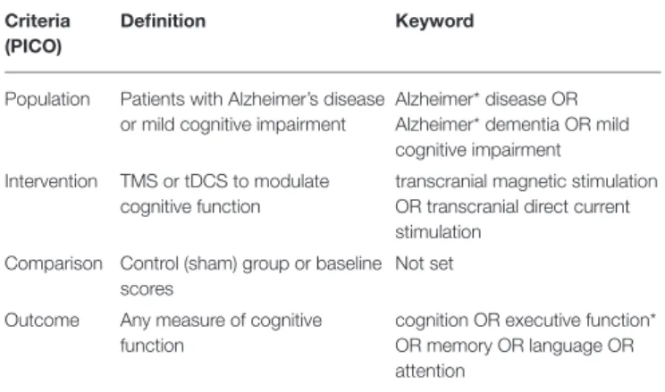
TABLE 1 |Search keywords in PICO format.
Criteria (PICO)
Definition Keyword
Population Patients with Alzheimer’s disease or mild cognitive impairment
Alzheimer* disease OR Alzheimer* dementia OR mild cognitive impairment Intervention TMS or tDCS to modulate
cognitive function
transcranial magnetic stimulation OR transcranial direct current stimulation
Comparison Control (sham) group or baseline scores
Not set
Outcome Any measure of cognitive function
cognition OR executive function*
OR memory OR language OR attention
The asterisk means that the keywords were truncated.
The keywords were determined according to the PICO (population, intervention, comparison, outcome) framework (Schardt et al., 2007) and were searched in the full text of the articles to increase the recall of relevant publications (Kostoff, 2010). The following keywords were applied: Alzheimer∗disease OR Alzheimer∗ dementia when searching for papers involving AD patients. Mild cognitive impairment was used to identify MCI research. For the intervention methods, the MESH terms, transcranial magnetic stimulation OR transcranial direct current stimulation were used. Finally, the following keywords were applied to define outcomes: cognition OR executive function∗ OR memory OR language OR attention. These elements were appended using AND operators (Table 1).
Eligibility Criteria
We aimed to identify original research articles examining the effects of two NIBS techniques (either TMS or tDCS) on any measures of cognitive function in AD or MCI patients. Correspondingly, the following inclusion criteria were determined prior to the literature search: (1) original research articles; (2) written in English; (3) involving human subjects diagnosed with AD or MCI; (4) using TMS or tDCS as an intervention to enhance cognition and; (5) applying any measures of cognitive function. We included clinical trials from the start dates of the databases published until 31 December 2018. As MCI can originate from a wide range of etiological backgrounds, we decided only to include studies that examined MCI with no specified subgroups or MCI due to AD. We decided not to exclude the articles that combined NIBS with other interventions such as cognitive training or ongoing medication, even without the presence of a NIBS-only condition. We argue that the inclusion of studies with combined therapies does not hinder the evaluation of the articles from a methodological point of view. No criteria regarding the design of the studies were determined. We excluded articles for (1) not reporting empirical research; (2) not being written in English; (3) involving animal models of dementia and; (4) not applying NIBS as an intervention aiming to enhance cognition. Conference abstracts
and supplementary reports that were not peer-reviewed were excluded due to their nature of limited methodological reporting.
Risk of Bias Assessment
As randomized-controlled trials (RCTs) are reported to be particularly common in the field of NIBS (Lange et al., 2017), we decided in advance to perform risk of bias assessment of the identified RCTs. To assess the risk of bias in parallel- group and crossover design RCTs, we administered Version 2 of the Cochrane risk-of-bias tool for randomized trials (RoB 2) recommended by the Cochrane Collaboration (Higgins et al., 2019; Sterne et al., 2019). This tool involves more domains than other widely used scales, thus more effectively evaluating the trials’ internal validity (Hartling et al., 2009). The five domains of RoB 2 are (1) randomization process (selection bias), (2) deviations from intended interventions (performance bias), (3) missing outcome data (attrition bias), (4) measurement of the outcome (detection bias), and (5) selection of the reported result (reporting bias). All domains were evaluated separately and ranked as presenting a low risk of bias, some concerns, or high risk of bias. Three levels regarding the overall risk of bias were possible: “Low,” containing no concerns on any of the examined domains; “Some Concerns” involving some concerns in at least one but less than three domains, and “High” if any of the domains involved a high risk of bias or more than three domains contained some concerns. The evaluation of the studies was conducted by two authors (AH and VLN). Any discrepancy was solved by discussion and the consensus results are presented.
Data Extraction
Single data extraction has been found comparable with the results of two independent data extractors in the direction, magnitude, and precision of estimates for a great number of outcomes (Buscemi et al., 2006); therefore, AH was responsible for the data extraction. Data were extracted from each eligible article regarding (1) the main characteristics of the study design and the sample; (2) information regarding the NIBS stimulation (Table 2) and; (3) steps to prevent bias (Table 3).
Study Characteristics, Methods, and Outcomes We extracted information on the study design including the intervention model and relevant study methods. The sample size and the mean age were collected to describe the sample characteristics. The use of the Mini-Mental State Examination (MMSE) as a screening test was found to be a common practice, thus we report its mean score indicating the severity of the cognitive symptoms in the examined samples. Regarding the outcomes, we examined the targeted cognitive domains and the specific tests that were used to measure the given function.
The concluded results of the studies were also collected. We examined the most important methodological characteristics of the identified studies most of which were also evaluated during the risk of bias assessment. We also extracted additional data from the retrieved studies, such as the applied diagnostic criteria for AD/MCI, as well as the time points of the applied cognitive assessment and other aspects affecting the effect estimates (e.g., the use of sample size estimation). In the case of repeated testing,
Frontiers in Human Neuroscience | www.frontiersin.org 3 May 2020 | Volume 14 | Article 179
zeretal.NIBSinDementia:MethodologicalIssu
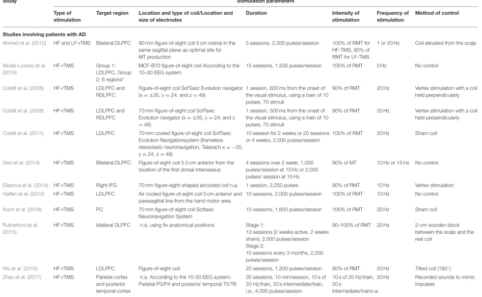
TABLE 2 |The stimulation parameters of the reviewed studies.
Study Stimulation parameters
Type of stimulation
Target region Location and type of coil/Location and size of electrodes
Duration Intensity of
stimulation
Frequency of stimulation
Method of control
Studies involving patients with AD
Ahmed et al. (2012) HF and LF-rTMS Bilateral DLPFC 90 mm figure-of-eight coil 5 cm rostral in the same sagittal plane as optimal site for MT production
5 sessions, 2,000 pulses/session 100% of RMT for HF-TMS, 90% of RMT for LF-TMS
1 or 20 Hz Coil elevated from the scalp
Alcalá-Lozano et al.
(2018)
HF-rTMS Group 1:
LDLPFC, Group 2: 6 regions*
MCF-B70 figure-of-eight coil According to the 10–20 EEG system
15 sessions, 1,500 pulses/session 100% of RMT 5 Hz No control
Cotelli et al. (2006) HF-rTMS LDLPFC and RDLPFC
Figure-of-eight coil SofTaxic Evolution navigator (x= ±35, y=24, and z=48)
1 session, 600 ms from the onset of the visual stimulus, using a train of 10 pulses, 70 stimuli
90% of RMT 20 Hz Vertex stimulation with a coil held perpendicularly
Cotelli et al. (2008) HF-rTMS LDLPFC and RDLPFC
70 mm figure-of-eight coil SofTaxic Evolution navigator (x= ±35, y=24, and z
=48)
1 session, 500 ms from the onset of the visual stimulus, using a train of 10 pulses, 70 stimuli
90% of RMT 20 Hz Vertex stimulation with a coil held perpendicularly
Cotelli et al. (2011) HF-rTMS LDLPFC 70 mm cooled figure-of-eight coil SofTaxic Evolution Navigationsystem (frameless stereotaxic neuronavigation, Talairach x= −35, y=24, z=48)
10 session for 2 weeks or 20 sessions or 4 weeks, 2,000 pulses/session
100% of RMT 20 Hz Sham coil
Devi et al. (2014) HF-rTMS Bilateral DLPFC Figure-of-eight coil 5.5 cm anterior from the location of the first dorsal interosseus
4 sessions over 2 week, 1,000 pulses/session at 10 Hz or 2,000 pulses/ session at 15 Hz
90% of MT 10 Hz or 15 Hz No control
Eliasova et al. (2014) HF-rTMS Right IFG 70 mm figure-eight-shaped aircooled coil n.a. 1 session, 2,250 pulses 90% of RMT 10 Hz Vertex stimulation Haffen et al. (2012) HF-rTMS LDLPFC Air cooled figure-of-eight coil 5 cm anterior and
parasagittal line from the hand motor area
10 sessions, 2,000 pulses/session 100% of RMT 10 Hz No control
Koch et al. (2018) HF-rTMS PC 70 mm figure-of-eight coil Softaxic
Neuronavigation System
10 sessions, 1,600 pulses/session 100% of RMT 20 Hz Sham coil
Rutherford et al.
(2015)
HF-rTMS bilateral DLPFC n.a. using fix anatomical positions Stage 1:
13 sessions (2 weeks active, 2 weeks sham), 2,000 pulses/session Stage 2:
10 sessions every 3 months, 2,000 pulses/session
90–100% of RMT 20 Hz 2-cm wooden block between the scalp and the real coil
Wu et al. (2015) HF-rTMS LDLPFC Figure-of-eight coil 20 sessions, 1,200 pulses/session 80% of RMT 20 Hz Tilted coil (180◦)
Zhao et al. (2017) HF-rTMS Parietal cortex and posterior temporal cortex
n.a. According to the 10-20 EEG system:
Parietal P3/P4 and posterior temporal T5/T6
30 sessions, 10 min/session, 10 s of 20 Hz/train, 20 s intermediate/train, i.e., 4,000 pulses/session
10 s of 20 Hz/train, 20 s
intermediate/trainn.a.
20 Hz Recorded sounds to mimic impulses
(Continued)
tiersinHumanNeuroscience|www.frontiersin.org4May2020|Volume14|Article17
Holczeretal.NIBSinDementia:MethodologicalIssues
TABLE 2 |Continued
Study Stimulation parameters
Type of stimulation
Target region Location and type of coil/Location and size of electrodes
Duration Intensity of
stimulation
Frequency of stimulation
Method of control
Bentwich et al. (2011) TMS-Cog 6 regions* 47–86 mm figure-of-eight coil NeuroNix system 5 sessions/week for 6 weeks, 1,300 pulses/session+cognitive training for 6 weeks, then bi-weekly sessions for 3 months
90% of MT (when stimulating Broca, R-dlPFC and L-dlPFC) 11%
of MT (when stimulating Wernicke, R-pSAC and L-pSAC)
10 Hz No control
Lee et al. (2016) TMS-Cog 6 regions* n.a. NeuroNix System 30 sessions, 1,200 pulses/session 90–110% of RMT 10 Hz Recorded sounds to mimic
impulses Nguyen et al. (2017) TMS-Cog 6 regions* Figure-of-eight coil NeuroAD system (NeuroNix) 6 weeks, 3 regions/day, 1,300
pulses/session+cognitive training
100% of RMT 10 Hz No control
Rabey et al. (2013) TMS-Cog 6 regions* Figure-of-eight coil NeuroAD system (NeuroNix) 6 weeks, daily sessions 1,300 impulses/session of rTMS+cognitive training for 6 weeks, then bi-weekly sessions for 3 months
90% of RMT at Broca’ area and leftLDLPFC/right DLPFCRDLPFC, 110% of RMT at Wernicke, and left/right pSAC
10 Hz Sham coil
Rabey and Dobronevsky (2016)
TMS-Cog 6 regions* Figure-of-eight coil NeuroAD system (NeuroNix) 30 sessions in 6 weeks, daily sessions of 1,300 pulses of rTMS+ cognitive training for 6 weeks
90–110% of RMT 10 Hz No control
Avirame et al. (2016) dTMS bilateral DLPFC H2-coil 6 cm anterior from the motor cortex 20 sessions, 2 or 3 times a week, 42 trains for 2 s in every 20 s, for 20 min
60% of MSO 10 Hz No control
Penolazzi et al. (2015) atDCS+ cognitive training
LDLPFC According to the 10-20 EEG system: anode: 5
×7 cm, F3 cathode: 10×10 cm, right supraorbital area
10 sessions, 20 min/session 2 mA 10 s active stimulation
Andrade et al. (2016) atDCS LDLPFC According to the 10-20 EEG system: anode: 5
×7 cm, F3 cathode: supraorbital area
10 sessions, 30 min/session 2 mA No control
Boggio et al. (2009) atDCS LDLPFC, left temporal cortex
According to the 10-20 EEG system: anode: 5 x 7 cm, L-DLPFC: F3, temporal cortex: T7 cathode: 5 x 7 cm, contralateral supraorbital area
3 sessions, 30 min/session 2 mA 30 s active stimulation
Boggio et al. (2012) atDCS Bilateral temporal cortex
According to the 10-20 EEG system: anode 5×7 cm, T3, T4 cathode 8×8 cm, over the right deltoid muscle
5 sessions, 30 min/session 2 mA 30 s active stimulation
(Continued)
FrontiersinHumanNeuroscience|www.frontiersin.org5May2020|Volume14|Article179
zeretal.NIBSinDementia:MethodologicalIssu TABLE 2 |Continued
Study Stimulation parameters
Type of stimulation
Target region Location and type of coil/Location and size of electrodes
Duration Intensity of
stimulation
Frequency of stimulation
Method of control
Bystad et al. (2016) atDCS Left temporal cortex
According to the 10-20 EEG system: anode:
5×7 cm, at T3 cathode: 5×7 cm, at Fp2
6 sessions, 30 min/session 2 mA 30 s active stimulation
Bystad et al. (2017) atDCS Left temporal lobe
According to the 10-20 EEG system: anode T3 cathode Fp2
Everyday sessions for 8 months, 30 min/session
2 mA No control
Suemoto et al. (2014) atDCS LDLPFC anode 5×7 cm, over DLPFC cathode 5×7 cm, right supraorbital region
6 sessions on every 2nd day, 20 min/session
2 mA 20 s active stimulation
Ferrucci et al. (2008) atDCS or ctDCS Bilateral temporoparietal cortex
According to the 10/20 EEG system: anode or cathode P3-T5 and P6-T4 cathode or anode right deltoid muscle
3 sessions, 15 min/session 1.5 mA 10 s active stimulation
Marceglia et al.
(2016)
atDCS or ctDCS Bilateral temporoparietal cortex
According to the 10-20 EEG system: anode 5×5 cm, P3-T5, P6-T4 cathode 8×8 cm, over the right deltoid muscle
2 sessions, 15 min/session 1.5 mA Comparison of atDCS and
ctDCS
Khedr et al. (2014) atDCS and ctDCS
LDLPFC anodal: 10 x 10 cm, right supraorbital region (10 x 10 cm) cathodal: 4 x 6 cm, left DLPFCLDLPFC (4 x 6 cm)
10 sessions, 25 min/session 2 mA 30 s active stimulation
Studies involving patients with MCI
Turriziani et al. (2012) LF rTMS LDLPFC and RDLPFC
70 mm figure-of-egiht coil According to the 10-20 EEG system: F3, F4
1 session/condition, 600 pulses/session
90% of RMT 1 Hz Tilted coil (no angle mentioned) Drumond Marra et al.
(2015)
HF-rTMS LDLPFC Figure-of-eight coil 5 cm in a parasagittal plane parallel to the point of maximum rMT
10 sessions, 2,000 pulses/session 110% of RMT 10 Hz Sham coil
Padala et al. (2018) HF-rTMS LDLPFC Figure-of-eight coil n.a. 10 sessions/condition, 3,000
pulses/session
120% of RMT 10 Hz Sham coil
Sole-Padulles et al.
(2006)
HF-rTMS LDLPFC Double-cone coil 5 cm anterior from the point of maximum MT
1 session, 3,000 pulses 80% of MT 5 Hz Coil positioned tangentially
Cotelli et al. (2012) HF rTMS Left inferior parietal lobule
70 mm cooled coil SofTaxic Evolution navigator system (x= −44, y= −51, z=43)
10 sessions, 2,000 pulses/session 100% of RMT 20 Hz No control
Cruz Gonzalez et al.
(2018)
atDCS+ cognitive stimulation
LDLPFC According to the 10–20 EEG system: anode: 7
×5 cm, F3 cathode: 7×5 cm, contralateral deltoid muscle
number of sessions randomized (min.
1 max. 5/condition), 30 min/session
2 mA 30 s of active stimulation
Meinzer et al. (2015) atDCS Left ventral IFG anode: 5×7 cm, left Brodmann areas (BA) 44/45 cathode: 10×10 cm, right supraorbital region
1 session, 20 min/session 1 mA 30 s of active stimulation
Murugaraja et al.
(2017)
atDCS LDLPFC According to the 10-20 EEG system: anode: 5
×7 cm, placed between F3 and FP1 cathode:
5×7 cm, right supra-orbital area
5 sessions, 20 min/session 2 mA No control
HF-rTMS, High frequency repetitive transcranial magnetic stimulation; LF-rTMS, Low-frequency repetitive transcranial magnetic stimulation; TMS-Cog, combination of high frequency transcranial magnetic stimulation and cognitive training; dTMS, deep transcranial magnetic stimulation; tDCS, transcranial direct current stimulation; atDCS, anodal transcranial direct current stimulation; ctDCS, cathodal transcranial direct current stimulation; DLPFC, dorsolateral prefrontal cortex; LDLPFC, left dorsolateral prefrontal cortex; RDLPFC, right dorsolateral prefrontal cortex; IFG, inferior frontal gyrus; PC, precuneus; L-pSAC, left parietal somatosensory association cortices; R-pSAC, right parietal
somatosensory association cortices; EEG, electroencephalography; MT, motor threshold; RMT, resting motor threshold; MSO, maximum stimulator output.*Six brain regions: Broca’s area, Wernicke’s area, RDLPFC, LDLPFC, R-pSAC,
and L-pSAC.
tiersinHumanNeuroscience|www.frontiersin.org6May2020|Volume14|Article17
Holczeretal.NIBSinDementia:MethodologicalIssues
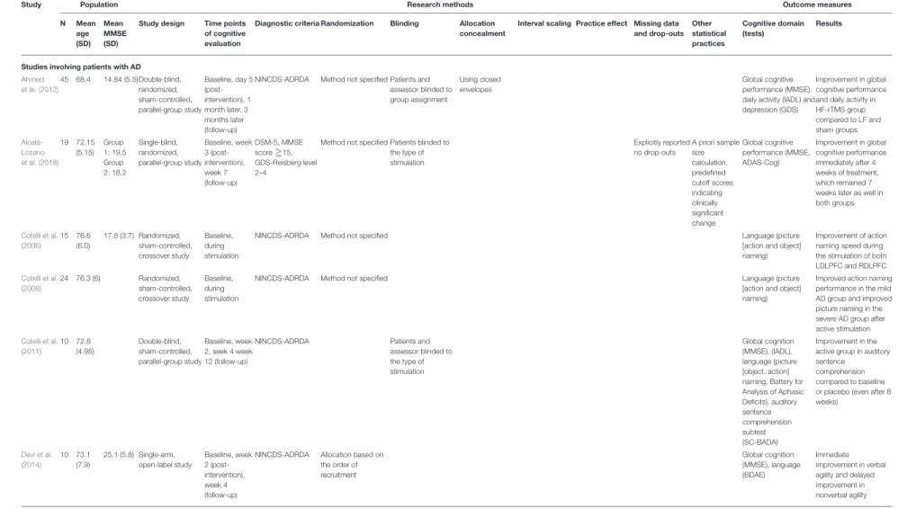
TABLE 3 |The methodical properties of the reviewed studies.
Study Population Research methods Outcome measures
N Mean
age (SD)
Mean MMSE (SD)
Study design Time points of cognitive evaluation
Diagnostic criteria Randomization Blinding Allocation concealment
Interval scaling Practice effect Missing data and drop-outs
Other statistical practices
Cognitive domain (tests)
Results
Studies involving patients with AD Ahmed
et al. (2012)
45 68.4 14.84 (5.5)Double-blind, randomized, sham-controlled, parallel-group study
Baseline, day 5 (post- intervention), 1 month later, 3 months later (follow-up)
NINCDS-ADRDA Method not specified Patients and assessor blinded to group assignment
Using closed envelopes
Global cognitive performance (MMSE), daily activity (IADL) and depression (GDS)
Improvement in global cognitive performance and daily activity in HF-rTMS group compared to LF and sham groups Alcalá-
Lozano et al. (2018)
19 72.15 (5.15)
Group 1: 19.5 Group 2: 18.2
Single-blind, randomized, parallel-group study
Baseline, week 3 (post- intervention), week 7 (follow-up)
DSM-5, MMSE score≧15, GDS-Reisberg level 2–4
Method not specified Patients blinded to the type of stimulation
Explicitly reported no drop-outs
A priori sample size calculation, predefined cutoff scores indicating clinically significant change
Global cognitive performance (MMSE, ADAS-Cog)
Improvement in global cognitive performance immediately after 4 weeks of treatment, which remained 7 weeks later as well in both groups
Cotelli et al.
(2006)
15 76.6 (6.0)
17.8 (3.7) Randomized, sham-controlled, crossover study
Baseline, during stimulation
NINCDS-ADRDA Method not specified Language (picture
[action and object]
naming)
Improvement of action naming speed during the stimulation of both LDLPFC and RDLPFC Cotelli et al.
(2008)
24 76.3 (6) Randomized,
sham-controlled, crossover study
Baseline, during stimulation
NINCDS-ADRDA Method not specified Language (picture
[action and object]
naming)
Improved action naming performance in the mild AD group and improved picture naming in the severe AD group after active stimulation Cotelli et al.
(2011)
10 72.8 (4.95)
Double-blind, sham-controlled, parallel-group study
Baseline, week 2, seek 4 week 12 (follow-up)
NINCDS-ADRDA Patients and
assessor blinded to the type of stimulation
Global cognition (MMSE), (IADL), language (picture [object, action]
naming, Battery for Analysis of Aphasic Deficits), auditory sentence comprehension subtest (SC-BADA)
Improvement in the active group in auditory sentence comprehension compared to baseline or placebo (even after 8 weeks)
Devi et al.
(2014)
10 73.1 (7.9)
25.1 (5.8) Single-arm, open-label study
Baseline, week 2 (post- intervention), week 4 (follow-up)
NINCDS-ADRDA Allocation based on the order of recruitment
Global cognition (MMSE), language (BDAE)
Immediate improvement in verbal agility and delayed improvement in nonverbal agility
(Continued)
FrontiersinHumanNeuroscience|www.frontiersin.org7May2020|Volume14|Article179
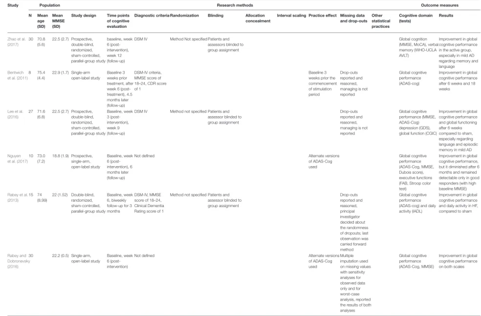
zeretal.NIBSinDementia:MethodologicalIssu TABLE 3 |Continued
Study Population Research methods Outcome measures
N Mean
age (SD)
Mean MMSE (SD)
Study design Time points of cognitive evaluation
Diagnostic criteria Randomization Blinding Allocation concealment
Interval scaling Practice effect Missing data and drop-outs
Other statistical practices
Cognitive domain (tests)
Results
Eliasova et al. (2014)
10 72 (8) 23 (3.56) Randomized, sham-controlled, crossover study
Baseline, retest within 30 min
Not defined Method not specified Tasks practiced
before trial commencement
Global cognitive performance (ACE-R, MMSE), memory (RCFT, WMS-III), attention, psychomotor speed, working memory (Stroop task, TMT-A), executive functions (TMT B, verbal fluency tasks)
Enhancement of attention and psychomotor speed after right IFG stimulation after active stimulation
Haffen et al.
(2012)
1 75 20 Case study 4 months
before intervention (baseline), 1 month after stimulation period, 5 months after stimulation period (follow-up)
NINCDS-ADRDA Baseline 4
months prior the commencement of stimulation period
Executive function (Isaacs Set Test), episodic memory (Memory Impairment Screen, Free and Cued Recall Test, Isaacs Set Test), information processing (TMT-A), visuospatial skills (copying geometric figure), naming
Improved performance on 8 of the 10 measures with maintained cognitive functioning at follow-up
Koch et al.
(2018)
14 70.0 (5.1)
26.1 (1.8) Double-blind, randomized, sham-controlled, crossover study
Baseline, week 2 (post- intervention)
Revised NINCDS-ADRDA criteria byDubois et al. (2016)
Method not specified Patients and assessor blinded to condition
Global cognition (ADCS-PACC, MMSE), attention and psychomotor speed (TMT) auditory verbal learning (RAVL-T), episodic memory (DSST) executive function (Modified Card Sorting test, Verbal fluency, FAB)
Improvement in active group in episodic memory, but not in global cognition and executive function
Rutherford et al. (2015)
11 57–87 Double-blind,
randomized, sham-controlled, crossover+ open-label study
Stage 1:
baseline, week 4 (post- intervention)
Diagnosed by neuropsychiatrist or neurologist or MOCA score between 5 and 26
Method not specified Patients and assessor blinded, the effectiveness to blinding was measured, when assessor was not blinded it got reported
Alternate versions of tasks used
Mean imputation used and reasons of drop-out reported
Calculating observed power of tests, average test-retest improvement calculated
Global cognitive performance and associative memory (ADAS-Cog, RMBC, spatial awareness, word–image association)
Improvement in global cognitive performance in the active group compared to sham, especially during the early stage of the treatment
Wu et al.
(2015)
54 15.25 (3.1)
15.25 (3.1)Double-blind, randomized, sham-controlled, parallel-group study
Baseline, week 4 (post- intervention)
NINCDS-ADRDA Standard table of random numbers
Patients and assessor blinded to group assignment
Patients and assessor blinded to the group assignment before starting the trial, method not specified
Using cutoff scores based on the findings of other studies
Behavioral pathology (BPSD) and global cognitive performance (ADAS-Cog)
Improvement of behavioral and global cognitive symptoms
(Continued)
tiersinHumanNeuroscience|www.frontiersin.org8May2020|Volume14|Article17
Holczeretal.NIBSinDementia:MethodologicalIssues
TABLE 3 |Continued
Study Population Research methods Outcome measures
N Mean
age (SD)
Mean MMSE (SD)
Study design Time points of cognitive evaluation
Diagnostic criteria Randomization Blinding Allocation concealment
Interval scaling Practice effect Missing data and drop-outs
Other statistical practices
Cognitive domain (tests)
Results
Zhao et al.
(2017)
30 70.8 (5.6)
22.5 (2.7) Prospective, double-blind, randomized, sham-controlled, parallel-group study
baseline, week 6 (post- intervention), week 12 (follow-up)
DSM IV Method Not specified Patients and assessors blinded to group assignment
Global cognition (MMSE, MoCA), verbal memory (WHO-UCLA AVLT)
Improvement in global cognitive performance in the active group, especially in mild AD regarding memory and language Bentwich
et al. (2011) 8 75.4
(4.4)
22.9 (1.7) Single-arm open-label study
Baseline 3 weeks prior treatment, after week 6 (post- treatment), 4.5 months later (follow-up)
DSM-IV criteria, MMSE score of 18–24, CDR score of 1
Baseline 3 weeks prior the commencement of stimulation period
Drop-outs reported and reasoned, managing is not reported
Global cognitive performance (ADAS-cog)
Improvement in global cognitive performance after 6 weeks and 18 weeks
Lee et al.
(2016)
27 71.6 (6.8)
22.5 (2.7) Prospective, double-blind, randomized, sham-controlled, parallel-group study
Baseline, week 3 (post- intervention), week 9 (follow-up)
DSM IV Method not specified Patients and assessor blinded to group assignment
Drop-outs reported and reasoned, managing is not reported
Global cognitive performance (MMSE, ADAS-Cog) depression (GDS), global function (CGIC)
Improvement in global cognitive performance and global functioning after 6 weeks compared to sham, especially regarding language and episodic memory in mild AD Nguyen
et al. (2017) 10 73.0
(7.2)
18.8 (1.9) Prospective, single-arm, open-label study
Baseline, week 6 (post- intervention), 6 months later (follow-up)
Not defined Alternate versions
of ADAS-Cog used
Global cognitive performance (ADAS-Cog, MMSE, Dubois score), executive functions (FAB, Stroop color test)
Improvement in global cognitive performance, but it diminished after 6 months and remained detectable only in good responders (with high baseline MMSE) Rabey et al.
(2013) 15 74
(8.99)
22 (1.52) Double-blind, randomized, sham-controlled, parallel-group study
Baseline, week 6, biweekly follow-up for 3 months
DSM-IV, MMSE score of 18–24, Clinical Dementia Rating score of 1
Method not specified Patients and assessor blinded to group assignment
Drop-outs reported and reasoned, principal investigator decided about the randomness of dropouts; last observation was carried forward method
Global cognitive performance (ADAS-cog) and daily activity (IADL)
Improvement in global cognitive performance and daily activity in HF, compared to sham
Rabey and Dobronevsky (2016)
30 22.2 (0.5) Single-arm, open-label study
Baseline, week 6 (post- intervention)
Not defined Alternate versions
of ADAS-Cog used
Multiple imputation used on missing values with sensitivity analyses for observed data only and for worst-case analysis, reported the results of both analyses
Global cognitive performance (ADAS-Cog, MMSE)
Improvement in global cognitive performance on both scales
(Continued)
FrontiersinHumanNeuroscience|www.frontiersin.org9May2020|Volume14|Article179
zeretal.NIBSinDementia:MethodologicalIssu TABLE 3 |Continued
Study Population Research methods Outcome measures
N Mean
age (SD)
Mean MMSE (SD)
Study design Time points of cognitive evaluation
Diagnostic criteria Randomization Blinding Allocation concealment
Interval scaling Practice effect Missing data and drop-outs
Other statistical practices
Cognitive domain (tests)
Results
Avirame et al. (2016)
11 76 (7) Single-arm
open-label study
Baseline, 2–3 weeks later (post- intervention)
Diagnosed by an expert neurologist and confirmed by a psychiatrist
Different stimuli within the tasks
Missing data reported and reasoned, managing is not reported
Global cognitive performance (Mindstreams, ACE)
Improvement of global cognition compared to baseline
Penolazzi et al. (2015)
1 60 23.2 Case study Two cycles of
baseline, week 4 (post- intervention), week 8 (follow-up), 2 months apart
Based on neuropsycholgical evaluation and neuroimaging
Patient blind to the stimulation, method not specified
Comparison to a normative score
Memory (Brief Neuropsychological Examination-2), psychomotor speed and executive function (TMT A and B, clock drawing)
Improvement on the trained tasks whith more enhancement when training was combined with active stimulation
Andrade et al. (2016)
1 73 Case study Baseline (1
week prior), 1 week after the intervention
NINCDS-ADRDA Baseline 1 week
prior to the commencement of the stimulation period
Global cognitive performance (ADAS-Cog), neuropsychiatric and behavioral symptoms (NPI, DAD, Blessed Dementia Scale)
Improvement of global cognitive performance, executive function and behavioral symptoms compared to baseline
Boggio et al. (2009)
10 79.1 (8.8)
17.0 (4.9) Single-blind, randomized, sham-controlled, crossover study
During stimulation
NINCDS-ADRDA Method not specified Patients blinded to the type of stimulation
Randomized use of alternate versions
Selective attention (Stroop test, Victoria version), working memory (Digit span test backward and forward), recognition memory (visual memory task using IBV software)
Improvement of visual recognition memory after LDLPFC and temporoparietal stimulation compared to sham
Boggio et al. (2012)
15 79.05 (8.2)
20 (3) Double-blind, randomized, sham-controlled, crossover study
Baseline, day 5 (post- treatment), week 2, week 4 (follow-up)
NINCDS-ADRDA and DSM-IV
Method not specified Patients and assessor blinded to group assignment
Randomized use of alternate versions of tasks
Global cognition (MMSE, ADAS-Cog), visual recognition (VRT), visual attention (VAT)
Improvement of memory performance in active stimulation group
Bystad et al. (2016)
25 72.5 (8.35)
20.6 (3.35)Double-blind, randomized, sham-controlled, parallel-group study
Baseline, day 6 (post- intervention)
Revised NINCDS-ADRDA
Computer randomized list containing 5-digit codes provided by the manufacturer of the tDCS device
Patients and assessor blinded to the type of stimulation
Assignment disclosed until the end of the intervention
Scaling according to standardized norm tables, transformation to z-scores
Two versions of CVLT-II used
Explicitly reported no drop-outs
Sample size based on other studies
Global cognitive performance (MMSE), Verbal learning (CVLT-II), Attention and executive function (TMT, clock-drawing test
No changes in either cognitive function
Bystad et al. (2017)
1 60 20 Case study Baseline, 5
months later (during stimulation period), 8 months later (post- intervention)
Revised NINCDS-ADRDA
Alternate versions used
Global cognition (RBANS)
Stabilized cognitive decline of patient with minor impairment of visuospatial function
(Continued)
tiersinHumanNeuroscience|www.frontiersin.org10May2020|Volume14|Article17