ANTIANGIOGENIC THERAPY IN ADVANCED LUNG ADENOCARCINOMA: EFFICACY AND KRAS MUTATION AS A PROGNOSTIC BIOMARKER
PhD thesis
Áron Kristóf Ghimessy
Clinical Medicine Doctoral School Semmelweis University
Supervisor: Balázs Döme, MD, PhD.
Official reviewers: Gergely Dénes Huszty MD, PhD, Zoltán Heiler MD, PhD,
Head of the Complex Examination Committee: Béla Szende, Professor emeritus
Members of the Complex Examination Committee: Krisztina Bogos MD, PhD.
Marcell A. Szász PhD
Budapest
2020
Table of contents
1. Abbreviations ... - 4 -
2. Introduction ... - 8 -
2.1. Lung cancer ... - 8 -
2.1.1. Epidemiology ... - 8 -
2.1.2. Etiology ... - 9 -
2.1.2.1. Smoking ... - 9 -
2.1.2.2. Environmental risk factors ... - 11 -
2.1.2.2.1. Radon ... - 11 -
2.1.2.2.2. Asbestos ... - 11 -
2.1.2.2.3. Air pollution ... - 11 -
2.1.2.3. Genetic risk factors ... - 12 -
2.1.2.4. Coexisting diseases and infections ... - 12 -
2.1.3. Histology and molecular background of lung cancer ... - 13 -
2.1.3.1. Adenocarcinoma ... - 15 -
2.1.3.2. Squamous Cell Carcinoma ... - 18 -
2.1.3.3. Large Cell Carcinoma ... - 19 -
2.1.3.4. Neuroendocrine tumors ... - 19 -
2.1.3.4.1. Small Cell Lung Cancer ... - 19 -
2.1.4. Screening, clinical diagnosis, staging and prognosis of lung cancer ... - 20 -
2.1.4.1. Screening ... - 20 -
2.1.4.2. Clinical diagnosis of lung cancer ... - 23 -
2.1.4.2.1. Functional evaluation of patients diagnosed with lung cancer ... - 25 -
2.1.4.3. Staging of lung cancer ... - 27 -
2.1.4.4. Prognosis of lung cancer ... - 30 -
2.1.4.4.1. Prognosis of SCLC ... - 30 -
2.1.4.4.2. Prognosis of NSCLC ... - 31 -
2.1.5. Treatment of lung cancer ... - 33 -
2.1.5.1. Treatment options for SCLC ... - 33 -
2.1.5.2. Treatment of NSCLC ... - 35 -
2.1.5.2.1. Treatment of early stage NSCLC ... - 35 -
2.1.5.2.2. Treatment of locally advanced (stage III) NSCLC ... - 43 -
2.1.5.2.1. Treatment of metastatic NSCLC ... - 44 -
2.2. Vascular endothelial growth factor and bevacizumab ... - 48 -
2.3. KRAS mutation and antivascular treatment ... - 50 -
3. Objectives ... - 53 -
3.1. The safety and efficacy of bevacizumab in addition to platinum based chemotherapy in patients with advanced NSCLC ... - 53 -
3.2. KRAS mutation as a biomarker for anti-VEGF therapy in NSCLC ... - 53 -
4. Methods ... - 55 -
4.1. General ethical consideration ... - 55 -
4.2. The safety and efficacy of bevacizumab in addition to platinum based chemotherapy in patients with advanced NSCLC ... - 56 -
4.2.1. Study design ... - 56 -
4.2.2. Patients ... - 56 -
4.2.3. Treatment ... - 56 -
4.2.4. Progression-free and overall survival ... - 57 -
4.2.5. Statistics ... - 58 -
4.3. KRAS mutation as a biomarker for anti-VEGF therapy in NSCLC ... - 58 -
4.3.1. Study population ... - 58 -
4.3.2. Molecular diagnosis ... - 60 -
4.3.3. Statistical methods ... - 61 -
5. Results ... - 62 -
5.1. The safety and efficacy of bevacizumab in addition to platinum based chemotherapy in patients with advanced NSCLC ... - 62 -
5.1.1. Baseline Characteristics of the Patients ... - 62 -
5.1.2. Treatment ... - 64 -
5.1.3. Efficacy analysis ... - 64 -
5.1.3.1. Progression-free survival ... - 64 -
5.1.3.2. Secondary endpoints ... - 67 -
5.1.3.2.1. Tumor response ... - 67 -
5.1.3.2.2. Overall survival ... - 69 -
5.1.3.2.3. Safety and adverse events ... - 70 -
5.2. KRAS mutation as a biomarker for anti-VEGF therapy in NSCLC ... - 73 -
5.2.1. Incidence of KRAS mutations in LADC patients treated with bevacizumab and chemotherapy ... - 73 -
5.2.2. The presence of KRAS mutations has clinical utility in predicting disease outcome in LADC patients receiving concurrent antiangiogenic and chemotherapy- 77 - 5.2.3. Distinct efficacy of BEV/CHT in advanced LADC patients with different subtype-specific KRAS mutations ... - 82 -
6. Discussion ... - 85 -
6.1. The safety and efficacy of bevacizumab in addition to platinum based chemotherapy in patients with advanced NSCLC ... - 85 -
6.2. KRAS mutation as a biomarker for anti-VEGF therapy in NSCLC ... - 91 -
7. Conclusion ... - 95 -
7.1. The safety and efficacy of bevacizumab in addition to platinum based chemotherapy in patients with advanced NSCLC ... - 95 -
7.2. KRAS mutation as a biomarker for anti-VEGF therapy in NSCLC ... - 95 -
8. Summary ... - 96 -
9. Összefoglalás ... - 97 -
10. References ... - 98 -
11. Bibliography of the candidate’s publications ... - 138 -
11.1. Publications related to the thesis ... - 138 -
11.2. Publications not related to the thesis ... - 138 -
12. Acknowledgements ... - 147 -
1. Abbreviations
AE adverse event
AIS adenocarcinoma in situ ALK anaplastic lymphoma kinase
ARID1A AT-rich interactive domain-containing protein 1A BEV bevacizumab
BRAF B-Raf proto-oncogene
CDKN2A cyclin-dependent kinase Inhibitor 2A CHT chemotherapy
CI confidence interval CK7 cytokeratin 7
COPD chronic obstructive pulmonary disease CR complete response
CRC colorectal cancer CRF case report form CT computer tomography
DLCO diffusing capacity of the lungs for carbon monoxide DNA deoxyribonucleic acid
ECOG Eastern Cooperative Oncology Group EGFR Epidermal Growth Factor Receptor EMA European Medicines Agency
ERBB2 v-erb-b2 avian erythroblastic leukemia viral oncogene homolog 2 ERK extracellular regulated kinases
ESMO European Society for Medical Oncology FDA Food and Drug Administration (U.S.) FEV1 forced expiratory pressure in 1 second FVC forced vital capacity
GABA gamma-aminobutyric acid GWAS genome wide association studies HIV human immunodeficiency virus
HLA-A human leukocyte antigens on the A locus
HRAS Harvey rat sarcoma viral oncogene homolog KEAP1 Kelch-like ECH-associated protein 1
KRAS Kirsten rat sarcoma 2 viral oncogene homolog LA Locally advanced
LADC lung adenocarcinoma LCC large cell carcinoma
LCNEC large cell neuroendocrine carcinoma LDCT low dose computer tomography
MAP2K1 mitogen-activated protein kinase kinase 1 MEK mitogen-activated protein kinase
MET met proto-oncogene (hepatocyte growth factor receptor) MGA MAX gene associated
MIA minimally invasive adenocarcinoma
MLL2 histone lysine methyltransferase gene KMT2D NA not applicable
nAChR nicotinic acetylcholine receptors NF1 neurofibromatosis type I
NFE2L2 Nuclear factor erythroid 2-related factor 2 NLST National Lung Screening Trial
NNK 4-(methylnitrosamino)-1-(13-pyridyl-1-butanone) NOTCH1 neurogenic locus notch homolog protein 1
NRAS Neuroblastoma rat sarcoma viral oncogene homolog NSCLC non-small cell lung cancer
nsNSCLC non-squamous non-small cell lung cancer OCS observational cohort study
OS overall survival
PAH polycyclic aromatic hydrocarbons PD progressive disease
PDYN prodynorphin
PET positron emission tomography PFS progression-free survival
PIK3CA phosphatidylinositol 4,5-bisphosphate 3-kinase catalytic subunit alpha PR partial response
PS performance status
PTEN phosphatase and tensin homolog
RAF rapidly accelerated fibrosarcoma, receptor tyrosine kinase effector RAS rat sarcoma viral analog
RB1 retinoblastoma protein 1 RBM10 RNA-binding protein 10 RCT randomized clinical trial
RECIST response evaluation criteria in solid tumors RET rearranged during transfection
RIT1 Ras-like without CAAX 1 ROS1 ROS proto-oncogene 1 RTK receptor tyrosine kinase SAE serious adverse event
SBRT stereotactic body radiotherapy SCLC small cell lung cancer
SD stable disease SD standard deviation SETD2 SET domain containing 2
SMARCA4 SWI/SNF related, matrix associated, actin dependent regulator of chromatin, subfamily a, member 4
SqCC squamous cell carcinoma STK11 serine/threonine kinase 11 TB tuberculosis
TCGA The Cancer Genome Atlas Program
TNM Tumor, node, metastasis - Internationally accepted classification of malignant tumors;
TP53 tumor protein p53
TSNA tobacco-specific N-nitrosamine TTF1 thyroid transcription factor 1 TTP time-to-progression
U2AF1 U2 auxiliary factor 1
VATS video-assisted thoracic surgery VEGF vascular endothelial growth factor WHO World Health Organization
WT wild type
2. Introduction
2.1. Lung cancer 2.1.1. Epidemiology
More than 8 million cancer deaths and 14 million new cancer cases were documented worldwide in 2012. Lung and breast cancer were diagnosed most frequently among these, and they represent the leading causes of cancer death overall. However, in more developed countries, lung cancer represents the leading cause of cancer death in women(1) and prostate cancer accounts for the most diagnosed malignancy.
Lung cancer became the second most common malignant tumor in the last century. In the 19th century and even in the beginning of the 20th century lung cancer was a very rare diagnosis. In the 1840s only 22 published cases could be found(2), while in 1912 Adler still only identified 374 published cases(3, 4). Recent statistics show that annually there were 1.825 million new lung cancer cases worldwide in 2012. This is a marked rise from 1.6 million new cases in 2008(5). 409 900 of these cases were reported in Europe which was 13% of all cancer cases(6), however it causes more deaths than breast, prostate and colon cancer combined. It is estimated that the total number of deaths caused by lung cancer was 1.589 million worldwide, which accounts for 17% of all cancer related deaths(7). The incidence of lung cancer still rises worldwide, although it shows great variances between countries.
Hungary has on of highest mortality rates of lung cancer in the world both in men and women. Hungary, unlike other developed countries, records a growing number of new cases. While the incidence hasn’t increased over the last few years in men, it continuously does in women(8). In 2014, the registered number of new cases of lung cancer in Hungary was 5189 (60% male, 40% female). Incidence is quite low in the population younger than 40 years (0-12%000), however it rises drastically to 250%000 in the male population between 50 and 54 years of age(9).
2.1.2. Etiology 2.1.2.1. Smoking
The use of tobacco attributes to 90% of all lung cancer cases, which makes it the single greatest risk factor regarding lung cancer. Smokers have a 15 fold chance of developing lung cancer compared to non-smoking population. Based on less univoque evidence, they presume that work-associated carcinogens attribute for 9-15%, radon released into air for 10% and pollution for 1-2% of cases.
Society-wide understanding of the relationship between smoking and lung cancer was achieved very slowly due to several factors. One of these factors is the long latency period between smoking initiation and the development of lung cancer and another interesting factor was the marketing activity of the tobacco industry(10). Tobacco had been used by people in Europe, America and Asia for centuries without significant incidence of lung cancer. Tobacco was regarded primarily as medicine or was used only in rituals. Tobacco was brought to Europe in the 15th century but cigarettes were only manufactured first in the 19th century. In this era, cigarettes were expensive and hand-rolled and only men used them occasionally(11, 12).
The world wars had a great role in the fact that smoking got popular first in men and later also in women. Smoking increased dramatically after both world wars because soldiers were handed free cigarettes and developed nicotine addiction. At this time the negative effects of smoking were not know to the wide public so soldiers subsequently brought the habit of smoking home. Early reports already suggested a link between smoking and cancer in the 1920s and 1930s but these reports were not perceived widely thus they had no effect on consumption(13-16). The first major epidemiological studies were released in 1950 by Doll and Hill (17) and Wyander and Graham (18). These definitely established the relation between smoking and lung cancer which led to statements Royal College of Physicians in Great Britain in 1962 (19) and the US Surgeon General in 1964 to warn the public about the dangers of smoking.
Smoking a cigar or a pipe is less dangerous than cigarettes because the tobacco smoke exposition is lower and deep inhalation is rare. The chance of developing lung cancer shows an exponential relationship with the number of cigarettes smoked and the total years of smoking. The length of smoking is of greater importance than the age of the patient and no matter how old someone is, quitting decreases the chance of lung cancer.
Passive smoking is also considered a risk for developing lung cancer, however a metanalysis comparing smoking and non-smoking couples found that passive smoking only increased the risk by 20%(20, 21). In Hungary an estimated 34% of the population is smoking (41% in male and 28% in female) which puts Hungary among the countries with the highest risk of lung cancer.
Nicotine, responsible for the addictiveness of cigarettes, is a natural alkaloid acting as an acetylcholine agonist that binds to the nicotinic acetylcholine receptors (nAChR) in the nervous system, causing release of neurotransmitters into the blood stream, including dopamine, serotonin, norepinephrine, endorphins, and gamma-aminobutyric acid (GABA). Nicotine upregulates nicotinic receptors and alters gene expression causing dependence and also helping progression of an existing tumor(22-24). Nicotine itself is not regarded as carcinogen, however there are at least 60 known carcinogens produced at tobacco combustion. The most significant are tobacco-specific N-nitrosamines (TSNAs), such as 4-(methylnitrosamino)-1-(13-pyridyl-1-butanone) (NNK) and polycyclic aromatic hydrocarbons (PAH), including benzo[a]pyrene and nitrates(25). The relationship of NNK to lung cancers, specifically adenocarcinomas have been demonstrated(26). Combustion of tobacco produce a smoke that has a vapor phase that produce 1015, and a particulate phase with 1017 free radicals per gram. The damage done by these free radicals is one of the methods of carcinogenesis, the other is DNA adduct and metabolite formation(24, 25). The carcinogenesis of smoking can be seen on Figure 1.
1. Figure – Carcinogenesis of smoking
2.1.2.2. Environmental risk factors 2.1.2.2.1. Radon
It has been known from the 15th century that mining carried the risk of lung disease. It was observed on miners working in the mountains on the German-Czech border that the incidence of “mountain disease” was very high. In these mines they produced cobalt, arsenic, bismuth, iron, silver and later in the 20th century radium. In the 20th century these mining communities had extremely high incidence of squamous cell lung cancer(4).
Residential exposition to radon occurs from soil. Radon is a radioactive gas occurring naturally from the earth’s crust from natural decay of uranium. Usually radon level can rise to unsafe measures in residential basements. Cigarette smoking in the same time increases the relative risk of lung cancer from radon(27-29).
2.1.2.2.2. Asbestos
Asbestos is the most common occupational exposure to carcinogens but especially in Eastern Europe asbestos can also be still found in residential buildings. Asbestos was used widely in constructions since the 19th century. It contains chrysotile fibers that have been shown to have association with lung disease and thoracic malignancies such as malignant mesothelioma and lung cancer. Residential and continuous occupational exposure to asbestos carries a 5-fold risk of lung cancer and have a synergistic effect with tobacco smoking(30, 31). A closed eternity factory still produce increased risk for asbestos exposure for residents living in nearby villages in Hungary’s Selyp.
2.1.2.2.3. Air pollution
An adult inhales approximately 10 000 liters of air per day which means that even a small concentration of carcinogens can cause changes in the cellular DNA with time. Air pollution in big cities consist of carcinogens released from combustion of fossil fuel, arsenic, nickel and chrome. A study conducted in six large cities in the USA found that the incidence of lung cancer was 40% higher in the most polluted city compared to the one with the least air pollution(32). The frequently cited paper of Doll and Petro estimated
that only 1-2% of lung cancer cases was due to air pollution in 1981(20). Their estimation is still most probably correct.
2.1.2.3. Genetic risk factors
The fact that not all smokers develop lung cancer establishes the role of genetic susceptibility to lung cancer. They observed that having a positive family history for lung cancer increases the risk 1.7 fold(33). Genome wide association studies (GWAS) identified the chromosome regions connected to increased risk of lung cancer: 5p15, 15q25-26 and 6q21(34, 35). Telomerase reverse transcriptase (TERT) is involved in cell replication and in the development of lung cancer it is associated with adenocarcinomas.
TERT is encoded on the 5p15 region. Chromosome locus 6p21 regulates G-protein signaling, and mutations increase the risk of lung cancer development in non-smokers.
The 15q25-26 chromosome locus is known for mutations that are positively linked to nicotine dependence and susceptibility for lung cancer(36, 37).
It is interesting that there have been a change in lung cancer histology throughout the years: in the early 20th century squamous cell lung carcinoma was the most frequent, while since the 1970s the incidence of lung adenocarcinoma rose and incidence of squamous cell carcinomas decreased. The background of this change is not entirely known, however several authors contribute this change to the wide use of filters in cigarettes. The use of filters decreased the amount of inhaled PAHs but on the other hand the deeper inhalation increased the amount of nitrogen-oxides and nitrosamines(32).
2.1.2.4. Coexisting diseases and infections
Previous lung injury caused by chronic obstructive pulmonary diseases (COPD) and lung fibrosis also plays a role in lung cancer development, however it is challenging to study their effect separately because smoking also has a role in COPD. Both diseases can cause lung injury through inflammatory pathways and it was also shown that infections such as tuberculosis (TB) were associated with a 1.7 fold increase in risk for lung cancer(38).
Human immunodeficiency virus (HIV) infection also carries a risk among other diseases for lung cancer. Lung cancer is the most common malignancy in patients with HIV infection and it also accounts for 30% of cancer deaths related with HIV(39, 40). The HIV virus itself has not been connected directly to oncogenesis, however the
immunosuppression caused by it defiantly plays a role since organ transplant recipients and HIV patients have a similar increase in cancer rates(41). Declining CD4 counts were found to be the reason for higher lung cancer rate. 42% of HIV patients also smoke, however HIV-infected patients have a 2.5 fold greater risk of developing lung cancer regardless of the smoking status(42).
2.1.3. Histology and molecular background of lung cancer
The histopathological diagnosis of lung cancer is based on morphologic features. Two main histopathological groups were made: small cell lung cancer (SCLC) and non-small cell lung cancer (NSCLC). SCLC accounts for approximately 15% of cases, while NSCLC accounts for 85%. NSCLC is usually subcategorized into adenocarcinoma, squamous cell carcinoma and large cell carcinoma, even though more and more evidence suggests that that these are molecularly heterogenous diseases(43).
Histological classification was last updated in 2015 by the World Health Organization (WHO). It recognizes the new molecular discoveries achieved since 2004.
1. Table – WHO 2015 classification of lung epithelial tumors
Adenocarcinoma Large cell carcinoma
Lepidic adenocarcinoma Adenosquamous carcinoma
Acinar adenocarcinoma Pleomorphic carcinoma
Papillary adenocarcinoma Spindle cell carcinoma Micropapillary adenocarcinoma Giant cell carcinoma
Solid adenocarcinoma Carcinosarcoma
Variants of adenocarcinoma Pulmonary blastoma
Invasive mucinous adenocarcinoma Other and unclassified carcinomas Mixed invasive mucinous and non-mucinous
adenocarcinoma Lymphoepithelioma-like carcinoma
Colloid adenocarcinoma NUT carcinoma
Fetal adenocarcinoma Salivary gland-type tumors
Enteric adenocarcinoma Mucoepidermoid carcinoma
Minimally invasive adenocarcinoma Adenoid cystic carcinoma
Non-mucinous Epithelial–myoepithelial carcinoma
Mucinous Pleomorphic adenoma
Preinvasive lesions Papillomas
Atypical adenomatous hyperplasia Squamous cell papilloma
Adenocarcinoma in situ Exophytic
Non-mucinous Inverted
Mucinous Glandular papilloma
Squamous cell carcinoma (SqCC) Mixed squamous cell and glandular papilloma
Keratinizing SqCC Adenomas
Non-keratinizing SqCC Sclerosing pneumocytoma
Basaloid SqCC Alveolar adenoma
Preinvasive lesion Papillary adenoma
SqCC in situ Mucinous cystadenoma
Neuroendocrine tumors Mucous gland adenoma
Small cell carcinoma
Combined small cell carcinoma
Large cell neuroendocrine carcinoma (LCNEC)
Combined LCNEC
Carcinoid tumors
Typical carcinoid
Atypical carcinoid
Preinvasive lesion
Diffuse idiopathic pulmonary neuroendocrine cell
Hyperplasia
2.1.3.1. Adenocarcinoma
Adenocarcinoma is currently the most common malignancy of the lung and it is a heterogenic group of tumors. In Hungary adenocarcinoma accounts for 47% of all lung cancer incidence(44). The common in all adenocarcinomas is the glandular structure and some level of mucin production. The growing pattern can be variable, the most common is lepidic growth that follows the alveolar wall, acinar, papillary or solid. Several of these patterns can be present within one tumor but usually one is dominant. The diagnosis of adenocarcinoma is often proven by immunohistochemistry (TTF1, Napsin-A, CK7) and mucin staining.
Adenocarcinoma in situ (AIS) was introduced as a new precancerous category. These tumors are smaller than 3 cm in diameter, they are solitary, the show clearly lepidic growth pattern and they do not invade the pleura, stromal cells or the vessels.
Minimally Invasive Adenocarcinoma (MIA) was also introduced as a new precancerous category. These tumors are not greater than 3 cm in diameter, they are solitary, they show dominantly lepidic growth pattern and they only show minimal (smaller than 5mm) stromal, vascular or pleural invasion.
The nomenclature do not use bronchiolo-alveolar carcinoma and mixed adenocarcinoma anymore(45).
2. Figure – Histologic subtypes of lung adenocarcinoma: A, Predominantly lepidic pattern with lepidic growth on the right with invasive acinar adenocarcinoma on the left; B, Proliferation along the alveolar wall (type II pneumocytes and Clara cells); C, Invasive acinar adenocarcinoma; D, oval-shaped malignant glands invading the fibrous stroma in invasive acinar adenocarcinoma; E, Papillary adenocarcinoma; F, Small papillary clusters of glandular cells in a micropapillary adenocarcinoma; G, Solid adenocarcinoma; H, Solid adenocarcinoma with mucin. Adopted from (46).
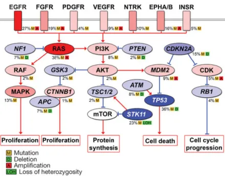
The most common significantly mutated pathways include the EGFR, FGFR, VEGFR, NTRK, EPHA/B and INSR genes as seen in Figure 3.
3. Figure – Significantly mutated pathways in lung adenocarcinoma. Oncoproteins are indicated in pink to red and tumor suppressor proteins are shown in light to dark blue. The darkness of the colors shows the number of tumors with genetic alterations.(47)
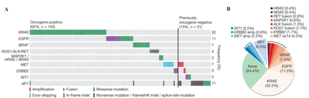
18 significant somatic mutations were identified in lung adenocarcinoma by comprehensive molecular profiling of 230 tumors by The Cancer Genome Atlas Program (TCGA) in 2014: TP53 (46%), KRAS (33%), KEAP1 (17%), STK11 (17%), EGFR(14%), NF1 (11%), BRAF (10%), SETD2 (9%), RBM10 (8%), MGA (8%), MET (7%), ARID1A (7%), PIK3CA (7%), SMARCA4 (6%), RB1 (4%), CDKN2A (4%), U2AF1 (3%), and RIT1 (2%). A mean of 8.9 mutations were reported per megabase which is considered very high. This widened the possibility of targeted therapy. 75% of the examined adenocarcinomas contained genetic changes to the RTK/RAS/RAF signaling pathway and 62% of them had driver genetic alterations that promote this cascade. The most common driver mutations were KRAS, EGFR and BRAF with 32, 11 and 7% respectively in this study. Other promoter mutations included MET exon 14 skipping (4.3%), ERBB2 mutation (1.7%), ROS1 fusion (1.7%), ALK fusion (1.3%), MAP2K1 mutation (0.9%), RET fusion (0.9%), NRAS mutation (0.4%), and HRAS mutation (0.4%).
4. Figure – A: Co-mutation plot of variants of known significance within the RTK/RAS/RAF pathway in lung adenocarcinoma. B: New candidate driver oncogenes (blue: 13% of cases) and known somatically activated drivers events (red: 63%) that activate the RTK/RAS/RAF pathway in the majority of the 230 adenocarcinoma cases. Adopted form the TCGA. (48)
The DNA copy numbers were also examined in the remaining 38% of cases were driver mutation was not found. It showed amplification of oncogenes in the RTK/RAS/RAF pathway: MET amplification (2.2%) and ERBB2 amplification (0.9%). New genetic alterations were also identified in this study: NF1 and RIT1 mutations, comprising 8.3%
and 2.2% respectively. Altogether 75% of lung adenocarcinomas have genetic driver alterations to the RTK/RAS/RAF pathway. This comprehensive study also conducted mRNA profiling, DNA methylation profiling and protein profiling(49).
2.1.3.2. Squamous Cell Carcinoma
In Hungary SqCC accounts for 25% of all lung cancer incidence(50). Squamous Cell Carcinomas are classified as keratinizing SqCC, non-keratinizing SqCC, and basaloid SqCC. Classification is only possible from resected samples. To detect basaloid SqCC immunohistochemistry testing of p63 and p40 is needed.
Comprehensive molecular testing of lung SqCC was performed in 2012 by TCGA. 178 cases of lung SqCC were profiled and complex genetic alterations were identified. Most patients were heavy smokers which caused the high number of genetic changes: a mean of 360 exonic mutations, 165 genetic rearrangements and 323 segments of copy number alterations per tumor. 11 statistically significant mutations were found: TP53, CDKN2A, PTEN, PIK3CA, KEAP1, MLL2, HLA-A, NFE2L2, NOTCH1, RB1, and PDYN. The incidence of TP53 was 90%.
This comprehensive study also conducted pathway analysis, mRNA profiling, DNA methylation profiling and protein profiling(51).
2.1.3.3. Large Cell Carcinoma
Large Cell Carcinomas (LCC) are rare, in Hungary LCC accounts for only 2% of all lung cancers. The diagnosis can only be made from resected specimen which can often cause a clinical problem is advanced cases were normally only fine needle or core biopsy would be taken. By definition LCC is a NSCLC that does not show signs of glandular or squamous differentiation(45). Immunohistochemistry has an important role in the diagnosis of LCC because most poorly differentiated tumors can be classified as either adenocarcinoma or SqCC with the right immunopanel (TTF1, Napsin-A, p40, p63, CK5/6).
2.1.3.4. Neuroendocrine tumors
This category “neuroendocrine tumors” was established in the 2015 WHO classification.
It consist of three subtypes: SCLC (20%), large cell neuroendocrine carcinoma (LCNEC – 3%), and carcinoid tumors. Carcinoids tumors can be divided in atypical (0.2%) and typical (2%) groups. Clinical behavior of neuroendocrine tumors can greatly vary, thus the distinction between high-grade neuroendocrine tumors, such as SCLC and LCNEC, and a carcinoid tumors is very important. The prognosis of the latter group is usually benign and these tumors frequently occur in non-smoking population, whereas high-grade neuroendocrine tumors are usually the most aggressive and the patients are usually heavy smokers.
In the pathological diagnosis of neuroendocrine tumors chromatin structure, the presence of necrosis and proliferative activity (mitosis number) are important.
2.1.3.4.1. Small Cell Lung Cancer
SCLC consists of small cells with typical dust-like chromatin, decreased cytoplasm and high mitosis number (›11/2mm2) is seen.
Two groups reported full genomic analysis of SCLC. SOX2 was reported as a frequently amplified gene in SCLC by Rudin et al(52). It was also shown that in vitro suppression of SOX2 blocked the proliferation of SOX2-amplified SCLC cell lines. Peifer et al. on
the other hand, found mutations in the CREBBP, EP300, and MLL genes that encode histone modifiers. This suggest that histone modification is an important process in SCLC(53). In 2015, George et al. found complex genetic alterations in SCLC, such as C:G>A:T transversions (in 28% of all cases), inactivation of TP53, RB1 and the NOTCH family genes (25% of all cases)(54, 55). These changes are often seen in heavy smokers.
2.1.4. Screening, clinical diagnosis, staging and prognosis of lung cancer 2.1.4.1. Screening
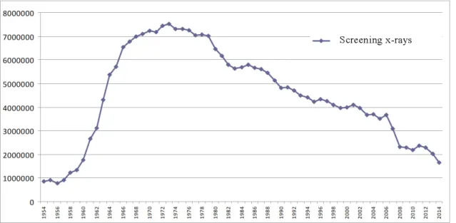
Screening for lung disease traditionally existed in most developed countries for the screening of infectious diseases such as tuberculosis (TB). Radiological screening with roentgen started widely after the II. World War when the price of the radiological examination was lowered(56). In Hungary, yearly lung screening was mandatory for every person of the age of 14 or older form 1960 to 2004. Screening examinations were done in a separate lung-screening network with yearly notifications via post.
5. Figure - Lung screening X-rays conducted in Hungary from 1954 to 2014(57)
Screening included chest x-ray and in some cases cytology and bacteriological culture from sputum. This system was primarily set up to find TB cases, however it helped in the discovery of lung cancer as well. With the notable decrease in TB cases and the worsening
financial system in the Hungarian health care, the screening network has been partially dismantled since 2004 and screening is not mandatory anymore. In the late 1990’s lung cancer screening became an important topic in the developed countries. The first country to introduce a modern screening system with computer tomography (CT) was Japan and the first dedicated program was the Early Lung Cancer Action Program in the USA(58, 59).
Several countries, including the USA, Netherlands, Japan, Denmark conducted observational trials and the conclusion was always the same: lung cancer can be found in an earlier stage with low-dose CT (LDCT) screening(60). To acquire reliable mortality data a randomized trial was warranted. The National Cancer Institute (US) founded the National Lung Screening Trial (NLST), a multi-center randomized study that was conducted from 2002 to 2009 and enrolled a total of 53,456 participants (from 2002 to 2004). Only heavy smokers (more than 30 pack year) were included between the ages of 55 to 75. Approximately 50% of cases were randomized to a chest x-ray arm, while the other 50% to a low dose screening arm. 309 deaths per 100,000 persons-year was observed in the chest radiography arm, while 247 in the LDCT arm, which means a 20%
relative reduction in lung cancer mortality. Since LDCT screening is a relatively expensive option, population wide LDCT screening is not affordable for most countries.
People should be selected on the basis of lung cancer risk assessment.
2. Table - Approximate 10-year risk of developing lung cancer.
Adopted from Bach PB et al. (61)
Duration of smoking
25 years 40 years 50 years
Age
Quit,
%
Still
smoking, %
Quit,
%
Still
smoking, %
Quit,
%
Still
smoking, % 1 pack per day smokers
55 <1 1 3 5 NA NA
65 <1 2 4 7 7 10
75 1 2 5 8 8 11
2 packs per day smokers
55 <1 2 4 7 NA NA
65 1 3 6 9 10 14
75 2 3 7 10 11 15
Several models were made to predict the risk of lung cancer development. Bach et al.
proposed a risk assessment model which primarily counts smoking history (in Table 2.).
Later the Spitz model (62) and the Liverpool Lung project (LLP) (63) model were also published. These models take several factors in consideration: age, age, smoking duration, exposure to carcinogens, diagnosed emphysema, prior diagnosis of pneumonia, prior diagnosis of malignant tumor, family history of cancer in first-degree relatives.
D’Amelio et al conducted a validation study including the Bach, the Spitz and the LLP
models. The latter two proved to be more discriminative, accurate and clinically more utilizable(64).
Screening with LDCT after lung cancer risk calculation is an affordable and efficient way of early lung cancer detection and thus a way to decrease lung cancer mortality. It raises some ethical considerations, however. Using the above mentioned risk assessment models mean that non-smokers will most probably be excluded from lung cancer screening. Considering the fact that non-smokers can also develop lung cancer, although the chance is thinner, raises concerns about the above described system. No financially feasible, more effective and ethically non-objectionable method has been developed however. In Hungary, currently no lung screening program is available and this situation must be addressed sooner than later.
2.1.4.2. Clinical diagnosis of lung cancer
Lung cancer detection in early stage is the most important step to achieve good results with treatment. 85% of the cases however, are only detected when symptoms occur and in 57% the diagnosis is only made when metastases are already present (65). The problem is that in most cases lung cancer does not cause early symptoms. Symptoms can be divided to subgroups (66):
• Symptoms related to endobronchial growth: Cough (8% to 75%), dyspnea (3% to 60%), pain, wheezing (0% to 2%), poststenotic pneumonia, hemoptysis (6% to 35%), stridor (0% to 2%).
• Symptoms related to intrathoracic extension: Chest pain (20% to 49%), hoarseness (recurrent nerve involved), diaphragm paralysis – raised diaphragm (phrenic nerve involved), upper airway inflow obstruction, dysphagia (esophagus involved), pleural effusion (carcinosis pleurae), pericardial effusion (pericardium involved), superior vena cava syndrome (v. cava superior involved), Horner triad, chronic shoulder pain (in Pancoast tumors).
• Systemic cancer symptoms: Weight loss (0% to 68%), fatigue, fever (0% to 20%), night sweats.
• Symptoms of distant metastases: Bone pain (6% to 25%), headache, neurological or psychiatric abnormalities, hepatomegaly, pathological fractures.
• Paraneoplastic syndromes: Cushing syndrome, Lambert-Eaton syndrome, Perre- Marie-Bamberger syndrome, syndrome of inappropriate ADH secretion, etc.
Lung cancer can be detected after the previously mentioned symptoms occur or in asymptomatic changes. Detection is most commonly done by chest x-ray or CT, however MRI (Magnetic Resolution Imaging) and PET (Positron Emission Tomography) are also more frequent.
Lung cancer diagnosis is often made from CT morphology and optionally the level of FDG uptake on PET, when a spiculated mass is seen, however pathological verification is also needed, primarily to distinguish between SCLC and NSCLC because the treatment strategies are very different. SCLC in most cases presents in a later stage with lymph node involvement. Generally small, central lung lesions with extensive lymphatic involvement always raise the concern for SCLC. The pathological diagnosis can be made from biopsies or after operation depending on the stage and treatment plan.
6. Figure – CT axial section of a spiculated mass in the left lower lobe (segment 6). Picture obtained from the Department of Thoracic Surgery, National Institute of Oncology, Budapest.
Lung cancer workup includes flexible bronchoscopy for the intrabronchial evaluation of anatomy and tumor spread. Bronchial washing and brushing can be done, which is ideal for intrabronchial or central tumors. Biopsies can be made through the bronchoscope with x-ray or ultrasound guidance (TBNA – trans-bronchial needle aspiration, EBUS – endo- bronchial ultrasound). The primary lesion and most regional lymph node stations can be reached with EBUS and also endoscopic ultrasound (EUS) guided biopsy can be of help.
In case of peripheral lesions, transthoracic fine needle aspiration or core biopsy under image guidance (usually CT) can be performed to get diagnosis and even the molecular characteristics of the tumor. Thoracentesis can also be of help in case of pleural fluid as a palliative and diagnostic tool. When none of the above mentioned methods yield a result, invasive, surgical techniques can be considered like mediastinoscopy, VATS biopsy or thoracotomy.
To provide adequate treatment it is important to assess how far the tumor have spread (staging) and to estimate the physical condition of the patient.
2.1.4.2.1. Functional evaluation of patients diagnosed with lung cancer
Physical evaluation of patients have to include the assessment of comorbidities, general performance (Karnofsky-index and the Eastern Cooperative Oncology Group (ECOG) status is most commonly used) and lung function. The most important comorbidities to take into account are cardiac, liver and renal diseases that can affect the feasibility and the result of certain treatment options. Cardiac echographia with the measurement of cardiac output and in selected cases cardiac stress test is often needed. Liver and renal function is tested with laboratory workup.
The evaluation of lung function is most important before surgical treatment, where the risk of the treatment and the quality of life achieved have to be taken into consideration.
Generally, Forced Expiratory Volume in 1 second (FEV1), Forced Vital Capacity (FVC) and Diffusing Capacity (DLCO) are measured. Patients with FEV1 of more than 80% of the normal (adjusted for size and age) and DLCO more than 80% are suitable for surgical treatment (including pneumonectomy) without further tests. In case either of these values are under 80% spiroergometry is recommended. If the maximum oxygen consumption is above 20mL/min/kg or 75% of normal, the patient can be referred for surgery (including
pneumonectomy). When the maximum oxygen consumption is below 40% of the expected (or ‹ 10mL/min/kg) functional inoperability is established. In case the maximum oxygen consumption is between 40% and 80%, postoperative FEV1 are calculated on the basis of quantitative ventilation and perfusion on lung scintigraphy. If the estimated FEV1
after the procedure is higher than 40% of normal, the operation can be performed; if it is below 40% functional inoperability has to be assumed.
7. Figure – Algorithm of functional evaluation recommendation of the European Society for Medical Oncology (ESMO); 2017 (67)
2.1.4.3. Staging of lung cancer
To evaluate the extent of the disease CT, PET CT, bone scintigraphy, brain MRI, abdominal ultrasound and biopsies are used. The staging is made following the actual international guideline. The latest staging system for lung cancer is the 8th tumor, node, metastasis (TNM) classification which was introduced after examining 77.156 cases and their outcome regarding survival and relapse (68).
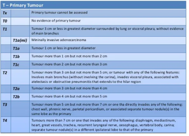
T stage is determined by the size of the primary tumor and its involvement of the adjacent structures:
3. Table - T value in the 8th edition of TNM staging. Adopted from: International Association for Study of Lung Cancer, 2015. [69]
N stage determines the lymph node involvement of lung cancer in the hilar and mediastinal regions. Lymph node involvement separates cases by prognosis and thus have a great effect on treatment.
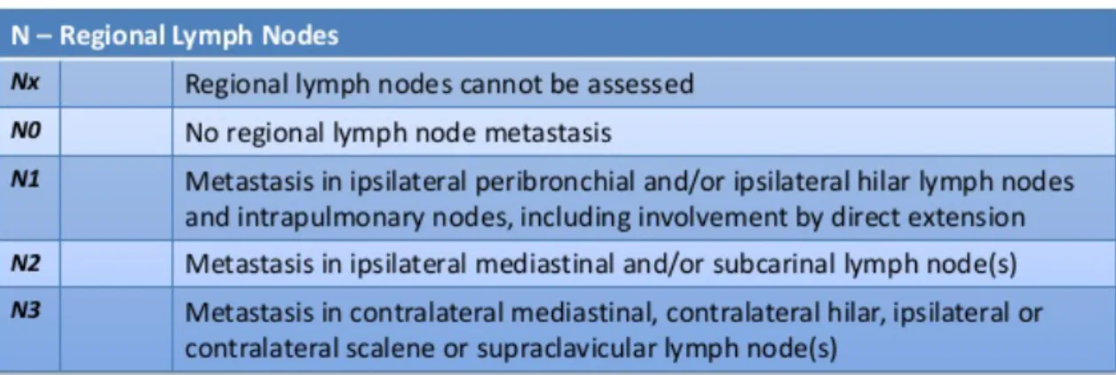
4. Table - N value in the 8th edition of TNM staging. Adopted from: International Association for Study of Lung Cancer, 2015. (69)
The N categories were made after the survival was assessed in pathological classified cases. N1 was divided into N1a (single station, ipsilateral hilum) and N1b (multiple stations); and N2 into N2a1 (single N2 station without N1 involvement), N2a2 (single N2 station with N1 involvement) and N2b (multiple N2 stations). The 5-year survival rates were the following: N1a, 59%; N1b, 50%; N2a1, 54%; N2a2, 43%; and N2b, 38%. A single-stage mediastinal nodal disease without N1 disease has the same prognosis as multiple N1 station involvement.
M stage defines distant metastases beyond the regional lymph nodes.
5. Table - M value in the 8th edition of TNM staging. Adopted from: International Association for Study of Lung Cancer, 2015. (69)
M1b was introduced as a separate category for the single extrathoracic metastases, since this patients had a significantly better survival (with a mean survival of 11.4 months instead of 6.3 months)(70). The treatment strategy also differs for these patients, since in
selected oligometastatic cases better survival can be achieved with aggressive local therapy, whereas in M1c cases systemic therapy has better results.
The appropriate stage is given from the TNM values according to the following table:
6. Table – Staging according to the 8th TNM. Adopted from: International Association for Study of Lung Cancer, 2015. (71)
T/M Subcategory N0 N1 N2 N3
T1 T1a IA IIB IIIA IIIB
T1b IA IIB IIIA IIIB
T1c IA IIB IIIA IIIB
T2 T2a IB IIB IIIA IIIB
T2b IIA IIB IIIA IIB
T3 T3 IIA IIIA IIIB IIIC
T4 T4 IIIA IIIA IIIB IIIC
M1 M1a IVA IVA IVA IVA
M1b IVA IVA IVA IVA
M1c IVB IVB IVB IVB
2.1.4.4. Prognosis of lung cancer
The prognosis depends highly on the histology, most importantly the distinction between SCLC and NSCLC, the biological behavior of the cancer and the stage.
2.1.4.4.1. Prognosis of SCLC
For the staging of SLCL currently the 8th TNM system is used, however another approach classifies SCLC into two groups: Limited Disease (LD) and Extensive Disease (ED). This classification reflects the clinical behavior and the spread of the tumor rather well, since LD patients have significantly superior survival when compared to ED patients. One contributing factor is that often small, accidentally found lesions turn out to be SCLC after final pathological examination. In these cases the surgical removement is done in a very early stage. The TNM system gives a more accurate estimation of the prognosis, however very few cases are found in early stages. When creating the 8th TNM for SCLC, Shirasawa et al. included 277 patients in a retrospective cohort and compared their prognosis. In this cohort 65.7% of the patients were classified as ED and 34.3% as LD, the OS was 37.2 (95% CI 25.7–48.7) and 13.7 (95% CI 11.9–15.5) months, respectively (this can be seen of Figure 8A). On Figure 8B. OS can be seen according to the TNM stages. OS drops markedly between stages, comparison of the curves reveals significant difference between every line (P values were the following: stage I vs II, P=0.04; I vs III, P=0.02; I vs IV, P<0.001; II vs III, P=0.47; II vs IV, P=0.009; III vs IV, P<0.001). It is important to know however, that the distinction between IVA and IB stage is also important because the change of M descriptor was also significantly associated with OS.
OS of stage IVB patients with the M1c descriptor was inferior to that of the stage III and IVA patients with the M0, M1a, or M1b descriptors (Figure 9A-B) (72).
8. Figure - Kaplan–Meier analysis-based estimates of survival based on staging system in SCLC patients (n=277). (A) Comparison of survival between LD (n=91; gray) and ED (n=186; black) SCLC patients (P<0.001). (B) Comparison of survival between patients in TNM stages I to IV.
Adopted from (72).
9. Figure - Kaplan–Meier analysis-based estimates of survival for ED SCLC patients. (A) Comparison of survival between patients in stages III (n=10, gray solid line), IVA (n=70, gray dotted line), and IVB (n=106, black solid line) (B) Comparison of survival according to the M descriptor of the 8th TNM. Adopted from (72).
2.1.4.4.2. Prognosis of NSCLC
The average 5-year survival in NSCLC is approximately 15%. This dismal result is due to the fact that most cases are found in latter stages (73) as previously mentioned. When
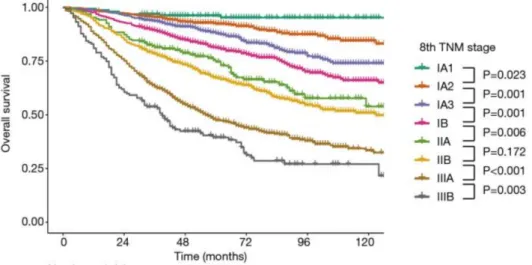
recently validation the 8th TNM Yun et al. compared the survival of 3950 following lung cancer resection. All TNM stages showed significant differences (except IIA and IIB where the difference was not significant). The 5-year median survival was 90.7%, 79.9%, 68%, 58.7% and 44.9% for IA, IB, IIA, IIB and IIIA stages, respectively according to the 7th TNM, however, reassessing the patients with the 8th TNM the 5 year median survival was quite different: 96.1%, 92.3%, 87.9%, 81.1%, 74.7%, 67.2%, 47.5% and 38.3% for IA1, IA2, IA3, IB, IIA, IIB, IIIA and IIIB stages, respectively. These results can be seen on Figure 10. The 8th TNM distinguishes better between both early and advanced stage NSCLC patients according to Yun et al (74).
10. Figure – Survival curves of OS according to the 8th TNM in the cohort of Yun et al. (74)
The 8th TNM was not yet validated in regards to stage IIIB and IV. However, available survival data shows significantly worse survival in these groups. The 5-year survival is reported to be 19%, while the median survival is 16 months in IIIB stage. The median survival drops to 6 months in IV. stage with the 5-year survival only 6%(75). This is already and improved results as 5-year survival virtually never existed in IV. stage before.
Further development and superior survival results are to be expected with the introduction of novel therapies such as immunotherapies.
2.1.5. Treatment of lung cancer 2.1.5.1. Treatment options for SCLC
The TNM staging system is useful to select patients for surgical treatment, however only very few cases are suitable for removal, reportedly less than 5%(76). Most of these patients are operated for an unknown lesion in the lung. These patients have superior outcome with 5-year survival up to even 50% is some reports (77, 78). Surgical approach can only be selected in these cases when lymph node involvement is ruled out with adequate mediastinal staging with PET-CT, EBUS and or mediastinoscopy. Postoperative adjuvant chemotherapy (CHT) is indicated even in T1-2,N0,M0 cases. Usually 4 cycles of CHT is administered. In case of unexpected N1 or N2 disease postoperative radiotherapy should be considered depending on the completeness of lymphadenectomy performed by the surgeon.
In cases with N2 disease, even without distant metastases (M0) surgery has no role. Total gross tumor volume proved to be an independent prognostic factor independent on the local therapy(79). N2 patients should undergo combined concurrent chemoradiotherapy.
Prophylactic cranial irradiation (PCI) should be considered at al T1-2,N0-1,M0 patients in case they responded to initial treatment.
Patients with T1-4,N0-3, M0 SCLC should be treated with chemotherapy and thoracic radiation concurrently in case the performance status (PS) allows. 70 Gy in daily fractions or 1.5 Gy in 30 fractions twice a day are usually selected options for radiotherapy. For the planning of radiotherapy to choose the target volume a PET CT is usually used.
For patients with metastatic SCLC the treatment is palliative. Most commonly a combination of CHT is used. A big meta-analysis reviewed the data of 19 randomized trials and shown that cisplatin was superior in terms of outcome in combination therapy when compared to other agents(80). Another collecting the data of 36 randomized trials concluded that etoposide, especially in combination with cisplatin showed longer OS than regimens without these two agents(81). The above mentioned results led to the clinical practice of etoposide-cisplatin combination therapy for SCLC as a standard of care.
Usually 4-6 cycles are administered in first line treatment.
PCI has led to significantly longer OS and less symptomatic brain metastases in randomized trials (82), thus patients responding well to initial CHT are usually offered PCI as well (25 Gy in 10 daily fractions or 20 Gy in 5 fractions).
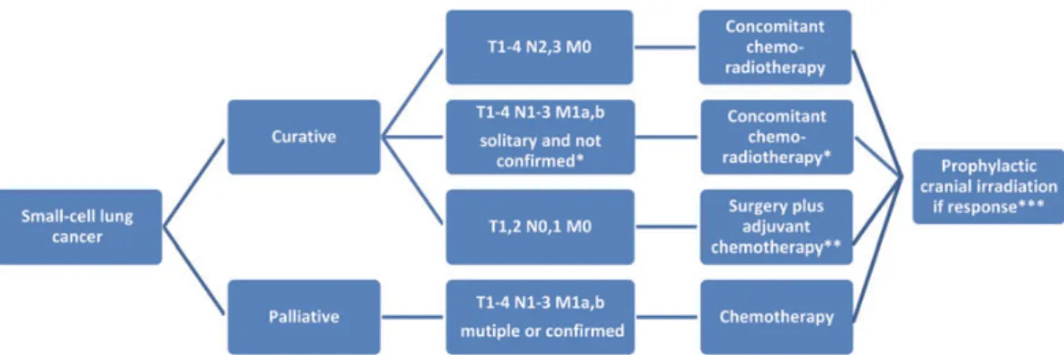
11. Figure – First line treatment algorithm for SCLC (ESMO 2013). Adopted from (83).
Second-line treatment for SCLC is only recommended in patients who respond to the first-line therapy. In patients who progress during chemotherapy and non-responders (early relapse within 6 weeks) outcomes are poor and further systemic therapy does not yield significant benefit. For these patients best supportive care is offered. Topotecan, a topoisomerase-I inhibitor, is a drug used for previously treated patients with SCLC. Its efficacy in terms of longer OS and better quality of life (better symptom control) was proven in a phase III trial (84). 8 years later the oral version of the drug was also tested in a phase III trial and was found to be equally efficient(85).
Targeted therapies were studied in extent for SCLC both in combination with chemotherapy and as a single agent, however very few studies managed to meet the primary endpoint. The examined drugs included: bevacizumab (86), vandetanib (87) and aflibercept (88) (anti-angiogenesis agents), panobinostat (89) (histon deacetylase inhibitor), oblimersen (90) (an agent targeting apoptosis) and cixutumumab and vismodegib (91) (agents targeting cell signaling). Alisertib, an aurora kinase inhibitor and sunitinib, a multi-targeted receptor tyrosine kinase inhibitor (TKI; targeting VEGFR, PDGFR and KIT) are the only agents showing preliminary signs of efficacy in SCLC(92,
93). The latter also showed improvements in PFS, however it is still not clear whether they will impact the standard of care.
2.1.5.2. Treatment of NSCLC
2.1.5.2.1. Treatment of early stage NSCLC
Surgical removal remains the first choice of treatment in localized, resectable NSCLC (Stage I and II). First-line surgery provides the best survival. As previously mentioned 5- year survival is between 90.7% (IA) and 58.7% (IIB)(74).
The choice of upfront surgery is made after staging, assessment of technical operability and tissue biopsy when needed. The algorithm of lymph node staging recommended by ESMO can be seen on Figure 15.
12. Figure – ESMO 2017: recommended algorithm for staging in patients with M0 NSCLC (FDG - fluorodeoxyglucose; LN - lymph node, NPV - negative predictive value; VAM - video-assisted mediastinoscopy) Adopted from (94).
Surgical techniques have improved significantly in the last twenty years. Most of the traditional thoracic operations were first described between 1900 and 1950. The first anatomical dissection of hilar structures, as lobar vein, artery and bronchus was first done in 1933 by Rienhoff, which was a big step to achieve fewer complications. The techniques improved quickly and anatomical segmentectomy (Churchill and Belsey in 1939) and even sleeve lobectomy was developed by 1947 (Price Thomas) (95). From the 1950s to the 1980s new surgical instruments were developed, but the main types of lung operations were not changed.
Wedge resection is an atypical resection of lung parenchyma most often done by surgical staplers. Atypical in this sense means that the resection line does not follow anatomical structures or borders. It is mainly used for inflammatory, benign, metastatic or unknown lung lesions. Segmentectomy means the selected resection of one or several pulmonary segments. This procedure requires precise surgical technique and was mainly used in situations when the pulmonary function did not allow more extensive parenchyma resection. Lobectomy is the gold standard operation for the removal of primary lung malignancies. Lobectomy means the central dissection and ligation or stapling of the lobar pulmonary artery(s), vein and bronchus. Lobectomy is also often used when the parenchyma of a complete or near complete lobe is damaged by inflammatory diseases or when a secondary tumor is deep in the parenchyma and a wedge resection is not possible. Pneumonectomy is the most extensive operation performed on a single lung, it means the complete removal of the lung on one side by centrally closing the main pulmonary artery, lower and upper lobe veins and the main bronchus.
13. Figure – The main four types of pulmonary resections (Education material from the University of South Carolina; access: cts.usc.edu)
All of the above mentioned procedures were done through a thoracotomy, when the thoracic cavity is approached through a rib space. To get appropriate access to preform complicated operations, long wounds had to be made and the ribs had to be either resected or pushed apart from each other. Thoracotomy was and still is a painful operation and it can have significant complications which sour the patients life. Most pain is caused by damaging the intercostal nerves. Neuralgic pain can last for years after the operation. A less invasive way to operate the chest was introduced very early: the first thoracoscopic procedures are attributed to Jacobeus (1910), who used a cystoscope to evacuate pleural fluids and to take pleural biopsies, although Sir Francis Richard Cruise was the first to apply and endoscope in the chest in 1865. Despite the early thoracic application of the technique, major thoracic procedures were only performed in the late 1980s, early ‘90s, after the video camera was invented. Rovario performed and described the first lobectomy
done by Video Assisted Thoracic Surgery (VATS) in 1991 (96). Later McKenna presented already 1000 cases of VATS lobectomy and in 2008 the first VATS sleeve lobectomy as well (97). VATS can be done through several small incisions, most commonly three, however newer techniques emerged: Gonzales-Rivas performed the first lobectomy in 2011 through one port (uniportal or single-port) (98). Ever since there was a shift towards single-port and less invasive surgery. At the beginning VATS was only used for small or benign lesions and there was a general view that VATS is less radical. However, later several study proved that OS was comparable after VATS lobectomy as with an open procedure (99-103) and the amount of lymph nodes taken out was also not different (104). In the US centers often choose another minimal-invasive technique instead of VATS, the Robotic Assisted Thoracic Surgery. Robotic surgery got widely accepted as a form of minimal-invasive surgery first in the US. Postoperative pain is reported to be comparable with VATS and the OS comparable with VATS and thoracotomy (105, 106).
14. Figure – Thoracotomy versus VATS. Comparison of (A) intraoperative access and incision and (B) postoperative wound length (Pictures taken in the Department of Thoracic Surgery, National Institute of Oncology, Budapest)
The real advantage of VATS or robotic thoracic surgery is shown in the early postoperative phase when patients are pain-free and can leave the hospital on the postoperative second day. This leads to less pulmonary complications (107), and some studies suggest that even less immunosuppression and thus better resistance against cancer (108). Ceppa et al. used the national database in the US, in their meta-analysis they showed that the greatest benefit of VATS can be seen in patients with poor pulmonary function. The incidence of pulmonary complications in patients with compromised pulmonary reserves, was higher after thoracotomy than after VATS (109), as can be seen on the figure below.
15. Figure – Pulmonary complications after thoracotomy (grey) and VATS (black). The difference of pulmonary complications after surgery is most significant in patients with poor pulmonary function (FEV1 lower than 50%). Adopted from (109).
It is not yet widely accepted that OS is better after VATS or robotic thoracic pulmonary resection and most guidelines do not differentiate, data suggest that minimal invasive resection should be the gold standard of care for early lung cancer (108).
16. Figure – OS is longer in patients who had VATS resection for early lung cancer when compared with those who underwent resection through thoracotomy (left). The difference is still visible after propensity crossmatching, although it is not statistically significant (right). (Figures adopted from Berry et al. (108))
Although imaging, surgical and staging techniques have significantly changed in the last two decades, still the Lung Cancer Study Group (LCSG) 821 trial remains the ground for surgical treatment. According to this trial lobectomy is the cornerstone of surgical therapy for stage I and II NSCLC, as local recurrence was found more frequently after less radical operations (wedge resection and segmentectomy) (110). This data and along that our current practice will most probably be updated as the new TNM distinguishes between several types of stage I NSCLC. Based on large collected cohorts segmentectomy seems equally appropriate for T1a tumors. In squamous cell carcinoma lobectomy was found to be superior even in early stages, however in adenocarcinoma equivalent results were reported after anatomical segmentectomy (111). Of note, in another study anatomical segmentectomy was also found to be equally effective in T1c adenocarcinoma (112).
Altogether it can be anticipated that in selected early NSCLC anatomical segmentectomy will be the choice of treatment, however no hard data is available to support this yet. Two major randomized studies are underway (CALGB 140503 and JCOG0802/WJOG4607L) (113, 114). The preliminary results have been published of the latter about the safety and feasibility of segmentectomies (simple and complex as well), their results show that
segmentectomy is not inferior in any of the examined parameters (complications, length of hospital stay, duration of air leak etc.) (115).
Lymph node management during surgery is also controversial. In early stages, the removal of lymph nodes is mainly necessary for staging purposes, to get a guaranteed R0 resection and an accurate mediastinal staging. The latest ESMO recommendation implies surgical evaluation of at least six lymph node stations, three of which are mediastinal, including the sub-carinal station. If all of these are negative on pathological report than R0 and N0 can be safely stated (116). It is widely accepted that in early stages, especially stage I OS and PFS, as well as local recurrence might not be influenced by the type of surgical lymph node assessment, however in stage II and particularly IIIA intraoperative nodal dissection is recommended (117). The extent of dissection is also up to debate, the most common technique is lobe specific lymph node dissection, though perioperative lymph node assessment (EBUS, mediastinoscopy) can also be of effect.
For high-risk patients, or for patients who are unwilling to take the risk of an operation, or patients with unresectable tumors primary stereotactic body radiotherapy (SBRT) can be recommended(67). The reported local control rate of SBRT is between 79-90.9% at 5 years (118, 119). The recommended dose is a biologically equivalent tumor dose of 100 Gy. The toxicity using this dose SBRT for stage I or II peripheral NSCLC is low and acceptable, even in elderly and patients with severe chronic obstructive pulmonary disease (COPD) (120). Data is available on outcomes of SBRT in patients who underwent the procedure for peripheral stage I tumors and are generally fit for surgery, however, there is no evidence to currently to recommend SBRT over up front surgery (121, 122).
One problem with SBRT is the lack of pathological confirmation after the procedure.
Randomized studies are being conducted, although three have failed to complete its plan (123). Two studies that have concluded reported comparable recurrence-free survival three years after the procedure . Given, that the toxicities are tolerable and the quality of life is good after SBRT, patient opinion should be more included in the discussion to recommend personalized treatment in the future.
17. Figure – SBRT distribution plan for NSCLC. Adapted from (124).
Multiple primary tumors should also be assessed with curative intent and complete surgical resection is recommended when feasible. In patients with borderline risk, combination of surgery and SBRT has been proven efficient(125).
In case a patient has contraindication against both surgery and SBRT radiofrequency ablation (RFA) might be the reasonable choice of treatment, however the level of supporting evidence is from observational cohorts only (126).
Postoperative radiotherapy is rarely indicated in stage I and II cases. It was found detrimental in cases without lymph node invasion (127). Cases with unexpected N1 and N2 disease are not clear. Postoperative radiotherapy (concurrent with chemotherapy) seem a reasonable choice when R0 resection is suspected (mainly because of N1 lymph nodes) or when the N2 lymph nodes are involved. To support this recommendation a large clinical trial is underway (128).
Adjuvant platinum based chemotherapy was found to have beneficial effect on survival for stage II NSCLC patients(129). Altogether a 4-5% improvement in 5-year survival was reported in N1 and N2 disease. The adjuvant regimen used was 300mg/m2 of cisplatin in three to four cycles (130). The best timing of adjuvant chemotherapy is not entirely clear, although most protocols say 6 weeks after surgery, recent data form the National Cancer Database (United States of America – US) show comparable results after longer interval between resection and chemotherapy (131).
Based on this knowledge, patients with completely resected stage II tumors are recommended adjuvant chemotherapy within 6-8 weeks after surgery. On the other hand,
in stage IA, chemotherapy has not been proven to be beneficial in an adjuvant setting(132). In IB a small benefit was seen in OS and an in depth analysis showed that this was due to patients with tumors >4 cm (133).
In stage IA NSCLC is therefore only followed up after resection, in IB, however adjuvant chemotherapy can be recommended in cases when the tumor size is over 3-4cm.
As of today, no evidence suggest that the use of targeted agents, such as TKIs for EGFR mutant cases, yield a benefit in an adjuvant setting. Prospective studies are being conducted, however until their result is presented, targeted agents are not recommended as adjuvant CHT.
There is less experience with neoadjuvant CHT in early NSCLC, most studies found no survival comparing neoadjuvant and adjuvant modalities (134, 135). In some, selected cases when downstaging or size reduction is needed for surgical removal, neoadjuvant regimens can potentially be of use (less extensive surgical procedure is possible after) (136).
Both adjuvant and neoadjuvant trials have been initiated using immunotherapy (anti-PD- 1 and anti-PD-L1 checkpoint inhibitors) for early lung cancer (trials NCT NCT02504372, NCT02273375 and NCT02998528) on the basis of the promising results in second line curative therapy. This might change the clinical practice in the near future.
2.1.5.2.2. Treatment of locally advanced (stage III) NSCLC
Stage III or locally advanced (LA) NSCLCs can be divided in two groups: surgically resectable LA NSCLC and unresectable LA NSCLC. The treatment strategy is understandably different.
Resectable LA NSCLC usually occurs in three instances: 1) single-station N2 lymph node involvement is suspected or confirmed by biopsy (other stations were found negative with PET and biopsy). In these cases complete lymphadenectomy and postoperative CHT has shown superior outcome compared to surgery or chemotherapy alone (137). However, it is not clear whether surgery or SBRT is the better option in these cases. In the limited number of studies comparing the two modalities of local control no clear advantage was shown for either (138, 139).
2) T4N0 tumors where R0 resection is considered feasible. In these cases up front surgery (generally open technique) and postoperative CHT is recommended. Potentially