R E S E A R C H Open Access
Ki-67 as a controversial predictive and
prognostic marker in breast cancer patients treated with neoadjuvant chemotherapy
Balázs Ács1†, Veronika Zámbó1†, Laura Vízkeleti1,2, A. Marcell Szász1,3, Lilla Madaras1, Gyöngyvér Szentmártoni4, Tímea Tőkés4, Béla Á. Molnár5, István Artúr Molnár5, Stefan Vári-Kakas6, Janina Kulka1and Anna-Mária Tőkés1,7*
Abstract
Background:Studies have partly demonstrated the clinical validity of Ki-67 as a predictive marker in the neoadjuvant setting, but the question of the best cut-off points as well as the importance of this marker as a prognostic factor in partial responder/non-responder groups remains uncertain.
Methods:One hundred twenty patients diagnosed with invasive breast cancer and treated with neoadjuvant
chemotherapy (NAC) between 2002 and 2013 were retrospectively recruited to this study. The optimal cut-off value for Ki-67 labeling index (LI) to discriminate response to treatment was assessed by receiver operating characteristic (ROC) curve analysis. Kaplan-Meier curve estimation, log-rank test and cox regression analysis were carried out to reveal the association between Ki-67 categories and survival (DMFS = Distant metastases-free survival, OS = Overall survival).
Results:Twenty three out of 120 patients (19.2%) achieved pathologic complete remission (pCR), whereas partial remission (pPR) and no response (pNR) to neoadjuvant chemotherapy (NAC) was detected in 60.8% and 20.0%, respectively. The distribution of subtypes showed a significant difference in pathological response groups (p< 0.001).
Most of the TNBC cases were represented in pCR group.
The most relevant cut-off value for the Ki-67 distinguishing pCR from pNR cases was 20% (p= 0.002). No significant threshold for Ki-67 was found regarding DMFS (p= 0.208). Considering OS, the optimal cut-off point occurred at 15%
Ki-67 (p= 0.006). The pPR group represented a significant Ki-67 threshold at 30% regarding OS (p= 0.001). Ki-67 and pPR subgroups were not significantly associated (p= 0.653). For prognosis prediction, Ki-67 at 30% cut-off value (p= 0.
040) furthermore subtype (p= 0.037) as well as pathological response (p= 0.044) were suitable to separate patients into good and unfavorable prognosis cohorts regarding OS. However, in multivariate analyses, only Ki-67 at 30% threshold (p= 0.029), and subtype (p= 0.008) were independently linked to OS.
Conclusions:NAC is more efficient in tumors with at least 20% Ki-67 LI. Both Ki-67 LI and subtype showed a significant association with pathological response. Ki-67 LI represented independent prognostic potential to OS in our
neoadjuvant patient cohort, while pathological response did not. Additionally, our data also suggest that if a tumor is non-responder to NAC, increased Ki-67 is a poor prognostic marker.
Keywords:Breast, Neoadjuvant chemotherapy, Ki-67, Predictive and prognostic markers
* Correspondence:tokesa1972@yahoo.co.uk
†Equal contributors
12nd Department of Pathology, Semmelweis University, 1091 Budapest, Üllői út 93, Hungary
7MTA-SE Tumor Progression Research Group, Semmelweis University, 2nd Department of Pathology, 1091 Budapest, Üllői út 93, Hungary Full list of author information is available at the end of the article;
© The Author(s). 2017Open AccessThis article is distributed under the terms of the Creative Commons Attribution 4.0 International License (http://creativecommons.org/licenses/by/4.0/), which permits unrestricted use, distribution, and reproduction in any medium, provided you give appropriate credit to the original author(s) and the source, provide a link to the Creative Commons license, and indicate if changes were made. The Creative Commons Public Domain Dedication waiver (http://creativecommons.org/publicdomain/zero/1.0/) applies to the data made available in this article, unless otherwise stated.
Background
Breast cancer is the most common malignant tumor among women all around the industrialized world [1].
Proliferation is an essential characteristic of all cancer types, including breast cancer [2]. The proliferation activity of different tumors assessed with immunohisto- chemical detection of the cell-cycle specific antigen Ki- 67 has been extensively studied in the last decade. Many studies have shown that Ki67 expression is a useful prognostic factor in breast cancer [3, 4]. Criscitiello et al.
reported that Ki67 expression can identify a subset of patients among Luminal-B and node positive breast can- cer cases who could benefit from addition of adjuvant chemotherapy to hormone therapy [5]. Contradicting this finding, Andre F. et al. reported that in the adjuvant setting Ki67 staining lacks analytical validity, moreover, no robust evidence indicates that Ki-67 staining predicts the efficacy of adjuvant chemotherapy [6]. Assessment of cellular proliferation by Ki-67 expression is not recom- mended in routine pathological evaluation by either International Ki-67 in Breast Cancer Working Group of the Breast International Group and North American Breast Cancer Group or the American Society of Clinical Oncology (ASCO) given the fact that it is not totally clear how Ki-67 measurements and thresholds could in- fluence clinical decisions [7].
Nowadays neoadjuvant chemotherapy (NAC) has be- come an accepted therapy choice, even in early stage breast cancers. In the last few years several research stud- ies have focused on the prognostic and predictive value of Ki-67 expression in neoadjuvant settings [8–10]. The International Ki-67 in Breast Cancer Group created guide- lines for the assessment of Ki-67 with recommendations on pre-analytical and analytical procedures, as well as on interpretation, scoring, and data handling [11].
Achieving pathologic complete remission (pCR) re- mains a subject of controversy in terms of definition and its evaluation methods. The predictive value of pCR in relation to patient outcome in various breast cancer bio- logical subtypes has been under debate. pCR has strongly predicted improved survival in triple negative breast cancer (TNBC) and HER2-enriched subgroups, while there are contradictory data related to the luminal subtypes [12, 13].
Even if significant efforts are currently made to select the group of patients who will benefit from NAC, there are no clear genomic markers that can predict the re- sponse rate [14]. There are contradictory data about Ki- 67 as a predictive factor [8, 9] for response to NAC and there are several questions to be answered, such as the standardization of the methodology used for Ki-67 de- tection, or how Ki-67 expression in core biopsies is rep- resentative of the whole tumor. It is becoming clear that there is an urgent need for standardized Ki-67
diagnostics or a need for use of other markers associated with cell proliferation. Klauschen et al. reported that the computer assisted Ki-67 scoring approach correlated with the clinical endpoints [15]. Balmativola et al. found that 18% threshold of Ki-67 positive cells accurately dis- criminated the pCR and non-pCR patient groups [8]. In a recent study by Magbanua et al. it is nicely presented that in addition to single-gene studies, genomic ap- proaches are needed to maximize the discovery of useful classifiers and druggable targets in the NAC setting [14].
The majority of previous research studies have focused mostly on the group of patients presenting pCR. Only a few studies have investigated the value of KI-67 in non- responders to NAC [8]. Even though partial responders to NAC represent the largest group with a very hetero- geneous population, and although patients with minimal residual cancer burden after neoadjuvant chemotherapy often have the same excellent prognosis as patients who achieve pCR, there are very few studies offering add- itional information about the likelihood of partial re- sponse to NAC [16, 17]. Until this will happen, we need to investigate more clearly the relationship between pre- therapeutic Ki-67 expression and response to neoadju- vant chemotherapy.
Our aim in this study was to find optimal cut-off values for Ki-67 expression that best correlates with re- sponse rates to NAC and with DMFS (distant metastasis-free survival) as well as with OS (overall sur- vival). We also investigated the association between Ki- 67, subtype and pathological response, as well as the prognostic potential of Ki-67 in partial responder pa- tients. Furthermore, the prognosis prediction potential of Ki-67 was correlated to that of the conventional clini- copathological factors in multivariate analysis.
Methods
A total of 120 patients diagnosed with invasive breast cancer and treated with NAC at Semmelweis University in Hungary between 2002 and 2013 were retrospectively recruited for this observational, cross-sectional cohort study. Patients were enrolled only if they had completed NAC, thereafter underwent surgery. The study was eth- ically approved by the Semmelweis University Institu- tional Review Board (SE-IKEB 120/2013).
The data recorded are partly presented in Table 1 and were as follows:
a. age at diagnosis, clinical and pathologic tumor size (cT and pT), nodal involvement (cN)
b. in the pre-treatment biopsy: histological type and nuclear grade, Ki-67 LI, Estrogen Receptor (ER), Progesterone Receptor (PgR), and human epidermal growth factor receptor 2 (HER2) status (based on
both immunostaining score and in situ hybridization analysis for HER2 score 2 cases)
c. degree of response to therapy was categorized following Pinder et al. [18] in the histological sections of the post treatment surgical specimens as follows: Pathologic complete response (pCR) was defined as no residual invasive tumour and the absence of any residual invasive tumor in the lymph nodes. Partial response to therapy (pPR), either
<10% of tumour remaining (pPRi), or 10–50%
tumour remaining (pPRii), or >50% of tumor remaining but some evidence of response to therapy is present (pPRiii). Non responders (pNR) were defined as no evidence of response to therapy, d. Surrogate molecular subtypes were defined based
on the 2013 St. Gallen Consensus Conference recommendations as Luminal-A, Luminal-B/
HER2-, Luminal-B/HER2+, HER2+ and triple negative categories [19, 20].
Immunohistochemical analysis
ER, PgR, HER2 status and Ki-67 LI were evaluated in the pre-treatment core biopsy specimens and in case of non pCR, on the surgical specimens as well, by immunohisto- chemistry (IHC). All immunohistochemical analyses were carried out at the 2nd Dept. of Pathology, Semmelweis University, Hungary with automated immunostainer sys- tem (Ventana Benchmark XT, Roche Diagnostics, Mannheim, Germany) according to the manufacturer’s in- structions, using the following antibodies: 1:200 anti-ER (clone 6 F11), 1:200 anti-PgR (clone 312) and 1:150 anti- HER2 (clone CB11) antibodies purchased from Novocas- tra Laboratories Ltd (Newcastle upon Tyne, UK), and 1:100 anti-Ki67 (clone MIB1) antibody purchased from DAKO Gmbh (Hamburg, Germany). The antigen retrieval method was performed in the immunostainer with CC1 antigen retrieval solution on pH9 at 42 °C for 30 min.
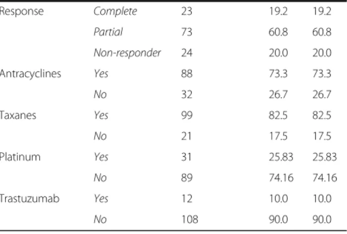
Table 1Clinicopathological data of analyzed cases Number of cases
Total
%
Valid
%
Age 40≥ 29 24.2 24.2
40 < 91 75.8 75.8
cT T1 19 15.8 15.8
T2 73 60.8 60.8
T3 15 12.5 12.5
T4 13 10.8 10.8
pT pT0 20 16.7 18.7
pT1 40 33.3 37.4
pT2 34 28.3 31.8
pT3 9 7.5 8.4
pT4 4 3.3 3.7
Unknown 13 10.8
cN N0 46 38.3 40.4
N1 55 45.8 48.3
N2 9 7.5 7.9
N3 4 3.3 3.5
Nx 6 5.0
pN pN0 51 42.5 48.1
pN1 36 30 33.9
pN2 13 10.8 12.3
pN3 6 5 5.7
Nx 14 11.7
Grade 1 1 0.8 0.9
2 46 38.3 41.8
3 63 52.5 57.3
Unknown 10 8.3
ER status Positive 80 66.7 66.7
Negative 40 33.3 33.3
PgR status 20% > 8 6.7 6.7
20%≤ 49 40.8 41.2
Negative 62 51.7 52.1
Unknown 1 0.8
HER2 status Positive 41 34.2 34.2
Negative 79 65.8 65.8
Histological type Lobular 6 5.0 5.1
IBC NOS 112 93.3 94.9
Other/Unknown 2 1.7
Molecular subtype
Luminal A 15 12.5 12.5
Luminal B/HER2- 38 31.7 31.7
Luminal B/HER2 +
27 22.5 22.5
HER2+ 14 11.7 11.7
Triple-negative 26 21.7 21.7
Table 1Clinicopathological data of analyzed cases(Continued)
Response Complete 23 19.2 19.2
Partial 73 60.8 60.8
Non-responder 24 20.0 20.0
Antracyclines Yes 88 73.3 73.3
No 32 26.7 26.7
Taxanes Yes 99 82.5 82.5
No 21 17.5 17.5
Platinum Yes 31 25.83 25.83
No 89 74.16 74.16
Trastuzumab Yes 12 10.0 10.0
No 108 90.0 90.0
The cut-off value for ER and PgR positivity was 1%
positive tumor cells with nuclear staining. Hormone receptor (HR) negativity was defined as being negative for both ER and PgR. HER2 IHC positivity was de- fined as score 3+ complete, strong membrane staining in >10% of tumor cells. For IHC 2+ samples, fluores- cent in situ hybridization (FISH) was performed to confirm gene amplification by using Zytovision ERBB2/CEN17 dual FISH probes. HER2 status was defined according to the ASCO/CAP guideline valid at the time of diagnosis (ASCO/CAP guideline 2007 and ASCO/CAP guideline 2013) [21, 22].
Ki-67 was scored as the percentage of positive tumor cell nuclei by counting a range of 400–500 cells (de- pending on the cellularity of the specimen), including also hot spot areas. The optimal cut-off value for Ki67 was defined by statistical analysis; however we also ap- plied the 20 and 30% threshold values as mostly recom- mended cut-off scores in the literature.
Clinical outcome assessment
Overall survival was defined as the elapsed time from the date of diagnosis of the tumor by core biopsy to the date of death, or when patients were last censored if still alive. DMFS was defined as time from the date of pri- mary diagnosis to the occurrence of first distant metas- tases. All patients were followed-up until the date of death or when censored at the date April 30, 2015 (me- dian follow up time for OS and DMFS was 60.5 and 59 months, respectively).
Statistical analysis
All analyses were carried out using SPSS version 22.0 statistical software (SPSS Inc, Chicago, IL, USA). Differ- ences in the distribution of characteristics between the parameters of patients with pCR or pPR and patients with pNR were evaluated using two-sided Fisher’s Exact Test. Two-sided Mann-Whitney-Wilcoxon test was used to define age distributions in pCR vs. pNR and vs. pPR.
The optimal cut-off value for Ki-67 percentage to dis- criminate response to treatment was assessed by receiver operating characteristic (ROC) curve analysis. To iden- tify the optimal Ki-67 threshold for NAC, only pCR and pNR cases were involved in ROC analyses, because pPR
status is considered as a soft endpoint. Kaplan-Meier curve estimation, log-rank test and Cox regression ana- lysis were carried out to analyze the association between Ki-67 categories and DMFS or OS. P values ≤0.05 were considered to be statistically significant.
Results
Baseline clinical and pathological data
Hundred and twenty patients met the inclusion criteria and their data were used in this study.
Mean patient age was 50.6 years (range, 29–74 years).
The majority of patients (59.6%) had node-positive dis- ease and cT2 tumors (60.8%). Tumors were ER-positive in 66.7% of cases and presented PgR positivity >20.0% in 41.2% of the analyzed samples. In 34.2% of cases HER2 positivity was detected. Of the 120 tumors, 12.5% were of Luminal-A, 31.7% of Luminal-B/HER2 negative, 22.5% of Luminal-B/HER2 positive, 11.7% of HER2+ and 21.7% of TNBC subtype (Table 1).
Twenty three out of 120 patients (19.2%) achieved pathologic complete remission (pCR), 73 (60.8%) showed partial remission (pPR), whereas no response to NAC (pNR) was detected in 24 cases (20.0%).
In the group of patients who obtained pPR, residual tumor was detected in lymph nodes only in 7 patients (9.6%), major response (>90% tumor regression) to NAC was observed in 8 cases (11.0%), a response rate between 50 and 90% was detected in 26 cases (35.6%), whereas a response rate <50% was observed in 32 cases (43.8%).
Non-responder patients (pNR, mean age: 56) showed significant difference (p= 0.008) in age compared to pa- tients achieving pathologic complete response (pCR, mean age: 46). This association between pNR patients’
age and pCR patients’ age was mainly confined to the HR+ patient population.
Significant difference in HR status was observed be- tween the pCR, pPR and pNR populations (p< 0.001):
approximately three-quarters of the samples within the pCR group were HR negative as contrasted to the pPR and pNR groups, in which cases the proportion of HR+ samples was more than 3-times higher (Table 2).
Table 2Association between age, hormone receptor positivity and therapy response Therapy response
category
N HR status (%) p1 Average age at diagnosis (year ± SD) p2
HR+ HR− HR+ HR− all
pCR 23 5 (21.7) 18 (78.3) <0.001 44.2 ± 12.6 46.4 ± 13.8 46.0 ± 13.3 0.008
pPR 73 55 (75.3) 18 (24.7) 50.0 ± 11.0 52.3 ± 12.8 50.4 ± 11.4
pNR 24 20 (83.3) 4 (16.7) 55.9 ± 10.7 53.3 ± 14.5 55.6 ± 11.0
HR–hormone receptor positive (+) or negative (−)
1two-sided Fisher’s exact test;2Kruskal-Wallis H-test with Dunn’s posthoc (multiple comparison) test for all samples without regarding the HR status
Defining cut-off points for Ki-67expression in the pCR and pNR groups
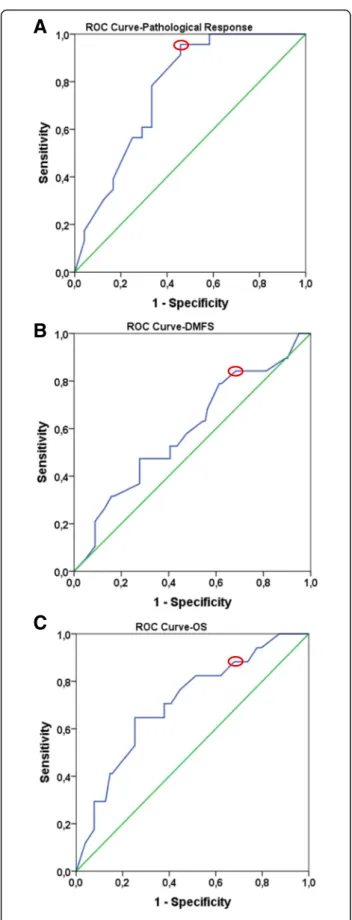
ROC curve analysis was used in order to identify the op- timal cut-off value of Ki-67 expression that could best predict response to NAC (Fig. 1a). The optimal Ki-67 cut-off value was 20% for distinguishing pCR from pNR patient cases (n= 47, AUC 0.767, sensitivity: 95.7%, spe- cificity: 54.3%,p= 0.002). (Fig. 1a).
Defining cut-off points for Ki-67 expression based on Survival (DMFS and OS)
We also investigated the optimal threshold values for Ki-67 regarding DMFS and OS. Based on DMFS, we were not able to detect a statistically significant cut-off value for Ki-67. The most relevant cut-off value was 20%
(n= 120, AUC 0.591, sensitivity: 82.2%, specificity 35.7, p= 0.208) (Fig. 1b). On the basis of OS data, the optimal cut-off point occurred at 15% for Ki-67 (n= 120, AUC 0.708, sensitivity: 92.3%, specificity 29.6, p= 0.006) (Fig. 1c).
Association between Ki-67, Subtype and Pathological Response
Pathological response and Ki-67 at investigated thresholds represented a significant association (Ki-67 15% p= 0.001, Ki-67 20% p= 0.010, Ki-67 30% p= 0.018). The proportion of Ki-67 low cases among non-responders was significantly higher compared to pPR and pCR cases (Additional file 1A). The distribu- tion of subtypes showed a significant difference in pathological response groups (p< 0.001). Most of the TNBC cases were represented in pCR group, while luminal-A cases mainly occurred in pPR and pNR groups (Additional file 1B). The Ki-67 expression at any investigated cut-off points and subtypes also rep- resented a significant correlation (p< 0.001 for all comparisons). Luminal-A subtype showed low Ki-67, while TNBC and HER2+ cases mostly had high Ki-67 (Additional file 1C). The association between Ki-67, subtype and pathological response was also investi- gated without Luminal-A cases, because NAC is not generally recommended in this subtype due to the high rate of pNR status in contrast with the favorable prognosis. Excluding Luminal-A cases, Ki-67 at any thresholds and pathological response did not show any significant association (Ki-67 15% p= 0.068, Ki-67 20% p= 0.122, Ki-67 30% p= 0.140) (Additional file 2A). Furthermore, Ki-67 expression at any investi- gated cut-off points also did not represent any signifi- cant linkage with subtypes (Ki-67 15% p= 0.410, Ki- 67 20% p= 0.158, Ki-67 30% p= 0.173) (Additional file 2C). In contrast to this, subtypes were significantly linked to the pathological response groups (p< 0.001).
The vast majority of Luminal-B cases were in pPR
and pNR groups, while TNBC cases mostly occurred in pCR subgroup (Additional file 2B).
Prognostic potential of Ki-67 status, Subtype and Pathological Response (DMFS and OS)
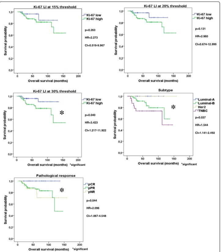
Neither Ki-67 at any thresholds nor subtype and not even pathological response were suitable to distinguish patient cohorts with different DMFS (Ki-67 15% p= 0.391, Ki-67 20%p= 0.185, Ki-67 30%p= 0.566, subtype p= 0.771, pathological response p= 0.280). Regarding OS Ki-67 at 15% (p= 0.263) and at 20% threshold failed (p= 0.131), but Ki-67 at 30% cut-off value (p= 0.040) furthermore subtype (p= 0.037) as well as pathological response (p= 0.044) were suitable to separate patients into good and unfavorable prognosis cohorts (Fig. 2).
When Luminal-A cases were excluded, neither Ki-67 at any cut-off points nor subtype not even pathological re- sponse were suitable to perform statistically significant splitting of our cohort into 2 patients’group with differ- ent DMFS (Ki-67 15% p= 0.426, Ki-67 20% p= 0.179, Ki-67 30% p= 0.642, subtype p= 0.488, pathological re- sponse p= 0.222,) or with different OS (Ki-67 15% p= 0.975, Ki-67 20%p= 0.518, Ki-67 30%p= 0.158, subtype p= 0.072, pathological response p= 0.058, Additional file 3). We also investigated the utility of Ki-67 at 15, 20 and 30% thresholds as potential independent pre- dictor of DMFS and OS adjusted by age, pathological response, hormone receptor status, subtypes, histo- logical grade, lymph node, cT and pT status. Neither Ki-67 at any thresholds nor any other clinicopatho- logical factors except pT status (p= 0.029) showed an independent association with DMFS (Table 3). How- ever, Ki-67 at 30% threshold (p= 0.029) and subtype (p= 0.008) were independently linked to OS (Table 3).
Without Luminal-A cases, Ki-67 at 30% cut-off point (p= 0.038) and subtype (p= 0.009) represented also an independent association with OS (Table 3).
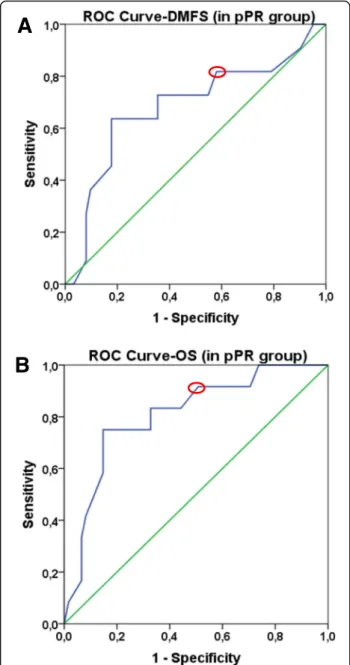
Ki-67 expression in the partial responder group
The prognostic potential of Ki-67 was also investi- gated in pPR subgroup that represents a heteroge- neous group with a response rate to NAC between 10 and 90%. Attempting to find the most relevant threshold for Ki-67, we could conclude that, the best cut-off value in pPR group based on DMFS was 20%
(n= 73, AUC 0.683, sensitivity: 82.4%, specificity 41.5%, p= 0.055, Fig. 3a), and 30% based on OS (n= 73, AUC 0.808, sensitivity: 92.2%, specificity 52.6, p= 0.001, Fig. 3b). No significant association was found between Ki-67 and pPR subgroups (pPRi, pPRii, pPRiii; p= 0.653)
For prognosis prediction, neither Ki-67 at any cut-off value (Ki-67 20% p= 0.233, Ki-67 30% p= 0.336), nor subtype (p= 0.218) not even pPR subgroups (p= 0.669)
were able to distinguish patient cohorts with different DMFS. Regarding OS, pPR subgroups (p= 0.590) and Ki-67 at 20% threshold failed (p= 0.095), but Ki-67 at 30% cut-off point (p= 0.037) and subtype (0.015) were suitable to separate patients into good and unfavorable prognosis cohorts (Fig. 4).
Discussion
Neoadjuvant systemic therapy is being increasingly used in the treatment of early stage breast cancer.
Despite several classification systems developed for the assessment of pathologic response to NAC there is a current lack in uniformity regarding the defin- ition of pathologic complete response [23, 24]. Since pCR is considered as the primary endpoint for re- sponse to chemotherapy, a majority of studies focus attention on pCR cases, while detailed analyses of partial responder or non-responder cases are relatively rare [8]. One of the hot topics in neoadjuvant therapy of breast cancer patients involves the question of reli- able prognostic and predictive markers. A high num- ber of studies have been conducted on the proliferation marker Ki-67, but due to the many con- troversies and debated issues this marker has not been fully integrated into clinical decision making.
Some of the questions about the performance of Ki- 67 as well as NAC in daily clinical practice concern the issue of cut points for Ki-67 and its use as prog- nostic or predictive marker. Different cut points are described, with values varying between 5 and 34%
[25], between 3 and 94% for pCR, and between 6 and 46% for DMFS [10, 26]. The 2013 St. Gallen consensus recommended a Ki-67 cut-off value of 14% for the separ- ation of luminal-A and B tumors, but in the footnote of the respective table there was a note indicating 20% as cut-off for“high”Ki67 LI. [19].
Our finding is in agreement with the results of Denkert et al. according to which Ki-67 is a mixed prognostic and predictive marker with its effect differing in opposite di- rections as regards prognosis and prediction [27].
Our study revealed that a Ki-67 cut-off value of about 20% distinguished pCR from pNR cases, whereas pa- tients exhibiting Ki-67 expression lower than 30% dem- onstrated a higher chance of better overall survival.
Increased Ki-67 LI was linked to worse OS, meaning that at least in some subgroups higher Ki-67 expression was related to increased response to NAC and was also
Fig. 1ROC curves to define optimal Ki-67 cut-off values for pathological response (a), DMFS (b), OS (c).Green linerepresents the diagonal reference line.Blue linecorresponds to ROC curve.Red circlesshow the optimal cut- off values based on the ROC curves
associated with worse prognosis. These data may suggest that if a tumor belongs to the group showing no re- sponse to NAC, increased Ki-67 is a marker of poor prognosis.
Denkert et al. also suggest that based on Ki-67 expres- sion there are three different groups of tumors, such as a group with low Ki-67 with good outcome, a group showing high Ki-67 and good outcome and a third
* *
*
*significant *significant
*significant p=0.263 HR=2.273 CI=0.519-9.967
p=0.131 HR=2.960 CI=0.674-12.999
p=0.040 HR=3.423 CI=1.217-11.922
p=0.037 HR=1.544 CI=1.141-2.492
p=0.044 HR=2.096 CI=1.067-4.548
Fig. 2Kaplan Meier plots of Ki-67, subtype and pathological response. Ki-67 at 30% cut-off value furthermore subtype and pathological response were suitable to separate good and unfavorable patient cohorts regarding OS
Table3MultivariateCoxregressionanalysisoftheKi-67andtheclinicopathologicalfactorsregardingdistantmetastasis-freesurvivalandoverallsurvival PrognosticfactorMultivariateCoxregressionregarding distantmetastasis-freesurvival(n=120)MultivariateCoxregressionanalysis regardingoverallsurvival(n=120)MultivariateCoxregressionwithoutLuminal-A casesregardingdistantmetastasis-freesur- vival(n=105)
MultivariateCoxregressionwithout Luminal-Acasesregardingoverallsurvival (n=105) HR95%CIp-valueHR95%CIp-valueHR95%CIp-valueHR95%CIp-value Age0.9640.653–1.4870.9191.5320.950–2.4700.0800.9330.565–1.5400.7871.4770.928–2.3500.100 pT2.3971.094–5.2520.0291.9310.914–4.0820.0851.8120.914–3.5920.0861.9030.895–4.0460.079 cT0.7710.356–1.6710.5101.1730.505–2.7260.7110.8310.341–2.0240.6851.1760.507–2.7240.706 IHCSubtype1.6760.436–6.4390.4522.2301.231–4.0430.0083.2630.369–22.8230.2872.1351.202–3.7850.009 Histologicalgrade1.2800.415–3.9450.6671.6410.559–4.8150.3671.3810.394–4.8360.6141.2780.453–3.6050.643 pN1.1460.681–1.9290.6071.4780.881–2.4790.1391.3340.765–2.3250.3091.4170.874–2.2960.157 cN1.6410.724–3.7170.2351.5020.694–3.2530.3021.9800.837–4.6820.1201.4850.685–3.2190.317 Hormonereceptorstatus0.9050.061–13.4560.9420.5610.201–1.5650.2702.8330.244–20.8080.6230.7840.289–2.1300.633 Pathologicalresponse0.8510.263–2.7490.7871.0560.376–2.9650.9180.8140.217–3.0490.7601.8990.300–2.6940.850 Ki-67LI_D15%1.6280.288–9.2090.5812.0220.373–10.9620.4142.1050.288–15.8430.3531.5680.287–8.5700.604 Ki-67LI_D20%2.5750.511–12.9620.2512.8470.528–15.3410.2233.8640.495–28.8090.1752.3380.434–12.6040.323 Ki-67LI_D30%1.2170.353–4.2010.7565.2861.189–23.4880.0291.3610.352–5.2550.6554.8501.089–18.3790.038 D15%=dichotomizedat15%threshold D20%=dichotomizedat20%threshold D30%=dichotomizedat30%threshold
group with high Ki-67 linked to poor outcome [27].
There are relatively few studies addressing the question of the role of Ki-67 in non-responder or pPR groups, even if the majority of cases treated with NAC show only partial response to chemotherapy.
In our study the majority of cases (60.83%) belonged to the pPR group. Based on Ki-67 expression, this group represented a mixture of tumors showing Ki-67 expres- sion ranging from 1 to 100%. We analyzed whether the group of patients showing a near complete pathologic
response (pPRi) showed higher Ki-67 expression com- pared to pPRii and pPRiii. According to our findings, there were no significant differences between these groups regarding Ki-67 expression. Based on the pa- tients’ follow-up data and using ROC analysis, the most relevant prognostic cut-off value for the Ki-67 in the pPR group was 20% based on DMFS and 30%
based on OS.
Balmativola et al. analyzed markers of non-response to NAC. Using ROC analysis, they identified a cut-off value of 18% for Ki-67 that performed well in differentiating the pNR and pCR + pPR categories [8].
In our study, Ki-67 expression was found to be higher than 20% in all patients achieving pCR and from the twenty-three pCR cases we detected distant metastases in one case only. In our study a Ki-67 LI of 20% was found capable of significantly distinguish- ing between the pCR and pNR groups. Despite this finding, however, it cannot be concluded that this is the only or best cut-off value for Ki-67, since the question then arises as to why a significant number of tumors with a Ki-67 value higher than 20% did not reach pCR.
Based on our results, both Ki-67 and subtype showed significant association with pathological response. How- ever, when Luminal-A cases were excluded, only subtype and pathological response were significantly linked, so we could conclude that subtype has a significant impact on the association between Ki-67 LI and pathological re- sponse. In contrast to this, both Ki-67 and subtype were independently associated with OS, while pathological re- sponse did not show significant relation with OS. Fur- thermore, both Ki-67 and subtype were suitable to separate pPR patients into good and unfavorable prog- nosis cohorts.
Conclusion
Neoadjuvant chemotherapy is more efficient in tu- mors presenting at least 20% Ki-67 expression. A cut- off value of 20% distinguished pCR from pNR cases.
Increased Ki-67 LI was linked to worse OS, meaning that at least in some subgroups higher Ki-67 expres- sion is related to increased response to NAC and is also associated with worse prognosis. Additionally, our data also suggest that if a tumor is non- responder to NAC, increased Ki-67 is a poor prognostic marker. Moreover, Ki-67 represented inde- pendent prognostic potential to OS, thus we can con- clude that Ki-67 has potential utility in the clinical management of breast cancer. However, we can also state that Ki-67 in itself is not suitable to decide whether a breast cancer patient should be treated with NAC or not.
A
B
Fig. 3ROC curves to define optimal Ki-67 cut-off values for DMFS, OS in pPR group.Green linerepresents the diagonal reference line.
Blue linecorresponds to ROC curve.Red circlesshow the optimal cut-off values based on the ROC curves
Limitations
The weakness of our retrospective study is the rela- tively low number of cases, for which reason i.) we could not define the optimal Ki-67 cut-off point for each subtypes. ii.) We could not investigate whether Ki-67 is suitable to predict pathological response in each subtype. Similarly, we could not address the question of the prognostic potential of Ki-67 for each subtype in breast cancer patients who underwent neo- adjuvant chemotherapy. iii.) We could not perform treatment-stratified analyses.
Additional files
Additional file 1:Contingency tables of Ki-67 LI, subtype and pathological response. (DOC 90 kb)
Additional file 2:Contingency tables of Ki-67 LI, subtype and pathological response without Luminal-A cases. (DOC 87 kb)
Additional file 3:Kaplan Meier plots of Ki-67, subtype and pathological response without Luminal-A subtype. When Luminal-A cases were excluded, neither Ki-67 at any cut-off points nor subtype not even pathological response were suitable to perform statistically significant splitting of our cohort into 2 patients’group with different OS. (DOC 305 kb)
Abbreviations
ASCO:American society of clinical oncology; cN: Clinical lymph nodal involvement; cT: Clinical tumor size; DMFS: Distant metastasis-free survival;
ER: Estrogen receptor; FISH: Fluorescent in situ hybridization; HER2: Human epidermal growth factor receptor 2; HR: Hormone receptor;
IHC: Immunohistochemistry; NAC: Neoadjuvant chemotherapy; OS: Overall survival; pCR: Pathologic complete remission; PgR: Progesterone receptor;
pNR: Non responders to neoadjuvant therapy; pPR: Pathologic partial remission; pT: Pathologic tumor size; ROC: Receiver operating characteristic;
TNBC: Triple negative breast cancer
Acknowledgments
The authors thank Rigóné Kálé Elvira for the careful reading and correcting the English of the manuscript. The authors thank Dr. Balazs Györffy for his accurate comments on data interpretation. AMS was supported by the János Bolyai Research Scholarship of the Hungarian Academy of Sciences.
p=0.037 HR=6.678 CI=1.358-18.338 p=0.095
HR=4.884 CI=0.422-28.357
*
p=0.015 HR=2.150 CI=1.216-3.803
*
*significant
*significant
p=0.590 HR=1.640 CI=0.681-3.949
Fig. 4Kaplan Meier plots of Ki-67, subtype and pathological response in pPR group. Ki-67 at 20% threshold and pPR subgroups failed, while Ki-67 at 30% cut-off point and subtype were suitable to separate good and unfavorable patient cohorts regarding OS
Funding
This work was supported by MKOT 2014–2016, SE-OTKA 2014–2015, SE-OTKA 2016–2017, KTIA_NAP_13-2014-0021 and OTKA grant no. K116151.
Availability of data and materials
The raw data are available upon request on the following e-mail address:
tokesa1972@yahoo.co.uk.
Authors’contributions
AMT, BA, VZ, JK, and AMSz conceived the study. BAM, IAM, GSz, ML and TT carefully selected the patients’clinical/oncological and pathological data and provided the patients follow up data. LV, BA and SVK performed data analysis. All authors were involved in data interpretation, writing the paper and had final approval of the submitted and published manuscript.
Competing interests
The authors declare that they have no competing interests.
Consent for publication Not applicable.
Ethics approval and consent to participate
The study was ethically approved by the Semmelweis University Institutional Review Board (SE-IKEB 120/2013). Additional patient consent for this retrospective study was not required.
Author details
12nd Department of Pathology, Semmelweis University, 1091 Budapest, Üllői út 93, Hungary.2MTA-SE-NAP Brain Metastasis Research Group, Semmelweis University, Budapest, Hungary.3MTA-TTK Lendület Cancer Biomarker Research Group, 1091 Budapest, Üllői út 93, Hungary.4Department of Clinical Oncology, Semmelweis University, 1083 Budapest, Tömőuta 25-29, Hungary.
51st Department of Surgery, Semmelweis University, 1082 Budapest Üllői út 78, Hungary.6University of Oradea, Romania, Faculty of Electrical Engineering and Information Technology, Str. Universitatii nr. 1, Oradea, Romania.
7MTA-SE Tumor Progression Research Group, Semmelweis University, 2nd Department of Pathology, 1091 Budapest, Üllői út 93, Hungary.
Received: 17 October 2016 Accepted: 2 February 2017
References
1. DeSantis C, Ma J, Bryan L, Jemal A. Breast cancer statistics, 2013. CA Cancer J Clin. 2014;64:52–62. doi:10.3322/caac.21203.
2. Luporsi E, Andre F, Spyratos F, Martin PM, Jacquemier J, Penault-Llorca F, Tubiana-Mathieu N, Sigal-Zafrani B, Arnould L, Gompel A, et al. Ki-67: level of evidence and methodological considerations for its role in the clinical management of breast cancer: analytical and critical review. Breast Cancer Res Treat. 2012;132:895–915. doi:10.1007/s10549-011-1837-z.
3. Yamamoto S, Ibusuki M, Yamamoto Y, Fu P, Fujiwara S, Murakami K, Iwase H. Clinical relevance of Ki67 gene expression analysis using formalin-fixed paraffin-embedded breast cancer specimens. Breast Cancer. 2013;20:262–70.
doi:10.1007/s12282-012-0332-7.
4. Nishimura R, Osako T, Nishiyama Y, Tashima R, Nakano M, Fujisue M, Toyozumi Y, Arima N. Prognostic significance of Ki-67 index value at the primary breast tumor in recurrent breast cancer. Mol Clin Oncol. 2014;2:
1062–8. doi:10.3892/mco.2014.400.
5. Criscitiello C, Disalvatore D, De Laurentiis M, Gelao L, Fumagalli L, Locatelli M, Bagnardi V, Rotmensz N, Esposito A, Minchella I, et al. High Ki-67 score is indicative of a greater benefit from adjuvant chemotherapy when added to endocrine therapy in luminal-B HER2 negative and node-positive breast cancer. Breast. 2014;23:69–75. doi:10.1016/j.breast.2013.11.007.
6. Andre F, Arnedos M, Goubar A, Ghouadni A, Delaloge S. Ki67–no evidence for its use in node-positive breast cancer. Nat Rev Clin Oncol. 2015;12:296–301.
doi:10.1038/nrclinonc.2015.46.
7. Polley MY, Leung SC, McShane LM, Gao D, Hugh JC, Mastropasqua MG, Viale G, Zabaglo LA, Penault-Llorca F, Bartlett JM, et al. An international Ki67 reproducibility study. J Natl Cancer Inst. 2013;105:1897–906. doi:10.
1093/jnci/djt306.
8. Balmativola D, Marchio C, Maule M, Chiusa L, Annaratone L, Maletta F, Montemurro F, Kulka J, Figueiredo P, Varga Z, et al. Pathological non-
response to chemotherapy in a neoadjuvant setting of breast cancer: an inter-institutional study. Breast Cancer Res Treat. 2014;148:511–23. doi:10.
1007/s10549-014-3192-3.
9. Voros A, Csorgo E, Kovari B, Lazar P, Kelemen G, Rusz O, Nyari T, Cserni G.
Different methods of pretreatment Ki-67 labeling index evaluation in core biopsies of breast cancer patients treated with neoadjuvant chemotherapy and their relation to response to therapy. Pathol Oncol Res. 2015;21:147–55.
doi:10.1007/s12253-014-9800-z.
10. Denkert C, Loibl S, Muller BM, Eidtmann H, Schmitt WD, Eiermann W, Gerber B, Tesch H, Hilfrich J, Huober J, et al. Ki67 levels as predictive and prognostic parameters in pretherapeutic breast cancer core biopsies: a translational investigation in the neoadjuvant GeparTrio trial. Ann Oncol. 2013;24:2786–93.
doi:10.1093/annonc/mdt350.
11. Klintman M, Dowsett M. Early Surrogate Markers of Treatment Activity:
Where Are We Now? J Natl Cancer Inst Monogr. 2015;2015:24–8. doi:10.
1093/jncimonographs/lgv002.
12. Wang-Lopez Q, Chalabi N, Abrial C, Radosevic-Robin N, Durando X, Mouret- Reynier MA, Benmammar KE, Kullab S, Bahadoor M, Chollet P, et al. Can pathologic complete response (pCR) be used as a surrogate marker of survival after neoadjuvant therapy for breast cancer? Crit Rev Oncol Hematol. 2015;95:88–104. doi:10.1016/j.critrevonc.2015.02.011.
13. Dowsett M, Nielsen TO, A’Hern R, Bartlett J, Coombes RC, Cuzick J, Ellis M, Henry NL, Hugh JC, Lively T, et al. Assessment of Ki67 in breast cancer:
recommendations from the International Ki67 in Breast Cancer working group. J Natl Cancer Inst. 2011;103:1656–64. doi:10.1093/jnci/djr393.
14. Magbanua MJ, Wolf DM, Yau C, Davis SE, Crothers J, Au A, Haqq CM, Livasy C, Rugo HS, Esserman L, et al. Serial expression analysis of breast tumors during neoadjuvant chemotherapy reveals changes in cell cycle and immune pathways associated with recurrence and response. Breast Cancer Res. 2015;17:73. doi:10.1186/s13058-015-0582-3.
15. Klauschen F, Wienert S, Schmitt WD, Loibl S, Gerber B, Blohmer JU, Huober J, Rudiger T, Erbstosser E, Mehta K, et al. Standardized Ki67 Diagnostics Using Automated Scoring-Clinical Validation in the GeparTrio Breast Cancer Study. Clin Cancer Res. 2015;21:3651–7. doi:10.1158/1078-0432.ccr-14-1283.
16. Symmans WF, Peintinger F, Hatzis C, Rajan R, Kuerer H, Valero V, Assad L, Poniecka A, Hennessy B, Green M, et al. Measurement of residual breast cancer burden to predict survival after neoadjuvant chemotherapy. J Clin Oncol. 2007;25:4414–22. doi:10.1200/jco.2007.10.6823.
17. Hatzis C, Symmans WF, Zhang Y, Gould RE, Moulder SL, Hunt KK, Abu-Khalaf M, Hofstatter EW, Lannin D, Chagpar AB, et al. Relationship between Complete Pathologic Response to Neoadjuvant Chemotherapy and Survival in Triple-Negative Breast Cancer. Clin Cancer Res. 2016;22:26–33. doi:10.
1158/1078-0432.ccr-14-3304.
18. Pinder SE, Provenzano E, Earl H, Ellis IO. Laboratory handling and histology reporting of breast specimens from patients who have received neoadjuvant chemotherapy. Histopathology. 2007;50:409–17. doi:10.1111/j.
1365-2559.2006.02419.x.
19. Goldhirsch A, Winer EP, Coates AS, Gelber RD, Piccart-Gebhart M, Thurlimann B, Senn HJ. Personalizing the treatment of women with early breast cancer: highlights of the St Gallen International Expert Consensus on the Primary Therapy of Early Breast Cancer 2013. Ann Oncol. 2013;24:2206– 23. doi:10.1093/annonc/mdt303.
20. Prat A, Cheang MC, Martin M, Parker JS, Carrasco E, Caballero R, Tyldesley S, Gelmon K, Bernard PS, Nielsen TO, et al. Prognostic significance of progesterone receptor-positive tumor cells within immunohistochemically defined luminal A breast cancer. J Clin Oncol. 2013;31:203–9. doi:10.1200/
jco.2012.43.4134.
21. Wolff AC, Hammond ME, Schwartz JN, Hagerty KL, Allred DC, Cote RJ, Dowsett M, Fitzgibbons PL, Hanna WM, Langer A, et al. American Society of Clinical Oncology/College of American Pathologists guideline
recommendations for human epidermal growth factor receptor 2 testing in breast cancer. J Clin Oncol. 2007;25:118–45. doi:10.1200/jco.2006.09.2775.
22. Wolff AC, Hammond ME, Hicks DG, Dowsett M, McShane LM, Allison KH, Allred DC, Bartlett JM, Bilous M, Fitzgibbons P, et al. Recommendations for human epidermal growth factor receptor 2 testing in breast cancer:
American Society of Clinical Oncology/College of American Pathologists clinical practice guideline update. J Clin Oncol. 2013;31:3997–4013. doi:10.
1200/jco.2013.50.9984.
23. Bossuyt V, Provenzano E, Symmans WF, Boughey JC, Coles C, Curigliano G, Dixon JM, Esserman LJ, Fastner G, Kuehn T, et al. Recommendations for standardized pathological characterization of residual disease for
neoadjuvant clinical trials of breast cancer by the BIG-NABCG collaboration.
Ann Oncol. 2015;26:1280–91. doi:10.1093/annonc/mdv161.
24. Provenzano E, Bossuyt V, Viale G, Cameron D, Badve S, Denkert C, MacGrogan G, Penault-Llorca F, Boughey J, Curigliano G, et al.
Standardization of pathologic evaluation and reporting of postneoadjuvant specimens in clinical trials of breast cancer: recommendations from an international working group. Mod Pathol. 2015;28:1185–201. doi:10.1038/
modpathol.2015.74.
25. Goldhirsch A, Wood WC, Coates AS, Gelber RD, Thurlimann B, Senn HJ.
Strategies for subtypes–dealing with the diversity of breast cancer:
highlights of the St. Gallen International Expert Consensus on the Primary Therapy of Early Breast Cancer 2011. Ann Oncol. 2011;22:1736–47. doi:10.
1093/annonc/mdr304.
26. Cserni G, Voros A, Liepniece-Karele I, Bianchi S, Vezzosi V, Grabau D, Sapino A, Castellano I, Regitnig P, Foschini MP, et al. Distribution pattern of the Ki67 labelling index in breast cancer and its implications for choosing cut-off values. Breast. 2014;23:259–63. doi:10.1016/j.breast.2014.02.003.
27. Denkert C, Budczies J, von Minckwitz G, Wienert S, Loibl S, Klauschen F.
Strategies for developing Ki67 as a useful biomarker in breast cancer. Breast.
2015. doi:10.1016/j.breast.2015.07.017
• We accept pre-submission inquiries
• Our selector tool helps you to find the most relevant journal
• We provide round the clock customer support
• Convenient online submission
• Thorough peer review
• Inclusion in PubMed and all major indexing services
• Maximum visibility for your research Submit your manuscript at
www.biomedcentral.com/submit
Submit your next manuscript to BioMed Central and we will help you at every step: