Summary. S100P - low molecular weight acidic protein has been shown to be involved in processes of proliferation, survival, angiogenesis, multidrug resistance and metastasis in various human malignancies. In breast cancer, S100P expression is associated with immortalization of neoplastic cells and aggressive tumour behaviour, indicating that this protein may have adverse prognostic value. We analyzed nuclear and cytoplasmic expression of S100P in 85 stage II breast cancer patients with a median follow up of 17 years. Immunohistochemical reactions were performed on paraffin sections of primary tumours, using monoclonal antibodies against S100P. We also studied prognostic value of S100P mRNA expression using the KM plotter which assessed the effect of 22,277 genes on survival in 2422 breast cancer patients. Moreover, the relationship was examined between expression of S100P in cells of four breast cancer cell lines and their sensitivity to the 11 most frequently applied cytotoxic drugs. Univariate and multivariate analyses showed that higher expression of nuclear S100P (S100Pn) was typical for cases of a shorter overall survival and disease-free time. KM plotter analysis showed that elevated S100P expression was specific for cases of a relapse-free survival and distant metastases-free survival. No relationship could be documented between expression of S100P and sensitivity of breast cancer cells to cytostatic drugs. We demonstrated that a high S100Pn expression level was associated with poor
survival in early stage breast cancer patients. Since preliminary data indicated that expression of S100P was up-regulated by activation of glucocorticoid receptor and several agents manifested potential to activate or inhibit S100P promoter activity, this protein might become a therapy target and warrants further studies with respect to its prognostic, predictive and potentially therapeutic value.
Key words:Breast cancer, Prognostic biomarker, S100P
Introduction
Breast cancer is the most common malignancy among women in the developed countries, with a general upward trend in incidence (Ferlay et al., 2004). The majority of patients are diagnosed with early stage breast cancer and are treated with radical intent. Adjuvant treatment is commonly used to reduce the risk of relapse and death. Results of prospective randomized trials confirmed the absolute reduction of breast cancer mortality in chemotherapy-treated patients by 10% and 3% and the risk of relapse by 12% and 4% in premenopausal and postmenopausal women, respectively. Endocrine treatment in patients with estrogen receptor (ER)-positive tumours reduced the risk of death and relapse by 31% and 39%, respectively (Early Breast Cancer Trialists’ Collaborative Group, 2005). One year of adjuvant trastuzumab in HER-2 positive subgroup added a supplementary benefit (Perez et al., 2007). However, taking into account the real risk of recurrence, in the current practice the majority of patients with early breast cancer receive adjuvant
Elevated nuclear S100P expression is associated with poor survival in early breast cancer patients
Adam Maciejczyk1, Aleksandra Łacko1,2, Marcin Ekiert1,2, Ewa Jagoda3, Teresa Wysocka3, Rafal Matkowski1,2, Agnieszka Hałoń6, Balázs Györffy4, Hermann Lage5and Pawel Surowiak3
1Lower Silesian Centre of Oncology, Wrocław, Poland, 2Department of Oncology, University School of Medicine, Wroclaw, Poland,
3Department of Histology and Embryology, University School of Medicine, Wroclaw, Poland, 4Research Laboratory for Pediatrics and Nephrology, Hungarian Academy of Sciences - Semmelweis University 1st. Dept. of Pediatrics, Semmelweis University, Budapest, Hungary, 5Institute of Pathology, Charité Universitätsmedizin Berlin, Germany and 6Department of Pathomorphology and Oncological Cytology, University School of Medicine, Wrocław, Poland
Offprint requests to: Prof. Paweł Surowiak, M.D., Department of Histology and Embryology, University School of Medicine, ul.
Chałubińskiego 6a, 50-356 Wrocław, Poland. e-mail:
pawel.surowiak@interia.pl
Cellular and Molecular Biology
treatment needlessly, either because they are cured with surgery alone or due to the lack of reliable predictive factors so they receive ineffective treatment and suffer from a relapse in spite of it. Therefore, an appropriate patient selection to adjuvant treatment becomes crucial.
According to current recommendations, indication for adjuvant therapy and choice of particular treatment modalities are based on the likelihood of response to specific therapies and magnitude of the risk of recurrence (Goldhirsch et al., 2009).
Prognosis of newly diagnosed breast cancer predominantly relies on stage, a few clinical factors, pathologic features of the tumour and status of hormonal and HER-2 receptors. However, breast cancer is a heterogenous disease, and even tumours sharing identical histological characteristics have a variable prognosis and may manifest also a variable response to therapy. Molecular features of malignant tumours are currently being comprehensively investigated. Both prognostic and predictive biomarkers are essential to individualize clinical management of the disease.
S100P is a low molecular weight acidic protein, which belongs to the EF-hand superfamily of calcium binding proteins. It was originally isolated from human placenta, and its presence was detected in various normal and malignant cells. S100P can be detected in the cytoplasm and/or cell nucleus and it is up-regulated in various human malignant tumours, such as pancreatic, lung, prostate, colorectal, ovarian and breast cancer (Guerreiro Da Silva et al., 2000; Mousses et al., 2002;
Logsdon et al., 2003; Diederichs et al., 2004; Wang et al., 2006; Fuentes et al., 2007). Several experimental studies showed that S100P, involved in processes of proliferation, survival, angiogenesis and metastasis, may contribute to development of a malignant phenotype (Arumugam et al., 2005; Wang et al., 2006; Surowiak et al., 2007; Parkkila et al., 2008). The S100P mechanism of action is not fully known. It was shown that S100P exerted both intracellular and extracellular functions (Arumugam et al., 2005). Different signalling pathways are involved in S100P transcriptional regulation, activated by hormones (glucocorticoids) and growth factors such as EGF (epidermal growth factor) and BMP (bone morphogenic factor). S100P is secreted to extracellular space where it binds to RAGE (receptor for advanced glycation end-product) and it seems to function as a signalling molecule (Donato, 2003;
Arumugam et al., 2004). Although it may be a biomarker of chemotherapy and endocrine therapy resistance, results of studies are contradictory. In our previous studies, performed on 30 cell lines, we compared gene expression profiles obtained using the Affymetrix U133A arrays with profiles of resistance to 11 cytostatic drugs applied in clinically relevant concentrations. In the study we demonstrated that augmented expression of S100P protein characterized cells resistant to cyclophosphamide, etoposide, methothrexate and mitoxantron (Györffy et al.,2006). In another study we have shown that elevated expression of S100P is an
unfavourable prognostic factor in ovarian cancer patients (Surowiak et al., 2007).
In breast tumours S100P expression is seen specifically on cancer cells, therefore it appears to be an indicator of malignancy (Guerreiro Da Silva et al., 2000). It has been shown that S100P overexpression is associated with poor prognosis (Wang et al., 2006).
In the present study, we tested the prognostic value of S100P for formalin-fixed paraffin-embedded (FFPE) tissue obtained from 85 patients with stage II primary ductal breast carcinoma with a median follow up of 17 years.
Material and methods Patients
Consecutive breast cancer patients (n=85) diagnosed with clinical stage II ductal breast cancer (stage pT1-3, pN0-1) treated in Lower Silesia Oncology Centre from January 1992 to June 1993 were identified. Patient characteristics are listed in Table 1. All patients treated with adjuvant chemotherapy received CMF regimen (cyclophosphamide, 5-fluorouracyl, methotrexate) for 6 cycles. Women with hormonally responsive disease received tamoxifen for 5 years. Median follow up time was 17 years for the whole group. Pathologic data were obtained from standard pathology reports from a local laboratory. Hormonal receptor status was examined by immunohistochemistry (IHC) and assessed using Allred score. HER-2 status was also assessed by IHC and scored by classical 0-3 score.
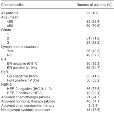
Table 1.Patient and tumour characteristics.
Characteristics Number of patients (%)
All patients 85 (100)
Age (mean)
<50 25 (29.4)
≥50 60 (70.6)
Grade
12 61 (71.8)
3 24 (28.2)
Lymph node metastases
Yes 36 (42.3)
No 49 (57.7)
ER ER negative (0-9 %) 30 (35.3)
ER positive (>10%) 55 (64.7)
PgRPgR negative (0-9%) 35 (41.2)
PgR positive (>10%) 50 (58.2)
HER-2
HER-2 negative (IHC 0, 1, 2) 66 (77.6)
HER-2 positive (IHC 3) 19 (22.4)
Adjuvant chemotherapy (alone) 21 (24.7) Adjuvant hormonal therapy (alone) 46 (54.1) Adjuvant chemoendocrine therapy 3 (3.6) No adjuvant systemic treatment 15 (17.6)
Immunohistochemistry
Formalin-fixed, paraffin embedded tissue was freshly cut (4 µm). The sections were mounted on Superfrost slides (Menzel Gläser, Germany), dewaxed wtih xylene, and gradually hydrated. Activity of endogenous peroxidase was blocked by 5 min exposure to 3% H2O2. All the studied sections were boiled for 15 min at 250W in the Antigen Retrieval Solution (DakoCytomation, Denmark). Then, immunohisto- chemical reactions were performed using the mouse monoclonal antibodies detecting S100P (BD Biosciences Pharmingen, Franklin Lakes, NJ, USA) at dilution 1:250 in Antibody Diluent, Background Reducing (DakoCytomation, Denmark), ER (monoclonal, clone 1D; dillution 1:100; Dako), PR (monoclonal clone 636;
dillution 1:100 x; Dako), HER-2 (HercepTest, based on the Dako A0485 polyclonal antibody, and the CB11 monoclonal antibody dillution 1:100; Dako). Tested sections were incubated with antibodies for 1 h at room temperature. Subsequent incubations involved biotinylated antibodies (15 min, room temperature) and streptavidin-biotinylated peroxidase complex (15 min, room temperature) (LSAB+, HRP, DakoCytomation, Denmark). DAB (DakoCytomation, Denmark) was used as a chromogen (7 min, at room temperature). All the sections were counterstained with Meyer’s hematoxylin (DakoCytomation, Denmark). In every case, control reactions were included, in which the specific antibody was substituted by the Primary Mouse Negative Control (DakoCytomation, Denmark) (Surowiak et al., 2007).
Control reactions
Control reactions for S100P were also performed on paraffin sections from six healthy human placentas and on paraffin sections from ten healthy human breast tissues (from the archive of the Department of Histology and Embryology, University School of Medicine, Wrocław, Poland) (Surowiak et al., 2007).
Cell lines and cell culture
In our study, we used 4 human breast cancer cell
lines: MCF-7, CAMA-1, SK-BR-3 and R-103. The cells were cultured in Leibovitz L-15 medium (Bio Whittaker, Walkersville, MD, USA) supplemented by 10% foetal calf serum (Gibco BRL, Grand Island NY, USA), 1 mM L-glutamine, 80 IE/l insulin, 2.5 mg/l transferrin, 1 g/l glucose, 1.1 g/l NaHCO3, 1% minimal essential vitamins and 20,000 kIE/l trasylol in a humified atmosphere in 5% CO2 at 37°C. Prior to resistance testing, Mycoplasma tests were performed using the Venor Mp kit, according to the manufacturer’s instructions (Minerva Biolabs GmbH, Berlin, Germany).
Resistance tests
Drugs were used in their commercially available form (except cyclophosphamide, which was used in its activated form). Each drug was applied to the cells in 3 concentrations (C1, C2, C3). C1=10-1x C2 and C3=10 x C2. Concentration C2 was deduced from levels assessed to be clinically achievable in tumour tissue, as discussed previously (Györffy et al., 2006) (Table 2). In each experiment, 500 cells/microtiter dish were seeded onto 96-well plates. After 2 days, precontrol cells were fixed and stained using sulforhodamine B (SRB). At the same time, triplicate cultures were prepared with all 11 studied drugs at C1, C2 and C3 concentrations. After 4 days, incubation was terminated by replacing the medium with 10% trichloroacetic acid, followed by incubation at 4°C for 1 hr. Subsequently, the plates were washed 5 times with water and stained by adding 100 µl 0.4% SRB (Sigma, St. Louis, MO, USA) in 1% acetic acid for 10 min at room temperature. Washing the plates 5 times with 1% acetic acid eliminated unbound dye. After air- drying and resolubilization of the protein-bound dye in 10 mM Tris-HCl (pH 8.0), absorbance was read at 562 nm in an Elisa-Reader (EL 340 Microplate Bio Kinetics Reader, BIO-TEK Instruments, Winooski, VT, USA).
The measurements were performed in triplicate in 3 independent experiments. For the calculation of the RI values, the averages of all 9 measurements were used.
The resistance index (RI) was estimated by the formula:
RI = (npost/npre) x [(n2-npre) / (npost-npre) x 100]
where npreis the medium absorbance value of precontrol at the C2 concentration, npostis the medium absorbance value of control and n2is the medium absorbance value of stained cells tested with the chosen concentration of the studied drug.
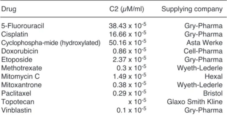
Table 2.Drugs used to establish resistance patterns and their C2 concentrations (the clinically available drug in the tumour).
Drug C2 (µM/ml) Supplying company
5-Fluorouracil 38.43 x 10⁻⁵ Gry-Pharma
Cisplatin 16.66 x 10⁻⁵ Gry-Pharma
Cyclophospha-mide (hydroxylated) 50.16 x 10⁻⁵ Asta Werke
Doxorubicin 0.86 x 10⁻⁵ Cell-Pharma
Etoposide 2.37 x 10⁻⁵ Gry-Pharma
Methotrexate 0.3 x 10⁻⁵ Wyeth-Lederle
Mitomycin C 1.49 x 10⁻⁵ Hexal
Mitoxantrone 0.38 x 10⁻⁵ Wyeth-Lederle
Paclitaxel 0.29 x 10⁻⁵ Bristol
Topotecan x 10-5 Glaxo Smith Kline
Vinblastin 0.1 x 10⁻⁵ Gry-Pharma
Table 3. Evaluation criteria of ABCC2 expression using IRS (ImmunoReactive Score) score.
Percentage of positive cells Points Intensity of reaction Points
No positive cells 0 No reaction 0
<10% positive cells 1 Weak reaction 1
10-50% positive cells 2 Moderate reaction 2 51-80% positive cells 3 Intense reaction 3
>80% positive cells 4
Immunocytochemistry
Immunostaining of S100P was performed using the studied panel of breast cancer cell lines. Cells were grown on microscopic slides and fixed in ice-cold methanol-acetone mixture (1:1) for 10 minutes.
Immunostaining reaction was performed in triplicate as described above.
Evaluation of reaction intensity
Intensity of immunohistochemical reactions was estimated independently by two pathologists. In doubtful cases a re-evaluation was performed using a double- headed microscope and staining was discussed until a consensus was achieved. In order to evaluate the S100P expression, a semi-quantitative scale of ImmunoReactive Score (IRS) was applied, which took into account intensity of the colour reaction (0: no reaction, 1: weak reaction, 2: moderate intensity, 3: intense reaction) as well as proportion of positive cells (0: no positive cells, 1: <10% positive cells, 2: 10-50% positive cells, 3: 51- 80% positive cells, 4: >80% positive cells). The final score represented the product of points given for individual characters and ranged between 0 and 12 (Table 3). Evaluation of estrogen and progesterone receptor expression was performed using standard methods. The staining intensity (0-3 scale) and proportion of positive cells (0-5 scale) were reported and the Allred score (AS) that combines the two was calculated. HER-2 status was evaluated using the FDA- approved scoring system of 0, 1+, 2+, and 3+ (0: no immunostaining, 1+: weak immunostaining, less than 30% of tumour cells; 2+: complete membranous staining, either uniform or weak in at least 10% of cells;
3+: uniform intense membranous staining in at least 30%
of cells).
Statistical analysis
All statistical analyses were conducted using Statistica 8.0 PL software (Statsoft, Poland). Disease- free survival (DFS) was defined as the time between the primary surgical treatment and date of relapse or death, whichever occurred first, and DFS was censored at last follow up for those who were alive without recurrence.
Overall survival (OS) was defined as the time between the primary surgical treatment and death from any cause, whichever occurred first, and OS was censored at last follow up for those who were alive.
Clinicopathologic variables considered in univariate analysis were: age at the time of primary surgery, which was a surrogate for the date of diagnosis (continuous variable), lymph node status (positive vs negative), hormonal receptor (HR) status (by IHC; positive vs negative), and HER-2 status (by IHC; positive [+3] v negative [0, 1 or 2]). Although according to current standards all tumours exhibiting any expression of HR
are considered endocrine responsive, for the purpose of this analysis we excluded tumours with low ER/PR level from HR- positive group and considered as a HR- positive only tumours with ≥10% stained malignant cells. Correlations between these factors and S100P were analysed using Chi2tests.
In order to estimate survival we used Kaplan Meier statistics, log rank tests, test F of Cox and Cox proportional hazard regression.
KM plotter online survival analysis
The KM plotter was capable of assessing the effect of 22,277 genes on survival in 2422 breast cancer patients (Gyorffy et al., 2010). A background database was established using gene expression data and survival information on 2422 patients downloaded from GEO (Affymetrix HGU133A and HGU133+2 microarrays).
The median relapse-free survival was 6.43 years. After quality control and normalization only probes present on both Affymetrix platforms were retained (n=22,277).
The background database was handled by a MySQL server, which integrates gene expression and clinical data simultaneously. In order to analyze the prognostic value of a particular gene, the samples were split into two groups according to the median (or upper / lower quartile) expression of the gene. The two groups could be compared in terms of relapse free survival, overall survival and distant metastasis free survival.
Results
Immunohistochemical staining of breast cancer for S100P
The immunohistochemical staining for S100P was predominantly nuclear (S100Pn). Of the 85 specimens evaluated, 23 (27.0%) showed no reaction. Sixty two specimens (73.0%) showed nuclear expression of S100P, 35 (41.2%) manifested borderline or weak expression (IRS: 1-4) and 27 of them (31.8%) showed strong expression (IRS: 6-12). For the purpose of analysis, patients with S100Pn negative tumours (IRS:0) and cancers with low expression (IRS:1-4) were combined.
Fifty eight tumours exhibited lack of expression or low nuclear expression of S100Pn, 27 (31.8%) manifested high expression (IRS: 6-12). Cytoplasmic expression of S100P (S100Pc) was seen in 59 (69.4%) of tumours, and the majority (61.2%) of samples were S100Pc negative or exhibited borderline or weak expression (n=52, 61.2%) (Fig. 1).
S100P expression and clinicopathologic data
The relationships between S100P expression and the clinicopathological data of studied patients was assessed using Chi2test. No correlation was seen between S100P expression and the assessed clinicopathologic
parameters (Table 4).
S100P expression versus overall survival (OS) and disease free survival (DFS) of the studied patients
In the Kaplan Meier analysis, OS and DFS were compared between groups with lower or negative S100p expression (IRS: 0 to 4 for S100Pn and 0 to 2 for S100Pc), and higher S100p expression (IRS: 6 to12 for S100Pn and 3 to12 for S100Pc). Patients with strong S100Pn expression had significantly shorter OS (P<0.00001) (Fig. 2) and DFS (P = 0.00002) (Fig. 3).
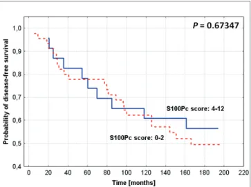
S100Pc expression exerted no impact on survival (Figs.
4, 5).
When we split the group into node-negative (N/-/) and node-positive (N/+/) cases, association between S100Pn and OS remained significant in both N/+/ and N/-/ subgroups (for N/+/ and N/-/ P=0.00002 and P=0.04105, respectively). However, high S100Pn was
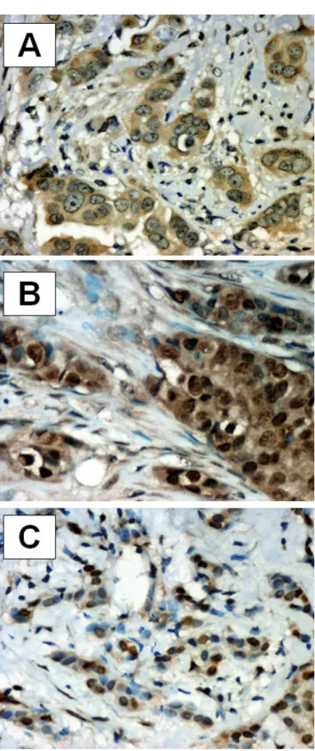
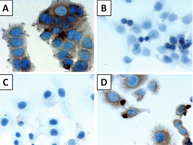
Fig. 1.Immunohistochemical localization of S100P expression in breast cancer: A.Cytoplasmic reaction; brown reaction product; S100Pn IRS=0, S100Pc IRS=8 (hematoxylin). B.cytoplasmic and nuclear reaction; brown reaction product; S100Pn IRS=9, S100Pc IRS=8 (hematoxylin). C. nuclear reaction; brown reaction product; S100Pn IRS=9, S100Pc IRS=2 (hematoxylin). A, B, x 400; C, x 200
Fig. 2. Kaplan-Meier curves for survival and expression of S100P in the studied group of breast cancer patients: nuclear S100P (S100Pn) expression and overall survival time (log-rank P<0.00001; Cox F test P=0.00159; regression of Cox proportional hazard P=0.00233).
Table 4. Correlation between S100P expression and various clinicopathologic parameters (Chi2test).
Characteristics S100 Pn P value S100Pc P value
Histologic grade 0.47621 0.40553
Lymph node metastases 0.22656 0.66007
ER (>10%) 0.27316 0.47341
PgR (>10%) 0.85228 0.59675
HER-2 0.27190 0.73906
Age (>50) 0.73025 0.63035
S100Pn: S100P nuclear staining; S100Pc: S100P cytoplasmic staining;
ER: estrogen receptor, PgR: progesterone receptor
associated with a significantly worse DFS only in N/+/
group (P=0.00012 ). In N/-/ patients this association was of borderline significance (P=0.05777) . A correlation with OS, DFS and S100Pc was found neither in N/+/, nor in N/-/ subgroup (for N/+/ and N/-/ : P>0.05).
Using univariate analysis, we also tested the
prognostic value of S100P in subgroups of patients manifesting various HER-2 expression and hormonal receptor status. The computations have shown, that elevated S100Pn expression is specific for cases of shorter OS and DFS in both in all studied subgroups (in HER/-/: P=0.00001 and P=0.00003 for OS and DFS,
Fig. 4.Kaplan-Meier curves for survival and expression of S100P in the studied group of breast cancer patients: cytoplasmic S100P (S100Pc) expression and overall survival time (log-rank P=0.81148; Cox F test P=0.42430).
Fig. 3.Kaplan-Meier curves for survival and expression of S100P in the studied group of breast cancer patients: nuclear S100P (S100Pn) expression and disease-free survival time (log-rank P=0.00002; Cox F test P=0.00002; regression of Cox proportional hazard P=0.00005).
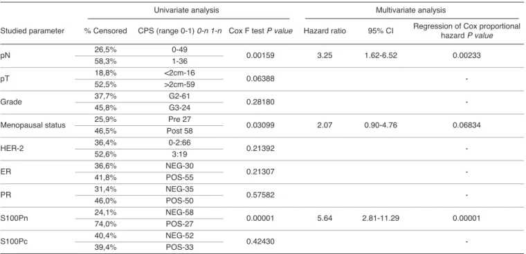
Table 5.Univariate and multivariate analysis of the relationships between various clinicopathologic parameters, nuclear S100P expression (S100Pn), cytoplasmic S100P expression (S100Pc) and the overall survival time of all the studied patients. The multivariate analysis was performed only for the factors significantly related to survival time in univariate analysis.
Univariate analysis Multivariate analysis
Studied parameter % Censored CPS (range 0-1) 0-n 1-n Cox F test P value Hazard ratio 95% CI Regression of Cox proportional hazard P value
pN 26,5%58,3% 0-491-36 0.00159 3.25 1.62-6.52 0.00233
pT 18,8% <2cm-16
0.06388 -
52,5% >2cm-59
Grade 37,7% G2-61 0.28180 -
45,8% G3-24
Menopausal status 25,9%46,5% Post 58Pre 27 0.03099 2.07 0.90-4.76 0.06834
HER-2 36,4% 0-2:66
0.21392 -
52,6% 3:19
ER 36,6% NEG-30 0.21307 -
41,8% POS-55
PR 31,4% NEG-35
0.57582 -
46,0% POS-50
S100Pn 24,1% NEG-58
0.00001 5.64 2.81-11.29 0.00001
74,0% POS-27
S100Pc 40,4% NEG-52 0.42430 -
39,4% POS-33
respectively, in ER/PR/+/: P=0.00002 and P=0.00027 for OS and DFS, respectively).
At the subsequent stage of analysis, using the Cox F test a relationship was examined between clinicopathological data of the patients and expressions of S100P on one hand and their OS and DFS on the other. The factors significantly correlated with patients’
survival including pN, menopausal status and expression of S100Pn (Table 5). Calculations conducted by Cox proportional hazard regression showed that only pN and S100Pn manifested a significant relationship with patients’ survival (Table 5). The analysis showed pN, menopausal status and S100Pn have relationship with the duration of DFS (Table 6).
At the final stage of Cox proportional hazard regression grouping variables were applied. The two selected parameters which in the studied group exerted a significant influence on patients’ survival included pN and menopausal status. The analysis confirmed that elevated expression of S100Pn was typical for patients with abbreviated OS and DFS (Table 7). On the case of S100Pc no significant relationship with patients’
survival could be demonstrated (Table 7).
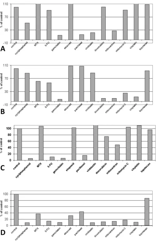
S100P expression and sensitivity of the breast cancer cell lines to cytostatic drugs
The studied cell lines demonstrated the following
intensity of reaction for S100P: MCF-7: S100Pn, IRS=0;
S100Pc, IRS=9; CAMA-1: S100Pn, IRS=0; S100Pc, IRS=1; SK-BR-3: S100Pn, IRS=0; S100Pc, IRS=0; R- 103: S100Pn, IRS=3; S100Pc, IRS=9. The results of
Fig. 5. Kaplan-Meier curves for survival and expression of S100P in the studied group of breast cancer patients: cytoplasmic S100P (S100Pc) expression and disease-free survival time (log-rank P=0.67347; Cox F test P=0.0.35074).
Table 6.Univariate and multivariate analysis of the relationships between various clinicopathologic parameters, nuclear S100P expression (S100Pn), cytoplasmic S100P expression (S100Pc) and the disease-free time of all the studied patients. The multivariate analysis was performed only for the factors significantly related to disease-free time in univariate analysis.
Univariate analysis Multivariate analysis
Studied parame-ter % Censored CPS (range 0-1) 0-n 1-n Test F Coxa P value Hazard ratio 95% CI Regression of Cox proportional hazard P value
pN 34,7% 0-49
0.00587 2.65 1.40-5.01 0.00932
61,1% 1-36
pT 25,0% <2cm-16
0.09534 -
59,3% >2cm-59
Grade 42,6% G2-61
0.21185 -
54,2% G3-24
Menopausal status 29,6% Pre 27
0.02144 2.13 1.07-4.63 0.04813
53,4% Post 58
HER-2 42,4% 0-2:66
0.17759 -
57,9% 3:19
ER 43,3% NEG-30
0.25118 -
47,2% POS-55
PR 45,7% NEG-35
0.36428 -
46,0% POS-50
S100Pn 36,2% NEG-58
0.00002 4.55 2.39-8.67 0.00005
66,7% POS-27
S100Pc 28,8% NEG-52
0.35074 -
72,7% POS-33
conducted cytotoxicity tests and the results of immunocytochemical reactions are depicted in Figs. 6, 7.
The investigations demonstrated no relationship between expression of S100P in breast cancer cells and sensitivity of the cells to cytostatic drugs.
KM Plotter online survival analysis
Using KM Plotter online survival analysis of the prognostic value of S100P mRNA (Affy id.: 204351_at) an elevated mRNA of S100P expression was shown to have a negative impact on patient’s relapse-free and distant metastases-free survival in the entire group, and of relapse-free time in the subgroups of estrogen receptor positive patients and lymph node negative patients (Table 8).
Discussion
Treatment tailored to specific characteristics of a
patient and a tumour may improve patients outcome. It can also alter management strategies. An understanding of cancer biology is crucial and should be translated into development of tests to identify prognostic and predictive molecular markers. It is hoped that molecular diagnostics will be more precise and reliable that currently known and commonly used clinicopathologic factors.
In the treatment of breast cancer prognostic factors it is necessary to determine prognosis and to identify women at high risk of recurrence, who may potentially benefit from adjuvant treatment. On the other hand, the ability to characterize patients who are cured with surgery alone or are candidates for less invasive therapy would spare avoidable adverse effects and a substantial cost of treatment. Indications for endocrine and anti- HER-2 treatment based on molecular predictors are clear. In the absence of predictive factors, indicators for chemotherapy are less certain. Additionally, the currently available prognostic factors fail to accurately
Fig. 6.Immunocytochemical location of S100P expression in cells of: MCF-7 breast cancer cell line (A), CAMA-1 breast cancer cell line (B), SK-BR-3 breast cancer cell line (C)and R-103 breast cancer cell line (D).
determine the risk of recurrence or to select candidates for adjuvant chemotherapy. The vast majority of women with breast cancer are treated to provide a benefit to few of the patients. A number of biomarkers are being investigated in recent years with a hope to provide prediction of outcome. So far, only a few (HER-2 status, expression of hormonal receptors, urokinase plasminogen activator and plasminogen activator inhibitor 1 [UPA/PAI-1], proliferation assessed by Ki67- labelling index and multigene assay) are recommended for routine practice. Clinical utility of other biomarkers, such as DNA/ploidy by flow cytometry, p53, cathepsin D, cyclin E, is limited due to insufficient evidence (Goldhirsch et al., 2009).
Features indicating metastatic potential and biological aggressiveness of the tumour might indicate the need for adjuvant chemotherapy.
Several in vitro and in vivo studies indicate that S100P is a measure of invasiveness and aggressive tumour behaviour in various tumour types, such as prostate, pancreatic, colorectal, lung and breast cancer (Guerreiro Da Silva et al., 2000; Mousses et al., 2002;
Logsdon et al., 2003; Diederichs et al., 2004; Wang et al., 2006; Fuentes et al., 2007). As we previously described, high S100 P score is also an adverse prognostic factor in ovarian cancer (Surowiak et al., 2007). S100P seems to be a reliable indicator of malignant evolution. Not expressed in normal breast tissue, S100P can be detected in hyperplasia, which is a lesion at high risk of malignancy in the forms of an invasive or in situ carcinoma (Guerreiro Da Silva et al., 2000). In the recently published analysis of suppression subtracted hybridization libraries genes associated with
breast cancer progression, S100 P was associated with aneuploidy in cell lines and relapse/death in patients (Barraclough et al., 2010). The prognostic value of S100P in breast cancer patients was examined in a retrospective analysis performed by Wang et al. in a series of 303 primary breast cancers (Wang et al., 2006).
In agreement with our findings they showed strong association with reduced survival in S100P positive tumours. They also found a correlation between high S100P and lymph node involvement. As our study was performed in a uniform stage II group, we were able to prove that S100P was an indicator of poor prognosis, regardless of stage. Although Wang et al. used different criteria assessing the level of expression, the frequency of negative, borderline positive and S100P positive tumours in their study were similar. They identified S100P, S100A4 and osteopontin as the most powerful independent indicators of death in the studied group (Wang et al., 2006). S100P was also found to be associated with HER-2 phenotype and ER expression (Mackay et al., 2003; Schor et al., 2006). It was suggested that HER-2 up regulated expression of S100P in breast tumours (Mackay et al., 2003). We failed to confirm any correlation between S100P and hormonal
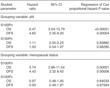
Table 7.Multivariate analysis of the relationships between nuclear (S100Pn) and cytoplasmic (S100Pc) expressions and the overall survival and disease-free survival time. The analysis included effect of grouping variables which were significantly linked to overall survival and disease-free survival time (pN and menopausal status).
Studied Hazard 95% CI Regression of Cox
parameter ratio proportional hazard P value Grouping variable: pN
S100Pn
OS 6.47 3.04-13.79 <0.00001
DFS 4.65 2.35-9.20 0.00004
S100Pc
OS 1.11 0.55-2.23 0.83882
DFS 1.03 0.54-1.97 0.66280
Grouping variable: menopausal status S100Pn
OS 5.74 2.86-11.54 0.00001
DFS 4.43 2.32 8.43 0.00006
S100Pc
OS 0.97 0.48-1.95 0.84033
DFS 0.93 0.49-1.97 0.67564
Table 8.KM Plotter analysis of prognostic significance of the S100P mRNA expression.
Studied parameter n P value
The entire group
Overall survival 464 0.2195
Relapse-free survival 2422 <0.0001
Distant metastases-free survival 673 0.0344 Estrogen receptor positive patients
Overall survival 134 0.6669
Relapse-free survival 1499 0.0004
Distant metastases-free survival 457 0.6473 Estrogen receptor negative patients
Overall survival 64 0.734
Relapse-free survival 442 0.9524
Distant metastases-free survival 101 0.1476 Lymph node positive patients
Overall survival 0 -*
Relapse-free survival 548 0.0833
Distant metastases-free survival 111 0.2143 Lymph node negative patients
Overall survival 198 0.3433
Relapse-free survival 1316 0.017
Distant metastases-free survival 447 0.1649 Truly prognostic datasets (patients not treated with systemic therapy)
Overall survival 609 0.3823
Relapse-free survival 609 0.0223
Distant metastases-free survival 609 0.1814 ER-positive endocrine treated datasets
Overall survival -* -
Relapse-free survival 414 0.6634
Distant metastases-free survival 414 0.3356
* Analysis not possible to perform.
receptors or HER-2.
We found that only nuclear expression of S100P had an impact on patients’ outcome. Lack of prognostic value of cytoplasmic S100P expression observed in our study is not clear. S100P is localized intracellularly in nucleus, cytoplasm or cell membrane and it can also be secreted extracellulary. It is likely that S100P
translocates within a cell and that it is a dynamic process, associated with its functional status and interactions (Guerreiro Da Silva et al., 2000; Sato et al., 2004; Parkkila et al., 2008). Nuclear translocation seems to be a result of interaction with S100P binding protein (S100PBPR) (Dowen et al., 2005), whereas binding to ezrin stimulates translocation to cell periphery
Fig. 7. Sensitivity of the studied cells to 11 cytostatic drugs of various groups: MCF-7 breast cancer cell line (A), CAMA-1 breast cancer cell line (B), SK-BR-3 breast cancer cell line (C)and R-103 breast cancer cell line (D).
(Koltzscher et al., 2003). This might explain contradictory data obtained in various studies.
In a few reports it was shown that S100P may also have predictive value, although these findings are controversial. S100P overexpression was associated with resistance to 5-fluorouracyl in pancreatic cancer cells and to irinotecan in prostate cancer (Arumugam et al., 2005; Basu et al., 2008). In our previous study performed on cell lines we found a correlation between overexpresion of S100P and resistance to cyclophosphamide, etoposide, methotrexate and mitoxantrone (Györffy et al., 2006). In contrast to these observations, studies on ovarian cancer cells exposed to carboplatin and paclitaxel, 5-fluorouracyl, etoposide and doxorubicin showed a possible chemosensitization effect of S100P (Wang et al., 2008). Similarly, studies on downregulation of S100P in colon cancer cell line 8307 have shown that S100P was associated with oxaliplatin sensitivity in these drug resistant cells (Tang et al., 2007). These data suggested that drug resistance/
sensitivity may be altered by S100P, but the mechanism of chemotherapy resistance is complex and it likely involves a variety of pathways. We found an association between decreased survival and high S100P score, irrespective of the adjuvant treatment. Small subgroups and uniform therapy did not allow an analysis assessing predictive value of S100P. However, since patients selection to adjuvant treatment is based on prognostic and predictive factors and biomarkers may have both a prognostic and a predictive value, a potential of S100P to predict benefits from certain chemotherapeutic agents warrants further studies.
Another, probably the most important issue which may have potential therapeutic implications, is a hypothesis that S100Psilencing may change aggressive tumour behaviour, and there are agents capable of inhibiting S100P. As a factor of poor prognosis, S100P may become an important target in breast cancer treatment. Most studies, however, focused on the concept that modulation of S100P may change response to cancer treatment. The S100P mechanism of action involves activation of RAGE receptor and, as a consequence, stimulation of different signalling pathways such as: nuclear factor - B (NFκB) mitogen- activated protein (MAP) kinase, which influence sensitivity/resistance to cytotoxic drugs (Fuentes et al., 2007). Observations from preclinical studies confirmed a correlation between S100P expression and presence of glucocorticoid receptor (GR). Activation of S100P by hydrocortisone and dexamethasone can be blocked by a GR antagonist, mifepristone (Gibadulinova et al., 2011).
A study in an animal model showed that inhibition of S100P and several other S100 molecules by cromolyn prevented activation of RAGE (Arumugam et al., 2006) and inhibited pancreatic cancer cell growth. Several pathway-implicated agents, such as phenothiasine, chlorpromazine and calmodulin inhibitor N-(6- aminohexyl)-5-chloro-1-naphtalenesulfonamide, may reverse resistance to cisplatin (Dairkee et al., 2009). The
concept of restoration of sensitivity to a certain cytotoxic agent by blocking S100P upregulation is certainly an interesting field to explore. Since transcriptional regulation of S100 P is complex and not fully understood, involving a variety of signalling pathways, depending on tumour type and other factors, these observations are very promising but demand confirmation in clinical studies.
In our investigations, both at the level of mRNA and at the level of protein S100P has been shown to be an unfavourable prognostic factor in breast cancer. In studies on the relationship between S100P expression in breast cancer cell lines and sensitivity of the cells to cytostatic drugs we have failed to demonstrate a relationship between expression of S100P and manifestation of resistance to the most popular cytostatic drugs. It should be concluded that in cases of breast cancer an elevated S100P expression is linked to a more aggressive tumour biology but not to its resistance to chemotherapy.
In conclusion, S100P expression provides important prognostic information in early breast cancer patients. It may be appropriate to validate the usefulness of S100P as a predictive marker. There is growing evidence that S100P inhibition by molecularly targeted agents may reverse unfavourable cancer cell phenotype, which in consequence improves patients outcome. If evidence from experiments indicating that modulation of S100P reverses drug resistance is confirmed in clinical studies, S100P would become a target allowing to overcome resistance to chemotherapy. So far, observations from studies performed on cell lines promote hypotheses.
Information from studies on the role of S100P in breast cancer may be useful in clarifying clinical significance of this family of proteins and in developing novel strategies in the treatment of breast cancer.
Acknowledgements. The study was supported by the grant No.
0638/B/P01/2008/35from the Ministry of Science and Higher Education (Poland).
References
Arumugam T., Simeone D.M., Schmidt A.M. and Logsdon C.D. (2004).
S100P stimulates cell proliferation and survival via receptor for activated glycation end products (RAGE). J. Biol. Chem. 279, 5059- 5065.
Arumugam T., Simeone D.M., Van Golen K. and Logsdon C.D. (2005).
S100P promotes pancreatic cancer growth, survival, and invasion.
Clin. Cancer Res. 11, 5356-5364.
Arumugam T., Ramachandran V. and Logsdon C.D. (2006). Effect of cromolyn on S100P interactions with RAGE and pancreatic cancer growth and invasion in mouse models. J. Natl. Cancer Inst. 98, 1806-1818.
Barraclough D.L., Sewart S., Rudland P.S., Shoker B.S., Sibson D.R., Barraclough R. and Davies M.P. (2010) Microarray analysis of suppression subtracted hybridisation libraries identifies genes associated with breast cancer progression. Cell Oncol. 32, 87-99.
Basu G.D., Azorsa D.O., Kiefer J.A., Rojas A.M., Tuzmen S., Barrett M.T., Trent J.M., Kallioniemi O. and Mousses S. (2008). Functional evidence implicating S100P in prostate cancer progression. Int. J.
Cancer 123, 330-339.
Dairkee S.H., Sayeed A., Luciani G., Champion S., Meng Z., Jakkula L.R., Feiler H.S., Gray J.W. and Moore D.H. (2009). Immutable functional attributes of histologic grade revealed by context- independent gene expression in primary breast cancer cells. Cancer Res. 69, 7826-7834.
Diederichs S., Bulk E. and Steffen B. (2004). S100 family members and trypsinogens are predictors of distant metastasis and survival in early-stage non-small cell lung cancer. Cancer Res. 64, 5564-5569.
Donato R. (2003). Intracellular and extracellular roles of S100 proteins.
Microsc. Res. Tech. 60, 540-551.
Dowen S.E., Crnogorac-Jurcevic T., Gangeswaran R., Hansen M., Eloranta J.J., Bhakta V., Brentnall T.A., Lüttges J., Klöppel G. and Lemoine N.R. (2005). Expression of S100P and its novel binding partner S100PBPR in early pancreatic cancer. Am. J. Pathol. 166, 81-92.
Early Breast Cancer Trialists' Collaborative Group (EBCTCG) (2005).
Effects of chemotherapy and hormonal therapy for early breast cancer on recurrence and 15-year survival: an overview of the randomised trials. Lancet 365, 1687-717.
Ferlay J.F., Bray P., Pisani P. and Parkin D.M. GLOBOCAN 2002 (2004). Cancer incidence, mortality and prevalence worldwide IARC cancerbase. Lyon. IARC Press.
Fuentes M.K., Nigavekar S.S., Arumugam T., Logsdon C.D., Schmidt A.M., Park J.C. and Huang E.H. (2007). RAGE activation by S100P in colon cancer stimulates growth, migration, and cell signaling pathways. Dis. Colon Rectum. 50, 1230-1240.
Gibadulinova A., Tothova V., Pastorek J. and Pastorekova S. (2011).
Transcriptional regulation and functional implication of S100P in cancer. Amino Acids. 41, 885-892.
Goldhirsch A., Ingle J.N., Gelber R.D., Coates A.S., Thürlimann B., Senn H.J. and Panel members (2009). Threshold for therapies:
highlights of the St Gallen International Expert Consensus on the Primary Therapy of Early Breast Cancer. Ann. Oncol. 20, 1319-1329.
Guerreiro Da Silva I.D., Hu Y.F. and Russo I.H. (2000). S100P calcium- binding protein overexpression is associated with immortalization of human breast epithelial cells in vitro and early stages of breast cancer development in vivo. Int. J. Oncol. 16, 231-240.
Györffy B., Surowiak P., Kiesslich O., Denkert C., Schäfer R., Dietel M.
and Lage H. (2006). Gene expression profiling of 30 cancer cell lines predicts resistance towards 11 anticancer drugs at clinically achieved concentrations. Int. J. Cancer 118, 1699-1712.
Györffy B., Lanczky A., Eklund A.C., Denkert C., Budczies J., Li Q. and Szallasi Z. (2010). An online survival analysis tool to rapidly assess the effect of 22,277 genes on breast cancer prognosis using microarray data of 1809 patients. Breast Cancer Res. Treat. 123, 725-731.
Koltzscher M., Neumann C., Konig S. and Gerke V. (2003) Ca- dependent binding and activation of dormant ezrin by dimeric S100P. Mol. Biol. Cell. 14, 2372-2384.
Logsdon C.D., Simeone D.M., Binkley C., Arumugam T., Greenson J.K., Giordano T.J., Misek D.E., Kuick R. and Hanash S. (2003).
Molecular profiling of pancreatic adenocarcinoma and chronic pancreatitis identifies multiple genes differentially regulated in pancreatic cancer. Cancer Res. 63, 2649-2657.
Mackay A., Jones C., Dexter T., Silva R.L., Bulmer K., Jones A., Simpson P., Harris R.A., Jat P.S., Neville A.M., Reis L.F., Lakhani S.R. and O'Hare M.J. (2003). cDNA microarray analysis of genes associated with ERBB2 (HER2/neu) overexpression in human mammary luminal epithelial cells. Oncogene 22, 2680-2688.
Mousses S., Bubendorf L., Wagner U., Hostetter G., Kononen J., Cornelison R., Goldberger N., Elkahloun A.G., Willi N., Koivisto P., Ferhle W., Raffeld M., Sauter G. and Kallioniemi O.P. (2002):
Clinical validation of candidate genes associated with prostate cancer progression in the CWR22 model system using tissue microarrays. Cancer Res. 62, 1256-1260.
Parkkila S., Pan P.W., Ward A., Gibadulinova A., Oveckova I., Pastorekova S., Pastorek J., Martinez A.R., Helin H.O. and Isola J.
(2008). The calcium-binding protein S100P in normal and malignant human tissues. BMC Clin. Pathol. 18, 8:2.
Perez E., Romond E. and Suman V. (2007). Updated results of the combined analysis of NCCTG N9831 and NSABP B-31 adjuvant chemotherapy with/without trastuzumab in patiens with HER2- positive breast cancer. J. Clin. Oncol. 25, 6s.
Sato N., Fukushima N., Matsubayashi H. and Goggins M. (2004).
Identification of maspin and S100P as novel hypomethylation targets in pancreatic cancer using global gene expression profiling.
Oncogene 23, 1531-1538.
Schor A.P., Carvalho F.M., Kemp C., Silva I.D. and Russo J. (2006).
S100P calcium-binding protein expression is associated with high- risk proliferative lesions of the breast. Oncol. Rep. 15, 3-6.
Surowiak P., Maciejczyk A., Materna V., Drag-Zalesińska M., Wojnar A., Pudelko M., Kedzia W., Spaczyński M., Dietel M., Zabel M. and Lage H. (2007). Unfavourable prognostic significance of S100P expression in ovarian cancers. Histopathology 51, 125-128.
Tang H., Liu Y.J., Liu M. and Li X. (2007). Establishment and gene analysis of an oxaliplatin-resistant colon cancer cell line THC8307/L- OHP. Anticancer Drugs 18, 633-639.
Wang G., Platt-Higgins A., Carroll J., de Silva Rudland S., Winstanley J., Barraclough R. and Rudland P.S. (2006). Induction of metastasis by S100P in a rat mammary model and its association with poor survival of breast cancer patients. Cancer Res. 66, 1199-1207.
Wang Q., He Z., Gao J., Hu S., Huang M., Liu M., Zheng J. and Tang H.
(2008). S100P sensitizes ovarian cancer cells to carboplatin and paclitaxel in vitro. Cancer Lett. 272, 277-284.
Accepted October 19, 2012