R E S E A R C H A R T I C L E
The genome sequence of a SNP type 3K strain of Mycobacterium leprae isolated from a seventh ‐ century Hungarian case of lepromatous leprosy
T. A. Mendum
1 |G. M. Taylor
1 |H. D. Donoghue
2 |H. Wu
1 |C. Szalontai
3 |A. Marcsik
4 |E. Molnár
4 |G. Pálfi
4 |G. R. Stewart
11Department of Microbial Sciences, Faculty of Health and Medical Sciences, University of Surrey, Guildford, UK
2Centre for Clinical Microbiology, Division of Infection and Immunity, Royal Free Campus, University College London, London, UK
3Salisbury Ltd., Budaörs, Hungary
4Department of Biological Anthropology, University of Szeged, Szeged, Hungary Correspondence
G Michael Taylor, Department of Microbial Sciences, Faculty of Health and Medical Sciences, University of Surrey, Guildford GU2 7XH, UK.
Email: gm.taylor@surrey.ac.uk
Funding information
British Academy, Grant/Award Number:
SG150114
Abstract
We report on a
Mycobacterium lepraegenome isolated from the remains of an individ- ual with lepromatous leprosy that were excavated from a seventh
‐century Hungarian cemetery. We determined that the genome was from a single nucleotide polymor- phism (SNP) type 3K0
M. lepraestrain, a lineage that diverged early from other
M. lepraelineages. This is one of the earliest 3K0
M. lepraegenomes to be sequenced to date. A number of novel SNPs as well as SNPs characteristic of the 3K0 lineage were confirmed by conventional polymerase chain reaction and Sanger sequencing.
Recovery of accompanying human DNA from the burial was poor, particularly when compared with that of the pathogen. Modern 3K0
M. lepraestrains have only been isolated from East Asia and the Pacific, and so these findings require new scenarios to describe the origins and routes of dissemination of leprosy during antiquity that have resulted in the modern phylogeographical distribution of
M. leprae.K E Y W O R D S
ancient DNA, Branch 0,Mycobacterium leprae, phylogeny, whole genome sequencing
1
|I N T R O D U C T I O N
The spread of leprosy around the world in antiquity has previously been inferred by characterizing the infrequent single nucleotide poly- morphisms (SNPs) of modern strains of Mycobacterium leprae, col- lected from across the world (Monot et al., 2005). More recently, the genotyping of isolates recovered from archaeological human remains displaying skeletal evidence of lepromatous leprosy (LL) has provided unique opportunities to refine our understanding of the distribution of M. lepraestrains seen in extant populations, as well as allowing analysis of strains from regions where the disease is no longer present.
In this respect, we have previously studied archaeological cases from various parts of Britain and Europe (Donoghue et al., 2015; Inskip et al., 2015; Taylor et al., 2013) as well as modern cases from regions where the disease is still common (Monot et al., 2009).
One of the cases identified previously was burial KD271, a seventh‐century male individual whose remains were excavated from
Kiskundorozsma (Szeged) in Hungary (Pálfi & Molnár, 2009). Study of a limited number of informative SNPs indicated that the strain of M. lepraepresent in KD271 was a type 3K on the original scheme of 16 genotypes (ranging from 1A–4P) proposed by Monot et al. (2009).
With the application of whole genome sequencing (WGS) to both modern and ancient cases of leprosy (Mendum et al., 2014;
Schuenemann et al., 2013), it has become clear that SNP type 3K iso- lates belong on a separate lineage of theM. lepraephylogenetic tree, now designated Branch 0. Although a number of modern examples of this lineage have been studied, all from East Asian locations (Benjak et al., 2018; Schuenemann et al., 2013), no ancient isolates have pre- viously been available for such genomic analysis. The current study presents WGS analysis of the strain from burial KD271 and provides a rare opportunity to study what is one of the oldest cases ofM. leprae to be sequenced. This is likely to represent a strain close to the most recent common ancestor of extant strains that is predicted to have existed only 3–4,000 years ago (Schuenemann et al., 2013). As DOI: 10.1002/oa.2673
Int J Osteoarchaeol. 2018;28:439–447. wileyonlinelibrary.com/journal/oa Copyright © 2018 John Wiley & Sons, Ltd. 439
previous biomolecular tests applied to KD271 have appeared in diverse sources, a summary of the conventional genotyping is brought together here, along with some novel observations. These serve both to provide context and as a useful comparator for the WGS study.
2
|M A T E R I A L S A N D M E T H O D S 2.1
|Sampling
The subject of the current study, burial KD271, was a mature male individual judged to be between 50 and 60 years of age at the time of death. The burial was excavated from the cemetery of Kiskundorozsma Szeged in Hungary, and the diagnosis of leprosy was made on osteological grounds (Pálfi & Molnár, 2009). For the present genomic study, a 50‐mg sample of bone from the palate of KD271 was ground into a fine powder in a sterilised pestle and mortar.
A second burial, Sk12, an adult male, without osteological signs of leprosy, acted as a control for the WGS protocol. Bone was sampled from around the vomer region (Schuenemann et al., 2013).
2.2
|DNA extraction
DNA was extracted using an in‐house version of the Boom method (Boom et al., 1990). In this procedure, bone powder (50 mg) was incu- bated in 1 ml of 1 × Tris‐EDTA buffer, containing 40 mAU/ml of pro- teinase K at 37 °C for 48 hr with occasional mixing. The sample was then centrifuged at 3,000 rpm for 3 min, and the supernatant was transferred into five volumes of 6 M guanidinium thiocyanate (GUSCN, product G9020 from US Biologicals, Salem, MA) containing 1% Triton X‐100 and buffered in 1 × Tris‐EDTA buffer adjusted to pH 6.5 with 3 M sodium acetate, pH 5.5. Bone powder was mixed with the GUSCN buffer on a mixing wheel for 1 hr at 4 °C. The sam- ples were then subjected to 3 freeze–thaw cycles. Bone powder was removed by centrifugation at 12,000 rpm for 5 min, and the superna- tant was transferred to a sterile 1.5‐ml Eppendorf tube. Pre‐washed silica suspension (40 μl of 0.5–10 μm, Sigma‐Aldrich, S5631) was added and kept in contact for 3 hr to maximise recovery of fragmented DNA. After centrifugation, silica was further washed twice with 1 ml aliquots of GUSCN extraction buffer, followed by three washes with 75% ethanol and finally with 1 ml of acetone. After thor- ough drying of the silica pellet, DNA residues were eluted in 60μl HPLC grade water at 55 °C. This was then subdivided into 2 × 30μl aliquots and stored in low retention plastic tubes to minimise loss of DNA through repeated freeze–thawing events.
2.3
|Mycobacterium leprae screening
Before undertaking WGS, we screened for evidence ofM. lepraeDNA in the new extract using a polymerase chain reaction (PCR) for the RLEP multi‐copy element. This method amplifies a 78 bp amplicon with product monitored with a specific 6‐fluorescein amidite (6‐FAM)‐FAM‐labelled hybridisation probe. Details of this and the primer sequences and conditions have been previously reported (Inskip et al., 2015).
2.4
|Variable number tandem repeat and SNP genotyping
The PCR methods for the variable number tandem repeat (VNTR) and individual SNP loci amplification and sequencing have been previously reported (Taylor et al., 2009; Taylor & Donoghue, 2011). The rpoT locus (ML1022) was PCR amplified and sequenced for the present study using the primers and conditions described in Taylor et al., 2009.
2.5
|Mycobacterium lepromatosis screening
The opportunity was taken to screen the new extract for any evidence of the second leprosy agent,Mycobacterium lepromatosis. The primers were modifications of those described by Singh et al., 2015, 5‐CTGT TCGTGAGGTACCGGTGAAA and 5′‐GTTCGGCCGGAGTGTAGGTGT TA. These amplify a 135 bp fragment from thehemN gene, present inM. lepromatosisbut absent inM. leprae. The PCR reagents and con- ditions were as described previously for M. leprae specific primers (Inskip et al., 2015), except that an annealing temperature of 56 °C was used.
2.6
|Screening of extract for Mycobacterium tuberculosis complex DNA
The KD271 extract was also tested for the presence ofMycobacterium tuberculosis(MTB) complex organisms using a real‐time PCR method for the IS1081repetitive element (Taylor, Murphy, Hopkins, Rutland,
& Chistov, 2007).
2.7
|Conventional PCR amplification and
sequencing of newly identified SNPs in the KD271 isolate of Mycobacterium leprae
Scrutiny of the WGS data from KD271 (Table 3) revealed 16 novel polymorphic SNPs not found in other sequenced strains. Conventional PCR amplification and sequencing was undertaken to confirm the WGS findings for three randomly selected SNPs. Sequences of primers and amplicon sizes are given in Table S1.
2.8
|Human DNA
An attempt was made to confirm the sex of individual KD271 using a PCR method based on a polymorphism in the amelogenin gene (Mannucci, Sullivan, Ivanov, & Gill, 1994). This generates two bands from males of 106 and 112 bp (AMELX and AMELY products, respec- tively), and a single AMELX product of 106 bp from females.
2.9
|WGS of Mycobacterium leprae genome from skeleton KD271
DNA from skeleton KD271 was enriched for M. leprae sequences using microarrays and sequenced as described previously for cases Sk2, Sk8, and the control Sk12 from medieval Winchester (Schuenemann et al., 2013) but with only a single round of microar- ray‐based enrichment. The raw sequence files were deposited in the Sequence Read Archive database with Submission ID SAMN08093649. After initial quality controls and alignment to the
M. lepraeTN reference genome (Schuenemann et al., 2013), SNPs were identified and accepted if they had a read depth of 3 or greater;
more than 75% of reads agreed; an alignment quality score (MQ)≥30;
and an absence of reads in the control sample Sk12.
Phylogenies were generated by aligning all SNPs from selected leprosy genomes (Data S1) using both the maximum likelihood and neighbour‐joining functions of MEGA7 (Kumar et al., 2016) and the Tamura 3‐parameter model (as determined to be the best fit by the model selection function of MEGA7). Equivalent loci inM. lepromatosis were determined by aligning M. lepraeTN, M. leprae Br423, and M. lepromatosisFJ924 (Han et al., 2015) with Mauve v2.4.0 (Darling, Mau, Blattner, & Perna, 2004).
3
|R E S U L T S
3.1
|Paleopathological lesions in KD271
The diagnosis of LL in individual KD271 was originally made on palaeopathological grounds. The lesions have been described in detail previously (Donoghue et al., 2005; Molnár, Marcsik, Bereczki, &
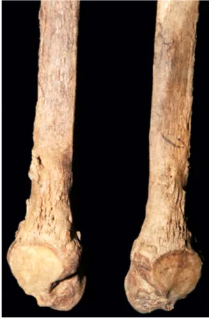
Donoghue, 2006), so only a summary is given here. The skull shows signs of the rhinomaxillary syndrome, with erosion and widening of the nasal margins and resorption of the anterior nasal spine (Figure 1 and inset). There is loss of bone from around the alveolar region on the maxillary process, and the upper incisor teeth have been lost ante mortem with remodelling of the tooth sockets (Figure 1). There is also pitting on the nasal surface of the palate due to the disease. Pitting due to periostitis is also present on some tarsal bones and on the surfaces of the tibiae and fibulae. Subperiosteal exostoses seen on the fibulae are more evident on the distal third of the bone shafts (Figure 2).
3.1.1
|Burial Sk12
This individual was found to be free of any macroscopic signs of the rhinomaxillary syndrome or other indications of leprosy on the hands, feet, or distal lower limbs (Taylor et al., 2013).
3.2
|Screening of KD271 extract for Mycobacterium leprae DNA
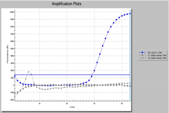
The extract prepared from the palatal region of the skull tested posi- tive using the RLEP PCR probe method (Figure 3). The control case was negative forM. lepraeDNA (not shown).
3.3
|SNP genotyping and multiple loci VNTR analysis
The results of SNP and VNTR typing are summarised in Table 1.
Figure S1 shows conventional Sanger sequencing of six informative SNP loci used in the Monot typing scheme (2009).
3.4
|Screening for Mycobacterium lepromatosis DNA
No PCR products were amplified from the extracts using thehemN primers specific forM. lepromatosis; therefore, there is no evidence that this individual was co‐infected with this pathogen. To show that this PCR method can amplify M. lepromatosis, an appropriately sized
FIGURE 1 The skull of KD271. Frontal view of skull from the burial (less mandible) showing evidence of rhinomaxillary syndrome, notably loss of several incisors, resorption of the anterior nasal spine, and widening of the piriform aperture. Inset highlights loss of anterior nasal spine [Colour figure can be viewed at wileyonlinelibrary.com]
FIGURE 2 The fibulae from KD271. KD271 fibulae showing subperiosteal exostoses on the distal third of the bone shafts [Colour figure can be viewed at wileyonlinelibrary.com]
amplicon (135 bp) was PCR amplified from partially purified M. lepromatosisDNA harvested from infected tissue of a Scottish red squirrel (Sciurus vulgaris; Figure S2).
3.5
|Screening for MTB complex DNA
The extract prepared from the palate of KD271 was negative for evi- dence ofM. tuberculosiscomplex DNA.
3.6
|Human DNA
No PCR products were obtained using the amelogenin PCR, which probably reflects the extremely fragmented nature of DNA in this skeleton.
3.7
|Genome sequencing of KD271
DNA extracts from skeletons KD217 and Sk12 were enriched for M. leprae sequences, PCR amplified and sequenced. Details of the FIGURE 3 Confirmation ofM. lepraeDNA in extracts from KD271. Real‐time amplification of RLEP polymerase chain reaction product (78 bp) monitored with a dual‐labelled fluorescent hybridisation probe. KD271 is shown in blue and negative controls in grey [Colour figure can be viewed at wileyonlinelibrary.com]
TABLE 1 Comparison of selected informative SNPs and VNTR loci determined by conventional and whole genome sequencing. Regions with insufficient or poor coverage in the WGS are indicated as not determined (nd)
SNP locus1 Amplicon size (bp) Nucleotide base SNP typing inference Nucleotide base by WGS
14,676 136 C Type 3 nd
1,642,879 122 T T
2,935,693 107 C nd
413,903 120 G 3I–3K G
591,858 107 C 3I–3L C
1,133,495 121 G 3J–3M G
2,312,066 120 G 3K–3M G
7,614 109 C Not 3I nd
1,113,926 117 A A
Overall 3K
VNTR loci Copies Copies by WGS
AGA(20) 2,785,364–2,785,494 Variable 16 nd
GTA(9) 2,583,816–2,583,839 Variable 24 nd
21–3,ML005872,683–73,686 96 2 2
rpoT, (sigA)ML1022 91 3 3
Note. SNP = single nucleotide polymorphism; VNTR = variable number tandem repeat; WGS = whole genome sequencing.
read depths, their percentage alignment to the M. lepraeTN refer- ence genome, percentage genome coverage, and average read length are given in Table 2. Samples from the KD271 burial gave an aver- age read depth of 11.94, whereas the Sk12 control sample's average read depth was 0.39% and less than 1% of reads aligned to the M. leprae genome. As observed previously (Mendum et al., 2014;
Schuenemann et al., 2013), the alignments were punctuated with regions with pan‐genus similarity that had large numbers of highly heterogeneous reads that presumably derive from environmental mycobacteria. The KD271 sequences had short read lengths (Sawyer, Krause, Guschanski, Savolainen, & Pääbo, 2012) typical of ancient DNA. These data confirm that there was little or no cross‐ contamination between samples, or from modern M. leprae DNA during the sample preparation.
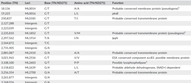
The WGS sequencing was in broad agreement with Sanger sequencing of targeted SNPs (Table 1), so validating both conven- tional typing and genome sequencing methods. However, the WGS had insufficient coverage for three of the nine Sanger‐determined SNP loci. Analysis of VNTR genotypes in KD271 was limited to the 21–3 locus (ML0058) and to therpoTlocus (ML1022), as insuffi- cient numbers of reads spanned the necessary regions for the other two VNTR loci, GTA(9) and AGA(20). Sixteen novel SNPs were iden- tified that have not been found in other published strains, including the 3K0 strains, S9, S10, CM‐1, or Kyoto‐1. None of the SNPs in coding regions are likely to have phenotypic effects. To validate the WGS data, three randomly chosen, newly identified SNPs were additionally confirmed by conventional PCR and Sanger sequencing (Table 3).
3.8
|Phylogenetic analysis of KD271
Phylogenetic analyses were consistent between both maximum likelihood and neighbour‐joining methods (Figures 4 and Figure S3). Both placed KD271 within the 3K0 lineage, branching from the common lineage before the S10/Kyoto‐1 branch but after the S9/CM‐1 branch (Avanzi et al., 2015; Honap et al., 2018; Mendum et al., 2014; Schuenemann et al., 2013). The branch lengths of KD271 were shorter than for the modern strains as would be expected for an ancient genome. KD271 was found to exhibit 26 SNPs, 24 of which were unique to the 3K0 group (S9, S10, CM‐ 1, and Kyoto‐1), six of these SNPs were uniquely shared with S10 and Kyoto‐1. In contrast, no SNPs were uniquely shared with S9 or CM‐1.
3.8.1
|rpoT locus
The presence of only three copies of GACATC in therpoTtandem repeat distinguishes KD271, as well as S9 and S10, all of which have earlier branch points, from the modern isolates from Japan, Korea, and parts of China that result from an apparently recent radiation of 3K0 strains, and all contain four copies of the hexanucleotide (Kai et al., 2013; Weng et al., 2013).
3.8.2
|ML0411 locus
Polymorphism was noted inML0411. LocusML0411is a serine rich, 45 kDa antigen (408 aa) recognised by B cells of the immune system.
It is a member of the PPE protein gene families of pathogenic mycobacteria, having the characteristic Pro‐Pro‐Glu motif at the N
TABLE 2 WGS sequencing and alignment statistics for KD271. Sequence reads were quality controlled and aligned to M. leprae TN and human genomes
Total number of reads
Percentage of reads aligning to the Mycobacterium lepraeTN genome
Percentage of reads aligning to the human genome
Average M. lepraeread depth
Average length of aligned reads
Percentage genome coverage≥3 reads, MQ score≥30
52,430,302 17.1% 0.9% 11.9 88.6 bp 83.8%
TABLE 3 SNPs Unique to KD271. SNPs confirmed using conventional PCR and Sanger sequencing Position (TN) Loci Base (TN/KD271) Amino acid (TN/KD271) Function
18,136 ML0014 C/T A/A Probable conserved membrane protein (pseudogene)1
19,223 ML0016 C/T L/L pknB
250,837 ML0185 C/T T/I Probable conserved transmembrane protein
2,127,288 Intergenic C/T 2,223,039 Intergenic C/T
2,235,810 ML1852 C/T V/M Probable conserved transmembrane protein (pseudogene)1
2,297,562 ML1914 T/A I/N lpqN
2,564,872 Intergenic T/C 2,731,405 Intergenic G/A
2,881,087 ML2410 G/A A/A Probable conserved transmembrane protein
3,021,965 ML2536 C/T V/V ESX conserved componenteccB3, possible membrane protein
3,108,100 ML2603 G/T P/P Possible lysophospholipase1
3,158,822 ML2639 G/A L/L Probable aldehyde dehydrogenase, (NAD+) dependent
3,256,354 ML2700 G/A A/T Probable conserved transmembrane protein
3,261,877 Intergenic G/A 3,268,026 Intergenic G/A
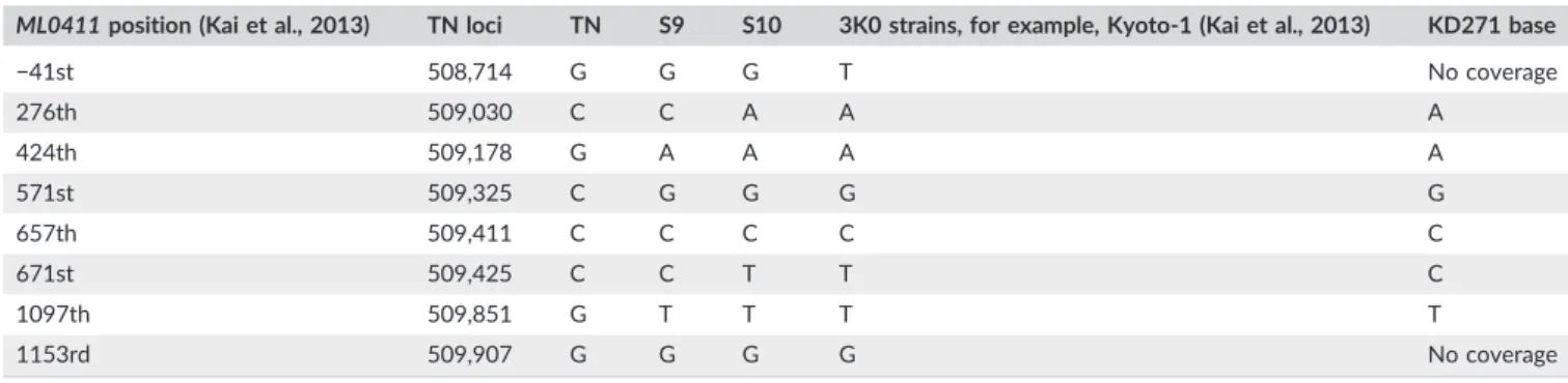
termini of the proteins. These proteins, along with the related PE fam- ily, are likely to give rise to antigenic variation and may modulate the immune response. ML0411 is the single most variable gene in the genome ofM. leprae. Comparison of the KD271 polymorphic SNPs inML0411(Table 4) with 3K0 strains from North‐east Asia (Kai et al., 2013) is compatible with its phylogenetic position as an early member of the 3K0 lineage.
3.8.3
|Subdivision of the Branch 0 lineage
Recent work shows that the type 3K strains of Branch 0 may be further subdivided into 3K0 and 3K1 lineages (shown as branch 5 in Figure 4),
depending on specific SNP subsets (Avanzi et al., 2015). These workers report that the 3K0 lineage displays a total of 20 specific SNPs, and the newly described branch 3K1 demonstrates 23 specific SNPs. The 3K common branch is characterised by C at position 711,197 and G at SNPs 563,796 and 57,633. Inspection of the data from KD271 shows the iso- late is consistent with 3K0 rather than 3K1 lineage (Table 5).
4
|D I S C U S S I O N
The subject of the current report, KD271, was a mature male individ- ual showing skeletal evidence of LL. The remains were excavated from
FIGURE 4 Phylogenetic relationships between selected modern (regular text) and ancient (bold text)Mycobacterium leprae strains. The phylogeny was inferred by the maximum likelihood method of MEGA7 (Kumar et al., 2016) and the Tamura 3‐ parameter model. The tree with the highest log likelihood value is shown. Bootstrap percentages from 1,000 replicates are shown next to the branches. The scale indicates the number of substitutions per site. All positions with less than 90% site coverage were eliminated.M. lepromatosiswas used as an outgroup (not shown). CM1 and Br15‐1 are derived from a cynomolgus macaque and a red squirrel, respectively [Colour figure can be viewed at wileyonlinelibrary.com]
TABLE 4 ML0411: Nucleotide bases in KD271 compared to the TN reference strain (SNP Type 1) and other 3K0 branch members
ML0411position (Kai et al., 2013) TN loci TN S9 S10 3K0 strains, for example, Kyoto‐1 (Kai et al., 2013) KD271 base
−41st 508,714 G G G T No coverage
276th 509,030 C C A A A
424th 509,178 G A A A A
571st 509,325 C G G G G
657th 509,411 C C C C C
671st 509,425 C C T T C
1097th 509,851 G T T T T
1153rd 509,907 G G G G No coverage
the cemetery of Szeged‐Kiskundorozsma‐Daruhalom dűlőII, Hungary in the summer of 2003. On the basis of the associated grave goods (Mészáros, Paluch, & Szalontai, 2005; Paluch & Szalontai, 2004;
Szalontai, 2012), the burial ground was in use in the early/middle Avar transition period, in the late seventh century. The cemetery contained 94 individuals, and skeletal signs of leprosy were noted in eight of these.
This presumptive diagnosis was previously confirmed by ancient DNA analysis in three cases, including the subject of the current work, KD271 (Molnár et al., 2006). Subsequently, this case was partially geno- typed for a later study of leprosy using conventional PCR and Sanger sequencing targetting phylogenetically informative SNP loci (Monot et al., 2009). This showed the isolate to be a type 3K strain ofM. leprae.
Since those initial investigations, the understanding of the phylogeography of leprosy has improved considerably, with the recog- nition of an additional lineage (Branch 0) into which the 3K strains may be placed (Schuenemann et al., 2013) and which branches earlier from the common lineage than the other groups. To extend our understand- ing of how KD271 and the other leprous remains at Szeged‐ Kiskundorozsma‐Daruhalom dűlőII relate to other ancient and mod- ern M. leprae strains, the genome of KD271 was sequenced. This genome is, to date, one of the oldest Branch 0M. lepraegenomes to have been studied and so is likely to represent a strain more similar to the notional most recent common ancestor (predicted to have existed ~3,000–4,000 years ago). From recent and ongoing studies on modern Branch 0 isolates, it is evident that there is a greater het- erogeneity within this lineage than first realised so that the type 3K strains may be further subdivided into 3K0 and 3K1, depending on specific SNP subsets (Avanzi et al., 2015). The genome sequencing, phylogenetic analysis, and SNP‐specific sequencing all confirm that KD271 falls within the currently known monophyleticM. lepraeradia- tion and does indeed represent a 3K strain, being placed on an early branch of the 3K0 lineage.
This position is reflected in the SNPs of the hypervariable gene, ML0411, which, although not identical to modern strains, was found to be consistent with its place on the 3K0 lineage.
The isolate affecting KD271, along with S9 and S10, the two other strains with early branch points in the 3K0 lineage, possessed only three copies of therpoThexanucleotide GACATC, rather than the four copies
typical of modern strains as described by Avanzi et al. (2015) and Kai et al., 2013. When first reported, this VNTR polymorphism was sug- gested as one, albeit limited, way of examining differences in the M. lepraegenome (Matsuoka et al., 2000). The locus may therefore be helpful in defining those strains mentioned above with a more recent radiation. However, the relevance of therpoTlocus is difficult to assess without further investigation of strains isolated from various regions of India (Lavania et al., 2007; Lavania et al., 2009) where strains with four copies have been described. This is because the recently described second agent of leprosy,M. lepromatosis(Han et al., 2008), also contains four copies of this sequence, as reported for cases in Western and Central Mexico (Han, Sizer, Velarde‐Félix, Frias‐Castro, & Vargas‐ Ocampo, 2012). So in studies where therpoTprimers recognise both pathogens, and where it was the only typing method used (Lavania et al., 2007; Lavania et al., 2009), further testing will be needed to distin- guish betweenM. lepromatosisand the 3K lineage ofM. lepraewith four copies of therpoThexanucleotide.
In the earlier study by Monot et al. (2009), two further LL cases from another Hungarian burial ground were also found to be positive forM. leprae. These were burials 222 and 503, a 45‐to 50‐year‐old male and a 30‐to 35‐year‐old female, respectively. These were both excavated from the 10th‐to 11th‐century Eastern Hungarian ceme- tery at Püspökladány‐Eperjesvölgy. They were found to be SNP types 3K and 3M, respectively. Interestingly, they were both subsequently shown to be co‐infected with M. tuberculosis (Donoghue, Marcsik, Molnár, Paluch, & Szalontai, 2005); hence, we checked burial KD271 for any signs of MTB complex DNA but found none. The lack of either MTB complex orM. lepromatosismycobacterial DNA is important for the accurate interpretation of WGS data.
The presence of 3K0 strains in seventh‐century Hungarian remains and of a 3K strain from the 10th centrury is consistent with two contrasting scenarios for the origins of geographical distibution of 3KM. lepraestrains. The global distribution of 3K0 and 3K1 strains is today restricted to regions of the Western Pacific such as Japan (except Okinawa), Korea, China, The Philippines, New Caledonia and Indonesia, among others (Avanzi et al., 2015; Honap et al., 2018; Kai et al., 2013; Monot et al., 2009; Weng et al., 2013). This could indicate that the 3K lineage originated in Northern or Eastern Asia. The TABLE 5 Distinguishing SNPs of the 3KI and 3KO strains. Three SNP loci associated with the 3K1 lineage and the equivalent positions in the 3KO burials KD271, S9, S10 and reference strains TN and Br4923. After Avanzi et al., 2015
Genome
ML0585c ML0466 ML0046c
Pseudogeneqor Hypothetical protein PseudogeneespJ
SNP 711,197 SNP 563,796 SNP 57,633
3K1 branch
Ryukyu‐2 C G G
3K0 branch
S9 C C T
S10 C C T
CM‐1 C C T
KD271 C C T
Other strains
TN T G T
Br 4923 T G T
Note. SNP = single nucleotide polymorphism.
presence of two type 3K cases (KD271 and 222) in early medieval Hungary would then suggest a route of dissemination from Asia to Central Europe, perhaps via trade links or migrations. This would be consistent with what is known of the origins of the Panonian Avars, who are believed to have reached the Hungarian plain from the Eur- asian steppe in the late sixth to early ninth centuries (Curta, 2006).
The other possibility is that Europe was a centre of dissemination of the ancestral 3K0 and related strains, some of which later became less common or even absent from Europe but persisted in East Asia and the Pacific. Determining the likelihood of each of these scenarios will require more sampling and characterization of both ancient and mod- ern strains.
5
|C O N C L U S I O N S
Ancient DNA analysis is a powerful approach for understanding past human diseases such as leprosy. In particular, it allows us to obtain strain typing data from geographical locations where the disease may no longer be found, to compare ancient with modern strain distri- butions and to assist with understanding earlier human migrations.
Recognition of greater diversity within 3K lineage strains (Branch 0), and its recognition as the deepest lineage, has come from WGS stud- ies applied to both ancient and modern cases. Burial KD271 repre- sents one of the earliest examples of this archaic lineage to be studied to date.
A C K N O W L E D G E M E N T S
We thank Dr. Charlotte Avanzi of The Global Health Institute, École Polytechnique Fédérale de Lausanne, 1015 Lausanne, Switzerland, for the generous gift of Mycobacterium lepromatosis genomic DNA, partially purified from red squirrel tissue. We also thank Dr. Tanvi Honap of the Department of Anthropology, University of Oklahoma for making available at short notice her sequence variant data. This study was supported in part by a Small Research Grant, reference SG150114, from the British Academy.
O R C I D
G. M. Taylor http://orcid.org/0000-0002-4215-3916
R E F E R E N C E S
Avanzi, C., Benjak, A., Kai, M., Busso, P., Singh, P., Matsuoka, M., & Cole, S.
T. (2015). Analysis ofMycobacterium lepraestrains from Japan: New trends in phylogeography. Copenhagen, Denmark: Eposter EP009 at 25th ECCMID. 25‐28thApril
Benjak, A., Avanzi, C., Singh, P., Loiseau, C., Girma, S., Busso, P.,…Cole, S.
T. (2018). Phylogenomics and antimicrobial resistance of the leprosy bacillus Mycobacterium leprae. Nature Communications, 9(1), 352.
https://doi.org/10.1038/s41467‐017‐02576‐z
Boom, R., Sol, C. J. A., Salimans, M. M. M., Jansen, C. L., Wertheim‐van Dillen, P. M. E., & van der Noordaa, J. (1990). Rapid and simple method for purification of nucleic acids. Journal of Clinical Microbiology, 28, 495–503.
Curta, F. (2006). Southeastern Europe in the Middle Ages, 500–1250. In Cambridge medieval textbooks. Cambridge: Cambridge University Press.
ISBN 978‐0‐521‐81539‐0
Darling, A. C., Mau, B., Blattner, F. R., & Perna, N. T. (2004). Mauve: Mul- tiple alignment of conserved genomic sequence with rearrangements.
Genome Research, 14, 1394–1403. https://doi.org/10.1101/
gr.2289704
Donoghue, H. D., Marcsik, A., Matheson, C., Vernon, K., Nuorala, E., Molto, J. E.,…Spigelman, M. (2005). Co‐infection ofMycobacterium tuberculo- sis and Mycobacterium leprae in human archaeological samples: A possible explanation for the historical decline of leprosy.Proceedings of the Royal Society B, 272, 389–394. https://doi.org/10.1098/
rspb.2004.2
Donoghue H. D., Marcsik A., Molnár E., Paluch T., Szalontai C.. 2005. Lepra nyomai a kiskundorozsmai avar temetőből Előzetes beszámoló. In Hadak útján” –Népességek és iparok a népvándorlás korában, 171‐ 186. [Osteological signs of leprosy from the Avar period series of Kiskundorozsma. Preliminary report. In: In the way of the armies–Pop- ulation and industries in the Migration Period] pp 171‐186.
Donoghue, H. D., Taylor, G. M., Marcsik, A., Molnár, E., Palfi, G., Pap, I.,… Spigelman, M. (2015). A migration‐driven model for the historical spread of leprosy in medieval Eastern and Central Europe.Infection, Genetics and Evolution, 31, 250–256. https://doi.org/10.1016/j.
meegid.2015.02.001
Han, X. Y., Mistry, N. A., Thompson, E. J., Tang, H.‐L., Khanna, K., & Zhang, L. (2015). Draft genome sequence of new leprosy agent“Mycobacte- rium lepromatosis”. Genome Announcements, 3, e00513–e00515.
https://doi.org/10.1128/genomeA.00513‐15
Han, X. Y., Seo, Y.‐H., Sizer, K. C., Schoberle, T., May, G. S., Spencer, J. S.,… Nair, R. G. (2008). A new Mycobacterium species causing diffuse lepro- matous leprosy.American Journal of Clinical Pathology,130, 856–864.
https://doi.org/10.1309/AJCPP72FJZZRRVMM
Han, X. Y., Sizer, K. C., Velarde‐Félix, J. S., Frias‐Castro, L. O., & Vargas‐ Ocampo, F. (2012). The leprosy agents Mycobacterium lepromatosis andMycobacterium lepraein Mexico.International Journal of Dermatol- ogy,51, 952–959. https://doi.org/10.1111/j.1365‐4632.2011.05414.x Honap, T. P., Pfister, L.‐A., Housman, G., Mills, S., Tarara, R. P., Suzuki, K.,… Stone, A. C. (2018). Mycobacterium leprae genomes from naturally infected nonhuman primates.PLoS Neglected Tropical Diseases,12(1), e0006190. https://doi.org/10.1371/journal.pntd.0006190
Inskip, S. A., Taylor, G. M., Zakrewski, S., Mays, S. A., Pike, A. W., Llewellyn, G.,…Stewart, G. R. (2015). Osteological, biomolecular and geochemical analysis of an early Anglo‐Saxon case of lepromatous leprosy. PLoS One, 13(10), e0124282. https://doi.org/10.1371/journal.
pone.0124282
Kai, M., Nakata, N., Matsuoka, M., Sekizuka, T., Kuroda, M., & Makino, M.
(2013). Characteristic mutations found in theML0411gene ofMyco- bacterium leprae isolated in Northeast Asian countries. Infection, Genetics and Evolution, 19, 200–204. https://doi.org/10.1016/j.
meegid.2013.07.014
Kumar, S., Stecher, G., & Tamura, K. (2016). MEGA7: Molecular evolution- ary genetics analysis version 7.0.Molecular Biology and Evolution,33, 1870–1874. https://doi.org/10.1093/molbev/msw054. Epub 2016 Mar 22
Lavania, M., Katoch, K., Singh, H., Das, R., Gupta, A. K., Sharma, R.,… Katoch, V. M. (2007). Predominance of three copies of tandem repeats inrpoTgene ofMycobacterium lepraefrom Northern India.Infection, Genetics and Evolution, 7, 627–631. https://doi.org/10.1016/j.
meegid.2007.05.011
Lavania, M., Lal, R., Joseph, G., Darlong, J., Abraham, S., Nanda, N. K., &
Jadhav, R. S. (2009). Genotypic analysis ofMycobacterium lepraestrains from different regions of India on the basis ofrpoT.Indian Journal of Leprosy,81, 119–124.
Mannucci, A., Sullivan, K. M., Ivanov, P. L., & Gill, P. (1994). Forensic appli- cation of a rapid and quantitative DNA sex test by amplification of the X‐Y homologous gene amelogenin.International Journal of Legal Medi- cine,106, 190–193. https://doi.org/10.1007/BF01371335
Matsuoka, M., Maeda, S., Kai, M., Nakata, N., Chae, G. T., Gillis, T. P.,… Kashiwabara, Y. (2000).Mycobacterium lepraetyping by genomic diver- sity and global distribution of genotypes. International Journal of Leprosy,68, 121–128.
Mendum, T. A., Schuenemann, V. J., Roffey, S., Taylor, G. M., Wu, H., Singh, P.,…Stewart, G. R. (2014). Mycobacterium leprae genomes from a Brit- ish medieval leprosy hospital: Towards understanding an ancient epidemic. BMC Genomics, 15, 270. https://doi.org/10.1186/1471‐ 2164‐15‐270
Mészáros P., Paluch T., Szalontai C. S. 2005. Avar kori temetők Kiskundorozsma határában. Előzetes beszámoló az M5 autópályán feltárt lelőhelyről.–MKCsM (2004) Szeged, pp. 145–162.
Molnár, E., Marcsik, A., Bereczki, Z., & Donoghue, H. D. (2006). Pathologi- cal cases from the 7th century in Hungary. In16thEuropean meeting of the Paleopathology Association. Fira, Santorini: Greece.
Monot, M., Honore, N., Garnier, T., Araoz, R., Coppee, J.‐Y., Lacroix, C.,… Cole, S. T. (2005). On the origin of leprosy.Science,308, 1040–1042.
https://doi.org/10.1038/ng.477
Monot, M., Honoré, N., Garnier, T., Zidane, N., Sherafi, D., Paniz‐Mondolfi, A., et al. (2009). Phylogeography of Leprosy. Nature Genetics, 41, 1282–1289. https://doi.org/10.1126/science/1109759
Pálfi, G., & Molnár, E. (2009). The paleopathology of specific infectious dis- eases from Southeastern Hungary: A brief overview. Acta Biologica Szegediensis,53, 111–116. (http://www.sci.u‐szeged.hu/ABS) Paluch, T., & Szalontai, C. S. (2004). Kiskundorozsma‐Daruhalom dűlőII. In
I. Kisfaludy (Ed.), Régészeti kutatások Magyaroszagon 2003 (pp.
293–329). Hungary, 2003: Archaeological Investigations.
Sawyer, S., Krause, J., Guschanski, K., Savolainen, V., & Pääbo, S. (2012).
Temporal patterns of nucleotide misincorporations and DNA fragmen- tation in ancient DNA. PLoS One, 7(3), e34131. https://doi.org/
10.1371/journal.pone.0034131
Schuenemann, V. J., Singh, P., Mendum, T. A., Krause‐Kyora, B., Jäger, G., Bos, K. I.,…Krause, J. (2013). Genome‐wide comparison of medieval and modern Mycobacterium leprae. Science, 341, 179–183. https://
doi.org/10.1126/science.1238286
Singh, P., Benjak, A., Schuenemann, V. J., Herbig, A., Avanzi, C., Busso, P.,… Cole, S. T. (2015). Insight into the evolution and origin of leprosy bacilli from the genome sequence ofMycobacterium lepromatosis.Proceedings of the National Academy of Science U S a.,112, 4459–4464. https://doi.
org/10.1073/pnas.1421504112
Szalontai C. 2012. Ismét az avar kori lepráról.—Again, about the leprosy in the Avar period. In Pető Zs. (ed)„Hadak Útján". A Népvándorláskor Fiatal kutatóinak XX. konferenciája. Budapest pp. 149–161.
Taylor, G. M., Blau, S., Mays, S. A., Monot, M., Lee, O. Y.‐C., Minnikin, D. E.,
…Rutland, P. C. (2009).Mycobacterium lepraegenotype amplified from an archaeological case of lepromatous leprosy in Central Asia.Journal of Archaeological Science, 36, 2408–2414. https://doi.org/10.1016/j.
jas.2009.06.026
Taylor, G. M., & Donoghue, H. D. (2011). Variable nucleotide tandem repeat (VNTR) typing ofMycobacterium lepraeisolates amplified from European archaeological human remains with lepromatous leprosy.
Microbes and Infection, 13, 923–929. https://doi.org/10.1016/j.
micinf.2011.05.003
Taylor, G. M., Murphy, E., Hopkins, R., Rutland, P. C., & Chistov, Y. (2007).
First report of Mycobacterium bovis DNA in archaeological human remains. Microbiology, 153, 1243–1249. https://doi.org/10.1099/
mic.0.2006/002154‐0
Taylor, G. M., Tucker, K., Butler, R., Pike, A. W. G., Lewis, J., Roffey, S.,… Stewart, G. R. (2013). Detection and strain typing of ancientMycobac- terium lepraefrom a medieval leprosy hospital.PLoS One,8, e62406.
https://doi.org/10.1371/journal.pone.0062406
Weng, X., Xing, Y., Liu, J., Wang, Y., Ning, Y., Li, M.,…Heiden, J. V. (2013).
Molecular, ethno‐spatial epidemiology of leprosy in China: Novel insights for tracing leprosy in endemic and non‐endemic provinces.
Infection, Genetics and Evolution, 14, 361–368. https://doi.org/
10.1016/j.meegid.2012.12.009
S U P P O R T I N G I N F O R M A T I O N
Additional supporting information may be found online in the Supporting Information section at the end of the article.
How to cite this article: Mendum TA, Taylor GM, Donoghue HD, et al. The genome sequence of an SNP type 3K strain of Mycobacterium lepraeisolated from a seventh‐century Hungar- ian case of lepromatous leprosy. Int J Osteoarchaeol.
2018;28:439–447.https://doi.org/10.1002/oa.2673