hyperplasia in rat carotid arteries after surgical endarterectomy
Kristo´f Hirschberg, MD,a,bTama´s Radovits, MD, PhD,a,bSivakkanan Loganathan,aLa´szlo´ Entz, MD, PhD,bCarsten J. Beller, MD,a Marie–Luise Gross, MD,cPeter Sandner, PhD,dMatthias Karck, MD,aand Ga´bor Szabo´, MD, PhDa
Objective:Long-term results of surgical vessel reconstruction are compromised by restenosis caused by neoin- timal hyperplasia. Recent studies suggest that reduced cyclic guanosine monophosphate signaling is associated with neointima formation. In a rat model of endarterectomy, we investigated the effect of pharmacologic inhibi- tion of cyclic guanosine monophosphate degradation on neointima formation by using the selective phosphodi- esterase-5 inhibitor vardenafil.
Methods:Carotid endarterectomy was performed in male Sprague–Dawley rats by means of incision of the right common carotid artery with removal of intima. Four groups were studied: unoperated control rats (n¼4), sham- operated rats (n¼9), control rats with endarterectomy (n¼9), or endarterectomized rats treated with vardenafil (10 mg/kg/day) postoperatively (n¼9). After 3 weeks, vessel compartment areas were measured by means of conventional microscopy with hematoxylin and eosin staining. Immunohistochemical analysis was performed to confirm neointima formation and the local cyclic guanosine monophosphate content. Plasma levels of cyclic guanosine monophosphate were determined by means of enzyme immunoassay. Student’sttest was used for sta- tistical evaluation.
Results:Immunohistochemical analysis demonstrated intensive staining for transforming growth factorb1 and a-smooth muscle actin in the control neointima. Vardenafil significantly reduced the stenosis grade (24.64%
7.46%vs 54.12%10.30%in the control group,P<.05) and expression of transforming growth factorb1, as well asa-smooth muscle actin, in the neointima. The immunohistochemical score for cyclic guanosine mono- phosphate was higher in the treated neointima (4.800.76 vs 2.840.40 in the control group,P<.05), and increased plasma cyclic guanosine monophosphate levels were found by means of enzyme immunoassay as well (84.6512.77 pmol/mL vs 43.503.30 pmol/mL in the control group,P<.05).
Conclusions:Treatment with vardenafil can be considered a new possibility to prevent neointimal hyperplasia after endarterectomy.
Carotid endarterectomy is the standard treatment of severe carotid stenosis.1Operative repair is not without potential complications, one of which is postoperative restenosis.
Despite technical problems, the main cause of early reste- nosis after surgical vessel reconstruction is neointimal hyperplasia. Risk factors for early restenosis include alter- ations in the complement system,2increased serum growth factor levels,3 or changes in the hemostatic factors.4 Ischemia–reperfusion injury and endothelial damage promote neointima formation. In contrast with early resteno- sis, late restenosis is due to the progression of primary atherosclerosis.5 Pathologically, the following processes
contribute to early restenosis: the migration of smooth mus- cle cells (SMCs) from the medial to the intimal layer, the proliferation of SMCs, the deposition of extracellular matrix material, and alterations in apoptosis of neointimal cells.6 Our previous studies showed that nitro-oxidative stress plays an important role in neointima formation after vascular injury, which could be effectively decreased by the inhibi- tion of the peroxynitrite–poly (adenosine diphosphate–
ribose) polymerase (PARP) pathway.7In addition, evidence is growing that reduced nitric oxide (NO)–cyclic guanosine monophosphate (cGMP) signaling plays an important role in the pathogenesis of neointima formation,8which could be prevented by local delivery of NO9or by using a short-acting NO donor in the perivascular area of the injured rat carotid artery.10 Furthermore, neointimal proliferation could also be attenuated by endothelial NO synthase gene transfer in a rat model of balloon injury.11Although there are several works that studied the effects of NO–cGMP signaling in the reduction of neointima formation through the adminis- tration of NO donors, to the best of our knowledge, there is little research on the effects of cGMP through its altered breakdown. A benzyl indazole derivative used in 2 different studies decreased neointimal hyperplasia; the drug has partial phosphodiesterase-5 inhibitory effects.12,13 Other
From the Departments of Cardiac Surgeryaand Pathology,cUniversity of Heidelberg, Heidelberg, Germany; the Department of Cardiovascular Surgery,bSemmelweis University, Budapest, Hungary; and Bayer HealthCare,dWuppertal, Germany.
This work was partially supported by Bayer HealthCare, Wuppertal, Germany. Peter Sandner, PhD, is an employee of Bayer HealthCare, Wuppertal, Germany.
Received for publication July 23, 2008; revisions received Sept 16, 2008; accepted for publication Oct 13, 2008.
Address for reprints: Kristo´f Hirschberg, MD, Experimental Laboratory of Cardiac Surgery, University of Heidelberg, Im Neuenheimer Feld 326, OG 2, 69120 Heidel- berg, Germany (E-mail:hirschbergkristof@gmail.com).
J Thorac Cardiovasc Surg 2009;137:1508-14 0022-5223/$36.00
CopyrightÓ2009 by The American Association for Thoracic Surgery doi:10.1016/j.jtcvs.2008.10.016
ET
Abbreviations and Acronyms
cGMP ¼cyclic guanosine monophosphate NO ¼nitric oxide
PARP ¼peroxynitrite–poly (adenosine diphosphate–ribose) polymerase PKG ¼protein kinase G
SMC ¼smooth muscle cell TGF ¼transforming growth factor
TUNEL¼terminal deoxynucleotidyl transferase–
mediated dUTP-biotin nick end labeling
phosphodiesterases have been shown to have beneficial effects on neointimal hyperplasia that are not selective for cGMP metabolism.14Vardenafil is a novel phosphodiester- ase-5 inhibitor drug that acts through the inhibition of cGMP degradation. This drug is already in clinical use for erectile dysfunction and pulmonary hypertension, but it has not been tested in cardiovascular diseases thus far or in the treatment of stenosis formation after intra-arterial injury.
Because decreased cGMP signaling plays a central role in neointima formation leading to luminal stenosis, we investi- gated, in our rat model of carotid endarterectomy, whether neointimal hyperplasia can be prevented by the treatment with vardenafil. We also wanted to test the effect of vardena- fil treatment on local and systematic cGMP levels. In addi- tion, we analyzed whether phosphodiesterase-5 inhibition affects neointimal proliferation and neointimal cell apopto- sis after endarterectomy.
MATERIALS AND METHODS Rat Carotid Endarterectomy Model
The protocol was approved by the Regional Ethical Committee for Laboratory Animal Use and conformed with the ‘‘Guide for the care and use of laboratory animals’’ published by the United States National Insti- tutes of Health (publication no. 85-23, revised 1996). Eighteen male Sprague–Dawley rats (250–300 g body weight) underwent right carotid endarterectomy with removal of the intima, and 9 rats underwent a sham operation. After adequate anesthesia with an intraperitoneal injection of ket- amine hydrochloride (100 mg/kg) and xylazine hydrochloride (5 mg/kg), the right common carotid artery was exposed by using a dissecting microscope through a midline cervical incision. A longitudinal arteriotomy was made with a corneal blade and extended to 6 mm with microscissors after clamping of the right common carotid artery. A sterile cotton-tipped applicator immersed in saponin (0.1%) was rubbed on the inner vessel surface to denude the endothelium. This chemical skinning technique was used to preserve vascular smooth muscle function postoperatively.15,16 The arteriotomy was closed with a running 9–0 Ethilon monofilament nylon suture (Ethicon, Inc, Somerville, NJ). The superficial cervical muscles and skin were closed with running 4–0 absorbable sutures. Animals were started postoperatively on a standard rat diet and provided with water ad libitum.
Experimental Groups
Animals were divided into 4 groups: (1) unoperated control rats (n¼4) for measurement of intact plasma; (2) sham-operated animals (n¼9) in which the carotid artery was not incised; (3) a control carotid endarterec-
tomy group (n¼9) treated with the vehicle orally; and (4) the treatment group (n¼9), which received the phosphodiesterase-5 inhibitor vardenafil (10 mg/kg daily, administered orally) dissolved in distilled water. This dose was drawn from recent literature reports showing efficacy of vardenafil in rat models.17
Perfusion Fixation Protocol
After 21 days, the carotid arteries were perfusion fixed and harvested.
First, the inferior vena cava was transected and the abdominal aorta was cannulated. Normal saline solution was infused at 100 mm Hg until the vena cava effluent ran clear, and then a solution of 4% formaldehyde was infused at a constant pressure of 100 mm Hg in an equal volume to the saline infusion (200 mL) to complete the perfusion-fixation process.
Histologic Analysis
Morphometric analysis was performed in 3 hematoxylin and eosin–
stained cross-sections of each animal (proximal, mid, and distal regions of the operated vessel segment) by using computer-aided planimetry (Q-Win; Leica, Wetzlar, Germany). Luminal, intimal, and medial dimen- sions were computed by using the internal and external elastic laminae as delimeters. The percentage of stenosis was calculated by using the ratio between the neointimal area and the original luminal area. In addition, the neointimal/medial area ratio was calculated. From the 3 cross-sections, the mean was calculated for each animal, and then again the mean was calculated for the different experimental groups (n¼9 animals per group), and these were compared between groups.
Immunohistochemical Staining
Immunohistochemical analysis was done in 3 representative cross-sec- tions of each animal by using the avidin–biotin method. Local cGMP content was determined to test the phosphodiesterase-5 activity. The primary antibody used for this staining was rabbit polyclonal anti-cGMP antibody (AbD Serotec, Dusseldorf, Germany). We used transforming growth factorb1 (TGF-b1) staining to confirm proliferation activity. The TGF-bsystem has been identified as a key defect of inhibitory systems, which regulates atherosclerosis and other injury-induced hyperplasias, such as restenosis. The primary antibody for TGF-b1 was rabbit polyclonal immunoglobulin G (Santa Cruz Biotechnology, Santa Cruz, Calif).
a-Smooth muscle actin staining has been used to prove that neointimal cells originate from the medial layer of the injured artery. The primary antibody for a-smooth muscle actin was mouse monoclonal immunoglobulin (a-Smooth Muscle Actin Immunohistology Kit; Sigma, Steinheim, Germany). The concentration that was optimal for staining was evaluated by testing different dilution series in a pilot study. The following dilutions of antibodies were used: 1:40 for TGF-b1 and 1:1000 for cGMP.a-Smooth muscle actin undiluted antibody solution was supplied in the immunohistol- ogy kit.18Negative controls were performed by omitting the primary anti- body. Immunohistochemical staining was evaluated by using the COLIM software package (Pictron Ltd, Budapest, Hungary). A digital camera was used to input microscopic pictures from a low-power (163–403) magnifi- cation of the whole section and a high-power (4003) examination of 5 adjacent fields. First, positively stained areas were separated from each other and from the background based on the intensity of different colors in the specimen. The colors were measured by using densitometric analysis and put in 4 color classes: 1 class for background staining and 3 classes for positively stained areas. On the basis of the measured intensity, the color classes were coupled with score values as follows: 0, no positive staining;
1, weak staining; 2, intermediate staining; and 3, extensive staining. The program automatically measured the area of the objects in each class in each field, assigned an area score (1,10%positive cells; 2, 11%to 50%positive cells; 3, 51%to 80%positive cells; 4,>80%positive cells), and calculated an average score for the whole picture (intensity score
ET
multiplied by area score, 0–12). Finally, each specimen was characterized with the average of the 5 adjacent fields.
Terminal Deoxynucleotidyl Transferase–Mediated dUTP-Biotin Nick End Labeling Assay
Terminal deoxynucleotidyl transferase–mediated dUTP-biotin nick end labeling (TUNEL) is a common method for detecting DNA fragmentation that results from apoptosis or oxidative DNA damage. The cells with clear nuclear labeling were defined as TUNEL positive. These TUNEL-positive cells were counted in 1000mm2of neointimal tissue segments and were compared between the control endarterectomized and treatment groups.
The number of tissue sections in each group was the same as for the morpho- metric analysis.6
Enzyme Immunoassay
Blood samples were collected from each animal in the unoperated, control endarterectomized, and treatment groups. Plasma was separated and placed immediately in liquid nitrogen. cGMP concentrations were determined by using competitive enzyme immunoassay with a cGMP enzyme immunoassay kit (Amersham, Piscatway, NJ).
Statistical Analysis
Values are expressed as means SEM. Statistical analysis was performed with the Origin 7 statistical software product (OriginLab, Northampton, Mass). Two group comparisons were determined by using the Student’sttest.
RESULTS
Effects of the Phosphodiesterase-5 Inhibition on Neointima Formation and on the Expression of TGF-b1 anda-Smooth Muscle Actin
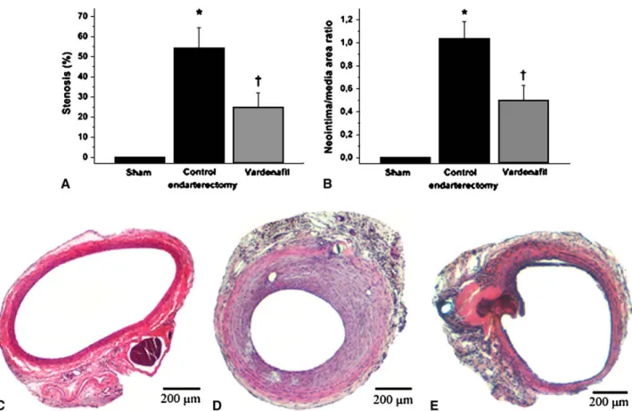
Three weeks after in vivo endothelial denudation, concen- tric neointima was found in the control endarterectomized group, resulting in a 54.12%stenosis of the original luminal area (Figure 1). In the phosphodiesterase-5 inhibitor–treated group, significant reduction of neointima formation was observed. Stenosis was 24.64%in this group (P<.05, treat- ment group compared with control endarterectomy group).
In addition, the neointimal/medial area ratio was calculated, which was found to be significantly reduced after phospho- diesterase-5 inhibition (0.50 vs 1.03 in the phosphodiester- ase-5 inhibitor–treated group vs the control endarterectomy group,P<.05). In the sham and unoperated groups, normal morphologic structure was found with a single endothelial cell layer.
Neointimal cells originate from the medial layer of the injured de-endothelialized vessel by means of migration and proliferation of vascular SMCs. To confirm this, we detected a-smooth muscle actin in the neointimas of the control and treated animals. Parallel to the extent of intimal
FIGURE 1. Morphometric results and representative histologic cross-sections from the different groups (hematoxylin and eosin staining). A, Percentage of stenosis (neointimal/original luminal area ratio). B, Neointimal/medial area ratio. C, Sham group. D, Control endarterectomy group with massive neointima proliferation. E, Significantly less neointima formation after treatment with vardenafil. *P<.05 vs sham operation;yP<.05 vs control endarterectomy.
ET
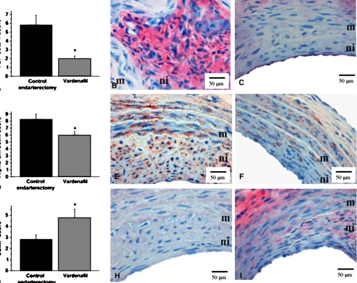
thickening during neointima formation, significantly less immunoreactive a-smooth muscle actin was found in the injured and treated vessels, whereas its levels were higher in the control group (5.98 0.51 vs 8.27 0.73 in the control group, P<.05, Figure 2). In addition, we used TGF-b1 immunostaining. Usually, marked TGF-b1 staining can be seen in proliferating tissues; therefore it is a useful marker of the proliferation activity in the neointima. An extensive TGF-b1 deposition was found in the control group, and the immunohistochemical score for TGF-b1 was significantly less in the vardenafil-treated group (2.0 0.3 vs 5.821.09 in the control group,P<.05).
Effects of the Phosphodiesterase-5 Inhibition on Neointimal and Plasma cGMP Levels
To confirm that the effect of vardenafil on neointima for- mation was related to the increase in cGMP levels seen in
our endarterectomy model, we determined neointimal cGMP content by means of immunohistochemistry, as well as by measuring circulating cGMP levels by means of enzyme immunoassay in deep-frozen plasma samples. As shown inFigure 2, immunoreactivity for cGMP was signi- ficantly higher in the neointimas of the vardenafil-treated group compared with those of the control endarterectomized group. Semiquantitative evaluation of all sections of both groups indicated a significantly higher score for the phos- phodiesterase-5 inhibitor–treated group compared with the control group (4.800.76 vs 2.84 0.40 in the control group,P<.05). The increased cGMP levels in the treatment group could be shown systematically as well. Plasma cGMP concentrations were almost twice as increased as in the con- trol groups (84.6512.77 pmol/mL vs 43.503.30 pmol/
mL and 38.20 2.18 in the control endarterectomy and unoperated control groups, respectively;P<.05;Figure 3).
FIGURE 2. Representative photomicrographs (B–C, E–F, and H–I) and semiquantitative scoring (A, D, and G) of immunohistochemical staining for trans- forming growth factorb1 (TGF-b1; red stain, A–C) anda-smooth muscle actin (Alpha-sm actin; brown stain, D–F) and for cyclic guanosine monophosphate (cGMP; red stain) in the neointima (G–I) are shown in the control endarterectomy group (B, E, and H) and the vardenafil treatment group (C, F, and I).
m, media;ni, neointima. *P<.05 vs control endarterectomy.
ET
On the other hand, there was no significant difference in plasma cGMP concentrations between the 2 control groups (43.50 3.30 in the control endarterectomy group vs 38.202.18 in the unoperated control group).
Effects of the Phosphodiesterase-5 Inhibition on Apoptosis of Neointimal Cells
To test a possible contribution of apoptosis to the decrease of neointimal hyperplasia after treatment with vardenafil, we performed the TUNEL assay on each section from both groups. As shown inFigure 4, we did not find a significant difference in the apoptotic cell counts in the same unit (1000 mm2) of the control and treated neointimas (4.230.43 vs 4.440.64 in the control group).
DISCUSSION
Recent studies show that reduced cGMP signaling is asso- ciated with neointimal proliferation, which is the main cause of restenosis after endarterectomy.8It has also been shown that an NO donor can prevent neointimal hyperplasia in a rat carotid artery injury model, as well as in a porcine cor- onary injury model.9,10Additionally, neointima formation could be reduced by adenovirus-mediated transfer of the endothelial NO synthase gene in a rat model of balloon injury.11To the best of our knowledge, it has not been tested whether the selective inhibition of phosphodiesterase-5, the enzyme which is responsible for cGMP degradation, would also be capable of preventing neointima formation after end- arterectomy. Some non–cGMP-specific phosphodiesterases have been tested and showed beneficial effects on neointi- mal hyperplasia.14 The benzyl indazole derivative YC-1 also decreased neointimal hyperplasia; nevertheless, this
drug has only a partial phosphodiesterase-5 inhibitory effect.12,13 The primary aim of our present study was to test the effectiveness of the novel selective phosphodiester- ase-5 inhibitor vardenafil in a rat model of carotid endarter- ectomy. We previously showed, in the same rat carotid endarterectomy model, that 21 days after endothelial denu- dation, significant neointimal hyperplasia and ongoing neo- intima development is detectable; therefore we used the same timeframe in this study as well.15 The main results of the present study are that neointima formation and expres- sion of different neointimal markers were attenuated by treatment with the phosphodiesterase-5 inhibitor vardenafil.
Treatment with vardenafil significantly increased plasma cGMP levels, as well as neointimal cGMP content, as deter- mined at the end of the 3-week follow-up period. Compara- ble neointimal reduction could be achieved in our previous studies by using the PARP inhibitor or the combination of brachytherapy and the PARP inhibitor.7,19
Endothelially derived NO is able to activate the soluble guanylate cyclase in vascular SMCs, which is responsible for cGMP synthesis. cGMP mediates its intracellular effects through activation of specific cGMP-dependent protein kinases (protein kinase G [PKG]). The main effects of cGMP, which could be involved in decreased neointima formation, are as follows: inhibition of migration and prolif- eration of vascular SMCs and alterations of oxidative and apoptotic signaling.20,21
A possible downstream signaling mechanism that contrib- utes to the beneficial effects of phosphodiesterase-5 inhibi- tion on neointima formation acts through PKG, as was shown by Sinnaeve and colleagues22 in a PKG-overex- pressed rat model. In another study by the same authors, the antiproliferative and antimigratory effects of soluble guanylate cyclase gene transfer after balloon injury in rats were demonstrated, which again confirms the role of
FIGURE 3. Cyclic guanosine monophosphate(cGMP)plasma concentra- tions (in picomoles per milliliter) measured by means of competitive en- zyme immunoassay in the different groups. *P<.05 vs unoperated control and control endarterectomy.
FIGURE 4. Terminal deoxynucleotidyl transferase–mediated dUTP-bio- tin nick end labeling(TUNEL)–positive cell count in 1000mm2of neointima in the control endarterectomy and vardenafil groups.
ET
cGMP signaling in the reduction of neointima formation.23 In connection with these results, Tulis and associates13 showed reduced postangioplasty stenosis after enhanced cGMP signaling by using a benzyl indazole derivate (YC-1) that can stimulate soluble guanylate cyclase (sGC) in vascu- lar endothelium and SMCs. The benzyl indazole derivate used in this study has, in addition, a phosphodiesterase-5–
inhibiting secondary effect as well. Liu and coworkers12 showed similar results on neointimal hyperplasia using YC-1. Several mechanisms for the potent protective actions of vardenafil on neointima formation can be suggested based on results from in vitro studies. Yu and colleagues21showed an inhibition of DNA synthesis associated with diminution in mitogen-activated protein kinase levels after enhanced cGMP signaling. Other work showed cGMP-induced inhibi- tion in anti-mitogen cyclin-dependent kinase activity and cyclin D1 expression, leading to the suppression of cell cycle in vascular cells.24
An interesting new finding of our study is a significant reduction of TGF-b1 immunoreactivity in the neointima after treatment with the phosphodiesterase-5 inhibitor vardenafil. It has been shown in several studies that the TGF-b system contributes to neointimal proliferation and that neointimal hyperplasia could be suppressed by using TGF-bantibody.25,26TGF-b1 is overexpressed in fibropro- liferative vascular lesions, allowing a slow but uncontrolled growth while overproducing extracellular matrix compo- nents. In addition, Saura and coworkers27 showed that endothelial cells treated with an NO donor exhibited a decreased response to TGF-b, resulting in a downregulation of TGF-b target genes.28 Taking these findings together with our present results, the following explanation can be given for the linkage between cGMP and TGF-b1 signal- ing during neointima formation: NO decreases TGF-b1 signaling through increases in cGMP levels, which finally leads to reduced vascular SMC proliferation. In our opinion the same linkage is present in the phosphodiesterase-5 inhibitor–treated neointima because treatment with varde- nafil leads to an increased cGMP signaling similar to the effect of NO. The observed reduction in TGF-b1 levels after treatment with vardenafil in our study might be a primary consequence of phosphodiesterase-5 inhibition because of interrupted cytokine production rather than a secondary phenomenon. Nevertheless, we have no direct evidence in the present study or in the literature that would support this hypothesis. In addition to the decreased TGF-b1 immunoreactivity after treatment with vardenafil, we found decreased expression in a-smooth muscle actin as well. Cell culture studies suggest that vascular SMC mi- gration is attenuated by the NO–cGMP–PKG pathway.20,29 On the other hand, cGMP-increasing agents have been found to prevent actin polymerization and thereby decrease vascular cell migration. We can speculate that decreased a-smooth muscle actin expression found in the treated neo-
intima in our study might be due to decreased proliferation and migration.
Chiche and associates30 showed proapoptotic effects of the NO–cGMP pathway in cultured rat pulmonary artery SMCs after adenovirus-mediated gene transfer of cGMP- dependent protein kinase. To test a possible contribution of apoptosis to the reduction of neointimal hyperplasia after treatment with phosphodiesterase-5 inhibitor, we performed the TUNEL assay. No effects were found in our in vivo model of restenosis, which leads us to the conclusion that attenuated neointima formation might be due to the decreased proliferation activity rather than to enhanced neo- intimal apoptosis. Nevertheless, we cannot exclude other mechanisms to be involved in the beneficial effects of phos- phodiesterase-5 inhibition on neointimal hyperplasia. Vaso- dilatation or phosphorylation of protein kinases could also influence this process. Unfortunately, we do not have any direct evidence regarding this issue in our present study;
further studies are necessary to investigate these issues.
Limitations of the Study
The carotid endarterectomy model used in this study does not resemble the complex situation of a severely diseased atherosclerotic artery in a patient. However, the native rat ca- rotid artery is an established model of intimal hyperplasia.15 In addition, it has to be noted that animal models of athero- sclerosis also lack direct comparability with the human situ- ation. Even knockout models have pathophysiologic and phenotypic differences regarding plaque cause and morphol- ogy. However, further studies with animal models of athero- sclerosis are warranted in the future to address this issue.
CONCLUSION
Treatment with the selective phosphodiesterase-5 inhibitor vardenafil significantly suppressed neointimal hyperplasia af- ter surgical endarterectomy in a rat model. These results sug- gest a potential therapeutic role of vardenafil in preventing neointima formation and restenosis after endarterectomy.
The expert technical assistance of H. Ziebart and G. Piecha is gratefully acknowledged.
References
1. North American Symptomatic Carotid Endarterectomy Trial Collaborators. Ben- eficial effect of carotid endarterectomy in symptomatic patients with high-grade carotid stenosis.N Engl J Med. 1991;325:445-53.
2. Szeplaki G, Hirschberg K, Gombos T, Varga L, Prohaszka Z, Dosa E, et al. Early complement activation follows eversion carotid endarterectomy and correlates with the time of clamping of the carotid artery.Mol Immunol. 2008;11:3289-94.
3. Szabo A, Laki J, Madsen HO, Dosa E, Prohaszka Z, Rugonfalvi-Kiss S, et al. Early rise in serum VEGF and PDGF levels predisposes patients with a normal MBL2 genotype to restenosis after eversion endarterectomy.Stroke. 2007;38:2247-53.
4. Dosa E, Szabo A, Prohaszka Z, Karadi I, Rugonfalvi-Kiss S, Apor A, et al.
Changes in the plasma concentration of soluble thrombomodulin in patients with severe carotid artery stenosis after eversion endarterectomy.Inflamm Res.
2005;54:289-94.
ET
5. Hunter GC, Edgar J, Poth Memorial/W.L. Gore and Associates, Inc, Lectureship.
The clinical and pathological spectrum of recurrent carotid stenosis.Am J Surg.
1997;174:583-8.
6. Ohwada T, Ishibashi T, Yaoita H, Shindo J, Noji H, Ohkawara H, et al. Different contribution of apoptosis to the antiproliferative effects of L-arginine, enalapril and losartan on neointimal growth inhibition after balloon arterial injury.Circ J. 2002;66:965-71.
7. Beller CJ, Radovits T, Kosse J, Gero D, Szabo C, Szabo G. Activation of the peroxynitrite-poly(adenosine diphosphate-ribose) polymerase pathway during neointima proliferation: a new target to prevent restenosis after endarterectomy.
J Vasc Surg. 2006;43:824-30.
8. Melichar VO, Behr-Roussel D, Zabel U, Uttenthal LO, Rodrigo J, Rupin A, et al.
Reduced cGMP signaling associated with neointimal proliferation and vascular dys- function in late-stage atherosclerosis.Proc Natl Acad Sci U S A. 2004;101:16671-6.
9. Yoon JH, Wu CJ, Homme J, Tuch RJ, Wolff RG, Topol EJ, et al. Local delivery of nitric oxide from an eluting stent to inhibit neointimal thickening in a porcine cor- onary injury model.Yonsei Med J. 2002;43:242-51.
10. Pearce CG, Najjar SF, Kapadia MR, Murar J, Eng J, Lyle B, et al. Beneficial effect of a short-acting NO donor for the prevention of neointimal hyperplasia.Free Radic Biol Med. 2008;44:73-81.
11. Janssens S, Flaherty D, Nong Z, Varenne O, van Pelt N, Haustermans C, et al.
Human endothelial nitric oxide synthase gene transfer inhibits vascular smooth muscle cell proliferation and neointima formation after balloon injury in rats.
Circulation. 1998;97:1274-81.
12. Liu YN, Pan SL, Peng CY, Guh JH, Huang DM, Chang YL, et al. YC-1 [3- (50-hydroxymethyl-20-furyl)-1-benzyl indazole] inhibits neointima formation in balloon-injured rat carotid through suppression of expressions and activities of matrix metalloproteinases 2 and 9.J Pharmacol Exp Ther. 2006;316:35-41.
13. Tulis DA, Durante W, Peyton KJ, Chapman GB, Evans AJ, Schafer AI. YC-1, a benzyl indazole derivative, stimulates vascular cGMP and inhibits neointima formation.Biochem Biophys Res Commun. 2000;279:646-52.
14. Yamamoto K, Onoda K, Sawada Y, Fujinaga K, Imanaka-Yoshida K, Yoshida T, et al. Locally applied cilostazol suppresses neointimal hyperplasia and medial thickening in a vein graft model.Ann Thorac Cardiovasc Surg. 2007;13:322-30.
15. Beller CJ, Kosse J, Radovits T, Krempien R, Gross ML, Berger I, et al. Adjunct brachytherapy: a new concept to prevent intimal hyperplasia after surgical endar- terectomy?Eur J Cardiothorac Surg. 2006;29:334-42.
16. Legan E, Sisson JA. Method to denude rat aortic endothelium with saponin for phos- phoinositide analysis in vascular smooth muscle.J Pharmacol Methods. 1990;23:31-9.
17. Filippi S, Morelli A, Sandner P, Fibbi B, Mancina R, Marini M, et al. Character- ization and functional role of androgen-dependent PDE5 activity in the bladder.
Endocrinology. 2007;148:1019-29.
18. Ehsan A, Sommer F, Schmidt A, Klotz T, Koslowski J, Niggemann S, et al. Nitric oxide pathways in human bladder carcinoma. The distribution of nitric oxide syn- thases, soluble guanylyl cyclase, cyclic guanosine monophosphate, and nitrotyr- osine.Cancer. 2002;95:2293-301.
19. Beller CJ, Kosse J, Radovits T, Gero D, Krempien R, Gross ML, et al. Poly(ADP- ribose) polymerase inhibition combined with irradiation: a dual treatment concept to prevent neointimal hyperplasia after endarterectomy.Int J Radiat Oncol Biol Phys. 2006;66:867-75.
20. Munzel T, Feil R, Mulsch A, Lohmann SM, Hofmann F, Walter U. Physiology and pathophysiology of vascular signaling controlled by guanosine 30,50-cyclic mono- phosphate-dependent protein kinase [corrected Circulation 2008;108:3165].
Circulation. 2003;108:2172-83.
21. Yu SM, Hung LM, Lin CC. cGMP-elevating agents suppress proliferation of vas- cular smooth muscle cells by inhibiting the activation of epidermal growth factor signaling pathway.Circulation. 1997;95:1269-77.
22. Sinnaeve P, Chiche JD, Gillijns H, Van Pelt N, Wirthlin D, Van De Werf F, et al. Overexpression of a constitutively active protein kinase G mutant re- duces neointima formation and in-stent restenosis. Circulation. 2002;105:
2911-6.
23. Sinnaeve P, Chiche JD, Nong Z, Varenne O, Van Pelt N, Gillijns H, et al. Soluble guanylate cyclase alpha(1) and beta(1) gene transfer increases NO responsiveness and reduces neointima formation after balloon injury in rats via antiproliferative and antimigratory effects.Circ Res. 2001;88:103-9.
24. Fukumoto S, Koyama H, Hosoi M, Yamakawa K, Tanaka S, Morii H, et al.
Distinct role of cAMP and cGMP in the cell cycle control of vascular smooth muscle cells: cGMP delays cell cycle transition through suppression of cyclin D1 and cyclin-dependent kinase 4 activation.Circ Res. 1999;85:
985-91.
25. Fu K, Corbley MJ, Sun L, Friedman JE, Shan F, Papadatos JL, et al. SM16, an orally active TGF-beta type I receptor inhibitor prevents myofibroblast induction and vascular fibrosis in the rat carotid injury model.Arterioscler Thromb Vasc Biol. 2008;28:665-71.
26. Wolf YG, Rasmussen LM, Ruoslahti E. Antibodies against transforming growth factor-beta 1 suppress intimal hyperplasia in a rat model.J Clin Invest. 1994;93:
1172-8.
27. Saura M, Zaragoza C, Herranz B, Griera M, Diez-Marques L, Rodriguez-Puyol D, et al. Nitric oxide regulates transforming growth factor-beta signaling in endothe- lial cells.Circ Res. 2005;97:1115-23.
28. Wang S, Wu X, Lincoln TM, Murphy-Ullrich JE. Expression of constitu- tively active cGMP-dependent protein kinase prevents glucose stimulation of thrombospondin 1 expression and TGF-beta activity.Diabetes. 2003;52:
2144-50.
29. Gurjar MV, Sharma RV, Bhalla RC. eNOS gene transfer inhibits smooth muscle cell migration and MMP-2 and MMP-9 activity.Arterioscler Thromb Vasc Biol.
1999;19:2871-7.
30. Chiche JD, Schlutsmeyer SM, Bloch DB, de la Monte SM, Roberts JD Jr, Filippov G, et al. Adenovirus-mediated gene transfer of cGMP-dependent protein kinase increases the sensitivity of cultured vascular smooth muscle cells to the antiproliferative and pro-apoptotic effects of nitric oxide/cGMP.J Biol Chem.
1998;273:34263-71.