https://doi.org/10.1007/s00439-020-02148-0 REVIEW
Molecular genetic diagnostics of hypogonadotropic hypogonadism:
from panel design towards result interpretation in clinical practice
Henriett Butz1,2,3 · Gábor Nyírő1,4,5 · Petra Anna Kurucz1 · István Likó2 · Attila Patócs1,2,3
Received: 8 January 2020 / Accepted: 5 March 2020
© The Author(s) 2020
Abstract
Congenital hypogonadotropic hypogonadism (CHH) is a clinically and genetically heterogeneous congenital disease. Symp- toms cover a wide spectrum from mild forms to complex phenotypes due to gonadotropin-releasing hormone (GnRH) deficiency. To date, more than 40 genes have been identified as pathogenic cause of CHH. These genes could be grouped into two major categories: genes controlling development and GnRH neuron migration and genes being responsible for neuroendocrine regulation and GnRH neuron function. High-throughput, next-generation sequencing (NGS) allows to ana- lyze numerous gene sequences at the same time. Nowadays, whole exome or whole genome datasets could be investigated in clinical genetic diagnostics due to their favorable cost–benefit. The increasing genetic data generated by NGS reveal novel candidate genes and gene variants with unknown significance (VUSs). To provide clinically valuable genetic results, complex clinical and bioinformatics work are needed. The multifaceted genetics of CHH, the variable mode of inheritance, the incomplete penetrance, variable expressivity and oligogenic characteristics further complicate the interpretation of the genetic variants detected. The objective of this work, apart from reviewing the currently known genes associated with CHH, was to summarize the advantages and disadvantages of the NGS-based platforms and through the authors’ own practice to guide through the whole workflow starting from gene panel design, performance analysis and result interpretation. Based on our results, a genetic diagnosis was clearly identified in 21% of cases tested (8/38).
Congenital hypogonadotropic hypogonadism (CHH)
Congenital hypogonadotropic hypogonadism (CHH) as a clinically heterogeneous entity
Congenital hypogonadotropic hypogonadism (CHH) is a genetic condition characterized by incomplete or absent puberty and infertility due to central (tertiary or hypotha- lamic) hypogonadism caused by gonadotropic hormone- releasing hormone (GnRH) deficiency. Three clinical forms are distinguished by the European consensus: (1) GnRH defi- ciency with defective sense of smell (Kallmann syndrome, KS), (2) isolated GnRH deficiency (normosmic CHH) and the third form when KS/CHH is part of a complex genetic syndrome (Boehm et al. 2015). CHH has an incidence of 1:125,000 in female and 1:30,000 in males indicating the male predominance (Stamou and Georgopoulos 2018). CHH has a heterogeneous clinical appearance, and lately the con- stitutional delay of growth and puberty (CDGP), the adult- onset hypogonadotropic hypogonadism and the hypotha- lamic amenorrhea are also considered as a milder end of the
Electronic supplementary material The online version of this article (https ://doi.org/10.1007/s0043 9-020-02148 -0) contains supplementary material, which is available to authorized users.
* Attila Patócs
patocs.attila@med.semmelweis-univ.hu
1 Department of Laboratory Medicine, Semmelweis University, Nagyvárad tér 4, Budapest 1089, Hungary
2 Hereditary Tumours Research Group, Hungarian Academy of Sciences and Semmelweis University, Budapest, Hungary
3 Department of Molecular Genetics, National Institute of Oncology, Budapest, Hungary
4 Molecular Medicine Research Group, Hungarian Academy of Sciences and Semmelweis University, Budapest, Hungary
5 2nd Department of Internal Medicine, Semmelweis University, Budapest, Hungary
spectrum (Stamou and Georgopoulos 2018). Most patients are diagnosed in adolescence due to delayed puberty. In male, neonate cryptorchidism and micropenis can be con- sidered as signs of CHH, but there are no specific signs of CHH in female neonates (Young et al. 2019). Prepubertal testes and undervirilized secondary sexual features are the most common symptoms in males, while absence of breast development and primary amenorrhea occur in females as a consequence of CHH (Young et al. 2019). The disease can be diagnosed in adulthood as well by low libido, infer- tility, bone loss and fractures when it is untreated (Young et al. 2019). Interestingly, in 10–20% of the cases, CHH is reported reversible, however, the pathophysiology behind this is not clearly revealed (Stamou and Georgopoulos 2018;
Young et al. 2019). To establish the biochemical diagnosis in infants is challenging as GnRH neurons are active only dur- ing mini-puberty (4–8 weeks after birth) and after that, their activity becomes quiescent until puberty. In adolescence, results of biochemical tests (basal and stimulated blood levels of sex hormones and gonadotropins), brain imaging for examination of olfactory bulbs, assessment of smell and evaluation of family history are parts of the routine medical investigations. (Naturally, additional work-ups, i.e. evalua- tion of bones, kidneys and sexual organs, are also required for diagnosis and for differential diagnostic purposes (Young et al. 2019). Constitutional delayed of growth and puberty (CDGP) is defined as the lack of the start of sexual matura- tion at an age > 2 SDs above the mean for a given population (Stamou and Georgopoulos 2018). There is no identifiable cause behind and finally puberty occurs. 50–80% of CDGP individuals have positive family history of the phenomenon, and approximately 10% of CHH patients have relatives with CDGP (Stamou and Georgopoulos 2018). Differentiating CDGP and CHH in adolescence is challenging as to date no gold-standard diagnostic test is known for this purpose (Young et al. 2019).
There are non-reproductive features as well that are commonly recognized in patients with CHH. Midline facial defects (cleft lip or palate), dental agenesis, unilateral renal agenesis, short metacarpals, hearing loss, synkinesia, cer- ebellar ataxia can appear additionally to CHH (Young et al.
2019). Furthermore, the disease can occur as part of com- plex genetic syndromes summarized in Table 1.
Diagnostics and genetic counseling is important in CHH as effective therapies are available for the development of secondary sexual features and fertility (Maione et al. 2018;
Young et al. 2019).
Adult onset of hypogonadotropic hypogonadism is a rare form of CHH. It is a non-reversible, long-lasting condition but the etiology and pathogenesis have to be investigated and demonstrated. The diagnosis can be made when all other acquired causes of hypogonadotropic hypogonadism (e.g. structural anomalies, infiltrative/inflammatory origin,
pituitary/CNS tumors etc.) have been excluded (Stamou and Georgopoulos 2018).
Genetic background of CHH
CHH is heterogeneous not only clinically but also geneti- cally. To date, more than 40 genes have been identified as pathogenic cause in the background of the disease (Boehm et al. 2015; Maione et al. 2018; Stamou and Georgopoulos 2018). Analysis the individual CHH genes (Table 1) one by one exceeds the goal of our study, but these are excel- lently reviewed in recent papers (Topaloglu and Kotan 2016; Topaloğlu 2018; Maione et al. 2018). Genes impli- cated in the pathogenesis of CHH are usually divided into two major categories (Boehm et al. 2015; Topaloğlu 2018;
Maione et al. 2018; Stamou and Georgopoulos 2018). The first group consists of genes that control development and GnRH neuron migration. Therefore, the pathogenic variants of these genes are frequently associated with anosmia and midline developmental anomalies (Table 1). The second group of genes is responsible for neuroendocrine physiology and GnRH neuron function (either by afferent modulators or by regulating GnRH secretion), these can be detected in normosmic CHH forms. Although there are genes with mul- tiple roles that participate in both mechanisms, their muta- tions can be often identified in both anosmic and normosmic forms (Boehm et al. 2015; Maione et al. 2018; Stamou and Georgopoulos 2018) (Table 1).
Autosomal dominant, autosomal recessive and X-linked inheritance have been identified, however, with the availability of high-throughput next-generation sequencing at least 20% of CHH cases have thought to be di- or oligogenic. In these cases, two or more gene variants can be identified in the same patient (Boehm et al. 2015) (Table 1). Still, in more than half of the CHH cases, there is no pathogenic mutation identified. Among the main genetic forms of CHH, the most common autosomal recessively inherited types are caused by GNRHR, KISS1R and TACR3 variants (Maione et al. 2018). Kallmann syndrome caused by ANOS1 gene mutations is inherited by X-linked recessive trait as it is located on chromosome X. FGFR1 and PROK2/PROKR2 lead to autosomal dominantly inherited type of CHH (Boehm et al. 2015; Maione et al. 2018). Regarding FGFR1, nearly half, regarding PROK2/PROKR2, nearly two- third of the cases exhibit incomplete penetrance and variable expressivity that complicate the determination of inheritance (Maione et al. 2018). Recently, a normosmic CHH patient was reported who inherited a pathogenic variant in GNRHR gene in a homozygous form due to the occurrence of uniparental isodisomy (Cioppi et al. 2019). (Uniparental disomy-UPD is a non-Mendelian inheritance pattern when an individual has inherited two copies of a specific chromosome (or part of it) from a single parent. When a chromosomal pair inherited from the same parent, it is called uniparental heterodisomy, when
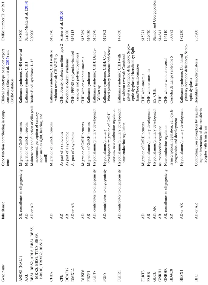
Table 1 Congenital hypogonadotropic hypogonadism gene characteristics Gene nameInheritanceGene function contributing in symp- tomsClinical phenotype based on expert consensus (Boehm et al. 2015) and OMIM database OMIM number ID or Ref ANOS1 (KAL1)XR, contributes to oligogenicityMigration of GnRH neuronsKallmann syndrome, CHH reversal308700 AXLADMigration of GnRH neuronsKallmann syndrome; CHHSalian-Mehta et al. (2014)
BBS1, BBS2, ARL6, BBS4, BBS5, MKK S, BBS7, TTC8, BBS9, BBS10, TRIM32, BBS12
AD or AR
Maintenance and function of cilia (cell mo vement, perception of sensory input such as sight, hearing, and smell)
Bardet-Biedl syndrome 1–12209900 CHD7ADMigration of GnRH neuronsKallmann syndrome; CHH with or without reversal; CHARGE sy612370 CPEARAs part of a syndromeCHH, obesity, diabetes mellitus type 2Alsters et al. (2015) DCAF17ARAs part of a syndromeWoodhouse-Sakati syndrome241080 DMXL2AD or ARAs part of a syndromeCHH; PEPNS (polyendocrine defi- ciencies and polyneuropathies)616113 DUSP6ADMigration of GnRH neuronsCHH with or without anosmia615269 FEZF1ARMigration of GnRH neuronsKallmann syndrome616030 FGF17AD, contributes to oligogenicityHypothalamus/pituitary developmentKallmann syndrome; CHH; Dandy- Walker sy615270 FGF8AD, contributes to oligogenicityHypothalamus/pituitary development,migration of GnRH neurons, neuroendocrine regulation
Kallmann syndrome; CHH; Com- bined pituitary hormone deficiency612702 FGFR1AD, contributes to oligogenicityHypothalamus/pituitary development, neuroendocrine regulationKallmann syndrome; CHH with or without reversal; Combined pituitary hormone deficiency; Septo- optic dysplasia; Hartsfield sy; Split hand/foot malformation
147950 FLRT3ADMigration of GnRH neuronsCHH with anosmia615271 FSHBARHypothalamus/pituitary developmentCHH without anosmia229070 GLCEAD, ARHypothalamus/pituitary developmentKS, CHHStamou and Georgopoulos (2018) GNRH1ARneuroendocrine regulationCHH614841 GNRHRAR, contributes to oligogenicityNeuroendocrine regulationCHH with or without reversal146110 HDAC8XRTranscriptional regulation, cell cycle progression and developmentCornelia de Lange syndrome 5300882 HESX1AD or ARHypothalamus/pituitary developmentKallmann syndrome; Combined pituitary hormone deficiency, Septo- optic dysplasia 182230 HFEAD or ARControlling iron absorption by regulat- ing the interaction of the transferrin receptor with transferrin
Hereditary hemochromatosis235200
Table 1 (continued) Gene nameInheritanceGene function contributing in symp- tomsClinical phenotype based on expert consensus (Boehm et al. 2015) and OMIM database
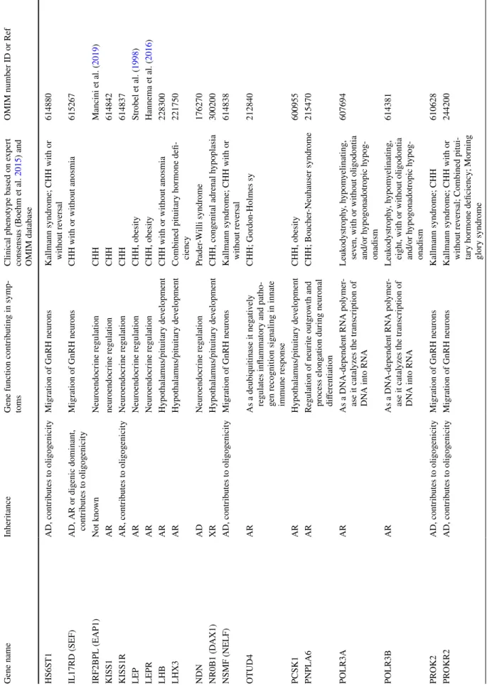
OMIM number ID or Ref HS6ST1AD, contributes to oligogenicityMigration of GnRH neuronsKallmann syndrome; CHH with or without reversal614880 IL17RD (SEF)AD, AR or digenic dominant, contributes to oligogenicityMigration of GnRH neuronsCHH with or without anosmia615267 IRF2BPL (EAP1)Not knownNeuroendocrine regulationCHHMancini et al. (2019) KISS1ARneuroendocrine regulationCHH614842 KISS1RAR, contributes to oligogenicityNeuroendocrine regulationCHH614837 LEPARNeuroendocrine regulationCHH, obesityStrobel et al. (1998) LEPRARNeuroendocrine regulationCHH, obesityHannema et al. (2016) LHBARHypothalamus/pituitary developmentCHH with or without anosmia228300 LHX3ARHypothalamus/pituitary developmentCombined pituitary hormone defi- ciency221750 NDNADNeuroendocrine regulationPrader-Willi syndrome176270 NR0B1 (DAX1)XRHypothalamus/pituitary developmentCHH, congenital adrenal hypoplasia300200 NSMF (NELF)AD, contributes to oligogenicityMigration of GnRH neuronsKallmann syndrome; CHH with or without reversal614838 OTUD4ARAs a deubiquitinase it negatively regulates inflammatory and patho- gen recognition signaling in innate immune response
CHH; Gordon-Holmes sy212840 PCSK1ARHypothalamus/pituitary developmentCHH, obesity600955 PNPLA6ARRegulation of neurite outgrowth and process elongation during neuronal differentiation CHH; Boucher-Neuhauser syndrome215470 POLR3AARAs a DNA-dependent RNA polymer- ase it catalyzes the transcription of DNA into RNA
Leukodystrophy, hypomyelinating, seven, with or without oligodontia and/or hypogonadotropic hypog- onadism 607694 POLR3BARAs a DNA-dependent RNA polymer- ase it catalyzes the transcription of DNA into RNA
Leukodystrophy, hypomyelinating, eight, with or without oligodontia and/or hypogonadotropic hypog- onadism
614381 PROK2AD, contributes to oligogenicityMigration of GnRH neuronsKallmann syndrome; CHH610628 PROKR2AD, contributes to oligogenicityMigration of GnRH neuronsKallmann syndrome; CHH with or without reversal; Combined pitui- tary hormone deficiency; Morning glory syndrome
244200
Table 1 (continued) Gene nameInheritanceGene function contributing in symp- tomsClinical phenotype based on expert consensus (Boehm et al. 2015) and OMIM database
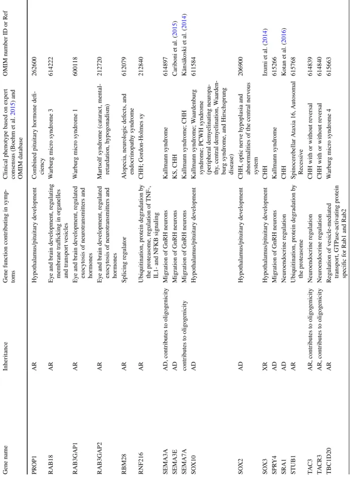
OMIM number ID or Ref PROP1ARHypothalamus/pituitary developmentCombined pituitary hormone defi- ciency262600 RAB18AREye and brain development, regulating membrane trafficking in organelles and transport vesicles
Warburg micro syndrome 3614222 RAB3GAP1AREye and brain development, regulated exocytosis of neurotransmitters and hormones Warburg micro syndrome 1600118 RAB3GAP2AREye and brain development, regulated exocytosis of neurotransmitters and hormones
Martsolf syndrome (cataract, mental- retardation, hypogonadism)212720 RBM28ARSplicing regulatorAlopecia, neurologic defects, and endocrinopathy syndrome612079 RNF216ARUbiquitination, protein degradation by the proteasome, regulation of TNF-, IL1- and NFKB signaling
CHH; Gordon-Holmes sy212840 SEMA3AAD, contributes to oligogenicityMigration of GnRH neuronsKallmann syndrome614897 SEMA3EADMigration of GnRH neuronsKS, CHHCariboni et al. (2015) SEMA7Acontributes to oligogenicityMigration of GnRH neuronsKallmann syndrome; CHHKänsäkoski et al. (2014) SOX10ADHypothalamus/pituitary developmentKallmann syndrome; Waardenburg syndrome; PCWH syndrome (peripheral demyelinating neuropa- thy, central demyelination, Waarden- burg syndrome, and Hirschsprung disease) 611584 SOX2ADHypothalamus/pituitary developmentCHH, optic nerve hypoplasia and abnormalities of the central nervous system
206900 SOX3XRHypothalamus/pituitary developmentCHHIzumi et al. (2014) SPRY4ADMigration of GnRH neuronsKallmann syndrome615266 SRA1ADNeuroendocrine regulationCHHKotan et al. (2016) STUB1ARUbiquitination, protein degradation by the proteasomeSpinocerebellar Ataxia 16, Autosomal Recessive615768 TAC3AR, contributes to oligogenicityNeuroendocrine regulationCHH with or without reversal614839 TACR3AR, contributes to oligogenicityNeuroendocrine regulationCHH with or without reversal614840 TBC1D20ARRegulation of vesicle-mediated transport, GTPase-activating protein specific for Rab1 and Rab2
Warburg micro syndrome 4615663
two identical chromosomes are inherited it is called uniparen- tal isodisomy (iUPD). This discovery further complicates the inheritance pattern of CHH and raises the possibility of the same phenomenon in case of other genes as well.
Individual relevance of genes in oligogenic CHH cases are needed to be interpreted with cautions. For instance, another candidate CHH gene NSMF (earlier NELF), listed in the expert consensus statement, has now a controversial role (Spilker et al. 2016). Several publications reported NSMF variants in CHH patients alone or in combination with a mutation in another gene (Miura et al. 2004; Pitteloud et al.
2007; Xu et al. 2011) underlining again its questionable role (Spilker et al. 2016).
Interestingly, rare variants of TAC3, TACR3 and other genes are suggested to be linked with CHH reversal that further raises the possibility of therapy discontinuation from time to time to test the reversibility of CHH in these carriers (Gianetti et al. 2010; Boehm et al. 2015).
Variants of known CHH genes have been investigated and identified in CDGP and in cases with hypothalamic amenor- rhea too. This suggests that the time of menarche and meno- pause are genetically determined which is strongly supported by family histories (Stamou and Georgopoulos 2018).
In CDGP, Zhu et al. identified that variants in CHH genes were enriched in CDGP family members compared to unaf- fected family members suggesting the genetic link between CHH and CDGP (Zhu et al. 2015). This is further supported by variants identified in CDGP patients in TAC3, TACR3, IL17RD, GNRHR, PROKR2, HS6ST1, FGFR1, FEZF1, AXL genes (Gianetti et al. 2012; Tusset et al. 2012; Zhu et al.
2015; Hietamäki et al. 2017; Cassatella et al. 2018). How- ever, results of Cassatella et al. demonstrated that CDGP and CHH have distinct genetic profiles that may facilitate the differential diagnosis in patients presenting with delayed puberty (Cassatella et al. 2018).
Hypothalamic amenorrhea is also a reversible dysfunc- tional feature that can be triggered by nutritional deficit, extensive exercise or psychological stress. Genetic vari- ants have been identified in FGFR1, PROKR2, GNRHR and ANOS1 genes suggesting that these mutations may contrib- ute to the variable functional changes in GnRH secretion (Caronia et al. 2011).
CHH can be also part of complex genetic syndromes which are summarized by Boehm et al. and genetic back- ground are summarized in Table 1 (Boehm et al. 2015).
Genetic testing and genetic counseling in CHH Testing strategies
Although high-throughput screening can be recommended, targeted panel testing, prioritization and gene selection based on clinical data are also possible (Boehm et al.
Table 1 (continued) Gene nameInheritanceGene function contributing in symp- tomsClinical phenotype based on expert consensus (Boehm et al. 2015) and OMIM database
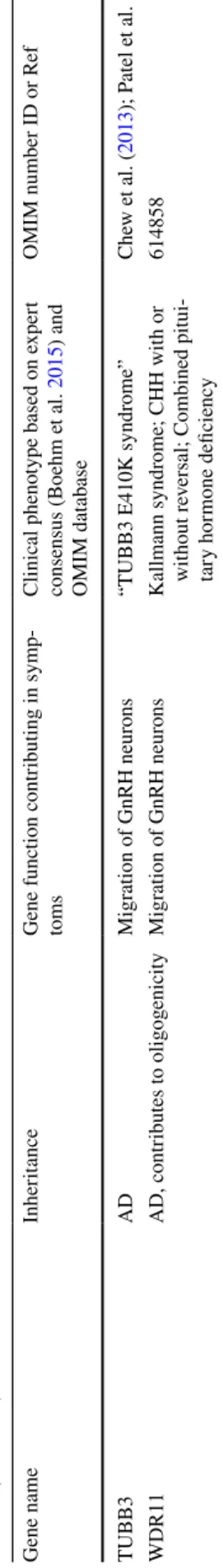
OMIM number ID or Ref TUBB3ADMigration of GnRH neurons“TUBB3 E410K syndrome”Chew et al. (2013); Patel et al. (2017) WDR11AD, contributes to oligogenicityMigration of GnRH neuronsKallmann syndrome; CHH with or without reversal; Combined pitui- tary hormone deficiency
614858
2015; Topaloğlu 2018; Stamou and Georgopoulos 2018).
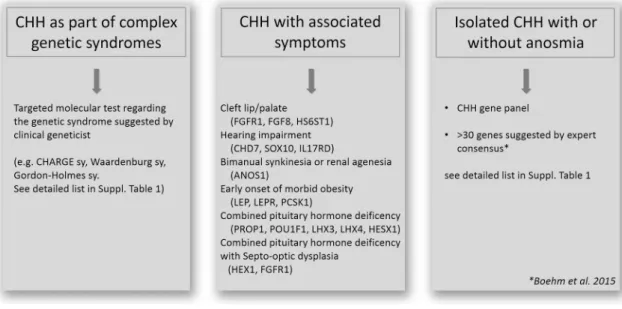
The first step is to exclude the presence of genetic syn- dromes based on clinical findings. When a clinical geneti- cist based on the whole clinical presentation indicates a specific syndrome (e.g. CHARGE sy., Bardet-Biedl sy., Gordon-Holmes sy., see details in Table 1) targeted gene testing is recommended. When complex syndromes can be excluded additional associated signs and symptoms can increase the probability of finding casual mutations (Boehm et al. 2015). For instance, besides anosmia/hypos- mia, bimanual synkinesia or renal agenesis can associ- ate with ANOS1 mutation (Fig. 1). Cleft palate/lip, dental agenesis and digital bone anomalies were frequently asso- ciated with CHH caused by mutations in genes of FGF8 signaling (FGFR1, FGF8, HS6ST1) (Costa-Barbosa et al.
2013; Boehm et al. 2015). Hearing impairment commonly appeared with CHH in CHD7, SOX10 or IL17RD mutation carriers (Costa-Barbosa et al. 2013; Boehm et al. 2015).
Additionally, early onset of morbid obesity with CHH could suggest variants in LEP, LEPR or PCSK1 genes (Jackson et al. 1997; Farooqi and O’Rahilly 2008). If CHH is associated with severe adrenal insufficiency congenital adrenal hypoplasia caused by NR0B1 (DAX1) is likely.
Combined pituitary hormone deficiency (CHPD) should also be clinically investigated/excluded as CHH and CHPD have overlapping genetic etiologies. If isolated CHPD is diagnosed, genetic testing of genes encoding the pituitary transcription factors (PROP1, POU1F1, LHX4, LHX3 and HESX1) should be recommended (Fang et al. 2016). How- ever, lately, variants of certain CHH genes including CHD7, PROKR2, WDR11, FGFR1 and FGF8 have also been impli- cated in CPHD (Raivio et al. 2012; Fang et al. 2016). Similar to CHH, CPHD is suggested to be a multifactorial disease
as symptoms frequently present incomplete penetrance even harboring the very same mutations (Raivio et al. 2012).
Genetic testing starts with evaluation of the inherit- ance pattern using pedigree analysis. However, Mendelian inheritance have been described for the majority of genes associated with CHH, some genes show different inherit- ance patterns (e.g. FGFR1: AD/AR/oligogenic/de novo;
PROK2/PROKR2: AD/AR/oligogenic), see Table 1 (Boehm et al. 2015; Maione et al. 2018).
Parallel with revolution of molecular genetic technolo- gies for patients with CHH multi-gene panel testing can be recommended, because there is a wide overlap between both symptoms and genetic background (Boehm et al. 2015;
Maione et al. 2018). Since expert consensus have been pub- lished in 2015 (Boehm et al. 2015) several high-throughput multi-gene panel studies were carried out. However, there is no consented gene list that should be offered for patients providing an accurate diagnosis for the majority of cases.
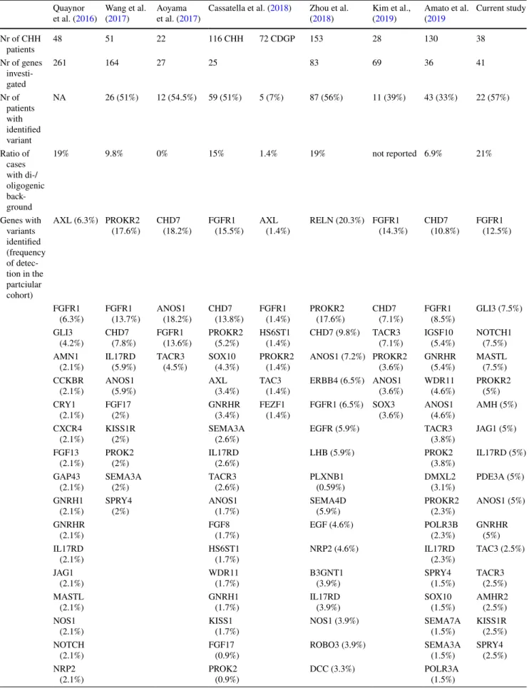
After publication of the expert consensus 7 CHH gene panel testing studies were reported (Table 2) (Quaynor et al. 2016;
Aoyama et al. 2017; Wang et al. 2017; Cassatella et al. 2018;
Zhou et al. 2018; Amato et al. 2019; Kim et al. 2019). In these studies, 25–261 genes were included as susceptibility genes of CHH. The positive detection rate varied between 33 and 56% (Table 2). Mutations in the FGFR1 gene were found the most commonly (in all eight studies), ANOS1 (in seven studies) and CHD7, PROKR2, TACR3 and IL17RD variants were also frequently detected (in six studies) among different groups (Table 2). Analyzing the detec- tion rate by patient number FGFR1 variants were detected most commonly, in an average of 11.4% of all investigated patients, CHD7, PROKR2 and ANOS1 in 8.4, 6.4 and 5.7%
of patients, respectively, across all studies. All other gene
Fig. 1 Genetic testing strategies in CHH (based on Boehm et al. 2015; Stamou and Georgopoulos 2018; Topaloğlu 2018)
Table 2 High-throughput NGS studies investigating CHH patients Quaynor
et al. (2016) Wang et al.
(2017) Aoyama
et al. (2017) Cassatella et al. (2018) Zhou et al.
(2018) Kim et al.,
(2019) Amato et al.
(2019 Current study Nr of CHH
patients 48 51 22 116 CHH 72 CDGP 153 28 130 38
Nr of genes investi- gated
261 164 27 25 83 69 36 41
Nr of patients with identified variant
NA 26 (51%) 12 (54.5%) 59 (51%) 5 (7%) 87 (56%) 11 (39%) 43 (33%) 22 (57%)
Ratio of cases with di-/
oligogenic back- ground
19% 9.8% 0% 15% 1.4% 19% not reported 6.9% 21%
Genes with variants identified (frequency of detec- tion in the partciular cohort)
AXL (6.3%) PROKR2
(17.6%) CHD7
(18.2%) FGFR1
(15.5%) AXL
(1.4%) RELN (20.3%) FGFR1
(14.3%) CHD7
(10.8%) FGFR1 (12.5%)
FGFR1
(6.3%) FGFR1
(13.7%) ANOS1
(18.2%) CHD7
(13.8%) FGFR1
(1.4%) PROKR2
(17.6%) CHD7
(7.1%) FGFR1
(8.5%) GLI3 (7.5%) GLI3
(4.2%) CHD7
(7.8%) FGFR1
(13.6%) PROKR2
(5.2%) HS6ST1
(1.4%) CHD7 (9.8%) TACR3
(7.1%) IGSF10
(5.4%) NOTCH1 (7.5%) AMN1
(2.1%) IL17RD
(5.9%) TACR3
(4.5%) SOX10
(4.3%) PROKR2
(1.4%) ANOS1 (7.2%) PROKR2
(3.6%) GNRHR
(5.4%) MASTL (7.5%) CCKBR
(2.1%) ANOS1
(5.9%) AXL
(3.4%) TAC3
(1.4%) ERBB4 (6.5%) ANOS1
(3.6%) WDR11
(4.6%) PROKR2 (5%) CRY1
(2.1%) FGF17
(2%) GNRHR
(3.4%) FEZF1
(1.4%) FGFR1 (6.5%) SOX3
(3.6%) ANOS1
(4.6%) AMH (5%) CXCR4
(2.1%) KISS1R
(2%) SEMA3A
(2.6%) EGFR (5.9%) TACR3
(3.8%) JAG1 (5%) FGF13
(2.1%) PROK2
(2%) IL17RD
(2.6%) LHB (5.9%) PROK2
(3.8%) IL17RD (5%) GAP43
(2.1%) SEMA3A
(2%) TACR3
(2.6%) PLXNB1
(0.59%) DMXL2
(3.1%) PDE3A (5%) GNRH1
(2.1%) SPRY4
(2%) ANOS1
(1.7%) SEMA4D
(5.9%) PROKR2
(2.3%) ANOS1 (5%) GNRHR
(2.1%) FGF8
(1.7%) EGF (4.6%) POLR3B
(2.3%) GNRHR (5%) IL17RD
(2.1%) HS6ST1
(1.7%) NRP2 (4.6%) IL17RD
(2.3%) TAC3 (2.5%) JAG1
(2.1%) WDR11
(1.7%) B3GNT1
(3.9%) SPRY4
(1.5%) TACR3 (2.5%) MASTL
(2.1%) GNRH1
(1.7%) IL17RD
(3.9%) SOX10
(1.5%) AMHR2 (2.5%) NOS1
(2.1%) KISS1
(1.7%) NOS1 (3.9%) SEMA7A
(1.5%) KISS1R (2.5%) NOTCH
(2.1%) FGF17
(0.9%) ROBO3 (3.9%) SEMA3A
(1.5%) SPRY4 (2.5%) NRP2
(2.1%) PROK2
(0.9%) DCC (3.3%) POLR3A
(1.5%)
Table 2 (continued) Quaynor
et al. (2016) Wang et al.
(2017) Aoyama
et al. (2017) Cassatella et al. (2018) Zhou et al.
(2018) Kim et al.,
(2019) Amato et al.
(2019 Current study PALM2
(2.1%) KISS1R
(0.9%) MTOR (3.3%) NSMF
(1.5%) PDE3A
(2.1%) TAC3
(0.9%) SEMA7A
(3.3%) IGFALS
(1.5%) PLEHKA5
(2.1%) DLX5 (2.6%) GNRH1
(1.5%)
RD3 (2.1%) GNRHR
(2.6%) FGF8
(1.5%) TRAPPC9
(2.1%) IGF1 (2.6%) TAC3
(0.8%) TSPAN11
(2.1%) KISS1R
(2.6%) RNF216
(0.8%)
PAX6 (2.6%) PNPLA6
(0.8%)
AXL (2%) OTX2
(0.8%)
CNTN2 (2%) IGSF1
(0.8%)
EBF2 (2%) FLRT3
(0.8%)
EFNA5 (2%) EBF2
(0.8%)
MET (2%) FGF17
(0.8%) PLXNA1 (2%)
SEMA3A (2%) SLIT2 (2%) TACR3 (2%) FEZ1 (1.3%) CCKAR
(1.3%) DCAF17
(1.3%) EDNRB
(1.3%) EPHA5 (1.3%) GHR (1.3%) HGF (1.3%) NRP1 (1.3%) WDR11
(1.3%) CASR (0.7%) GH1 (0.7%) GNRH1
(0.7%) LEPR (0.7%) LIF (0.7%) NELF(NSMF)
(0.7%) PROK2 (0.7%) STS (0.7%) TLE4 (0.7%)
variants were found less than an average of 3% in these patients (see details in Table 2). Di- and oligogenic cases occurred approximately between 10 and 20% of all cases.
Genetic counseling
Genetic screening is essential in CHH as it can be treated and patients could have a good reproductive prognosis upon treatment (see details in (Boehm et al. 2015; Maione et al.
2018). Genetic counseling should give information on her- itability for other family members too, and also required before family planning (Maione et al. 2018).
In certain cases, heritability can be determined rela- tively easily. For instance, in case of GNRH1/GNRHR, TAC3/TACR3, KISS1/KISS1R, autosomal recessive inherit- ance pattern is characteristic, while ANOS1 is inherited as an X-linked trait (Maione et al. 2018). However, for genes of which variants inherited by an autosomal dominant way, the penetrance and expressivity can be variable. In case of FGFR1 nearly half, regarding PROK2/PROKR2 nearly two- third of the cases exhibit incomplete penetrance and variable expressivity complicating the determination of the inherit- ance pattern (Maione et al. 2018). Regarding certain genes (e.g. FGFR1), de novo mutations are also relatively com- mon that has to be taken into consideration when analyzing pedigrees.
Additionally, together with the availability of NGS, the main challenge is to distinguish true oligogenicity from rare variants which appear as incidental findings and are not related to the phenotype. In determination of oligogenic- ity, genotype–phenotype co-segregation should be assessed by investigating both the affected and healthy family mem- bers. In addition, in diagnosis of oligogenicity, Maione et al.
(2018) suggested that oligogenic load has to be correlated with phenotype severity. There are several complicating fac- tors (small families, not available or not compliant family members, incomplete penetrance and variable expressivity) in segregation analysis, still, it is one of the most important way to identify the closest evidence of pathogenicity clini- cally besides in vitro and in vivo studies (Oliver et al. 2015;
Maione et al. 2018). Additionally, in clinical interpretation of variants of unknown significance (VUSs), clinical data (genotype–phenotype segregation) are of utmost important.
Once heritability is assessed, risk of disease transmission can be discussed according to the Mendelian rules.
Prognosis has also to be discussed as approximately 20%
of the cases appear to be spontaneously reversible. From
genetic point of view, to date TAC3 and TACR3 loss of func- tion variants were described to be associated with CHH reversal (Gianetti et al. 2010), but with the increasing data provided by high-throughput NGS platforms, the number of genes connected to this phenomenon will probably increase as well.
Next‑generation sequencing allows
evaluation of sequence variants of several genes at the same time in a cost‑effective way
Formerly, genetic testing was confined to rare genetic dis- orders due to their complexity, labour intensity and cost.
Now, NGS-based methods are widely available allowing to test even hundreds of genes at the same time. Therefore, NGS has been rapidly integrated into laboratory diagnos- tics workflows for identification of germline mutations in inherited diseases. Due to its time and cost effectiveness, it is especially useful in cases when several genes have been identified in the background of a certain genetic condition such as CHH.
NGS‑based platform options for clinical genetic diagnostics
Although the technology allows to investigate the sequence of the whole genome (WGS, whole genome sequencing) or exome (WES, whole exome sequencing) currently, the most prevalent applications of NGS in clinical practice are the evaluation of certain genes using targeted gene panels (Di Resta et al. 2018).
As WGS covers the whole genome (coding and noncoding regions) it may seem the most preferable choice in identifica- tion of pathogenic gene mutations in inherited diseases. The advantage of WGS is that library preparation is straightfor- ward as it does not require target enrichment. Additionally, data obtained from WGS can easily be used for detection of CNVs. However, among NGS approaches it gives the least average depth of coverage and it is still a costly tech- nology (Di Resta et al. 2018). Also, from clinical point of view, the interpretation of noncoding variants and variants of unknown significance (VUSs) make its utility limited.
WES aims to cover all coding regions in the genome.
Exome contains all of the protein-coding regions of genes and it comprises ~ 1–2% of the genome, yet contains ~ 85%
Table 2 (continued) Quaynor
et al. (2016) Wang et al.
(2017) Aoyama
et al. (2017) Cassatella et al. (2018) Zhou et al.
(2018) Kim et al.,
(2019) Amato et al.
(2019 Current study TYRO3 (0.7%)
of known disease causing mutations. Its cost is also more preferable and it is a more feasible option comparing to WGS (Di Resta et al. 2018). Usually, the average exome coverage of a WES test is 90–95% due to sequence complex- ity. WES is sometimes used by clinical laboratories by inter- preting only genes which have been already associated with any disease. When mutation has not been identified data analysis can be extended to the remaining exome regions.
It has been shown that WES provides diagnosis in approxi- mately of 11–40% of cases where the clinical diagnosis were uncertain (Sawyer et al. 2016). Furthermore, because the depth of coverage for WES is not uniform the sensitivity is usually lower compared to those observed in case of targeted disease panels.
Customized targeted gene panels offer the ability to per- form fast and low-cost screening option, therefore, currently, it is the most widely used NGS approach in clinical practice (Di Resta et al. 2018; Wang et al. 2018; Graziola et al. 2019).
By focusing on a limited set of genes selected for certain clinical condition, it is able to provide high coverage that increases analytical sensitivity even in detection of mosai- cism. Furthermore, because the role of genes included in these panels are known to be associated with the particular condition the detection rate (positive finding) is also higher compared to WES (Di Resta et al. 2018; Wang et al. 2018;
Graziola et al. 2019). Targeted panels give the advantage to avoid incidental, secondary findings and to decrease the number of VUSs detected.
Therefore, when the genetic background is well-defined, targeted testing of a gene panel could offer at a relatively low-cost sensitive detection of genetic variants responsible for a disease. However, when no suspect genes stand behind the clinical phenotype, exome sequencing may provide a wider screening option, but in these cases, trio sequencing would allow a more comprehensive result compared to the
“only” individual sequencing.
Workflow of an NGS‑based genetic analysis
NGS-based sequencing analysis comprises of three steps:
(1) library preparation, (2) parallel sequencing and (3) data analysis and variant interpretation (Oliver et al. 2015).
Molecular genetic analysis is routinely performed using DNA extracted from peripheral blood or buccal mucosa.
In our example, we used DNA samples of 38 consecutive patients and 2 family members with hypogonadotropic hypo- gonadism referred to our diagnostics laboratory. Fourteen patients developed the disease ≤ 18 years [2 girls and 12 boys with an average age of 16.2 year (± 2.1 years)]. Twenty-four patients developed disease in adult age (3 females, 21 males with an average age of 31.8 years (± 12.7 years). Patient characteristics, clinical findings, laboratory results and imaging studies are included in Supplementary Table 1. Our
study was approved by the Scientific and Research Commit- tee of the Medical Research Council of Ministry of Health, Hungary (67/PI/2012). All samples were obtained after acquiring written informed consent from all adult patients and permissions were given by parents of all minors. For NGS-based technologies, the amount and quality of input DNA is an essential factor. Degradation or low concentration of DNA may jeopardize the analysis.
For any NGS-based strategy, library preparation is a key step in the laboratory workflow. The instrumentation deter- mines the library preparations, because high-throughput instruments allow larger analysis. Barcodes (unique, short sequences) are used to label different samples enabling pooling patients’ samples into one reaction and decreasing the per-sample cost. Library preparation methods can be grouped into two main categories by principle used for gene amplifications: (1) PCR-based and (2) hybridization-based methods (Butz and Patócs 2019). Although processes using hybridization-based capture are more time consuming and labour intensive, those have the advantage of having greater tolerance against sequence variations (sequence variants and copy-number alteration).
The sequencing characteristics (read length, output read number, cost and run time) of each platform can be different that are needed to be taken into consideration.
For an in-house panel design (gene selection), the rec- ommendation of the European Society of Human Genetics should be followed. Only genes with known relationship between genotype and phenotype should be included in the analysis for diagnostic purposes (Matthijs et al. 2016). Also, the guideline states that “to avoid irresponsible testing, for the benefit of the patients, ‘core disease gene list’ should be established by the clinical and laboratory experts” (Matthijs et al. 2016). Therefore, consensus statements and guidelines, OMIM (Online Mendelian Inheritance in Man) database and literature search should be assessed to assemble genes in a diagnostic panel. For CHH, there is an available European Consensus Statement (Boehm et al. 2015) which was used as a primary guide during our panel design too.
Accordingly, our panel was designed during the first half of 2017. Some CHH-related genes were left out, mostly those which have been already introduced earlier into clini- cal practice in our laboratory (e.g. genes responsible for combined pituitary deficiency or adrenal diseases) (Halász et al. 2006; Bertalan et al. 2019) or due to the capacity of the applied method. Genes associated with complex syndromes were not present either in our selection owing to our patient profile. Finally, 41 genes were analyzed (see in Supplemen- tary Table 2 and in Table 3).
We selected NimbleGene approach to create the appro- priate hybridization capture probe set for our gene list using NimbleDesign Software (https ://seque ncing .roche .com/en/
produ cts-solut ions/by-categ ory/targe t-enric hment /softw are/
nimbl e-desig n-softw are.html) targeting the region of inter- ests (exons + /– 30 bp/exon). Capture probe synthesis was done by the supplier. Library was prepared following double capture; the library quantification was performed follow- ing the manufacturer’s instructions (NimbleGen SeqCap EZ
Library protocol). Sequencing runs were done on Illumina MiSeq instrument using MiSeq Reagent Micro Kit v2.
Sequencing data processing, performance analysis
During NGS, huge amount of data is produced which require special bioinformatics handling and analysis (Biesecker and Green 2014), therefore, appropriate hardware, software and expert personnel are required for data analysis (Oliver et al.
2015). Currently, there is no gold standard, freely available tool or filtering settings for bioinformatics analysis related to clinical applications of NGS. Each laboratory has to develop and validate its own pipeline (Oliver et al. 2015).
First step of sequencing data analysis is base calling that is integrated into the instrument’s software. During the next step, raw sequence reads are aligned to the reference human genome (Sayitoğlu 2016). Quality filtering of read align- ment defines sensitivity and specificity of the test. Using very strong filtering could lead to loss of variants, while inclusive filters can minimize false negative results but it will increase the burden of confirmatory analysis. Both cov- erage depth and uniformity are important regarding detec- tion accuracy. In germline testing, a minimum of 20 reads/
alleles are required for diagnostic purposes. On the other hand, as read/error ratio increases with the increase of cover- age practically 300–500 reads/target has been suggested to be enough for diagnostics (Strom 2016; Deans et al. 2017;
Butz and Patócs 2019). Even if the coverage is adequate, it is important to evaluate coverage uniformity in order not to miss certain regions falling below the detection cut-off, because variants not detected will not be further analyzed (Rizzo and Buck 2012). In certain cases, due to sequence complexity, 1–2% of the targeted region may not be covered (Rizzo and Buck 2012).
Variant calling is performed to identify alterations com- pared to the reference sequence (Oliver et al. 2015). In this step, false sequence variants are omitted by investigating variant allele frequency (VAF) (Lee et al. 2014; Deans et al.
2017). (VAF is the percentage of sequence reads divided by the overall coverage of the particular locus. In germline testing, VAF represents diploid zygosity (near 0 and 100%
for homozygosity and near 50% for heterozygosity). Unfor- tunately, results of different variant calling algorithms do not correlate well, therefore, to maintain technical validity, confirmatory tests are recommended (Trubetskoy et al. 2015;
Matthijs et al. 2016; Muller et al. 2016). In germline NGS applications, Sanger sequencing is generally accepted for validation.
As In Vitro Diagnosis (IVD) proved NGS-based assays are not widely available, each laboratory has to develop and validate their own protocols including from sam- ple and library preparation, bioinformatics analysis and quality assurance (Rehm et al. 2013). In our analysis, we
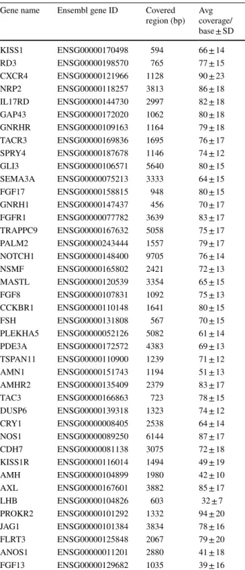
Table 3 CHH gene list and panel performance indicated by coverage (mean read/base ± SD)
Gene name Ensembl gene ID Covered
region (bp) Avg coverage/
base ± SD
KISS1 ENSG00000170498 594 66 ± 14
RD3 ENSG00000198570 765 77 ± 15
CXCR4 ENSG00000121966 1128 90 ± 23
NRP2 ENSG00000118257 3813 86 ± 18
IL17RD ENSG00000144730 2997 82 ± 18
GAP43 ENSG00000172020 1062 80 ± 18
GNRHR ENSG00000109163 1164 79 ± 18
TACR3 ENSG00000169836 1695 76 ± 17
SPRY4 ENSG00000187678 1146 74 ± 12
GLI3 ENSG00000106571 5640 80 ± 15
SEMA3A ENSG00000075213 3333 64 ± 15
FGF17 ENSG00000158815 948 80 ± 15
GNRH1 ENSG00000147437 456 70 ± 17
FGFR1 ENSG00000077782 3639 83 ± 17
TRAPPC9 ENSG00000167632 5058 75 ± 17
PALM2 ENSG00000243444 1557 79 ± 17
NOTCH1 ENSG00000148400 9705 76 ± 14
NSMF ENSG00000165802 2421 72 ± 13
MASTL ENSG00000120539 3354 65 ± 15
FGF8 ENSG00000107831 1092 75 ± 13
CCKBR1 ENSG00000110148 1641 80 ± 15
FSH ENSG00000131808 567 70 ± 15
PLEKHA5 ENSG00000052126 5082 61 ± 14
PDE3A ENSG00000172572 4383 69 ± 13
TSPAN11 ENSG00000110900 1239 71 ± 12
AMN1 ENSG00000151743 1194 51 ± 13
AMHR2 ENSG00000135409 2379 83 ± 17
TAC3 ENSG00000166863 723 78 ± 15
DUSP6 ENSG00000139318 1323 74 ± 12
CRY1 ENSG00000008405 2538 64 ± 14
NOS1 ENSG00000089250 6144 87 ± 17
CDH7 ENSG00000081138 3075 72 ± 18
KISS1R ENSG00000116014 1494 49 ± 19
AMH ENSG00000104899 1980 42 ± 10
AXL ENSG00000167601 3882 85 ± 17
LHB ENSG00000104826 603 32 ± 7
PROKR2 ENSG00000101292 1332 94 ± 20
JAG1 ENSG00000101384 3834 78 ± 16
FLRT3 ENSG00000125848 2067 79 ± 20
ANOS1 ENSG00000011201 2880 41 ± 18
FGF13 ENSG00000129682 1035 39 ± 16
followed the Genome Analysis Tool Kit (GATK) Best Practices guideline using the germline short variant dis- covery (SNPs + Indels) algorithm (DePristo et al. 2011).
A minimum coverage of 20 reads was applied as detec- tion filter. In our gene panel, all regions were covered by 71 ± 14 reads/base (see details regarding each gene in Table 3).
The accuracy depends on the depth of sequence cov- erage therefore NGS gene panels show the highest diag- nostic accuracy (Oliver et al. 2015). Indeed, in a recent study, comparing different exome sequencing platforms found that 93.2% of the investigated regions were cov- ered > 10 reads (Kong et al. 2018) (of the covered regions the sensitivity was reported 97.5–99.99%). Comparably, in our panel, 97.2% of the investigated regions (86,329 bp) was covered > 20 read/base. Of the investigated region, 14,532 bp was assessed by Sanger sequencing as well, and all detected variants were identified by both approach, therefore the specificity of our panel was 100%.
Variant interpretation
Variant interpretation are guided by expert recommenda- tions for clinical diagnostics [American College of Medi- cal Genetics and Genomics (ACMG), European Society of Human Genetics (ESHG)] which should be followed for all laboratories offering NGS-based diagnostics (Rehm et al.
2013; Richards et al. 2015; Matthijs et al. 2016).
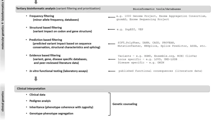
WGS usually identifies 3–4 million, while WES detects usually 15,000–20,000 variants. Therefore, variant prioriti- zation and interpretation are needed to determine the one or the few pathogenic variants responsible for disease (Fig. 2).
First step is to assess the prevalence of certain variants in general population-based databases to filter out frequent variants assuming that pathogenic variants are not common in the broad population. However, in oligogenic diseases, relatively frequent variants can have additional or genetic modifier effect on the phenotype (Maione et al. 2018).
Analyzing the functional consequence of a certain vari- ant may help the interpretation. Using various algorithms
Fig. 2 Process of molecular genetic testing by NGS from NGS data analysis to variant interpretation. See details in the text