1
Béla Gyurcsik
University of Szeged
2020
2
This teaching material has been made at the University of Szeged and supported by the European Union. Project identity number: EFOP-3.4.3-16-2016-00014
3
Preface
What is the meaning of the modern chemistry, or rather the modern chemist? In view of the author a description like this could be applied for all the experimental sciences. First of all, the modern researcher recognises a scientific/practical problem. Let’s take the development of a drug molecule against cancer as an example. To solve this problem, we have to understand that it shall be approached from various aspects, and in fact it is an interdisciplinary task. We need to apply all the necessary tools (knowledge, instruments) of the chemistry, but also of other scientific fields. In the following is a list of few aspects of this research without an endeavour of completeness. Informatics helps the design of the drug molecule taking into account the many factors by various programs. Biochemistry provides information on the pathway of the drug action and metabolism. Biophysics and bioanalytical experiments tell about the structure and speciation of the drug molecule under various conditions – e.g. upon interaction with physiological solution, membranes, receptors and target biomolecules. Mathematics contributes in validation of the experimental results through statistical analysis of the data. It is also necessary to think about the practical applicability, economical aspects etc. including specialists in medicine and business and many more. Chemists will synthesize and characterize the drug molecule and encapsulate it to reach the biological target. However, we can easily recognize that without the know-how listed above this will not be a successful project. Thus, we need to collaborate with researchers from different fields.
4
As a chemist, the author has frequently faced difficulty to discuss with biologists, physicists, medical doctors, etc, and recognized that it is very important to precisely understand each other’s scientific language. An adventurous postdoctoral research trip to a famous Japanese university, Tokyo Institute of Technology, within the frame of a UNESCO fellowship, provided a chance to the author to get insight into the life of a virology laboratory, extensively applying molecular biology tools in their experiments. We have been mutually teaching chemistry and biology to each other with the Japanese members of the laboratory.
The acquired knowledge made possible to synthesize, purify and characterize biological macromolecules, such as proteins and DNA, as well as, to carry out their targeted modification and investigate the effect of the changes on the structure, interaction and function. This experience proved to be fruitful in the establishment of new bioinorganic chemistry approach at University Szeged, namely the study of biomolecules and their derivatives themselves, instead of classical modelling studies.
Recognizing the usefulness of the interdisciplinary outlook, the author decided to invent a new course for the non-biology students to assist the development of their collaborative problem solving approach. Students learn about basics of molecular biology tools that can be applied in advanced chemical and biochemical research. The goal of this electronic book is to provide written material in support of the university course under the same name. The course is primarily suggested for chemistry and info-bionics master and doctoral students.
Nevertheless, its biological analytical part serves as a guide to the students in biology, molecular bionics and pharmaceutics through the instrumental methods of the biomolecule identification, purification and analysis.
5
Contents
GENERAL LEARNING OUTCOMES 6
1. INTRODUCTION 10
2. THE OVERLAP OF THE CHEMICAL AND BIOLOGICAL
SCIENCES 13
3. DNA AND THE RECOMBINANT DNA TECHNOLOGY 27
4. DNA REPLICATION AND THE POLYMERASE CHAIN REACTION 46 5. PRIMER DESIGN FOR THE POLYMERASE CHAIN REACTION 65 6. IDENTIFICATION OF PCR PRODUCTS – AGAROSE GEL
ELECTROPHORESIS 82
7. RESTRICTION ENDONUCLEASES AND DNA CLONING 99
8. TRANSCRIPTION AND TRANSLATION 129
9. PROTEIN IDENTIFICATION AND PURIFICATION 149
SUGGESTED READING 162
6
General learning outcomes
This book is intended to help students reaching the following general learning outcomes:
Knowledge
The students define the possibility of the overlap of biology and chemistry, list the appropriate borderline research fields.
The students know the biological tools suitable to solve the given complex interdisciplinary problem.
The students summarize the newly introduced/ acquainted methods suitable to study the properties of biological macromolecules.
The students denominate modern procedures, by the help of which macromolecules of biological importance (DNA, RNA, protein) can be synthesized.
The students list the opportunities of applications of the tools of molecular biology in the field of chemistry research.
The students understand the overlap of the various scientific disciplines.
The students explain the effect of molecular biology on the development of modern chemical research.
7
The students know the methods of examination used in molecular biology.
The students are aware of the background and application opportunities of various microscopic methods.
The students know the theory and practice of the molecular biology tools for studying the structure of protein molecules.
Skills
The students analyse the possibilities of the collaboration with researchers representing various scientific disciplines.
The students communicate with researchers representing biology and other scientific disciplines to solve a complex interdisciplinary problem.
The students evaluate the capability of biological and chemical procedures in the separation and investigation of DNA or protein molecules.
The students carry out the targeted modification of a protein molecule based on its genetic code in theory and practice.
The students formulate their interdisciplinary projects in bioinorganic chemistry, drug design, understanding biological processes, the mechanism of drug action, etc. - all projects involving macromolecules such as proteins and DNA.
The students efficiently discuss with collaborators from biology, pharmacology and medicine field of sciences.
8
The students select the appropriate molecular biology tools that can be applied in advanced chemical and biochemical research to solve the given complex problem.
The students select the appropriate procedure for the purpose of the detection, identification and purification of DNA or protein molecules.
The students explain the role of the metal ions in biological systems including examples for the role of the free metal ions, metalloproteins and metalloenzymes.
The students select the appropriate methods to investigate the amino acid composition, amino acid sequence.
The students distinguish the various levels of the protein structure, list methods by means of which these structural organizations can be studied.
The students estimate the secondary structure of the protein molecules.
The students demonstrate by examples the possibilities of the modification of biological macromolecules (DNA and protein), including point mutations, deletions or insertions and fusions.
The students explain the steps of the design of biological macromolecules.
Attitude
The students pay attention to the precise application of the chemistry and molecular biology terminology.
9
The students make effort to apply the interdisciplinary approach in their study and research.
The students help the colleagues from the biology field in understanding the methods of chemical approach to a biological experiment.
The students are motivated to acquire new information on diverse scientific fields.
The students are critical during the evaluation of the literature.
Responsibility and autonomy
The students collaborate with colleagues from biology or various research areas upon recognizing an interdisciplinary research problem.
The students independently apply the proper terminology of chemistry and molecular biology, and explains the terminology to either chemistry or biology orientated colleagues.
Facing an interdisciplinary problem, the students independently develop their knowledge on the borderline scientific areas.
The students understand that the complex research project has to be conducted in collaboration with researchers from other scientific disciplines.
The students can critically evaluate the results of the complex experiments including high number of the degree of freedom.
10
1. Introduction
At the beginning of the scientific research it was common the observe the universe as a whole and to collect knowledge in every aspect of the observed objects. The polyhistors intended to understand and explain natural phenomena based on their knowledge regardless of the fields of sciences evolved since then.
The exponential development of science has been realized, however, in extreme differentiation of various fields. Nowadays students and researchers experience difficulties in understanding each others’ presentations, since the research topics are extremely specific to one subject or to one new experimental technique. A biologist colleague of the author told already several years ago that his life-long research topic will be the study of the digestive disorders of the shrimps living in the deep oceans. Indeed, we can meet with such focused projects in an uncountable amount of research articles. These pieces of isolated works may contribute to the understanding of the nature, but they also may cause difficulty and confusion, if they diverge far from the coherency of the multiple processes in our environment.
It might be even worse if the article contains mistakes, misleading information and/or the experiments are described in a way that they are not reproducible. These phenomena are unfortunately not rare. The research is becoming business in an increasing extent. It is natural that the researchers would like to benefit from their discoveries helping to invent new technologies, environmental protection, drugs for diseases leading to better life of people.
11
However, the introduction of money directly in the publication process lead to controversy concerning the scientific level of the articles appeared in various journals. Also the researchers are pressed to publish more and more since often the indicators such as the number of the published papers, or their citations are takes as the measure of the value of their scientific research. Based on these numbers they are advanced or awarded grants etc. Because of the high number of manuscripts, it is difficult to select proper researchers for the evaluation process being experts on the specific field. Therefore, increasing amount of incorrect information will necessarily appear in the scientific literature. We should think about the request of Max Delbrück, a Nobel-prize holder in his letter to the wife of his friend and colleague, Seymour Benzer – as it is described in the book Csillagórák a tudományban by Venetianer Pál. Citing this part of the letter: "Dear Dotty, please tell Seymour to stop writing so many papers. If I gave them the attention his papers used to deserve, they would take all my time. If he must continue, tell him to do what Erns Mayr asked his mother to do in her long daily letter, namely, underline what is important."
It is also worth mentioning a determinative episode from Delbrück’s carrier taken from his biography at the Nobel Prize website for improving the future opportunity of the reader. He has spent three postdoctoral years (1929-1932) abroad, in England, Switzerland, and Denmark. The stay in England, with its immersion into a new language and a new culture, had a vast effect on widening his outlook on life. In Switzerland and Denmark the associations with Wolfgang Pauli and Niels Bohr shaped his attitude toward the pursuit of truth in science.
Apart from the above discrepancies, the high amount of the published information (even restricted to the scientific literature) makes it impossible to
12
collect all this in a single mind, thus the era of polyhistors is over. Nevertheless, the nature can not take our limitations into account: the problems are complex, as it is schematically shown in Fig. 1.
Figure 1. Schematic representation of the complexity of the natural systems, and the need for the collaboration of the researchers for various research fields for correct problem solving.
Therefore, to keep the life going on on earth, we need to collect the scientific knowledge. It is not enough e.g. to develop a more economic technology, but the multiple consequences of its practical application on the environment should be strictly verified. We need to solve the problem, how to gather together enough and reliable information to be able to come to a right decision. Informatics can be an invaluable tool in collecting and analysing the data, but the knowledge to develop such methods and to correctly understand this can only arise from the collaboration of the researchers from diverse scientific fields.
13
2. The overlap of the chemical and biological sciences
Students who study this chapter will acquire the following specified learning outcomes:
Knowledge
The students define the possibility of the overlap of biology and chemistry, list the appropriate borderline research fields.
The students list the opportunities of applications of the tools of molecular biology in the field of chemistry research.
The students explain the effect of molecular biology on the development of modern chemical research.
Skills
The students formulate their interdisciplinary projects in bioinorganic chemistry, drug design, understanding biological processes, the mechanism of drug action, etc. - all projects involving macromolecules such as proteins and DNA.
The students communicate with researchers representing biology and other scientific disciplines to solve a complex interdisciplinary problem.
14
The students analyse the possibilities of the collaboration with researchers representing various scientific disciplines.
Attitude
The students make effort to apply the interdisciplinary approach in their study and research.
The students help the colleagues from the biology field in understanding the methods of chemical approach to a biological experiment.
The students are motivated to acquire new information on diverse scientific fields.
The students pay attention to the importance of interdisciplinary research.
Responsibility and autonomy
Facing an interdisciplinary problem, the students independently develop their knowledge on the borderline scientific areas.
The students understand that the complex research project has to be conducted in collaboration with researchers from other scientific disciplines.
15
With the development of the science including knowledge and research infrastructure an increasing demand arose for collaboration among various disciplines to solve a specific problem. As the result of this evolutionary process, the interdisciplinary outlook has become indispensable in scientific work.
Nowadays a large number of procedures, which were inaccessible just several years ago, are routinely carried out. This provides a unique chance to extend our experimental repertoire and to promote our research into the domain of modern science. Day by day we witness the appearance of new names for various interdisciplinary fields followed by the new editions of scientific journals and books.
Bioinorganic chemistry is an interdisciplinary science emerged to connect the knowledge accumulated in inorganic chemistry and biology. The knowledge about the presence of the inorganic compounds, with main emphasis on the metal ions has been extended by the development of new analytical methods. Several metal ions have been found to be an integrated part of the biological fluids, humors, cells, and biomolecules. At the same time few metal ions acquired from the environment may exert beneficial or toxic effect. It became the important to keep tabs on the modes of their intake, distribution and function of all these metal ions to understand their biological role. An inorganic chemist has the knowledge about the physicochemical properties of the elements and their compounds, which can be used to explain the strength of the interactions, the local structure of the metal ion surroundings, and the reactivity of the metal ions and/or the molecules interaction with them.
16
Metal ions may have been involved in the formation of the first biomolecules through complex formation processes, which arranged the ligand molecules in a favourable steric position for their reaction, as well as catalysing certain acid-base and redox reactions. With the appearance of more complex biomolecules the first specific metalloproteins and among these the metalloenzymes could evolve. The metal ions may stabilize the structure of these macromolecules, but also may participate in their various functions such as electron or oxygen carrier, activator of small inert molecules, or being part of the catalytic active centre. Thus, the properties of metal ions will largely influence the behaviour of the coordinated molecules. In the following few selected examples of the metal ion containing molecules will be listed with their most important bioinorganic chemistry relations.
Probably the most well-known metal ion containing protein in human organism is haemoglobin. Haemoglobin serves as an oxygen carrier. Fig. 2. shows the image of a haemoglobin monomer based on its crystal structure. It can be seen that the iron-containing heme group is attached to the protein through a coordinative bond between the metal ion and a histidine side-chain.
Haemoglobin binds the oxygen molecule at the metal ion centre. The cooperative oxygen binding of the four subunits will enable the oxygen binding at high partial oxygen partial pressure, while providing oxygen to myoglobin at low partial oxygen pressure. The basis of the cooperativity lays in the induced movement of the metal ion upon oxygen binding, which induces the conformational change in the neighbouring subunit increasing its oxygen binding ability.
17
Figure 2. PyMol image of a haemoglobin monomeric based on its crystal structure (coordinates have been downloaded from RCSB Protein Databank, as it has been done for all further PDB files. PDB Id: 1HCO). Heme group is in the centre of the molecule, with an Fe(II) atom as an orange sphere. A histidine side- chain (yellow sticks) is creating the coordinative connection between the protein and the heme group.
Zinc fingers are abundant proteins in living organisms. Zinc(II) ions serve structural role in these proteins. Upon zinc(II) binding the originally unordered protein structure turns into a finger-like ββ motif (Fig. 3.). These fingers are able to interact with DNA molecule specifically recognizing selected DNA sequences.
As such zinc finger proteins serve mostly as transcription activator factors inducing the synthesis of RNA from its DNA template as the first step of protein synthesis.
18
Figure 3. PyMol image of a zinc finger protein based on its crystal structure in complex with DNA (PDB Id: 1MEY). The protein consists of three finger units and is represented by green cartoon. The Zn(II) ions are shown as grey spheres each one coordinated to two histidine and two cysteine side-chains (green sticks).
The backbone of the DNA molecule is orange.
The metal ion within a finger unit is coordinated to two imidazole nitrogens in the side-chains of two histidines and two thiolate groups in the side-chains of two cysteines. This type of complex formation saturates the zinc(II) coordination sphere and neutralizes the charge of the metal ion. In such environment zinc(II) does not show catalytic behaviour, such as it does in the active centre of several nucleases. Thus, an unwanted side-reaction, leading to the damage of the DNA during the transcription process is excluded.
Hydrogenases are bacterial enzymes utilizing the redox reaction between the protons and hydrogen as the source of energy or for the storage of energy. Fig.
4. shows a cartoon of a [NiFe] hydrogenase enzyme, which contains an irons sulphur cluster at its active centre containing a nickel(II) ion as well. As the enzymes catalysing redox reactions this protein contains metal ions, which can
19
change their oxidation states in the active site. The electron transport within the protein structure is ensured by the organized chain of the iron-sulfur clusters. The electron transport is governed by the redox potential of each cluster.
Figure 4. PyMol image of the [NiFe] hydrogenase from Desulfovibrio desulfuricans based on its crystal structure (PDB Id: 1E3D). The protein is represented by green cartoon. The metal clusters are shown as spheres.
Metal ions also have a role in unequal electrolyte distribution across the cell membranes with various roles. These phenomena are related to the stability of the cell membrane, changing of the membrane potential leading to the action potential in nerve cells, etc. It is well-known that the concentration of the sodium ion is different at the two sides of the cell membrane. It is much smaller within the cells, than outside. Potassium ions show opposite gradient. These concentration gradients must be maintained for the cell survival. Therefore, the organism uses energy to pump the metal ions against the concentration gradient – a process
20
called active transport. A Mg2+ ion is participating in the catalysis of ATP/ADP conversion (Fig. 5).
Figure 5. PyMol image of the sodium-potassium pump (left) discovered by Jens Christian Skou (right, photo downloaded from the official website of the Nobel Prize), who received Nobel prize in chemistry for the first discovery of an ion- transporting enzyme, Na+, K+-ATPase. The protein is represented by green cartoon based on its crystal structure (PDB Id: 3B8E). The helix bundle at the right side of the structure is the cross-membrane section. Metal ions are shown as spheres.
Numerous further metal ion containing biomolecules can be itemized, but it would be out of the scope of the subject of this e-book. Nevertheless, these examples show the diversity and power of bioinorganic chemistry.
One of the two main approaches of the bioinorganic chemistry is the study of metal complexes and artificial systems modelling the biological structure, function and environment. This approach involves mainly chemical methods of synthesis and study. It has the advantages that the simple/cheap substances
21
applied in experiments can be synthesized/obtained in a scale that is enough for detailed physicochemical investigations. Simple, well determined systems are studied and straightforward conclusions can be obtained. Fig. 6. represents an example from the author’s research study on the metal ion peptide system, in which modelling of a nuclease active centre was carried out by a heptapeptide, which coordinates the zinc(II) ion by three histidine side-chain, similarly to many hydrolytic enzymes. This study involved investigation methods commonly used in the modelling in bioinorganic chemistry, such as pH-potentiometric titrations, spectrophotometry and circular dichroism spectroscopy. By these methods we can determine the total concentration of the components, such as e.g. the metal ion, and also the detailed speciation of its different complexed forms under varying conditions. Spectroscopic methods provide opportunity to get insight into the structure of individual complex species, either within the metal ion microenvironment or for the whole molecule. Based on these, one can select appropriate conditions for the further experiments for functional modelling of the studied biological system. In this example, it is shown that the three-histidine coordinated complexes form in the pH range between ~ 5 and ~ 9 (Fig. 6A). The result of the DNA cleavage study revealed that at least one of these complexes is catalytically active (Fig. 6B).
Modelling is also essential for the development of bioinspired molecules, being able to selectively bind metal ions for metal ion sensing, detecting or accumulating purpose. These agents can be used in various practical applications such as environmental analysis, technology, medicine, etc. The Catalytically active bioinorganic models are promising for the future economic and environmentally friendly technologies.
22
Fig. 6 A) The speciation diagram of the zinc(II)–peptide 1:1.1 system. The scheme of the metal complex, in which the coordination is achieved by three imidazole-nitrogen from the side-chains of histidines is shown above the appropriate species. B) The nuclease activity assay of the zinc(II)–peptide complexes is demonstrated by their action on a pUC18 circular DNA. The result of the reaction was visualised by agarose gel electrophoresis – one of the methods discussed later in this e-book. The peptide and zinc(II) together caused an enhanced hydrolytic activity at pH 7.1 demonstrated by the detection of the relaxed and even the linear forms of the DNA in lane 5.
0 20 40 60 80 100
3 5 7 9 11
pH
% Zn(II)
100 111
101 1-11
1-21
102
ZnLH(OH) → {3×ImN, OH-} ZnLH → {3×ImN}
HN CH C
H2C O
N NH N
C O
NH H2C CH C
O
HN N
N C O NH CH CH2 C
O N
NH ZnII OH- Ac-Lys
Gln-NHGln-NH22 Ac-Lys
A
No 1 2 3 4 5 6 7 pH 8.1 7.1 7.6 8.1 7.1 7.6 8.1 Cu(II) - + + + - - - Zn(II) - - - - + + + AcKHPHPHQ - + + + + + + DNA + + + + + + + Relaxed circular DNA
Linear DNA Supercoiled DNA
B
23
Although we can learn from the modelling experiments, here researchers study isolated systems, and the results are difficult to translate to the real biological environment.
The second main approach is the study of macromolecular systems themselves. These experiments include mainly the metalloproteins and metalloenzymes, but in a broader sense all the metal ion binding biomolecules.
These studies will provide direct information on the biologically active substances, but their investigation is difficult with the conventional physicochemical methods. The main reason for this is that the synthesis of the macromolecules poses difficulties. The solid phase peptide synthesis is not suitable for preparation of proteins consisting of hundreds of amino acids. Even the careful synthesis of a 100 amino acid long peptide would yield a mixture of peptides, with similar properties, which would be extremely difficult to purify even using the most advanced separation techniques. Thus, the resulting substance would be too expensive for further experiments.
The Department of Biochemistry at University of Szeged is located in the Institute of Biology. Therefore, chemistry orientated students have only a limited knowledge on this field, which could help them to solve such problems. In this e- book it will be demonstrated that by using the routine methods of the molecular biology the level of the chemical research can be increased. Bacteria can e.g. be used as the most advanced "peptide synthesizers" that can produce proteins with precise sequence. Substitution reactions are often applied in organic chemistry to modify compounds. However, the exchange of a single selected amino acid side- chain within a protein is an almost impossible task by using chemical reactions.
24
Such modification can be easily executed by the tools of recombinant DNA technology starting out from the gene of the protein as it will be demonstrated later. Similarly, it is impossible to find suitable instrumental separation technique (HPLC or electrophoresis) for the separation of a mixture of DNA molecules with identical length of e.g. 300 bp (where bp stands for the 2'-deoxynucleotide pairs commonly called base pairs). This task can also be easily solved in the knowledge of the rules of the DNA cloning procedure.
In the following chapters, numerous examples of the biological tools will be described and explained, which can be applied in chemistry research. Not only the bioinorganic chemists can utilize these methods, but many others. Counting just few examples:
(i) The drug design e.g. relies essentially on the interactions of the synthetic organic drug molecules with biological macromolecules. Therefore, the drug design is essentially based on the knowledge on the structure and function, and in particular the first step of verification is based on the studies on interactions of these molecules with drugs.
Figure 7. A future photosynthetic car designed by Shanghai Automotive Industry Corporation (source: https://phys.org/news/2010-05-yez-car.html)
25
(ii) Biosensors are emerging tools of analytical chemistry, as in is demonstrated by the exponentially increasing number of published articles in this field.
(iii) Modern technologies (nitrogen fixation, solar energy utilization (Fig.
7), etc.) can arise from the knowledge acquired in the detailed studies and targeted modifications of the natural systems.
26
Monitoring questions
- How can be a complex research problem in natural sciences solved?
- List examples of overlaps between various research areas in selected research problems.
- What are the roles of metal ions in biological systems? List examples for each type of role.
- What is bioinorganic chemistry?
- What are the methods of studies in bioinorganic chemistry?
- How can be the biological tools applied in chemistry?
27
3. DNA and the recombinant DNA technology
Students who study this chapter will acquire the following specified learning outcomes:
Knowledge
The students know the basics of recombinant DNA technology.
The students identify the building blocks of the DNA molecule.
The students are aware of the DNA base pairing principles.
The students explain the structure of the DNA double helix.
The students list the main elements of the recombinant DNA technology.
Skills
The students write the sequence of the complementary strand of the DNA based on the knowledge of base pairing.
The students select the complementary (unusual) codes of the nucleobases even if the definition of the nucleobase is not unique.
The students analyse the possibility of the specific and non-specific DNA-protein interactions based on the DNA structure.
28
Attitude
The students pay attention to the importance of correct writing of the DNA sequence and its complementary sequence.
The students realize the importance of the antiparallel arrangement of the two DNA strands in the double helix.
The students make an effort to realize the importance of the achievements in the history of the DNA research.
Responsibility and autonomy
The students independently search and identify the chemical structures of the DNA components.
The students independently develop their knowledge on the new research on the area of DNA-based drug design.
29
"Is life just a game where we make up the rules, While we're searching for something to say, Or are we just simply spiralling coils, Of self-replicating DNA?"
Monty Python – The Meaning of Life
The above cited text is the perfect introduction to this chapter defining the recombinant DNA technology (Fig. 8.), which is based on the central dogma of the molecular biology.
Figure 8. Schematic description of the main processes applied by the recombinant DNA technology.
DNA
Transcription RNA
Regulation: off on
Replication
Reverse
transcription DNA DNA DNA
Translation Protein
Sequencing Transformation
Transduction
30
It means that (i) the information which is stored in DNA is transferred to a newly synthesized DNA molecule in a process called replication; (ii) the information which is stored in DNA is transferred to a newly synthesized RNA molecule in a process called transcription; (iii) the information which is stored in RNA is transferred to newly synthesized protein molecule in a process called translation, being the most abundant processes in the cells. Nevertheless, the transfer of the information in DNA → protein, RNA → RNA, RNA → DNA relations may also occur in nature and have their own importance.
As it can be concluded from Fig. 8, the starting point of the protein synthesis – a key molecule in chemistry research – is the DNA. The consequence of this is that the knowledge about DNA should be deep enough to handle such a chemically unfriendly substance. For a long time, researchers could not find suitable tools to better understand its structure and to carry out suitable reactions with it. DNA in the cells is usually a very long and stable polymer consisting of only four (chemically not very) different subunits. This makes it extremely difficult to analyse, synthesize and purify. On the other hand, it is well-known how the recent routine methods of molecular biology revolutionized the knowledge on this research area, as well as the other scientific fields through the interdisciplinary researches. It is very clear that the many discoveries on this field deserved Nobel prizes.
First, the DNA molecule will be introduced in the following. A chemist considers the DNA as a usual chemical identity consisting of atoms, which are bound together with primary and secondary bonds. The building blocks of the DNA polymer are the 2'-deoxyribonucleoside monophosphates (dNMPs), shown in Fig. 9. Their common part is the central 2'-deoxyribose, which lacks the
31
hydroxyl group on the 2'-carbon atom. The hydroxyl group at the 5'-carbon atom is phosphorylated. The glycosidic carbon is substituted by a heteroaromatic compound, a nucleobase, which can either be an adenine, guanine, thymine or cytosine. Accordingly, the individual building blocks are the 2'-deoxyadenosine 5'-monophosphate or simply deoxyadenosine monophosphate (dAMP), deoxyguanosine monophosphate (dGMP), deoxythymidine monophosphate (dTMP) and deoxycytidine monophosphate (dAMP). Adenine and guanine are purine bases, while thymine and cytosine are pyrimidine bases. As an easy research task, find the structural formulae of these molecules.
Figure 9. PyMol images of the building blocks of DNA, 2'-deoxyribonucleoside monophosphates from the crystal structure of a DNA molecule (PDB Id: 1ZEW).
The abbreviations are explained in the text. Note the numbered carbon atoms of the central 2’-deoxyribose rings, as it will be often referred to these numbers later.
dAMP
dTMP dCMP
dGMP
5' 5'
5' 5'
3'
3' 3'
3'
2'
2' 2'
2'
32
Figure 10. PyMol images of a 2'-deoxyribonucleoside and a 2'-deoxyribonucleotide. The difference between the two molecules is the lack of the phosphate group in the former. The data for the figure have been obtained from the crystal structure of a DNA molecule (PDB Id: 1ZEW).
dNMPs can be more simply named as 2'-deoxynucleotides. Removing the 5'-phosphates the resulting molecules are named as 2'-deoxynucleosides (Fig. 10.). The building blocks of DNA are connected together through phosphodiester bonds formed in condensation reaction between the 5'-phosphate and the 3'-hydroxy group of the 2'-deoxyribonucleoside monophosphates (Fig. 11.). In fact, soon it will be shown that 2'-deoxyribonucleoside triphosphates (dNTPs) are used as reactive species in DNA synthesis, to provide the necessary energy for the reaction.
2'-deoxyribonucleotide 5'
3' 2'
nucleobase phosphate
2'-deoxyribose 2'-deoxyribonucleoside
33
Figure 11. PyMol image of a single strand DNA chain from the crystal structure of a DNA molecule (PDB Id: 1ZEW) showing the phosphodiester bonds forming sugar-phosphate backbone of the DNA molecule. Note the direction of the molecule is determined by the 5'-phosphate and the 3'-hydroxy groups. The text will often refer to the DNA 5' and 3' termini later.
The DNA chain formed in this way, possesses a free 5'-phosphate and a free 3'-hydroxy group (Fig. 11.). These groups determine a direction of the DNA chain. The sequence of the DNA is defined as the order of 2'-deoxyribonucleotide units read from the 5'-end of the DNA towards the 3'-end. As an easy exercise try to write the sequence of the DNA chain depicted in Fig. 11. For easier reading of the sequence the 2'-deoxyribonucleotides are usually abbreviated with a one-letter code each. These codes refer to the nucleobase (the subunits, which are different among the 2'-deoxyribonucleotide molecules) within each
5'
3'
Nucleobases
Sugar-phosphodiesterbackbone
34
2'-deoxyribonucleotide. Accordingly, for the 2'-deoxyribonucleotides containing adenine, guanine, thymine or cytosine nucleobases A, G, T or C is written, respectively.
A specific and somewhat surprising property of the single strand DNA molecules (such as it is shown in Fig. 11.) that they can bind to a specific second DNA chain to form a double strand DNA. The two DNA chains must be in a specific relationship i.e, they must be complementary to each other to form the double strand. Fig. 12. shows how the double strand DNA is formed. It can be seen that the two chains are bound together through multiple hydrogen bonds formed between the nucleobases. It can immediately be recognised that for optimal arrangement of these subunits, the base pairs always consist of one purine and one pyrimidine base. More specifically A is always paired with T and G is always paired with C. It is said that A and T, as well as G and C are complementary to each other. Because of their different size this arrangement provides similar distance between the two chains along the DNA sequence. It is also worth mentioning that the two strands of the double strand DNA have antiparallel arrangement, meaning that the directions of the two strands are opposite to each other. Based on this knowledge try to write the DNA sequence of the complementary strand of the DNA shown in Fig. 11.
The two strands form a double helix with a narrow and a broad groove, named minor and major grooves, respectively. This arrangement is also important from the point of view of the interactions of the double strand DNA with macromolecules, such as proteins. Later it will be shown that the proteins recognising specific DNA sequences should interact with nucleobases, and this is only achievable though the major groove in which the nucleobases are exposed to
35
proteins. Contrary the minor groove is too narrow for a protein to access nucleobases thus, the interactions will mostly be established with the sugar- phosphate backbone, which is uniform along the DNA chain.
Figure 12. PyMol image of a double strand DNA from its crystal structure (PDB Id: 2M54). Note that the direction of the two single strand DNA molecules is opposite/antiparallel.
It is also important to mention that two hydrogen bonds are formed between A and T and three between G and C (Fig. 13). It is not difficult to prevision that the strength of the binding is higher between G and C than between A and T, which will also have consequences concerning the behaviour of DNA in its certain reactions. It is e.g, easier to deform the DNA structure at the A/T-rich motifs than at C/G-rich sequences.
5' 3'
3' 5'
major groove
minor groove
minor groove
36
Figure 13. Hydrogen bonds formed between the nucleobases within a double strand DNA.
Table 1. Codes of 2'-deoxynucleotides, including those allowing variation in the sequence or showing ambiguity in the sequence.
Code Equivalent Complementary code
A A T
C C G
G G C
T T A
M A or C K
R A or G Y
W A or T W
S C or G S
Y C or T R
K G or T M
V A or C or G B
H A or C or T D
D A or G or T H
B C or G or T V
X or N A or C or G or T N or X
Adenine Thymine Guanine Cytosine
37
It is worth mentioning that new codes have been introduced for the 2'- deoxnucleotides allowing for the ambiguity in the DNA sequence (Table 1.). The significance of these codes will be demonstrated later.
The backbone of the DNA chain is highly negatively charged, as it carries one charge at each phosphodiester bond. For two long DNA chains forming a double strand DNA this results in strong repulsive force. This has to be overcompensated to stabilize the double helix. It is obvious that positively charges ions can shield this high negative charge. Later it will be demonstrated that DNA molecule can be easily precipitated in the presence of salt, which neutralizes the solid substance. However, in addition to the above mentioned hydrogen bonding interactions the hydrophobic interactions between the neighbouring nucleobase residues contribute also to the stabilization of the double strand DNA. The planar nucleobases are arranged to be parallel within the core of the helix and in this way establish stacking interactions.
The knowledge about the DNA listed above was not available until ~ 70 years ago, in spite of the fact that in 1869 Friedrich Miescher isolated a phosphorous containing material from the cell nucleus and published in 1871 (Fig. 14.). It was essential to prove that the isolated substance was free of protein impurity, which could turn his conclusions false. He supposed that this "nuclein"
is needed for cell division process, and predicted that the knowledge of the interactions between the materials from the nucleus, proteins and their direct metabolic products will gradually enlighten the internal processes of the cells. The experiments were carried out in Felix Hoppe-Seyler’s laboratory in Tübingen.
Prior to becoming the chemical laboratory of Tübingen University in 1823, the room was Tübingen castle’s laundry.
38
The deoxyribonucleic acids then escaped the researchers attention until 1944, when Oswald T. Avery, Colin MacLeod, and Maclyn McCarty suggested that the material responsible for the heredity of various properties in bacteria is DNA instead of proteins, as thought earlier. Not much later the structure of the DNA has been solved.
Figure 14. Friedrich Miescher and his paper about the discovery of "nuclein"
(DNA) in Hoppe-Seyler’s Medicinisch-chemische Untersuchungen (Miescher, F., 1871. Über die chemische Zusammensetzung der Eiterzellen. Med.-Chem.
Unters. 4, 441–460).
Rosalind Elsie Franklin (1920 – 1958) was a chemist X-ray crystallographer who contributed to the understanding of the molecular structures of DNA (deoxyribonucleic acid), RNA (ribonucleic acid), viruses, coal, and
39
graphite. She published her results about the structure of sodium thymonucleate fibres determined by X-ray diffraction in 1953 (Fig. 15.).
Figure 15. Rosalind Elsie Franklin and her paper about the X-ray diffraction experiments in Acta Crystallographica.
Next, James Dewey Watson and Francis Harry Compton Crick published the structure of the DNA double helix in 1953 in Nature (Fig. 16.). This achievement deserved a Nobel prize in physiology or medicine in 1962. In the same issue of Nature subsequent articles by Wilkins, Stokes and Wilson, as well as by Franklin and Gosling also discuss the structure of the DNA. Rosalind Elsie Franklin was an X-ray crystallographer, whose X-ray diffraction experiments essentially contributed to the understanding of the molecular structures of DNA Her results leading to the fruitful discussions and finally the discovery of the structure of DNA were largely recognised after her death in 1958. In fact, she might have had the Nobel prize shared with the scientists in Fig. 16, but the Nobel prize is not awarded posthumously. Therefore, she could never receive the deserved highest honour of the scientific community for her work.
40
Figure 16. A) From top to bottom: James Dewey Watson and Francis Harry Compton Crick the Nobel prize holders in physiology or medicine 1962 "for their discoveries concerning the molecular structure of nucleic acids and its significance for information transfer in living material." (Photo from the Nobel Foundation archive.) B) The schematic of the DNA structure as published in Nature in April, 1953. The figure was taken from the original article: Nature, 1953, vol. 171, pp.737-738.
The next important step in understanding the properties of the DNA was the cracking of the genetic code in early 1960-s. For this achievement Robert W.
Holley, Har Gobind Khorana and Marshall W. Nirenberg received the Nobel Prize in Physiology or Medicine in 1968; while J. Heinrich Matthaei a German biochemist, a post-doctoral visitor in the laboratory of Marshall Warren Nirenberg also significantly contributed to this work. Resolving the genetic code – i.e. the determination of the nucleotide sequences corresponding to the amino acids – was
B
A
41
the most challenging genetic project at that time. The revealed code proved to be essentially the same for nearly all organisms.
Figure 16. Top left to right: Robert W. Holley, Har Gobind Khorana and Marshall W. Nirenberg the Nobel prize holders in physiology or medicine 1968 "for their interpretation of the genetic code and its function in protein synthesis." (Photo from the Nobel Foundation archive.) Bottom: J. Heinrich Matthaei in Nirenberg’s laboratory (source: https://www.telegraph.co.uk/news/science/science- news/8546830/Genes-and-DNA-meet-the-first-man-to-read-the-book-of-
life.html).
In the next figure (Fig. 17.) the most important milestones of the research results related to DNA are collected in chronological order taken from the excellent review on DNA by Ralf Dahm from the Max Planck Institute for Developmental Biology, Tübingen, Germany.
42
Figure 17. The timeline of the milestones in DNA research taken from Developmental Biology, 2005, vol. 278, pp. 274-288.
43
The figure summarizes a huge success of the researchers in understanding the structure and function of the DNA molecule.
Although the sequence of the human genome (total human DNA) is already known, the research on DNA is far from being finished. Bioinorganic chemists also participate to this research. DNA is e.g, a target of metal ion containing anticancer drugs, because the components of DNA can bind metal ions, which affect its structure and function.
The double strand DNA discussed so far, can also form higher order structures, including its complex with histone proteins. These proteins organize DNA into nucleosomes, which form chromatins, and by further condensation the chromosomes in the human cell nuclei. The double helix of the DNA has a diameter of ~ 2 nm. The distance between to nucleobases is ~ 0.34 nm.
Approximately ten nucleotides form on full turn of the DNA strand, i.e, the length of a full turn is ~ 3.4 nm. As an example, the X chromosome in human cells consists of ~ 156 Mbp (millions of base pairs). This size of DNA double helix would be 53 mm long in its linear form. When organized into a chromosome, its dimensions will be ~ 2×8 m, which can be fit into the cells. The size of human genome is ~ 3200 Mbp, organized into 23 chromosome pairs (the 23rd being the XX for females and XY for males). This long DNA contains short regions, which encode for proteins, as it will be shown later.
It is worth mentioning that the DNA double helix can exist in various conformations. A-DNA, B-DNA, C-DNA, D-DNA, E-DNA, L-DNA, P-DNA, S- DNA, Z-DNA, etc were described so far, but from biological point of view the
44
first three have most significant appearances. The most common type, described by Watson and Crick is the B-DNA.
In the next chapter the principle of the transfer of the information from a DNA molecule to a newly synthesized DNA molecule will be discussed. The process, called replication is an important element of the recombinant DNA technology.
45
Monitoring questions
- What are the components of the DNA molecule?
- What is the difference between nucleosides and nucleotides?
- What are the rules of base complementarity and what is the chemistry behind these characteristics?
- Write the complementary sequence of the following DNA strands:
5'-TTAGCCGGTAAGGCCTAT-3' 5'-AGACCMGGTBTAGTGCY-3’
- What is the meaning of the unusual codes in the above DNA sequence?
- How is the double strand DNA built up from the two single strands? List the stabilization and destabilization effects in the double strand DNA.
- Who was awarded Nobel prize for the breaking of the genetic code?
- Characterize the size of the DNA by numbers.
46
4. DNA replication and the polymerase chain reaction
Students who study this chapter will acquire the following specified learning outcomes:
Knowledge
The students understand the concept of DNA replication.
The students list the enzymes needed for DNA replication within the cells.
The students are aware of the function of the various enzymes in DNA replication.
The students know the theoretical background of PCR
Skills
The students design a PCR experiment listing the materials required to perform such an experiment.
The students analyse the product pattern of a PCR.
The students calculate the amount of the PCR product in each cycle of the reaction.
The students list the possible practical applications of PCR.
47
Attitude
The students realize the importance of the proper experiment design for PCR and explain this to their colleagues.
The students are motivated to discuss the possibilities of using PCR in their research work.
Responsibility and autonomy
The students design an experimental protocol for PCR on their own.
The students realize the necessity of the responsible evaluation of the PCR results in practice, such as inherited diseases, parenthood affiliation, criminalistics, etc.
The students independently study about the further opportunities of PCR
48
The complementarity of the nucleobases within the DNA sequence guarantees that the nucleotide sequence of the parent DNA double helix i.e, the genetic information, will be precisely copied into the newly synthesized molecule.
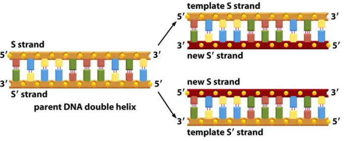
The nucleotide sequence in one strand of the double strand DNA determines the nucleotide sequence of the complementary strand. Therefore, both strands of the DNA can serve as the template for the DNA synthesis, resulting in the same double strand DNA product. This process is visualized schematically in Fig. 18.
As an exercise, identify the nucleobases in the figure.
Figure 18. The schematic representation of the duplication of the DNA molecule.
The figure is taken from The molecular biology of the cell, Garland Publishing Inc, New York, London, 1989.
This seemingly simple process utilizes a large number of proteins/enzymes in living cells. In the following the focus will be set strictly on the replication process. First the replication in the cell will be summarized briefly. The long double strand DNA molecule has to be separated into single strand DNA
49
molecules behaving as templates in the area of the replication process. For this the cells apply the helicase enzymes. These enzymes use the energy of adenosine triphosphate (ATP) to separate the two DNA strands. The crystal structure of helicase isolated from T7 phage shows that the molecule consists of six subunits, but it is not sixfold symmetric (Fig. 19.). The conformation of the hexamer depends on the form of the substrate bound. In opposite positions pairwise it is either ATP or ADP bound or the substrate binding site is empty. The three sites interconvert in the coordinate fashion through conformational changes, causing a rotation-like movement and oscillation of the enzyme resulting in unwinding the double strand of the DNA. Close to the helicase a replication fork is formed.
Figure 19. The crystal structure of the helicase enzyme isolated from T7 phage (PDB Id: 1CR0).
The new DNA strand is built up by another enzyme, called DNA polymerase. However, this enzyme can only build up the new strand starting from an existing initial sequence. This sequence, further denoted as primer, is
50
synthesized by the DNA primase enzyme. From this sequence the DNA polymerase starts to synthesize the new strand from the appropriate energy-rich building blocks, the 2'-deoxyribonucleoside triphosphates (dNTPs). The 5'- triphosphate group will be the reactive side of the new building blocks, which will react with the free 3'-hydroxy group of the primer or the new growing strand, if the correct base pairing is established. This means that the new strand is always growing in the 5' → 3' direction as shown in Fig. 20.
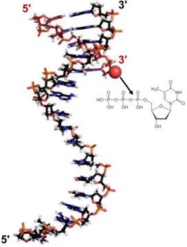
Figure 20. The crystal structure of the template strand DNA with a primer sequence (with red backbone) hybridized to the 3’ terminus of the template strand.
In this way the 3' terminus (symbolized by red ball) of the primer is directed toward the template, thus it can react with the incoming dNTP, by the help of the DNA polymerase enzyme. The primer is thus, prolonged by stepwise attaching the incoming dNTPs to the 3’-OH. A pyrophosphate is released during each phosphodiester bond formation. (PDB Id: 2M54).
3'
5' 3'
5'
51
An important consequence of this rule is that following the helicase, one of the two DNA polymerases can continuously build the new strand at the template strand directed with its 5' end to the polymerase – called leading strand template.
The other template strand, however, has opposite direction – called lagging strand template –, which requires the second DNA polymerase to build the new strand in backward direction compared to the path of the helicase. As the consequence of this, the synthesis of the second strand very complicated (see Fig. 21.). Further proteins and enzymes participate in it, and the new strand is synthesized piece by piece.
Figure 21. The schematic representation of the DNA replication fork with the protein machinery attached to the template strands. The growing new strands are in red, while the template strands are orange. The figure is taken from The molecular biology of the cell, Garland Publishing Inc, New York, London, 1989.
52
The new DNA fragments, called Okazaki fragments which are finally linked together by the help of a ligase enzyme. Nevertheless, the process is well optimized and it is so efficient, that the two strands can grow in parallel.
The DNA replication is extremely precise and quick process in the cells.
The helicase is rotating with approximately the speed of a jet engine. The polymerase enzyme catalyzes the formation of ~ 5000-10000 phosphodiester bonds in one minute, and allows on average for only one mistaken base pairing in every 10000th case. Some DNA polymerases possess so-called proofreading activity, by means of which they can correct even this error in the new DNA sequence. By means of this safety function they allow erroneous base pairing only in every 108 nucleotide unit.
The results of the research on the field of the DNA replication conducted in the laboratories of Max Delbrück, Alfred D. Hershey and Salvador E. Luria also deserved Nobel prize (Fig. 22.).
Figure 22. The Nobel Prize in Physiology or Medicine 1969 was awarded jointly to Max Delbrück, Alfred D. Hershey and Salvador E. Luria (from left to right)
"for their discoveries concerning the replication mechanism and the genetic structure of viruses." (Photo from the Nobel Foundation archive.)
53
The DNA replication within the cells is often used to multiply DNA in recombinant DNA technology, but in this way only the DNA recognized by the cell as its own component will be multiplied. Elaborating the method of efficient DNA replication in a test tube would allow researchers to multiply any desired DNA sequence. Certainly, the final goal of these experiments is to produce recombinant proteins from recombinant DNA molecules. By this technology, the modification of proteins, including mutation, truncation, fusion to other proteins would become feasible. The first step of this is to synthesize and modify DNA sequences coding for these proteins. Recombinant DNA molecules are DNA segments from different biological sources combined together to obtain new genetic material. Such recombinant DNA could be a bacterial DNA including a target gene for protein expression in bacterial cell.
To replicate the DNA in a test tube, it is necessary to avoid the use of various enzymes, since this would make the reaction very expensive. It is possible to separate the two strands of any template DNA by increasing the temperature of a reaction mixture to ~ 100C. This, however, would be deleterious for the DNA polymerase, the presence of which is the minimal requirement for DNA synthesis.
The solution to this problem was the discovery of the heat-resistant DNA polymerase (Taq polymerase) isolated from the Thermus Aquaticus extremophyl bacterial species living in hot springs and geysers. This enzyme can survive the increased temperature for separating the double strand of the DNA. It is worth mentioning that Science nominated the Taq polymerase enzyme as the molecule of the year in 1989. The other brick in the wall was the brilliant idea of Karry B.
Mullis (Fig. 23.), working as a chemist at Cetus Corporation in USA.
54
Figure 23. Kary Banks Mullis (1944-2019) obtained Nobel Prize in Chemistry in 1993 "for contributions to the developments of methods within DNA-based chemistry - for his invention of the polymerase chain reaction (PCR) method", and his scientific paper describing the method. (Photo from the Nobel Foundation archive.)
He introduced a pair of primers to determine the termini of the new DNA molecule to be amplified in subsequent cycles, periodically changing the temperature. Repeating the cycles of separating the double strand DNA into single strand templates, the hybridization of the primers a lover temperature, and the new DNA strand prolongation by the help of the DNA polymerase enzyme, the exponential amplification of the target DNA has been achieved. The method has been published in 1986 in a paper in Cold Spring Harb Symp Quant Biol, vol. 51, pp. 263-273. These discoveries lead to the polymerase chain reaction (PCR) and the Nobel Prize for its invention in chemistry in 1993. The PCR process revolutionized the fields of biochemistry and molecular biology, and its invention had also strong economical consequences: the Cetus company, as the owner of
55
the right of the technique from 1989, has sold the patent of PCR and Taq polymerase in 1992 to Hoffmann-LaRoche company for 300,000,000 USD.
If the genome carrying the gene of the target protein is available, selection of the gene, and its mutations can be carried out by polymerase chain reaction.
The PCR reaction mixture contains the following components:
- template DNA molecules;
- the heat stable polymerase enzyme in a Mg2+-containing buffer, e.g. Taq polymerase;
- DNA primers: short synthetic 2'-deoxyoligonucleotides that can hybridize to the single strand DNA template and allow the polymerase to start the synthesis of the complementary strand;
- 2'-deoxynucleoside triphosphates (dNTPs: dCTP, dATP, dGTP and dTTP) as building blocks.
The reaction is driven by heat control (Fig. 24A.). In the first step of the cycle, the denaturation, the reaction mixture is heated to > 90 °C. The template DNA dissociates to single strands at around the so-called melting temperature.
Then the temperature is lowered to a point close to the melting point of the primers (usually ~50-65 °C) so that they can hybridize to the single stranded template (annealing). In the third step of the cycle the temperature is adjusted to the optimum for the polymerase enzyme function, which is ~72 °C in case of the Taq polymerase, and the elongation of the primers towards the 3' end of the new strand using the dNTPs as building blocks results in the new double strand DNA molecule. These three steps are then repeated in the PCR instrument (Fig. 24B.).
This instrument is adjusting the preprogrammed temperature very precisely and
56
the temperature change is very quick. It is essential to use special thin-walled test tubes for efficient heat transfer.
A
B
Figure 24. A) The temperature program of a PCR used to introduce mutation to the end of the sequence and scheme of the reaction. Each blue lines indicate a strand of the template DNA. The red and green endings are restriction enzyme cleavage sites, carried by the primer molecules. The mutation introduced by primers is in yellow. These will be explained later in more detail in the section dealing with the design of the primers. (Taken from the PhD dissertation of Eszter Németh, written in the laboratory of the author of this e-book.) B) The PCR instrument used in the laboratory of the author.
57
The products of each cycle formed in the elongation step serve as templates in the next cycles. The amount of the PCR products is mathematically doubled in each cycle. Thus, the amount of DNA is exponentially increasing: the number of products from one template molecule will be ~2n where n is the number of cycles.
As already mentioned the primers determine the termini of the newly amplified DNA molecule. Thus the termini can be selected by the appropriate selection of the primer sequences. By means of this, any target gene can be selected from a genome for amplification (Fig. 25.).
At the beginning of the reaction from the original template strands longer PCR products are formed than the desired ones, as there is no barrier for the polymerase to stop the process. The prolongation will be terminated by the increase of the temperature at the beginning of the next cycle. From the next cycles the PCR products formed in the previous cycles will also serve as templates. The PCR from these templates will yield the product with the precisely the selected length – now determined by the end of the new template strands (see Fig. 25.). The "long products" can for only from the original template, i.e. their amount is increasing linearly. Therefore, after 20-25 cycles these can be neglected in comparison to the exponentially increasing amount of the selected DNA section. In the 25th cycle starting out from a single DNA molecule we can obtain 50 single strand DNA molecules of undetermined length, and ~ 33.55 millions of the targeted double strand DNA with the desired length. Thus, the selected section of the DNA is efficiently multiplied in PCR.

![Figure 4. PyMol image of the [NiFe] hydrogenase from Desulfovibrio desulfuricans based on its crystal structure (PDB Id: 1E3D)](https://thumb-eu.123doks.com/thumbv2/9dokorg/1190594.87828/19.892.220.683.274.576/figure-pymol-image-hydrogenase-desulfovibrio-desulfuricans-crystal-structure.webp)