https://doi.org/10.1007/s11033-019-04688-9 SHORT COMMUNICATION
Novel frameshift mutation of the NR0B1(DAX1) in two tall adult brothers
Rita Bertalan1,2 · Zsuzsa Bencsik3 · Piroska Mezei4 · Zsolt Vajda5 · Henriett Butz6,7 · Attila Patócs6,7
Received: 2 October 2018 / Accepted: 7 February 2019 / Published online: 6 July 2019
© The Author(s) 2019
Abstract
NR0B1 (nuclear receptor subfamily 0, group B, member 1) is a transcription factor encoded by DAX1 (dosage-sensitive sex reversal, adrenal hypoplasia critical region, on chromosome X, gene 1) responsible for the development and maintenance of the steroidogenic tissues. In humans the DAX1 mutations cause congenital adrenal hypoplasia (AHC) and hypogonadotropic hypogonadism (HHG) in boys. Here we report two brothers who were assessed by endocrinologist at the age of 51 and 43 because of their serious osteoporosis. They had been substituted with prednisolone since the age of 4 and 9 years because of their primary adrenal insufficiency (PAI). Due to their late puberty caused by HHG at the age of 16 and 17 years their heights were − 3.1 and − 3.3 SD, but then they had a significant growth during their adulthood and reached the + 1.85 SD and + 3.78 SD respectively. During this period, they received glucocorticoid supplementation, but the treatment of their HHG was inadequate. At the age of 51 and 43 years insulin tolerance test (ITT) and gonadotropin releasing hormone (GnRH) test confirmed their PAI and HHG. Genetic test performed at this time revealed a novel, four nucleotides deletion (del.586- 571c.GGGC or 572-575c.GGGC) of DAX1 gene. The two brothers with AHC and HHG caused by a novel DAX1 mutation, reached tall final heights, despite of the disadvantageous prednisolone treatment during their childhood. We assume that the long-term lack of the sexual hormone substitution was a significant reason of their above average height as well as their serious osteoporosis.
Keywords Novel NR0B1(DAX1) gene mutation, AHC · Adrenal hypoplasia congenita, HHG · Hypogonadotropic hypogonadism
* Rita Bertalan
bertalan.rita@med.semmelweis-univ.hu Zsuzsa Bencsik
bencsik.zsuzsanna@szentdonatkorhaz.hu Piroska Mezei
pmezei@mail.fmkorhaz.hu Zsolt Vajda
zsvajda@heimpalkorhaz.hu Henriett Butz
butz.henriett@med.semmelweis-univ.hu Attila Patócs
patocs.attila@med.semmelweis-univ.hu
1 1st Department of Pediatrics, Semmelweis University, Bókay
2 Csolnoky Ferenc Hospital, Kórház Street 1, Veszprém 8200, Hungary
3 Szent Donát Hospital, Honvéd Street 2-3, Várpalota 8100, Hungary
4 Fejér County Szent György University Teaching Hospital, Seregélyesi Street 3, Szekesfehervar 8000, Hungary
5 Pál Heim Children’s Hospital, Üllői Street 86, Budapest 1089, Hungary
6 Momentum Hereditary Endocrine Tumours Research Group Semmelweis University, Szentkirályi Street 46, Budapest 1088, Hungary
7 Department of Laboratory Medicine, Semmelweis University, Szentkirályi Street 46, Budapest 1088, Hungary
Introduction
The DAX1 is located on the short arm of X chromosome and is also known as NR0B1 (OMIM*300473), encoding an orphan nuclear hormone receptor (NHR) [1].
DAX-1 is expressed in embryonic stem cells, steroido- genic tissues (gonads, adrenals), ventromedial hypothala- mus as well as pituitary gland in developing and adult tissues [2, 3]. The exact mechanism of DAX1 in different transcriptional processes remains unclear, but it has shown that it can function as a repressor during the transcription or can have a role as an RNA shuttling protein [1, 4–9].
In case of the loss of DAX1 function due to mutations develops an AHC and HHG in boys, although the pheno- typic variation of severity and the age of manifestation are highly variable [10–12]. Even animal models indicate that DAX1 also plays a primary role in the development of testis and spermatogenesis [13].
In AHC patients the permanent zone of adrenal cortex is absent and abnormally large fetal adrenal cells persist, resulting in a structural and functional disorganization of the adrenal gland with consequent different severity of glu- cocorticoids, mineralocorticoids and androgens deficien- cies [14, 15]. In patients with AHC the current treatment indicates maintenance of glucocorticoid and mineralocor- ticoid replacement and appropriate symptomatic treatment during adrenal crises [12, 16].
HHG is caused by combined and variable deficiency of hypothalamic failure in gonadotropin-releasing hormone (GnRH) producing and pituitary defect in responsiveness to GnRH, resulting in reduced gonadotropin production [17]. Treating HHG in affected boys requires gonadotro- pins and/or sexual hormone substitution during puberty and lifelong [12].
Here we report two brothers who had been substituted with glucocorticoids since their childhood due to their AHC and partially with sexual hormone since their late teenage because of their HHG. The final diagnosis was achieved by genetic testing demonstrating a novel, four nucleotides deletion of DAX1.
Case Reports
Patient 1The 51-age patient was referred to the endocrinologist because of a serious osteoporosis (T-score lumbar: − 2.3 hip: − 2.5). At this point his height was188 cm and he had a hypogonad phenotype with gynecomastia and hypoplas- tic testes (2 ml × 2 ml).
He was born at term from an uneventful pregnancy with 4400 gramm birthweight, from non-consanguineous healthy average height parents. However, during family history turned out that but one of his maternal uncles died in infancy.
After an unremarkable early childhood, the 4-year-old boy was hospitalized several times because of the repeat- ing classical symptoms of hypadrenia (hyperpigmenta- tion, failed to thrive, stomachache, vomiting). Based on the clinical findings and laboratory results the diagnosis of primary hypadrenia was established. The possibil- ity of 21-hydroxylase deficiency was excluded, but he received antituberculotic treatment. Karyotype revealed 46,XY male genotype. Per os prednisolone 2.5 mg/die and decosterone intramuscular injection (1 × 5 mg/week) were introduced.
Due to bilateral cryptorchidism he was treated with intramuscular HCG (human choriogonadotropin) but because of the inefficacy of this treatment he underwent an orchidopexy at age 11 years.
His puberty was delayed and since the age of 23 years he received regular HCG treatment (3000 IU/2 months) and then the level of testosterone achieved the 9 nmol/l, testis and pubic hair were growing. Because of compli- ance problem from the age of 25 years until 51 years he remained untreated with testosterone replacement, but he has been receiving glucocorticoid supplementation (hydro- cortisone 40 mg/day).
His growth curve was unusual (13 years 147 cm: − 1.26 SD, 16 years 152 cm: − 3.1 SD, 23 years 166 cm: − 1.68 SD) but at adult age (51 years) he reached 188 cm (1.85 SD) and his weight was 95 kg (BMI: 26.9 kg/m2).
Exactly at this age the diagnoses of PAI and HHG were confirmed by gonadotropin releasing hormone (GnRH) and the Insulin tolerance tests (ITT) (Table 1). These tests revealed a low level of the gonadotropic hormones, peak luteinizing hormone (LH): 0.3 IU/l and peak Fol- licle stimulating hormone (FSH): 1.6 IU/l, low cortisol (3.5 nmol/l), elevated ACTH (Adrenocorticotropic hor- mone) (> 330 pg/ml) and low growth hormone (GH) level (peak: 0.25 mIU/l). The brain magnetic resonance (MR) imaging showed normal brain anatomy and a lack of pitui- tary anomalies.
Molecular genetic examination revealed a novel, four nucleotides deletion of DAX1 gene confirming the diag- nosis of congenital adrenal hypoplasia.
From that time his treatment regimen includes hydro- cortisone 30 mg/day (13 mg/m2/day hydrocortisone), 0.1 mg fludrocortisone/day, mesterolone 50 mg/day, alen- dronic acide 70 mg/week and vitamin D 3000 IU/day.
Upon androgen substitution (testosterone undecanoate 1000 mg) he had a priapism and he refused this therapy.
Patient 2
He is a younger brother of the Patient 1 and he was also evaluated by the endocrinologist at the age of 43 years because of a serious osteoporosis (T-score, lumbar − 2.6, hip: − 2.5). His height was 200 cm presenting with a hypo- gonad phenotype: gynecomastia, 2 ml testes and a hypo- plastic penis.
He was born from an uneventful pregnancy at term with 4700 gr birth weight. At the age of 9 years, based on the clinical symptoms (easily exhausted, failure to thrive, stom- achache, repeated vomiting) and the PAI of his brother, this diagnosis was also established for him. Karyotype revealed a 46,XY male genotype and 21-hydroxylase deficiency were excluded. Per os prednisolone 5 mg/die and decosterone intramuscular injection 1 × 5 mg/week were commenced.
As with his older brother, his puberty was also delayed and the patient was treated with HCG 3000 IU/2 months from the age of 17 years. Due to this treatment testis became larger and secondary signs of male gender developed. A few years later because of the adherence problem he stopped the HCG, although he still stayed on glucocorticoid treatment (hydrocortisone 20 mg/day).
His registered heights were: at 7 years 125 cm (0.47 SD), 10 years: 131 cm (− 1.27 SD), 17 years: 155 cm (− 3.0 SD), 43 years: 200 cm (+ 3.78 SD) and weight 120 kg (BMI:
30 kg/m2).
At the age of 43 the diagnosis of PAI and HHG were proved by GnRH and ITT test. (Table 1). These tests revealed a low level of the gonadotropic hormones (peak LH:
0.3 IU/L, peak FSH: 1.4 IU/l), low cortisol (27.3 nmol/l), elevated ACTH (111.2 pg/ml) and low GH level (peak:
0.11 mIU/l). Similarly, to his brother the brain MR imaging revealed normal brain anatomy and a lack of any pituitary anomalies. In addition a molecular genetic examination showed the similar mutation detected in his brother.
From the age of 43 years his treatment included hydro- cortisone 20 mg/day (8 mg/m2/day) hydrocortisone, mester- olone 50 mg/day, 0.1 mg fludrocortisone/day and vitamin D 30,000 IU/week. Moreover the androgen substitution (testosterone undecanoate 1000 mg) caused the same pain- ful priapism as his brother, he also refused the replacement therapy of male sexual hormone.
Molecular genetic analysis of DAX1 gene
Informed consent was obtained from the brothers and their mother for publication of their medical details. Molecu- lar genetic examination of DAX1 was performed within the frame of the routine clinical genetic testing service at Clinical Genetics and Endocrinology Laboratory Sem- melweis University following the institutional protocol.
Genomic DNA was extracted from peripheral blood
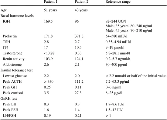
Table 1 Hormone
concentrations in fasting and after endocrine stimulation (ITT and GnRH) tests in Patient 1 and Patient 2
ACTH Adrenocorticotropic hormone, FSH Follicle stimulating hormone, fT4 free thyroxine, GnRH Gon- adotropin releasing Hormone, GH Growth hormone, IGFI Insulin-like growth factor, LH Luteinizing hor- mone, TSH Thyroid stimulating hormone
Patient 1 Patient 2 Reference range
Age 51 years 43 years
Basal hormone levels
IGFI 169.5 96 92–244 UG/l
Male: 35 years: 80–240 ng/ml Male: 45 years: 70–210 ng/ml
Prolactin 171.8 371.8 54–380 mIU/l
TSH 2.8 2.7 0.35–4.94 mIU/l
fT4 17 10.5 9–19 pmol/l
Testosterone < 0.28 0.33 5.8–28.1 nmol/l
Renin activity 103.9 124.1 0.2–5.7 ng/ml/h
Aldosterone 2.6 2.1 30–400 pg/ml
Insulin tolerance test
Lowest glucose 2.2 2.0 < 2.2 mmol/l or half of the initial value
Peak ACTH > 330 111.2 7.2–63.3 pg/ml
Peak GH 0.25 0.11 0–6 ng/ml
Peak cortisol 3.5 27.3 8–25 μg/dl
GnRH test
Peak LH 0.3 0.3 1.7–8.6 IU/l
Peak FSH 1.6 1.4 1.5–12 IU/l
LH/FSH 0.19 0.21 > 1
leukocytes using the DNA isolation kit for mammalian blood (Boehringer Mannheim Corporation, Indianapolis, IN, USA) and DNA samples were frozen at − 20 °C until they were analyzed. Mutation analysis of DAX1 gene was carried out using polymerase chain reaction (PCR) followed by direct bidirectional sequencing of the PCR- amplified DNA. The oligonucleotide primer pairs used for PCR amplification of exons 1 and 2 of DAX1 gene were as following:
1 AF: 5′-CGT GCG CGC TAG GTA TAA AT-3′
1 AR: 5′-GTT TGC TTT GCG CTC GTC-3′
1 BF: 5′-CAG GGC AGC ATC CTC TAC A-3′
1 BR: 5′-AAG CAG CAG CGG TAC AGA AG-3′
1 CF: 5′-CTT CTG TAC CGC TGC TGC TT-3′
1 CR: 5′-CCA CAG TCT CGA ACT GCA AG-3′
1 DF: 5′-CTT GCA GTT CGA GAC TGT GG-3′
1 DR: 5′-TTT TGT GAG CTG GGA AAA GC-3′
2 F: 5′-TTT TTC TCC CTC CAG ACG TG-3′
2 R: 5′-TCT TTT GCC CAC AGC TCT TT-3′
The PCR-amplified DNA was purified using a commer- cial kit (High pure PCR product, Roche Diagnostics, Man- nheim, Germany). For direct sequencing of the whole cod- ing region of DAX1 gene, Sanger sequencing was used All sequences were compared to the NCBI reference sequence NG_009814.1.
Results
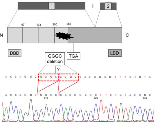
The clinical and endocrinological characteristics of the patients are summarized in Table 1. Direct sequencing of the NR0B1 (DAX1) gene revealed in both brothers a novel, four nucleotides deletion in the first exon (Fig. 1).
The GGGC deletion is located in a sequence repetition GGG CGG GC, so deletion can be del.568-571c.GGGC or del.572-575.c.GGGC. This four nucleotides containing dele- tion results in a frameshift and consequently the protein is terminated at an “ambush” Stop codon 788-790c. (Fig. 1).
This mutation was also found in a heterozygous state in the patients’ healthy mother.
This variant has not been reported in any public databases such as dbSNP (http//:http://www.ncbi.nlm.nih.gov/proje ct/
SNP), ClinVar (https ://www.ncbi.nlm.nih.gov/clinv ar/) or ExAC (http//exac.broadinstitute.org).
Discussion
DAX is a member of the nuclear hormone receptor fam- ily, although the homology is significant only in the puta- tive carboxyl-terminal ligand binding domain (LBD). The DNA binding N terminal domain is substituted by an unu- sual novel domain that contains three and half repeats of a
Fig. 1 A novel four nucleotides deletion del.586-571c.GGGC or 572-575c.GGGC of DAX1 gene in homozygous form
65–67 amino-acid motif containing two putative zinc fingers [18, 19].
In the current cases the deletion (c.568-571/572-575) locates just before the start of the ligand-binding domain (codon 196) (Fig. 1). Even the protein translation continues the four nucleotide deletion results in a frameshift and the structure of this part of the protein is not intact anymore.
The “ambush” Stop codon at nucleotides 788–790 nucleo- tides cause deleterious damage of the ligand binding domain and this variant is considered pathogenic based on ACMG (American College of Medical Genetics) guideline.
DAX1 has a role in the maintenance of certain adult tis- sues, according to the Human Protein Atlas (http://www.
prote inatl as.org/ENSG0 00001 69297 -NR0B1 /tissu e). It is expressed only in the adrenal gland, testis and ovaries, mak- ing impossible to test the functional consequences of the 4 nucleotides deletion on RNA level.
Several cases have been published when the LBD was abolished and the symptoms of hypadrenia were presented only in childhood [18]. Nevertheless, even carrying the same mutation the age of the appearance of clinical symptoms can be very various [20]. One of the possible explanations of these facts is the differences in salvage mechanisms for persistence of fetal adrenal, which suggests that other fac- tors, including epigenetics and protein modifications may be involved in the process [21, 22].
Affected boys with HHG fail to undergo puberty and require sexual hormone substitution. Repeated injections of hCG (human Choriogonadotropin) are generally successful in stimulating the testosterone production of the testis, but in practice the more accepted method is to use external tes- tosterone replacement treatment to induce puberty, and the maintenance of sex-hormone replacement during adulthood [16]. In our patients the hCG treatment was successful, but after a two-year period the patients refused the intramuscular treatment and they stayed on the glucocorticoid, but without male sexual hormone replacements for two decades. Then in their adulthood induced testosterone undecanoate caused a priapism and due to this painful experience, they have also refused the androgen substitution therapy. According to patients they had never had an active sexual life. We suppose the long insufficient androgen replacement led to late fusion of the epiphysis consequently the patients reached their final height only at the end of their twenties and a serious osteo- porosis developed in their forties.
According to Kletter and his colleagues in congenital adrenal hypoplasia with gonadotropin deficiency, the defi- cit in pituitary hormones is selective for gonadotropins (LH, FSH) and the other pituitary hormones’ production remains intact (ACTH, GH, TSH and PRL) [14]. In our patients the diagnosis was proved at the ages of 51 and 43 years, as an ITT and GnRH test were implemented and beside the AHC and HHG, a low level of GH was established coincidentally.
The recommendation of Bornstein and colleagues does not suggest the measurement of growth hormone level in the case of AHC including the DAX1 mutation suspect patients [23]. During the childhood of the brothers in Hun- gary the only accessible glucocorticoid product was the 5 mg prednisolone tablet. It is well known that the accurate dosing of this drug is difficult. Despite of this challenge, since introducing the prednisolone treatment the brothers had not had adrenal crisis and they did not show symp- toms of overdose. From their young adult ages, they were treated with hydrocortisone. The Patient 1 required higher doses to be asymptomatic, but the Patient 2 was treated in substitution dose of hydrocortisone supplemented both of them with mineralocorticoid. Despite of the disadvanta- geous prednisolone treatment on the bone maturation the final adult heights of the brothers were + 1.85 and + 3.78 SD.Calliari and colleagues published a case about a boy with DAX1 mutation and below average adult height. In this case the growth hormone level was not measured and the induction of puberty was started at 12 years of age due to psychological aspects [24]. In two patients published by Rojek the growth hormone deficiency was diagnosed at the age of 13 and 18 years respectively. In the second case, due to the retarded bone age, the latterly induced sexual hormone replacement and growth hormone therapy in the patient still showed growth at 23 years of age [25].
In cases of glucocorticoid treatment, the GH deficiency should be interpreted as a consequence of the excess of glucocorticoids substitution [25]. Although in our cases the suppressed ACTH level has not been proved, we could not exclude the harmful effect of glucocorticoids. However the fact, that the patients had still significant growth after 16 years of age (35 and 45 cm) does not confirm the over- dose of glucocorticoid administered during this period.
In our patients, beside the low normal range meas- ured IGF level, significantly low GH concentration were detected during the ITT, which can be explained by the fact that they were not on adequate male sexual hormone therapy before and during the test. Nevertheless, we sup- pose the main reason of the elevated BMI is their sedimen- tary lifestyle which may be related to the mesterolone ther- apy instead of a proper male sexual hormone replacement.
In conclusion, in our two male patients with AHC and HHG, caused by a novel DAX1 mutation despite of the long prednisolone treatment in their childhood they had reached a tall adult height. While the brothers had a significant growth during their adulthood, we assume the lack of the sexual hormone substitution was the main reason of their above average height as well as their serious osteoporosis.
Acknowledgements Open access funding provided by Semmelweis University (SE).
Compliance with ethical standards
Conflict of interest The authors declare that they have no conflict of interest.
Open Access This article is distributed under the terms of the Crea- tive Commons Attribution 4.0 International License (http://creat iveco mmons .org/licen ses/by/4.0/), which permits unrestricted use, distribu- tion, and reproduction in any medium, provided you give appropriate credit to the original author(s) and the source, provide a link to the Creative Commons license, and indicate if changes were made.
References
1. Zanaria E, Muscatelli F, Bardoni B et al (1994) An unusual mem- ber of the nuclear hormone receptor superfamily responsible for X-linked adrenal hypoplasia congenita. Nature 372:635–641 2. Zhang J, Liu G, Ruan Y, Wang J et al (2014) Dax1 and Nanog act
in parallel to stabilize mouse embryonic stem cells and induced pluripotency. Nat Commun 5:5042
3. Ikeda Y, Swain A, Weber TJ et al (1996) Steroidogenic factor 1 and Dax-1 colocalize in multiple cell lineages: potential links in endocrine development. Mol Endocrinol 10:1261–1272 4. Ferraz-de-Souza B, Martin F, Mallet D et al (2009) CBP/
p300-interacting transactivator, with Glu/Asp-Rich C-terminal domain, 2, and Pre-B-cell leukemia transcription factor 1 in human adrenal development and disease. J Clin Endocrinol Metab 94:678–683
5. Xu B, Yang WH, Gerin I, Hu CD, Hammer GD, Koenig RJ (2009) Dax-1 and steroid receptor RNA activator (SRA) function as tran- scriptional coactivators for steroidogenic factor 1 in steroidogen- esis. Mol Cell Biol 29:1719–1734
6. Ito M, Yu R, Jameson JL (1997) DAX-1 inhibits SF-1-mediated transactivation via a carboxy-terminal domain that is deleted in adrenal hypoplasia congenita. Mol Cell Biol 17:1476–1483 7. Zazopoulos E, Lalli E, Stocco DM, Sassone-Corsi P (1997) DNA
binding and transcriptional repression by DAX-1 blocks steroido- genesis. Nature 390:311–315
8. Lalli E, Bardoni B, Zazopoulos E, Wurtz JM, Strom TM, Moras D, Sassone-Corsi P (1997) A transcriptional silencing domain in DAX-1 whose mutation causes adrenal hypoplasia congenita. Mol Endocrinol 11:1950–1960
9. Lalli E, Ohe K, Hindelang C, Sassone-Corsi P (2000) Orphan receptor DAX-1 is a shuttling RNA binding protein associated with polyribosomes via mRNA. Mol Cell Biol 20:4910–4921 10. Muscatelli F, Strom TM, Walker AP et al (1994) Mutations in the
DAX-1 gene give rise to both X-linked adrenal hypoplasia con- genita and hypogonadotropic hypogonadism. Nature 372:672–676 11. Guo W, Mason JS, Stone CG Jr et al (1995) Diagnosis of X-linked
adrenal hypoplasia congenita by mutation analysis of the DAX1 gene. JAMA 274:324–330
12. Jadhav U, Harris RM, Jameson JL (2011) Hypogonadotropic hypogonadism in subjects with DAX1 mutations. Mol Cell Endo- crinol 346(1–2):65–73
13. Yu RN, Ito M, Saunders TL, Camper SA, Jameson JL (1998) Role of Ahch in gonadal development and gametogenesis. Nat Genet 20:353–357
14. Kletter GB, Gorski JL, Kelch RP (1991) Congenital adrenal hypo- plasia and isolated gonadotropin deficiency. Trends Endocrinol Metab 2:123–128
15. Lalli E, Sassone-Corsi P (2003) DAX-1, an unusual orphan recep- tor at the crossroads of steroidogenic function and sexual differ- entiation. Mol Endocrinol 17:1445–1453
16. Suntharalingham JP, Buonocore F, Duncan AJ, Achermann JC (2015) DAX-1 (NR0B1) and steroidogenic factor-1 (SF-1, NR5A1) in human disease. Best Pract Res Clin Endocrinol Metab 29:607–619
17. Habiby RL, Boepple P, Nachtigall L, Sluss PM, Crowley WF Jr, Jameson JL (1996) Adrenal hypoplasia congenita with hypogon- adotropic hypogonadism: evidence that DAX-1 mutations lead to combined hypothalmic and pituitary defects in gonadotropin production. J Clin Invest 98:1055–1062
18. Krone N, Riepe FG, Dörr HG, Morlot M et al (2005) Thirteen novel mutations in the NR0B1 (DAX1) gene as cause of adrenal hypoplasia congenita. Hum Mutat 25:502–503
19. Zhang YH, Guo W, Wagner RL, Huang BL et al (1998) DAX1 mutations map to putative structural domains in a deduced three- dimensional model. Am J Hum Genet 62:855–864
20. Calliari LE, Longui CA, Rocha MN, Faria CD et al (2007) A novel mutation in DAX1 gene causing different phenotypes in three siblings with adrenal hypoplasia congenita. Genet Mol Res 6:277–283
21. Achermann JC, Meeks JJ, Jameson JL (2001) Phenotypic spec- trum of mutations in DAX-1 and SF-1. Mol Cell Endocrinol 185:17–25
22. Piferrer F (2013) Epigenetics of sex determination and gonado- genesis. Dev Dyn 242:360–370
23. Bornstein SR, Allolio B, Arlt W et al (2016) Diagnosis and treat- ment of primary adrenal insufficiency: an endocrine society clini- cal practice guideline. J Clin Endocrinol Metab 101:364–389 24. Calliari LE, Rocha MN, Monte O, Longui CA (2013) Mild adrenal
insufficiency due to a NROB1 (DAX1) gene mutation in a boy presenting an association of hypogonadotropic hypogonadism, reduced final height and attention deficit disorder. Arq Bras Endo- crinol Metabol 57:562–655
25. Rojek A, Obara-Moszynska M, Malecka E, Slomko-Jozwiak M, Niedziela M (2013) NR0B1 (DAX1) mutations in patients affected by congenital adrenal hypoplasia with growth hormone deficiency as a new finding. J Appl Genet 54:225–230
Publisher’s Note Springer Nature remains neutral with regard to jurisdictional claims in published maps and institutional affiliations.