HUMAN GENETICS•ORIGINAL PAPER
A novel WDR62 missense mutation in microcephaly with abnormal cortical architecture and review of the literature
Melinda Zombor1&Tibor Kalmár1&Nikoletta Nagy2&Marianne Berényi3&Borbála Telcs3&Zoltán Maróti1&
Oliver Brandau4&László Sztriha1
Received: 27 May 2018 / Revised: 5 January 2019 / Accepted: 17 January 2019 / Published online: 1 February 2019
#Institute of Plant Genetics, Polish Academy of Sciences, Poznan 2019
Abstract
Autosomal recessive primary microcephaly (MCPH) is a group of rare neurodevelopmental diseases with severe microcephaly at birth. One type of the disorder, MCPH2, is caused by biallelic mutations in theWDR62gene, which encodes the WD repeat–
containing protein 62. Patients withWDR62mutation may have a wide range of malformations of cortical development in addition to congenital microcephaly. We describe two patients, a boy and a girl, with severe congenital microcephaly, global developmental delay, epilepsy, and failure to thrive. MRI showed hemispherical asymmetry, diffuse pachygyria, thick gray matter, indistinct gray-white matter junction, and corpus callosum and white matter hypoplasia. Whole exome sequencing revealed the same novel homozygous missense mutation, c.668T>C, p.Phe223Ser in exon 6 of theWDR62gene. The healthy parents were heterozygous for this mutation. The mutation affects a highly conserved region in one of the WD repeats of the WDR62 protein. Haplotype analysis showed genetic relatedness between the families of the patients. Our findings expand the spectrum of mutations randomly distributed in theWDR62gene. A review is also provided of the brain malformations described inWDR62mutations in association with congenital microcephaly.
Keywords Microcephaly . Malformations of cortical development . Whole exome sequencing .WDR62mutation . Global developmental delay
Introduction
Microcephaly is defined as an occipitofrontal head circumfer- ence below the third percentile or more than two standard deviations (SD) below the mean for sex, age, and ethnicity.
It can be associated with delayed motor and cognitive
development, various neurological signs, intellectual disabili- ty, epilepsy, and autism and accounts for a significant propor- tion of neurodevelopmental disorders in childhood.
Microcephaly may develop prenatally or postnatally and may have genetic or non-genetic cause. Any condition that affects important processes of brain growth, including
Communicated by: Michal Witt
* László Sztriha
sztriha.laszlo@med.u-szeged.hu Melinda Zombor
zombor.melinda@med.u-szeged.hu Tibor Kalmár
kalmar.tibor@med.u-szeged.hu Nikoletta Nagy
nikoletta.nagy@gmail.com Marianne Berényi berenyi@ella.hu Borbála Telcs boritel@gmail.com
Zoltán Maróti
maroti.zoltan@med.u-szeged.hu Oliver Brandau
oliver.brandau@bioscientia.de
1 Department of Pediatrics, University of Szeged, Temesvári krt.
35-37, Szeged 6726, Hungary
2 Department of Medical Genetics, University of Szeged, Szeged, Hungary
3 Department of Developmental Neurology, St. Margaret Hospital, Budapest, Hungary
4 Centogene AG, Rostock, Germany
progenitor cell proliferation, cell differentiation, and cell death, can lead to microcephaly (Alcantara and O’Driscoll 2014; Barbelanne and Tsang 2014; Zaqout et al. 2017).
Anomalies causing microcephaly may exclusively affect ce- rebral development (non-syndromic microcephaly) or may be associated with dysmorphic features and extracerebral malformations (syndromic microcephaly). The spectrum of phenotypes and associated disorders ofBmicrocephaly^ is wide with more than 1300 entries recorded to April 2018 in The Online Mendelian Inheritance in Man (OMIM) database.
Although a wide spectrum of genetic defects can result in microcephaly, traditionally, a group of microcephalies is dis- tinguished as autosomal recessive primary microcephaly (MicroCephaly Primary Hereditary, MCPH) (Barbelanne and Tsang2014; Zaqout et al.2017). At least 17 genetic loci (MCPH1–17) have been implicated in MCPH, all of which have now been connected to single genes:MCPH1,WDR62, CDK5RAP2, KNL1, ASPM, CENPJ, STIL, CEP135, CEP152, ZNF335, PHC1, CDK6 and CENPE, SASS6, MFSD2A,ANKLE2,CIT(Zaqout et al.2017).Many of the proteins encoded by these genes interact with the centrosome, which organizes the separation of chromosome copies during cell division (Alcantara and O’Driscoll2014; Barbelanne and Tsang2014; Zaqout et al.2017).
Mutations in WDR62, encoding WD repeat–containing protein 62, are responsible for MCPH2, which is the second most frequent form of MCPH after MCPH5 caused byASPM mutations. Over 40 pathogenic mutations in WDR62 have already been published. In addition to microcephaly, a wide range of cortical malformations was also described in these patients (Bacino et al.2012; Banerjee et al.2016; Bastaki et al.
2016; Bhat et al.2011; Farag et al.2013; Sajid Hussain et al.
2013; Kousar et al.2011; McDonell et al.2014; Memon et al.
2013; Miyamoto et al.2017; Murdock et al.2011; Najmabadi et al.2011; Nardello et al.2018; Naseer et al.2017; Poulton et al.2014; Rupp et al.2014; Wang et al.2017).
We report on two patients, a boy and a girl with the same novel missense mutation in WDR62, revealed by whole ex- ome sequencing. Both of them have pachygyria and thick cortex in addition to severe congenital microcephaly, short stature, epilepsy, and severe developmental delay.
Clinical report
Patient 1This 5-year-old boy was born at term from the third pregnancy with Cesarean section to a 32-year-old mother and 37-year-old father. The parents are consanguineous of Romani ethnicity (Fig.1a). Apgar scores were 8 and 8 at 1 and 5 min, respec- tively. Severe microcephaly was noted at birth with head cir- cumference of 30 cm (−3.5SD). The birthweight was 2900 g
(−1SD) and length 50 cm (0.1SD). The pregnancy was unre- markable, with no report of infection, alcohol use, or sub- stance abuse. The parents had unremarkable medical histories, normal head size and normal intellect. They have a healthy son and a healthy daughter. Microcephaly progressed, with head circumference of 40 cm (−6SD) at 2 years and 41.5 cm (−6SD) at 4 years of age. The patient also had short stature, with a height of 89 cm (−3.4SD) and a weight of 12 kg (−2.4SD) at 4 years of age. Sloping forehead and dis- proportionately large face and ears as compared to the skull were observed. He followed objects and responded to loud sounds. Global hypotonia was present with preserved deep tendon reflexes. Motor and intellectual development was se- verely impaired with inability to sit and stand unsupported, or reach out for objects at the age of 5 years. He had no words, but showed emotions. Infantile spasms began at 4 months of age and were well controlled by vigabatrin, which was tapered off and discontinued by 3 years of age. One year later, com- plex partial seizures appeared and the EEG showed interictal short paroxysms of bilateral spike and wave discharges.
Valproate treatment was initiated and proved to be successful.
Metabolic screening (plasma amino acids and urine organic acids) was negative.
MRI at age of 5 months showed hemispherical asymmetry (R > L) and abnormal cortical pattern. Diffuse pachygyria was observed with a few broad gyri, thick gray matter, and shallow sulci. The gray-white matter junction appeared indistinct at some areas. Moderate hypoplasia of the corpus callosum was seen. The white matter was thin in association with an asymmetrical (L > R) dilatation of the lateral ventricles. The myelination of the corpus callosum and internal capsule was appropriate for the infant’s age. Moderate cerebellar hypopla- sia was also seen. The Virchow-Robin spaces were dilated.
The basal ganglia, brainstem, and hippocampus were pre- served (Fig.2a, b).
Patient 2
This 4-year-old girl was born at term from the first preg- nancy with Cesarean section because of fetal bradycardia to a 17-year-old mother and 20-year-old father. The par- ents are of Romani ethnicity; they deny consanguinity (Fig. 1b). Apgar scores were 7, 9, and 10 at 1, 5, and 10 min, respectively. The pregnancy was complicated with urinary tract infection. Severe microcephaly was noted at birth with head circumference of 28 cm (−
5SD). Her birthweight was 2490 g (−0.4SD) and length 46 cm (−1.7SD). There was no evidence of inborn error of metabolism, intrauterine infection, alcohol use, or sub- stance abuse. The parents had unremarkable medical his- t o r i e s , n o r m a l h e a d s i z e a n d n o r m a l i n t e l l e c t . Microcephaly progressed, with head circumference of 39 cm (−5.9SD) at 2 years and 40 cm (−6.6SD) at
4 years of age. The patient also had short stature, with height of 88 cm (−3.4SD) and weight of 12.4 kg (−
2.0SD) at 4 years of age. On examination at the age of 14 months, she had severe convergent squint, but Fig. 1 Pedigree of patient 1 (a) and patient 2 (b). Sanger sequencing of
part of exon 6 of theWDR62gene shows the homozygous T to C muta- tion at position 668 of the coding DNA sequence in the patients. The 668 positions of the coding DNA sequences are indicated by arrows. The
mutation was heterozygous in the parents and brother of patient 1 (Y = T/C). A normal sequence is also shown in an unrelated control subject (c).
The mutation affects one of the WD40 repeats in the WDR62 protein (d)
followed objects and responded to loud sounds. Her mo- tor and cognitive development was severely delayed with inability to sit, stand, or reach out for objects. There was a moderate decrease in the muscle tone with slight left- sided weakness and preserved deep tendon reflexes. No further development was observed until the last follow- up at 4 years of age. Complex partial seizures started after 3 years of age, and the interictal EEG showed bi- lateral spike and wave discharges. The epilepsy was con- trolled with valproate treatment.
MRI at the age of 4 years showed hemispherical asymmetry (L > R) and abnormal cortical pattern similar to patient 1. Diffuse pachygyria, thick gray matter, and shallow sulci were observed. The white matter was thin with more or less age-appropriate myelin formation. The lateral ventricles were dilated in an asymmetrical (R > L) manner. On T2 images, a narrow periventricular band with high signal intensity was observed adjacent to the occipital horn of the right lateral ventricle. The Virchow- Robin spaces were dilated. The corpus callosum, basal ganglia, hippocampi, brainstem, and cerebellum were preserved (Fig. 2C, D).
The family members of the two patients were unaware of any relatedness.
Molecular analysis
Routine chromosomal analysis by G-banding showed normal karyotype in both patients. DNA was isolated from the periph- eral blood. Array comparative genomic hybridization using the Agilent 180K oligo-array showed normal genomic copy number in both patients.
Whole exome sequencing (WES) of affected probands and unaffected parents was performed with CentoXome® at Centogene AG (Rostock, Germany). Genomic capture was carried out with Illumina’s Nextera Rapid Capture Exome Kit. Massively parallel sequencing was done using NextSeq500 Sequencer (Illumina) in combination with the NextSeq™500 High Output Kit (2×150 bp). Raw sequence data analyses, including base calling, de-multiplexing, align- ment to the hg19 human reference genome (Genome Reference Consortium GRCh37), and variant calling, were performed using an in-house bioinformatics pipeline. For var- iant filtration, all disease-causing variants reported in Fig. 2 MRI of patient 1 (a, b) at
the age of 5 months and patient 2 (c, d) at the age of 4 years. The T2-weighted axial images dem- onstrate hemispherical asymme- try, diffuse pachygyria with a few broad gyri and shallow sulci, wide gray matter, and indistinct white- gray matter border in certain areas. The white matter is thin and the ventricles are asymmetrically enlarged. The Virchow spaces are dilated
HGMD®, ClinVar, or in CentoMD® as well as all variants with minor allele frequency (MAF) of less than 1% in ExAc database were considered. Variants that possibly impair the protein sequence, i.e., disruption of conserved splice sites, missense, nonsense, read-throughs, or small insertions/dele- tions, were prioritized. All relevant inheritance patterns were considered. All candidate pathogenic variants not previously identified were confirmed by conventional PCR amplification and Sanger sequencing. Segregation of these changes with the disease was assessed for all available family members.
We identified the same homozygous variant, c.668T>C, p . P h e 2 2 3 S e r i n e x o n 6 i n t h e W D R 6 2 g e n e (NM_001083961.1) in both patients. The detected variant was also found in heterozygous state in the patients’parents and the brother of patient 1, whereas it was absent in his sister (Fig.1a, b, c). To date, this variant has not been described in the Exome Aggregation Consortium, Exome Sequencing Project, or the 1000 Genome Browser. This variant is located in a highly conserved nucleotide (phyloP, 4.48) with large physicochemical differences between the exchanged amino acids phenylalanine and serine (Alamut v.2.7.1). Prediction programs Polyphen2, SIFT, and MutationTaster predicted pathogenicity of the missense variant which affects the WD40 repeat region of the protein (Fig.1d).
Haplotype analysis of the families
Since the two families were unaware of any relation between them, we performed a haplotype analysis to investigate their potential genetic relation. Plink (version v1.90b4.9) was used to convert variants in the region of interest to PED and MAP files from the joint VCF file (Chang et al.2015). Haplotype analysis was performed by Merlin (version 1.1.2.) software with theB–best^option using the PED, DAT, and MAP files prepared manually from the plink output files (Abecasis et al.
2002). HaploPainter (version 1.043) was used to visualize the haplotypes in the families (Thiele and Nürnberg2005). Our analysis showed that both families carry exactly the same haplotype for the entireWDR62gene (around 55 kilobases) as shown in Fig.3. Our results suggest that the two families are closely related genetically.
Discussion
The humanWDR62 gene maps to chromosome 19q13.12, consists of 32 exons, and encodes a 1523 amino acid protein containing several WD40 repeats (Bilgüvar et al. 2010;
Nicholas et al.2010; Yu et al.2010). We found the same novel missense mutation in theWDR62gene in two patients from related families with microcephaly in association with diffuse pachygyria, thickened cortex, and indistinct gray-white matter junction. Wide spectrum of cortical malformations has been
reported inWDR62mutations. Apart from pachygyria, thick- ened cortex and indistinct gray-white matter junction, band heterotopia, polymicrogyria, schizencephaly, and asymmetry of hemispheres have also been observed (Tables1 and 2).
Neuropathology in a fetus with WDR62 mutation revealed severe disruption of cortical neuronal architecture, immature radial columnar organization, and heterotopia in the interme- diate zone (Yu et al.2010).
The frameshifts, missense, nonsense, and splice site muta- tions in theWDR62gene are randomly distributed (Tables1 and2). It has been suggested initially that missense mutations may cause a deficiency of neurogenesis resulting in primary microcephaly, but nonsense mutations may cause a more se- vere microcephaly phenotype with addition of a cerebral cor- tex lamination defect (Nicholas et al.2010). Later studies, however, did not recognize any genotype-phenotype correla- tion. The novel missense mutation c.668T>C, p.Phe223Ser in our patients is associated with severe defects in cortical archi- tecture. It affects one of the WD40 repeat regions of the WDR62 protein. WD40 repeat is a short structural motif of approximately 40 amino acids, often terminating in a tryptophan-aspartic acid (W-D) dipeptide. The common func- tion of all WD40 repeat proteins is coordinating multiprotein complex assemblies, where the repeating units serve as a rigid scaffold for protein interactions (Li and Roberts2001).
Central to the mitotic process is the formation and mainte- nance of a microtubule-based spindle apparatus organized by the centrosomes (Prosser and Pelletier2017). The centrosome contains a pair of cylindrical centrioles in an orthogonal con- figuration and each made primarily of nine microtubule trip- lets. The centrioles are surrounded by pericentriolar matrix of proteins and centrosomal satellites. The satellites are granular structures implicated in the trafficking of material involved in centriole assembly (Bettencourt-Dias et al.2011). It is note- worthy that the two centrioles differ in their structure and function. The older, Bmother^ centriole possesses subdistal appendages, where microtubules are docked, and distal ap- pendages, which are important for docking to the plasma membrane. In contrast, the younger Bdaughter^ centriole, which is formed during the preceding S phase, lacks these structures (Bettencourt-Dias et al.2011). Full acquisition of appendages by the daughter centriole is not achieved until at least one-and-a half cell cycles later. Centrosome replication during each cell cycle leads to asymmetric centrosome inher- itance, that is, the formation of two centrosomes: one of which retains the original old mother centriole (that is, theBmother^
centrosome) while the other receives the newBmother^cen- triole (that is, the daughter centrosome). Asymmetric centro- some inheritance maintains neural progenitors in the neocor- tex (Wang et al.2009).
At the onset of mitosis, centrosomes separate and the pericentriolar matrix expands through the coordinated activa- tion and recruitment of spindle pole proteins (Fujita et al.
2016). Centrosomal duplication results in the generation of a bipolar mitotic spindle. The mitotic spindle is an array of microtubules, which are assembled from dimers ofα- and β-tubulins, initiated by aγ-tubulin ring complex. The chro- mosomes attach to bundles of microtubules via kinetochores, which are multiprotein complexes that assemble on the cen- tromere of each sister chromatid (Prosser and Pelletier2017).
A coordinated interplay between proteins, including WDR62, i.e., a large network of protein-protein interactions, is essential for normal centrosomal function. It has been demonstrated recently that four of the primary microcephaly-associated pro- teins, such as CDK5RAP2, CEP152, WDR62, and CEP63, assemble in a step-wise hierarchical manner. Both the microcephaly-associated proteins and their centriolar satellite partner proteins are required for the centrosomal localization of CDK2, a cyclin-dependent kinase, which has a role in both centriole duplication and cell cycle progression (Kodani et al.
2015; Meraldi et al.1999). Loss of any of the microcephaly- associated proteins, like loss of functioning WDR62 in our patients, disrupts centriole duplication or stability (Fujita
et al.2016; Kodani et al. 2015; Meraldi et al.1999). The regulation and subcellular localization of WDR62 is cell cycle dependent. Studies by immunocytochemistry revealed that WDR62 protein showed cytosolic distribution in the inter- phase but it accumulated strongly at the spindle poles during mitosis (Bogoyevitch et al.2012; Farag et al.2013; Nicholas et al.2010; Sgourdou et al.2017; Yu et al.2010). Fibroblasts from patient with homozygous WDR62 mutation or cells transfected with missense and frameshift mutations in WDR62failed to show protein expression at the spindle poles (Farag et al.2013; Nicholas et al.2010; Sgourdou et al.2017).
WDR62 recruitment coincides with increased activity of Aurora A kinase, a centrosomal and spindle-associated protein that regulates spindle architecture and stability during mitosis (Carmena et al. 2009). It potentiates the recruitment of WDR62 to the spindle pole and is essential for mitotic spindle regulation (Lim et al.2016).
The mitotic processes are dependent also upon the highly conserved chromosomal passenger complex, consisting of Aurora B kinase, inner centromere protein (INCENP), Fig. 3 Haplotype analysis of the
two families (a family 1, b family 2). Except the 19:36558314T>c unique variant, all the other examined SNPs are listed by their reference numbers. The identical haplotypes are colored matched.
The haplotype linked with the causative mutation is colored magenta
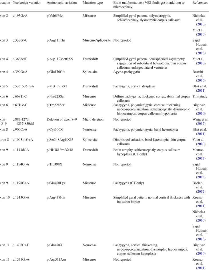
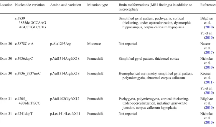
Table 1 HomozygousWDR62mutations and associated brain malformation patterns
Location Nucleotide variation Amino acid variation Mutation type Brain malformations (MRI findings) in addition to microcephaly
References
Exon 2 c.193G>A p.Val65Met Missense Simplified gyral pattern, polymicrogyria, schizencephaly, dysmorphic corpus callosum
Nicholas et al.
(2010) Yu et al.
(2010)
Exon 3 c.332G>C p.Arg111Thr Missense/splice-site Not reported Sajid
Hussain et al.
(2013) Exon 4 c.363delT p.Asp112MetfsX5 Frameshift Simplified gyral pattern, hemispherical asymmetry,
suggestion of subcortical heterotopia, thin corpus callosum, enlarged lateral ventricles
Yu et al.
(2010)
Exon 4 c.390G>A p.Glu130Glu Splice-site Agyria-pachygyria Bastaki
et al.
(2016)
Exon 5 c.535_536insA p.Met179fsX21 Frameshift Pachygyria, cortical dysplasia Bhat et al.
(2011) Exon 6 c.668T>C p.Phe223Ser Missense Diffuse pachygyria, thickened cortex, abnormal corpus
callosum
This study Exon 6 c.671G>C p.Trp224Ser Missense Pachygyria, polymicrogyria, cortical thickening,
under-opercularization, schizencephaly, dysmorphic hippocampus, corpus callosum hypoplasia
Bilgüvar et al.
(2010) Exon
8–9
c.883-1273_
1237-850del
Deletion of exon 8–9 Micro deletion Not reported Wang et al.
(2017) Exon 8 c.900C>A p.Cys300X Nonsense Pachygyria, polymicrogyria, band heterotopia Bhat et al.
(2011) Intron 8 c.1043+1G>A p.Ser348ArgfsX63 Splice-site Diminished sulcation, band heterotopia, thin corpus
callosum
Yu et al.
(2010) Exon 9 c.1143delA p.His381ProfsX48 Frameshift Brain atrophy, schizencephaly, corpus callosum
hypoplasia (CT only)
Memon et al.
(2013)
Exon 9 c.1194G>A p.Trp398X Nonsense Not reported Sajid
Hussain et al.
(2013)
Exon 9 c.1198G>A p.Glu400Lys Missense Pachygyria (CT only) Bacino
et al.
(2012) Exon 10 c.1313G>A p.Arg438His Missense Simplified gyral pattern, normal cortical thickness with
indistinct border
Kousar et al.
(2011) Nicholas et al.
(2010) Sajid
Hussain et al.
(2013) Exon 11 c.1408C>T p.Gln470X Nonsense Pachygyria, cortical thickening,
under-opercularization, dysmorphic hippocampus, corpus callosum hypoplasia
Bilgüvar et al.
(2010)
Exon 11 c.1531G>A p.Asp511Asn Missense Not reported Kousar
et al.
(2011)
Table 1 (continued)
Location Nucleotide variation Amino acid variation Mutation type Brain malformations (MRI findings) in addition to microcephaly
References
Nicholas et al.
(2010) Exon 12 c.1576G > T p.Glu526X Nonsense Pachygyria, cortical thickening, dysmorphic
hippocampus, corpus callosum hypoplasia
Bilgüvar et al.
(2010) Exon 12 c.1576G > A p.Glu526Lys Missense Pachygyria, cortical thickening,
under-opercularization, corpus callosum hypoplasia
Bilgüvar et al.
(2010) Exon 12 c.1606G > T p.Glu536X Nonsense Pachygyria, thickened cortex, corpus callosum
dysplasia
Poulton et al.
(2014)
Exon 14 c.1821dupT p.Arg608SerfsX26 Frameshift Details not reported McDonell
et al.
(2014) Exon 15 c.1942C > T p.Gln648X Nonsense Hemispherical asymmetry, ill-defined gyral pattern
(CT)
Kousar et al.
(2011) Exon 17 c.2115C > G p.Gly705Gly Splice-site Cerebellar atrophy, cortical structure not reported Najmabadi
et al.
(2011) Intron
21
c.2520 + 5G > T p.Asp823AlafsX5 Splice-site Not reported Wang et al.
(2017) Exon 22 c.2527dupG p.Asp843GlyfsX3 Frameshift Hemispherical asymmetry, ill-defined gyral pattern Rupp et al.
(2014) Exon 22 c.2588G > A p.Arg863His Missense Polymicrogyria, incomplete opercularization Poulton
et al.
(2014)
Exon 22 c.2667_2668GA > TT p.Met[889Ile;Lys890X] Nonsense Not reported Wang et al.
(2017) Exon 23 c.2863delGACA p.Asp955AlafsX112 Frameshift Pachygyria, thickened cortex, corpus callosum
dysplasia
Poulton et al.
(2014) Sgourdou
et al.
(2017) Intron
23
c.2867 + 4_
c.2867 + 7delGGT- G
p.Ser956CysfsX38 Splice-site Band heterotopia, thin corpus callosum (Appearance of the cortex not reported)
Yu et al.
(2010)
Exon 27 c.3232G > A p.Ala1078Thr Missense Not reported Nicholas
et al.
(2010) Intron
27
c.3335 + 1G > C p.? Splice site Polymicrogyria, gray-white matter blurring Nardello et al.
(2018)
Exon 28 c.3361delG p.Ala1121GlnfsX6 Frameshift Not reported Sajid
Hussain et al.
(2013)
Exon 29 c.3503G > A p.Trp1168X Nonsense Not reported Sajid
Hussain et al.
(2013)
Exon 30 p.Gly1275AlafsX21 Frameshift
survivin, and borealin (van der Waal et al.2012). The chro- mosomal passenger complex associates with the inner centro- mere until metaphase and then transfers to the spindle midzone, equatorial cell cortex, and midbody in late mitosis and cytokinesis. Aurora B functions include regulation of chromosome interactions with microtubules, chromatid cohe- sion, spindle stability, and cytokinesis (Carmena et al.2009).
Brain size at birth is primarily dependent on the ability of neuroprogenitor cells to proliferate and self-renew. While sym- metrical division of a neuroprogenitor cell results in the gener- ation of two identical neuroprogenitor cells (thereby increasing
the progenitor pool), asymmetrical division leads to the produc- tion of one progenitor cell (thereby maintaining the progenitor pool) and a committed precursor, which eventually undergoes migration and differentiates into neuron (Barbelanne and Tsang 2014). In vivo experiments on mice with knockdown or genetic inactivation ofWdr62and in vitro tests on cells withWDR62/
Wdr62mutations led to significant progress in the understand- ing the pathogenesis of microcephaly in patients withWDR62 mutations (Bogoyevitch et al. 2012; Chen et al. 2014;
Jayaraman et al.2016; Sgourdou et al.2017). Impaired prolif- eration of neural progenitors and reduced brain size were
Table 2 Compound heterozygousWDR62mutations and associated brain malformation patterns Location Nucleotide
variation
Amino acid variation Mutation type Brain malformations (MRI findings) in addition to microcephaly
Reference
Exon 1 c.28G>T p.Ala10Ser Missense Abnormal gyral pattern (dysgyria), corpus callosum dysgenesis, cerebellar atrophy
Banerjee et al. (2016)
Exon 2 c.189G>T p.Glu63Asp Missense
Exon 7 c.731C>T p.Ser244Leu Missense Not reported Miyamoto et al. (2017)
Exon 20 c.2413G>T p.Glu805X Nonsense
Exon 10 c.1313G>A p.Arg438His Missense Small frontal lobes, simplified hippocampal gyration, corpus callosum hypoplasia, cerebellar hypoplasia (US only)
Farag et al. (2013) Exon 23 c.2864_2867delACAG p.Asp955AlafsX112 Frameshift
Exon 17 c.2083delA p.Ser696AlafsX4 Frameshift Polymicrogyria, hemispherical asymmetry, heterotopia, abnormal corpus callosum
Murdock et al. (2011) Exon 23 c.2472_2473delAG p.Gln918GlyfsX18 Frameshift
Table 1 (continued)
Location Nucleotide variation Amino acid variation Mutation type Brain malformations (MRI findings) in addition to microcephaly
References
c.3839_
3855delGCCAAG- AGCCTGCCCTG
Simplified gyral pattern, pachygyria, cortical thickening, under-opercularization, dysmorphic hippocampus, corpus callosum hypoplasia
Bilgüvar et al.
(2010) Yu et al.
(2010)
Exon 30 c.3878C > A p.Ala1293Asp Missense Not reported Naseer
et al.
(2017) Exon 30 c.3936dupC p.Val1314ArgfsX18 Frameshift Simplified gyral pattern, thickened cortex Nicholas et al.
(2010) Exon 30 c.3936_3937insC p.Val1314ArgfsX18 Frameshift Hemispherical asymmetry, simplified gyral pattern,
polymicrogyria, abnormal corpus callosum
Kousar et al.
(2011) Yu et al.
(2010) Exon 31 c.4205_
4208delTGCC
p.Val1402GlyfsX12 Frameshift Pachygyria, polymicrogyria, cortical thickening, under-opercularization, indistinct gray-white junction, corpus callosum hypoplasia
Bilgüvar et al.
(2010)
Exon 31 c.4241dupT p.Leu1414LeufsX41 Frameshift Not reported Nicholas
et al.
(2010)
observed in these animals (Chen et al.2014; Jayaraman et al.
2016; Sgourdou et al.2017). Abnormalities in the centriole duplication, spindle pole orientation, and symmetric/
asymmetric division of neural progenitor cells and defects in the mitotic progression were noticed (Bogoyevitch et al.2012;
Chen et al.2014; Jayaraman et al.2016; Sgourdou et al.2017).
Premature delamination of progenitors from the germinal zones and increased apoptosis were also suggested in these experi- ments as the cause of reduced brain size (Bilgüvar et al.2010;
Bogoyevitch et al.2012; Chen et al.2014; Farag et al.2013;
Jayaraman et al.2016; Nicholas et al.2010; Sgourdou et al.
2017; Yu et al.2010). Downregulated Aurora-A-kinase activity was also found inWdr62mutant mouse embryonic fibroblasts, and investigations on isolated neural progenitor cells suggested thatWdr62andAurora Amay genetically interact to regulate mitotic progression of neural progenitor cells (Chen et al.2014).
A recent study revealed more details of the premature de- pletion of progenitor cells and mitotic progression defects in mice with truncated Wdr62 transcripts (Wdr621-21/1-21) (Sgourdou et al.2017). Centrosomes with differently aged mother centrioles are differentially inherited by the two daughter cells of asymmetrically dividing radial glia progeni- tors in the developing neocortex. Whereas the centrosome with the less mature new mother centriole migrates away from the ventricular surface and is largely inherited by differentiat- ing cells, the centrosome with the more mature old mother centriole stays at the ventricular zone surface and is predom- inantly inherited by renewing radial glia progenitors. WDR62 loss in mutantWdr6221-21/1-21mice disrupted asymmetric cen- trosome inheritance: the percentage of centrosomes retaining the old mother centriole decreased in the proliferating zones, while the percentage of centrosomes with new mother centri- oles increased. The opposite was found in the cortical plate suggesting abnormal migration and possibly differentiation.
This disturbed asymmetric centrosome inheritance may lead to premature depletion of progenitor cells from the ventricular zone and microcephaly (Sgourdou et al.2017). It has also been demonstrated that WDR62 protein can interact with the chromosomal passenger complex. Depletion of any chromo- somal passenger complex component disrupts mitotic pro- gression. WDR62 disruption caused a modest decrease in ki- netochore levels of Aurora B kinase, and a significant increase in kinetochore levels of survivin in fibroblasts from a patient with homozygous Asp955AlafsX112 mutation inWDR62 suggesting perturbed kinetochore function (Sgourdou et al.
2017). It has also been suggested that the mitotic delay of neural progenitors caused by WDR62 disruption may contrib- ute to the structural abnormalities observed in patients with WDR62mutations (Sgourdou et al.2017).
However, human brain disorders can be poorly recapitulat- ed in the mouse. Mice have smooth cerebral cortex that is 1000 times smaller than the abundantly gyrified human cor- tex. Cortical thinning was found after knockdown or genetic
inactivation ofWdr62in mice (Chen et al.2014; Jayaraman et al. 2016; Sgourdou et al.2017) in contrast to thick gray matter and cortical malformations in humans with WDR62 mutations (Tables1and2). This contradiction highlights that loss ofWdr62/WDR62may elicit divergent brain phenotypes in mice and humans. Recent experiments inAspmknockdown ferret, a species with a larger, gyrified cortex and greater neu- ral progenitor cell diversity than mice, suggested evolutionari- ly divergent functions ofAspmin the corticogenesis of mice and ferret (Johnson et al. 2018). Since both microcephaly proteins, Wdr62 and Aspm, define a mother centriole complex regulating centriole biogenesis, apical complex, and cell fate (Jayaraman et al.2016), similar divergence inWdr62/WDR62 function can also be hypothesized. Further experiments in ferret might elucidate more details of the abnormal corticogenesis inWDR62mutations (Johnson et al.2018).
Conclusions
Mutations inWDR62are the second most common cause of autosomal recessive microcephaly. The microcephaly is often associated with pachygyria, cortical thickening, and indistinct gray-white matter border, as in patients in this report; howev- er, a variety of other structural cortical malformations can also occur. Genotype-phenotype correlation cannot be found.
Recent investigations highlighted that WDR62 protein plays an essential role in the centrosome function and neural pro- genitor cell cycle; however, even these elegant experiments failed to explain accurately the genesis of the diverse cortical malformations. Discovery of more aspects of WDR62 func- tion in different animal models may clarify the mechanism of phenotypic heterogeneity.
Acknowledgements The authors thank the patients’parents for their participation in this study.
Authors’contributions MZ examined the patients and was a major con- tributor in writing the manuscript; MB and BT interpreted the patients’ data; TK, NN, and ZM contributed to gene analysis; OB supervised laboratory work; LSZ analyzed MRI data and designed the study. All authors read and approved the final manuscript.
Compliance with ethical standards
Conflict of interests The authors declare that they have no conflicts of interest.
Ethical approval and consent to participate The parents of both patients gave written informed consent to enter the study, which was approved by the Ethics Committee of the Faculty of Medicine, University of Szeged (Szeged, Hungary, Reference no: 18/2016-SZTE).
Publisher’s noteSpringer Nature remains neutral with regard to jurisdic- tional claims in published maps and institutional affiliations.
References
Abecasis GR, Cherny SS, Cookson WO, Cardon LR (2002) Merlin–rapid analysis of dense genetic maps using sparse gene flow trees. Nat Genet 30:97–101
Alcantara D, O’Driscoll M (2014) Congenital microcephaly. Am J Med Genet C Semin Med Genet 166C:124–139
Bacino CA, Arriola LA, Wiszniewska J, Bonnen PE (2012)WDR62 missense mutation in a consanguineous family with primary micro- cephaly. Am J Med Genet A 158A:622–625
Banerjee S, Chen H, Huang H, Wu J, Yang Z, Deng W, Chen D, Deng J, Su Y, Li Y, Wu C, Wang Y, Zeng H, Wang Y, Li X (2016) Novel mutations c.28G>T (p.Ala10Ser) and c.189G>T (p.Glu63Asp) in WDR62 associated with early onset acanthosis and hyperkeratosis in a patient with autosomal recessive microcephaly type 2.
Oncotarget 7:78363–78371
Barbelanne M, Tsang WY (2014) Molecular and cellular basis of auto- somal recessive primary microcephaly. Biomed Res Int.https://doi.
org/10.1155/2014/547986
Bastaki F, Mohamed M, Nair P, Saif F, Tawfiq N, Aithala G, El-Halik M, Al-Ali M, Hamzeh AR (2016) Novel splice-site mutation inWDR62 revealed by whole-exome sequencing in a Sudanese family with primary microcephaly. Congenit Anom 56:135–137
Bettencourt-Dias M, Hildebrandt F, Pellman D, Woods G, Godinho SA (2011) Centrosomes and cilia in human disease. Trends Genet 27:
307–315
Bhat V, Girimaji SC, Mohan G, Arvinda HR, Singhmar P, Duvvari MR, Kumar A (2011) Mutations inWDR62, encoding a centrosomal and nuclear protein, in Indian primary microcephaly families with corti- cal malformations. Clin Genet 80:532–540
Bilgüvar K, Öztürk AK, Louvi A, Kwan KY, Choi M, Tatli B, Yalnizoğlu D, Tüysüz B, Çağlayan AO, Gökben S, Kaymakçalan H, Barak T, Bakircioğlu M, Yasuno K, Ho W, Sanders S, Zhu Y, Yilmaz S, Dinçer A, Johnson MH, Bronen RA, Koçer N, Per H, Mane S, Pamir MN, Yalçinkaya C, KumandaşS, Topçu M, Özmen M, Šestan N, Lifton RP, State MW, Günel M (2010) Whole-exome sequencing identifies recessiveWDR62mutations in severe brain malformations. Nature 467:207–210
Bogoyevitch MA, Yeap YYC, Qu Z, Ngoei KR, Yip YY, Zhao TT, Heng JI, Ng DCH (2012) WD40-repeat protein 62 is a JNK- phosphorylated spindle pole protein required for spindle mainte- nance and timely mitotic progression. J Cell Sci 125:5096–50109 Carmena M, Ruchaud S, Earnshaw WC (2009) Making the Auroras
glow: regulation of Aurora A and B kinase function by interacting proteins. Curr Opin Cell Biol 21:796–805
Chang CC, Chow CC, Tellier LC, Vattikuti S, Purcell SM, Lee JJ (2015) Second-generation PLINK: rising to the challenge of larger and richer datasets. Gigascience.https://doi.org/10.1186/s13742-015- 0047-8
Chen JF, Zhang Y, Wilde J, Hansen K, Lai F, Niswander L (2014) Microcephaly disease geneWdr62regulates mitotic progression of embryonic neural stem cells and brain size. Nat Commun.https://
doi.org/10.1038/ncomms4885
Farag HG, Froehler S, Oexle K, Ravindran E, Schindler D, Staab T, Huebner A, Kraemer N, Chen W, Kaindl AM (2013) Abnormal centrosome and spindle morphology in a patient with autosomal recessive primary microcephaly type 2 due to compound heterozy- gousWDR62gene mutation. Orphanet J Rare Dis.https://doi.org/
10.1186/1750-1172-8-178
Fujita H, Yoshino Y, Chiba N (2016) Regulation of the centrosome cycle.
Mol Cell Oncol.https://doi.org/10.1080/23723556.2015.1075643 Jayaraman D, Kodani A, Gonzalez DM, Mancias JD, Mochida GH,
Vagnoni C, Johnson J, Krogan N, Harper JW, Reiter JF, Yu TW, Bae B, Walsh CA (2016) Microcephaly proteins Wdr62 and Aspm
define a mother centriole complex regulating centriole biogenesis, apical complex, and cell fate. Neuron 92:813–828
Johnson MB, Sun X, Kodani A, Borges-Monroy R, Girskis KM, Ryu SC, Wang PP, Patel K, Gonzalez DM, Woo YM, Yan Z, Liang B, Smith RS, Chatterjee M, Coman D, Papademetris X, Staib LH, Hyder F, Mandeville JB, Grant PE, Im K, Kwak H, Engelhardt JF, Walsh CA, Bae BI (2018) Aspm knockout ferret reveals an evolutionary mech- anism governing cerebral cortical size. Nature 556:370–375 Kodani A, Yu TW, Johnson JR, Jayaraman D, Johnson TL, Al-Gazali L,
Sztriha L, Partlow JN, Kim H, Krup AL, Dammermann A, Krogan NJ, Walsh CA, Reiter JF (2015) Centriolar satellites assemble centrosomal microcephaly proteins to recruit CDK2 and promote centriole duplication. elife.https://doi.org/10.7554/eLife.07519 Kousar R, Hassan MJ, Khan B, Basit S, Mahmood S, Mir A, Ahmad W,
Ansar M (2011) Mutations inWDR62gene in Pakistani families with autosomal recessive primary microcephaly. BMC Neurol.
https://doi.org/10.1186/1471-2377-11-119
Li D, Roberts R (2001) WD-repeat proteins: structure characteristics, biological function, and their involvement in human diseases. Cell Mol Life Sci 58:2085–2097
Lim NR, Yeap YYC, Ang CS, Williamson NA, Bogoyevitch MA, Quinn LM, Ng DCH (2016) Aurora A phosphorylation of WD40-repeat protein 62 in mitotic spindle regulation. Cell Cycle 15:413–424 McDonell LM, Chardon JW, Schwartzentruber J, Foster D, Beaulieu CL,
FORGE Canada Consortium, Majewski J, Bulman DE, Boycott KM (2014) The utility of exome sequencing for genetic diagnosis in a familial microcephaly epilepsy syndrome. BMC Neurol.https://
doi.org/10.1186/1471-2377-14-22
Memon MM, Raza SI, Basit S, Kousar R, Ahmad W, Ansar M (2013) A novelWDR62mutation causes primary microcephaly in a Pakistani family. Mol Biol Rep 40:591–595
Meraldi P, Lukas J, Fry AM, Bartek J, Nigg EA (1999) Centrosome duplication in mammalian somatic cells requires E2F and Cdk2- cyclin A. Nat Cell Biol 1:88–93
Miyamoto T, Akutsu SN, Fukumitsu A, Morino H, Masatsuna Y, Hosoba K, Kawakami H, Yamamoto T, Shimizu K, Ohashi H, Matsuura S (2017) PLK1-mediated phosphorylation of WDR62/MCPH2 en- sures proper mitotic spindle orientation. Hum Mol Genet 26:4429–
4440
Murdock DR, Clark GD, Bainbridge MN, Newsham I, Wu YQ, Muzny DM, Cheung SW, Gibbs RA, Ramocki MB (2011) Whole-exome sequencing identifies compound heterozygous mutations inWDR62 in siblings with recurrent polymicrogyria. Am J Med Genet A 155A:
2071–2077
Najmabadi H, Hu H, Garshasbi M, Zemojtel T, Abedini SS, Chen W, Hosseini M, Behjati F, Haas S, Jamali P, Zecha A, Mohseni M, Püttmann L, Vahid LN, Jensen C, Moheb LA, Bienek M, Larti F, Mueller I, Weissmann R, Darvish H, Wrogemann K, Hadavi V, Lipkowitz B, Esmaeeli-Nieh S, Wieczorek D, Kariminejad R, Firouzabadi SG, Cohen M, Fattahi Z, Rost I, Mojahedi F, Hertzberg C, Dehghan A, Rajab A, Banavandi MJS, Hoffer J, Falah M, Musante L, Kalscheuer V, Ullmann R, Kuss AW, Tzschach A, Kahrizi K, Ropers HH (2011) Deep sequencing reveals 50 novel genes for recessive cognitive disorders. Nature 478:57–63 Nardello R, Fontana A, Antona V, Beninati A, Mangano GD, Stallone MC, Mangano S (2018) A novel mutation ofWDR62gene associ- ated with severe phenotype including infantile spasm, microcephaly, and intellectual disability. Brain and Development 40:58–64 Naseer MI, Rasool M, Sogaty S, Chaudhary RA, Mansour HM,
Chaudhary AG, Abuzenadah AM, Al-Qahtani H (2017) A novel WDR62mutation cause primary microcephaly in a large consan- guineous Saudi family. Ann Saudi Med 37:148–153
Nicholas AK, Khurshid M, Désir J, Carvalho OP, Cox JJ, Thornton G, Kausar R, Ansar M, Ahmad W, Verloes A, Passemard S, Misson JP, Lindsay S, Gergely F, Dobyns WB, Roberts E, Abramowicz M,
Woods CG (2010)WDR62is associated with the spindle pole and is mutated in human microcephaly. Nat Genet 42:1010–1014 Poulton CJ, Schot R, Seufert K, Lequin MH, Accogli A, D’Annunzio G,
Villard L, Philip N, de Coo R, Catsman-Berrevoets C, Grasshoff U, Kattentidt-Mouravieva A, Calf H, de Vreugt-Gronloh E, van Unen L, Verheijen FW, Galjart N, Morris-Rosendahl DJ, Mancini GMS (2014) Severe presentation ofWDR62mutation: is there a role for modifying genetic factors? Am J Med Genet Part A 164A:2161– 2171
Prosser SL, Pelletier L (2017) Mitotic spindle assembly in animal cells: a fine balancing act. Nat Rev Mol Cell Biol 18:187–201
Rupp V, Rauf S, Naveed I, Windpassinger C, Mir A (2014) A novel single base pair duplication in WDR62 causes primary microceph- aly. BMC Med Genet.https://doi.org/10.1186/s12881-014-0107-4 Sajid Hussain M, Marriam Bakhtiar S, Farooq M, Anjum I, Janzen E,
Reza Toliat M, Eiberg H, Kjaer KW, Tommerup N, Noegel AA, Nürnberg P, Baig SM, Hansen L (2013) Genetic heterogeneity in Pakistani microcephaly families. Clin Genet 83:446–451
Sgourdou P, Mishra-Gorur K, Saotome I, Henagariu O, Tuysuz B, Campos C, Ishigame K, Giannikou K, Quon JL, Sestan N, Caglayan AO, Gunel M, Louvi A (2017) Disruptions in asymmetric centrosome inheritance and WDR62-Aurora kinase B interactions in primary microcephaly. Sci Rep.https://doi.org/10.1038/srep43708
Thiele H, Nürnberg P (2005) HaploPainter: a tool for drawing pedigrees with complex haplotypes. Bioinformatics 21:1730–1732
van der Waal MS, Hengeveld RCC, van der Horst A, Lens SMA (2012) Cell division control by the chromosomal passenger complex. Exp Cell Res 318:1407–1420
Wang X, Tsai JW, Imai JH, Lian WN, Vallee EB, Shi SH (2009) Asymmetric centrosome inheritance maintains neural progenitors in the neocortex. Nature 461:947–955
Wang R, Khan A, Han S, Zhang X (2017) Molecular analysis of 23 Pakistani families with autosomal recessive primary microcephaly using targeted next-generation sequencing. J Hum Genet 62:299– 304
Yu TW, Mochida GH, Tischfield DJ, Sgaier SK, Flores-Sarnat L, Sergi CM, Topçu M, McDonald MT, Barry BJ, Felie J, Sunu C, Dobyns WB, Folkerth RD, Barkovich AJ, Walsh CA (2010) Mutations in WDR62encoding a centrosome-associated protein, cause micro- cephaly with simplified gyri and abnormal cortical architecture.
Nat Genet 42:1015–1020
Zaqout S, Morris-Rosendahl D, Kaindl AM (2017) Autosomal recessive primary microcephaly (MCPH): an update. Neuropediatrics 48:
135–142