ORIGINAL ARTICLE
Renal Cell Carcinoma with Clear Cell Papillary Features: Perspectives of a Differential Diagnosis
Áron Somorácz1 &Levente Kuthi2&Tamás Micsik3&Alex Jenei2&Adrienn Hajdu2&Brigitta Vrabély3&
Erzsébet Rásó1&Zoltán Sápi3&Zoltán Bajory4&Janina Kulka1&Béla Iványi2
Received: 22 May 2019 / Accepted: 1 October 2019
#The Author(s) 2019
Abstract
Thirty-one cases of low-grade renal cell carcinoma (RCC) with clear cells and tubulopapillary/papillary architecture were analyzed retrospectively with immunohistochemical and genetic markers to gain more experience with the differential diagnosis of such cases. All samples coexpressed CK7 and CA9; the TFE3 or TFEB reactions were negative; the CD10 and the AMACR stainings were negative in 27 cases and 30 cases, respectively. The FISH assays for papillary RCC, available in 27 cases, and deletion of chromosome 3p, available in 29 cases, gave negative results. The results for 3p deletion,VHLgene mutation orVHL gene promoter region hypermethylation testing, along with the diffuse CD10-positivity in 2 cases confirmed 21 cases as clear cell papillary RCC (CCPRCC; CK7+, CA9+; no 3p loss, noVHLabnormality) and 10 cases as clear cell RCC (CCRCC; CK7+, CA9+; no 3p loss,VHLabnormality mutation/hypermethylation present). In CCPRCCs, the representative growth pattern was branching tubulo-acinar, commonly accompanied by cyst formation. The linear nuclear arrangement or cup-shaped staining of CA9 did not necessarily indicate CCPRCC, and the absence of these did not exclude the diagnosis of CCPPRC. One tumor infiltrated the renal sinus; the others exhibited pT1 stage; and metastatic outcome was not recorded. The CCRCC cases were in pT1 stage; 6 exhibited cup-shaped staining of CA9, and 1 displayed lymph node metastasis at the time of surgery. Distant metastatic disease was not observed. In summary, theVHLabnormalities distinguished the subset of CCRCC with diffuse CK7- positivity and no 3p loss from cases of CCPRCC.
Keywords Clear cell carcinoma . Clear cell papillary carcinoma . Cytokeratin 7-positivity . Differential diagnosis .VHLgene
Introduction
Clear cell papillary renal cell carcinoma (CCPRCC) is an in- frequent subset of RCC [1,2]. Although CCPRCC shares
histopathogical features with clear cell RCC (CCRCC), pap- illary RCC and Xp11.2 translocation RCC, its immunohisto- chemical coexpression of cytokeratin 7 (CK7) and carbonic anhydrase 9 (CA9), and negativity for CD10, alpha-methyl- CoA racemase (AMACR), and TFE3 usually clarifies the di- agnosis [3–7]. The renal angiomyoadenomatous tumor (RAT) is now regarded as being in the spectrum of CCPRCC [8–10].
Genetically, CCPRCCs lack chromosome 3p deletion orVHL gene mutation orVHLpromoter hypermethylation, the hall- marks of CCRCC, and have no loss of chromosome Y or gain of chromosome 7 and 17, the hallmarks of papillary RCC [2–4,11–13].
In surgical pathology practice, the separation of CCPRCCs from CCRCCs can pose certain difficulties. The distinction is crucial, because CCPRCCs have a very limited potential for metastasis (fatal outcome has been reported only in two pa- tients out of 400 [14]), whereas in low-grade CCRCCs distant metastases can occur several years after nephrectomy. To learn more about the differential diagnosis of low-grade RCCs with Áron Somorácz and Levente Kuthi contributed equally.
Electronic supplementary materialThe online version of this article (https://doi.org/10.1007/s12253-019-00757-3) contains supplementary material, which is available to authorized users.
* Áron Somorácz somoracz14@gmail.com
1 2nd Department of Pathology, Semmelweis University, Üllői út 93, Budapest H-1091, Hungary
2 Department of Pathology, University of Szeged, Szeged, Hungary
3 1st Department of Pathology and Experimental Cancer Research, Semmelweis University, Budapest, Hungary
4 Department of Urology, University of Szeged, Szeged, Hungary https://doi.org/10.1007/s12253-019-00757-3
CCPRCC features, a series of such tumors were subjected to a retrospective immunohistochemical analysis, applying CK7, CA9, CD10, AMACR, TFEB and TFE3 immunostainings, and the immunophenotypes were correlated with the results of genetic markers for CCRCC or papillary RCC.
Materials and Methods
Case Selection and Review Process
This study was conducted with the permission of the Medical Research Council (17489-4/2017/EKU). The hematoxylin and eosin-stained slides of 2326 consecutive RCC samples were reexamined for clear cell papillary RCC-like tumors, including low-grade nuclei, the presence of any degree of tubulopapillary growth pattern of tumor cells with clear cyto- plasm, linear arrangement of nuclei away from the basal mem- brane, along with the presence of a leiomyomatous stroma.
Demographical and clinical data were collected from the da- tabase management systems of Semmelweis University and University of Szeged.
Tissue Microarray and Immunohistochemical Reactions
Tissue microarray blocks were prepared for immunohisto- chemistry with TMA Master (3DHISTECH) applying a 2 mm core diameter. One to four representative cores were t h e n p u n c h e d o u t f r o m t h e d o n o r b l o c k s . Immunohistochemical staining for CA9, CK7, CD10, AMACR, TFEB and TFE3 were performed (see the dilutions and sources in Supplementary Table1). The epitope retrieval was performed for each antibody according to the manufacturer’s recommendations. The reactions were con- ducted using Autostainer (Dako). Afterwards, slides were evaluated microscopically by estimating the proportion (%) of immunopositive cells. Staining in over 50% of the tumor cells, in 10 to 50% of tumor cells, or in less than 10% of the tumor cells, was interpreted as diffusely or focally positive or negative, respectively.
Fluorescent in Situ Hybridization (FISH)
FISH assays were carried out to detect either the loss of chro- mosome 3p and chromosome Y or gain of chromosome 7 and 17. Tissue sections were cut from the TMA blocks and deparaffinized. The assays were done using aVHL/cen3 probe (ZytoLight® SPEC VHL/CEN3 Dual Color Probe, Zytovision,) and centromeric probes for chromosome 7, 17 and Y (Cytocell) according to the manufacturer’s instructions.
Slides were digitalized by using a Pannoramic Midi slide scanner (3DHISTECH), and reactions were evaluated using
a Pannoramic Viewer (3DHISTECH) in the following way.
Fifty tumor cells from each case were examined and were compared with the same number of cells of the peritumoral tissue, which served as an internal control. The cutoff values of chromosomal gain and/or loss were set at the mean ±3SD of the corresponding control values, as done in previous studies [5]. The analysis of 3p deletion was also performed based on a published method [15].
VHL Gene Sequence Analysis and VHL Gene Promoter Hypermethylation
A PCR-based amplification method was used forVHLgene mutation analysis as earlier described [16]. The VHLexons were amplified via specific primer pairs (Supplementary Table2). In the case of pathological mutation, the apparently tumor-free renal tissue was analyzed as well. A GenomeLAB DTCS - Quick Start Kit (Beckman Coulter) was used for DNA sequencing. The latter was carried out according to the manufacturer’s instructions using the GenomeLab GeXP Genetic Analysis System (Beckman Coulter). The methyla- tion status of VHL gene promoter region was determined using the methylation-specific PCR method. The extracted genomic DNA was modified using the EpiJET Bisulfite Conversion Kit (ThermoFischer Scientific), and followed by PCR-based amplification with methylation-specific primer pairs (Supplementary Table3). The methylation status (non- methylated, methylated) was determined by gel electrophore- sis of the PCR products, as reported previously [17].
Criteria for Diagnosing a Tumor as CCPRCC
The diagnosis of CCPRCC was made if the above-mentioned morphology together with characteristic immunophenotype (CK7- and CA9-positivity, negative CD10 or at most focal CD10-positivity, negative TFE3 and TFEB stainings), along with the lack of genetic alterations indicating CCRCC (3p deletion, VHLmutation, VHL promoter hypermethylation), and PRCC (7 and 17 trisomy, loss of Y) were detected.
Tumors with the same morphology, CK7 and CA9 coexpression, but with diffuse CD10-positivity or with altered VHLstatus were classed as CCRCC.
Results
Using the inclusion criteria, we retrieved 31 samples. All tu- mors coexpressed CK7 and CA9. The TFE3 and TFEB reac- tions were uniformly negative; and the CD10 and the AMACR reactions were negative in 27 and 30 cases, respec- tively. The FISH assays for papillary RCC, available in 27 cases, and deletion of chromosome 3p, available in 29 cases, yielded negative results. The histomorphology, the results for
VHL mutation and VHL methylation testing, and the immunophenotype confirmed 21 cases as CCPRCC and 10 cases as CCRCC. The principal characteristics of the two sub- sets are summarized in Tables1and2.
Features of CCPRCCs
Here, 21 tumors were examined, and the specimens were ob- tained from 12 females and 9 males. The mean age was 60 years (with range 28 to 84 years). Partial nephrectomy was performed in 4 patients and radical nephrectomy in 17 pa- tients; and one tumor developed in a transplanted kidney.
Twenty cases were incidental findings of imaging performed for non-urological symptoms.
Gross Findings
All the tumors were solitary, and the mean size was 23 mm (with range 6 to 65 mm). Cystic change was present in 12 samples; and multilocular cystic mass existed in cases 7 and 15. The tumorous parenchyma was grey-white to yellow- brown, occasionally with small hemorrhagic foci.
Microscopic Findings
Each tumor was circumscribed, and at least one thin fibrous, or fibromuscular capsule was present, except in one case. The capsule was thick (400-800μm) and contained smooth mus- cle in 13 tumors. A minimal infiltration of renal sinus fat was observed in Case 3 (Fig.1a, b). Vascular invasion was not detected in any of the cases examined. The dominant growth pattern was tubulo-acinar (15 tumors), with cyst formation in a continuum from microscopic to macroscopic cystic spaces in 12 samples. Also, a papillary architecture was observed in 14 tumors and was detected mainly focally, except in one case where it was predominantly seen. Solid areas with compact cell nests and trabeculae were seen in 11 samples, and it was mostly made up of a small proportion of the tumorous paren- chyma. Foamy macrophages, psammoma bodies and necrosis were absent. Although the clear cell phenotype was a distinc- tive feature, the tumor cells displayed various cytological characteristics. Most of them were cuboidal or columnar, but flattened forms in cysts, as well as elongated cells arranged focally in a fascicular pattern in solid areas were also encoun- tered. Some tumor cells–especially in solid areas–had eo- sinophilic cytoplasm. Prominent nucleoli were not present.
The linear arrangement of nuclei together with its orientation away from the basement membrane was observed in 16 tu- mors (from a minimal to an extensive presence). Mitotic fig- ures were occasionally seen. Stromal smooth muscle was found in 18 cases including two samples with abundant myo- matous stroma (cases 1 and 7).
Immunohistochemical and Molecular Profile
All tumors exhibited a strong and diffuse CK7 expression.
Immunoreaction for CA9 resulted in diffuse staining in 17 tumor samples, and focal staining in 4 tumor samples. The Bcup-shaped^pattern was detected in 17 cases, visible mainly in the tubular and cystic areas. Focal CD10-positivity was found in 2 samples. Weak granular, diffuse AMACR- positivity was noted in Case 14.
The mutation status of theVHLgene was investigated in 11 samples, and in Case 12, single nucleotide polymorphism (SNP) in untranslated region (UTR) was found. TheVHLgene promot- er hypermethylation status was analyzed in 16 samples; and none of these harbored promoter region hypermethylation.
Follow-up
The median time was 52.5 months (with range 1 to 184 months). Three patients did not have a follow-up, and two patients died in non-cancer-related causes. No evidence of tumor progression and recurrence was documented in the data of the 18 surviving patients.
Features of CCRCCs Mimicking CCPRCC
In this group, we analyzed 10 cases, and the mean age was 51 years (with range 37 to 69 years) with 5 female and 5 male patients. Half of the cases were treated via partial nephrectomy.
Tumor-related symptoms were registered in two patients. In Case 23, a CCRCC (CA9 and CD10: diffusely positive; CK7 nega- tive) was resected from the contralateral kidney two months after the first surgery. And in Case 24, a metastatic perihilar lymph node was removed together with the tumorous kidney (Fig.1c-f).
Gross Findings
The mean size of the tumors was 29 mm (with range 15 to 50 mm). Cystic change was noticed almost in all cases (9/10).
The cut surface was indistinguishable from those seen in CCPRCC.
Microscopic Findings
A capsule containing smooth muscle was present in 6 cases.
The predominant growth pattern was tubulo-acinar (5/10), followed by cystic (2/10), papillary (1/10) and solid (1/10).
In Case 29, the distribution of tubulo-acinar, papillary and cystic pattern was the same. The tumors were composed of clear cytoplasm cells with focal eosinophilic granulations.
Also, an apical linear nuclear arrangement was noted in 6 cases, and 2 cases contained a smooth muscle rich stroma.
The infiltration of the renal vein, renal sinus and perinephric fat tissue was not observed.
Immunohistochemical and Molecular Profile
There was a coexpression of CK7 in a diffuse fashion and CA9 in a diffuse (8 cases) or focal fashion (2 cases). The cup-shaped distribution of CA9 was present in 6 cases. Diffuse CD10-positivity was observed in cases 23 and 24.
TheVHL gene mutation status was analyzed in 9 sam- ples, and in cases 22, 23 and 31 a pathogenic mutation was identified that was not present in the tumor-free renal parenchyma (Fig. 2). The sequencing revealed an
SNP without any clinical significance in Case 24. The remaining 5 tumors analyzed harbored no genetic change. Also, the VHL gene promoter hypermethylation was tested in 8 cases, and 7 of them possessed promoter region hypermethylation.
Follow-up
All the cases had follow-up data with a median of 31.6 months (with range 3 to 100 months). None of them experienced any recurrence and cancer-related death.
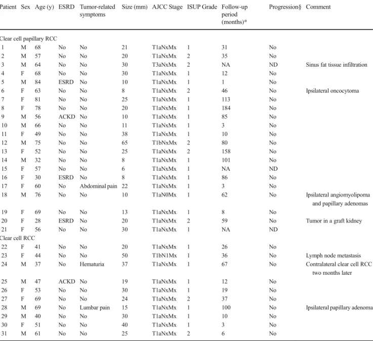
Table 1 Clinicopathological features of the cases examined Patient Sex Age (y) ESRD Tumor-related
symptoms
Size (mm) AJCC Stage ISUP Grade Follow-up period (months)*
Progression§ Comment
Clear cell papillary RCC
1 M 68 No No 21 T1aNxMx 1 31 No
2 M 57 No No 20 T1aNxMx 2 35 No
3 M 64 No No 30 T3aNxMx 2 NA ND Sinus fat tissue infiltration
4 F 68 No No 30 T1aNxMx 1 12 No
5 M 84 ESRD No 10 T1aNxMx 1 1 No
6 F 63 No No 8 T1aNxMx 2 46 No Ipsilateral oncocytoma
7 F 81 No No 25 T1aNxMx 1 113 No
8 F 78 No No 20 T1aNxMx 1 184 No
9 M 56 ACKD No 10 T1aNxMx 1 85 No
10 M 66 No No 11 T1aNxMx 1 3 No
11 F 49 No No 38 T1aNxMx 1 10 No
12 M 75 No No 65 T1bNxMx 2 80 No
13 F 52 No No 25 T1aNxMx 2 158 No
14 M 32 No No 8 T1aNxMx 1 101 No
15 F 57 No No 6 T1aNxMx 1 NA ND
16 F 30 ESRD No 8 T1aNxMx 1 86 No
17 F 60 No Abdominal pain 22 T1aNxMx 1 3 No
18 M 76 No No 10 T1aN0Mx 1 62 No Ipsilateral angiomyolipoma
and papillary adenomas
19 F 69 No No 13 T1aNxMx 1 8 No
20 F 28 ESRD No 20 T1aNxMx 2 59 No Tumor in a graft kidney
21 F 56 No No 30 T1aNxMx 1 NA ND
Clear cell RCC
22 F 41 No No 20 T1aNxMx 1 26 No
23 F 44 No No 50 T1bN1Mx 1 36 No Lymph node metastasis
24 M 37 No Hematuria 37 T1aNxMx 1 67 No Contralateral clear cell RCC
two months later
25 M 47 ACKD No 19 T1aNxMx 1 12 No
26 F 53 No No 30 T1aNxMx 1 19 No
27 F 69 No No 24 T1aNxMx 2 37 No
28 M 69 No Lumbar pain 15 T1aNxMx 1 100 No Ipsilateral papillary adenoma
29 M 40 No No 30 T1aNxMx 1 10 No
30 F 51 No No 40 T1aNxMx 1 3 No
31 M 61 No No 25 T1aNxMx 2 6 No
*Follow-up, determined from the surgery to the last follow-up; §Progression, assessed by radiological and/or autopsy data Mmale;Ffemale;ESRDend-stage renal disease;ACKDacquired cystic kidney disease;NAnot available;NDno data
Discussion
The WHO classification of RCCs defines the subsets via a synthesis of histopathological, immunohistochemical, and ge- netic data [1]. In the current study, we focused on the discrim- ination of CCPRCC from other tumor types by applying the well-known immunohistochemical markers supplemented
with a molecular analysis that seeks to find chromosomal abberations andVHL abnormalities (including mutations as well as methylation analysis). We made a formal diagnosis of CCPRCC when both the immunohistochemical and the genetic tests were in complete accordance with the histology.
All the RCC subtypes with clear cell phenotype (i.e.TFE3 or TFEB translocation RCCs, TCEB1-mutated RCC [18], Table 2 Morphological, immunohistochemical, and molecular characteristics of the cases examined.
Patient Architecture of tumor volume (%) Immune profile (%) Molecular characteristics Tubular Papillary Cystic Solid LiN CK7 CA9 CA9 cup-shaped CD10 AMACR +7 +17 -
Y - 3p
VHLmut VHLmet
Clear cell papillary RCC
1 90 - - 10 No Diff Diff Yes Neg Neg - - - - wt ua
2 88 2 - 10 Yes Diff Diff Yes Foc Neg - - - - wt -
3 95 - - 5 Yes Diff Diff Yes Neg Neg nd nd nd nd nd nd
4 59 1 20 20 Yes Diff Diff Yes Neg Neg - - - wt -
5 100 - - - Yes Diff Diff Yes Neg Neg - nd - - nd nd
6 95 5 - - Yes Diff Diff Yes Foc Neg - - - wt -
7 44 5 50 1 No Diff Diff Yes Neg Neg - - - wt -
8 50 50 - - Yes Diff Diff Yes Neg Neg - - - ua -
9 80 10 - 10 No Diff Diff Yes Neg Neg - - - - wt -
10 95 5 - - Yes Diff Diff Yes Neg Neg - - - - wt -
11 50 40 10 - Yes Diff Diff Yes Neg Neg - - - wt -
12 50 - 50 - Yes Diff Diff Yes Neg Neg - - - - 5’UTR SNP# -
13 - 80 20 - No Diff Diff Yes Neg Neg - - - ua -
14 80 10 10 - Yes Diff Diff Yes Neg Poz§ - - - - wt -
15 - 20 80 - Yes Diff Diff No Neg Neg - - - ua nd
16 95 - - 5 Yes Diff Diff No Neg Neg - - ua nd
17 50 20 30 - No Diff Diff No Neg Neg - - - wt -
18 90 - 5 5 Yes Diff Foc Yes Neg Neg nd nd nd nd nd -
19 45 1 50 4 Yes Diff Foc Yes Neg Neg - - - nd -
20 89 - 1 10 Yes Diff Foc Yes Neg Neg - - - ua -
21 85 1 5 10 Yes Diff Foc No Neg Neg - - - ua -
Clear cell RCC
22 50 5 35 10 Yes Diff Diff Yes Neg Neg - - - muta +
23 50 20 30 - Yes Diff Diff Yes Diff Neg nd nd - mutb nd
24 10 50 20 20 No Diff Diff No Diff Neg - - - - 5’UTR SNP¶ -
25 80 10 10 - No Diff Diff Yes Neg Neg - - - - wt +
26 95 1 - 4 Yes Diff Diff Yes Neg Neg - - - wt +
27 20 10 70 - Yes Diff Diff Yes Neg Neg - - - wt +
28 40 - 60 - Yes Diff Diff Yes Neg Neg - - - - ua +
29 20 40 40 - Yes Diff Diff No Neg Neg - - - - wt +
30 30 - 20 50 No Diff Foc No Neg Neg - - - wt +
31 90 - - 10 Yes Diff Foc No Neg Neg - - - - mutc nd
LiNlinear nuclear arrangement from basement membrane;+7 and +17trisomy of chromosome 7 and 17, respectively;-Ydeletion of chromosome Y;-3p deletion of chromosome 3p;VHLmut, von Hippel-Lindau gene mutation status;VHLmet, von Hippel-Lindau gene methylation status;ndnot determined;wtwild type;uaunsuccessful analysis;5’UTR SNPsingle nucleotide polymorphism in 5’untranslated region;diffdiffuse;focfocal;neg negative (less than or equal to 10%)
§weak granular positivity;#exon 3 could not be amplified;¶ exon1b could not be amplified;a c.221T>A/p.V74N;bc.625C>T/p.G209*;c c.354_
361delCTTCAGAGinsT
RCC with 8p monosomy [19], RCC with prominent smooth muscle stroma (RCCSMS) [20–25] and RCC associated with von Hippel-Lindau syndrome) can exhibit a CCPRCC-like histomorphology [26,27], but CCRCC cases pose the biggest difficulty because this tumor type is the most common and it also has some morphological similarities. CCRCCs are viewed as tumors that are CA9+ and CD10+, and display no more than a focal CK7 positivity. In contrast, the
immunophenotype of CCPRCCs is CK7+, CA9+, and CD10-. Perhaps diffuse and strong CK7 positivity is consid- ered the most important and an obligatory diagnostic criterion for CCPRCC. Nevertheless, the lack of a widespread CD10 reaction is also required.
We performed our case selection based on histological features suggestive of CCPRCC, and all the tumors displayed diffuse CK7 staining. In two of them, however, Fig. 1 a-b Case 3 with sinus fat invasionThe tumor showed a
branching tubular pattern and an immunophenotype characteristic for CCPRCC (basolateral CA9 reaction in the insert) (a). Superficial infiltration of sinus fat was seen (b).Figure1c-f Case 23 with lymph node metastasisThe tumor displayed the morphological features of CCPRCC, partly with papillary architecture (c). The lymph node
metastasis was mainly cystic; with some papillary infoldings (insert in figure d) (d). The tumor exhibited a diffuse CK7 positivity (e), but extensive CD10 staining was also observed (f). The latter, together with theVHLgene mutation detected in this tumor were not consistent with the diagnosis of CCPRCC
CD10 positivity was also diffuse, which supported our diagnosis of CCRCC. In another subset of our cases, the morphology and immunophenotype wholly favoured the diagnosis of CCPRCC; however, either VHL muta- tion (3 cases) or VHL promoter hypermethylation (7 cases) was present. We accepted the view of Hes et al.
who recommended not classifying cases with any VHL gene abnormality as CCPRCC [28]. Based on their ap- proach, our tumors with altered VHL status were classi- fied as CCRCC.
After performing an immunohistochemical and molecular analysis, our selected cases with histology of CCPRCCs were subdivided into two groups. These are CCPRCCs (21 cases), and CCRCCs with diffuse CK7 positivity (10 cases).
Features of CCPRCC Cases
The characteristic pattern of these tumors was branching tubulo-acinar that was commonly accompanied by cyst for- mation. Papillary areas, however, were detected as a minor Fig. 2 Case 22 exhibiting morphology and immunophenotype
completely consistent with CCPRCC, but containing aVHLgene mutation.The tumor had a thick fibromyomatous capsule and it was composed of both solid and cystic areas (a). Branching tubular
architectural pattern was the most characteristic (b). The tumor cells were diffusely positive for CK7 (c); and negative for CD10 (d). CA9 immunoreaction also resulted in a diffuse staining with a basolateral pattern (e).VHLgene mutation was detected by direct sequencing (f)
component except in three cases. As similar findings on the extent of papillarity were obtained by Aydin et al. [5] and by Williamson et al. [29], we conclude that CCPRCC with pre- dominant papillarity probably occurs quite rarely. Therefore, the WHO designation of this entity seems inaccurate, and the appellationBtubulopapillary^would perhaps be more apt, as was suggested by Aydin et al [5].
Linear nuclear arrangement away from the basement mem- brane is regarded as characteristic for CCPRCC [30].
Actually, 16/21 of CCPRCCs and 7/10 CCRCCs with diffuse CK7-positivity harbored this phenomenon (from a minimal to extensive presence). Dhakal et al. examined 37 tumors with a morphologic overlap between CCPRCC and CCRCC fea- tures, and linear nuclear arrangement was not the exclusive feature of cases classified as CCPRCC [31]. In another series of CCPRCC, Williamson at al. noticed linear nuclear arrange- ment only in 24/55 cases [32]. These findings suggest that linear nuclear arrangement is an overemphasized phenome- non, since its absence does not exclude the possible diagnosis of CCPRCC, and its presence does not necessarily support the diagnosis of CCPRCC.
The cup-shaped expression of CA9 was not uniformly present in our series. A diffuse cup-shaped expression was observed in 17/21 samples, while a dominant box-shaped staining with a focal cup-shaped expression was noted in 4/
21 samples. Upon reviewing the literature, a cup-shaped ex- pression involving 50% of tumor cells was reported by Rohan et al. in 3 out of their 9 cases [6]; and Dhakal et al. noted a cup- like expression of CA9 in 74% of their cases [31]; and Aydin et al. did not mention this feature at all in their 36 cases [5].
Since in our experience the cup-shaped staining pattern cannot be discerned unambiguously in solid areas, the absence of cup-shaped expression should be interpreted with caution when making a concrete diagnosis of CCPRCC for a specific case. A diffuse and weak granular AMACR-positivity was seen in one case. We reviewed the immunoprofile of the pub- lished CCPRCC cases and, albeit rarely, AMACR-positivity was reported [5,6,11,29,32–34]; hence if it is present, it does not necessarily contradict the diagnosis of CCPRCC. Focal CD10-positivity was encountered in two, otherwise complete- ly typical CCPRCC, andVHLgene abnormalities were not present in these samples. The focal extent of CD10 expression
may indicate the possibility of RAT, because a lack of a cystic component viewed microscopically, and the triple coexpression of CK7, CA9 and a certain degree of CD10 were noted in a series of RCC cases classified as RAT [35].
Our CCPRCC group comprised 19 pT1a, 1 pT1b and 1 pT3a cases, respectively. To our knowledge, ours is the first reported case with infiltration outside of the kidney parenchy- ma. Also, in Case 20 the tumor developed in a transplanted kidney. In a recently published review, Dhakal at al. [36] sum- marized the findings of 24 articles that reported tumors in transplanted kidneys, but among the 48 tumors described, not one was CCPRCC. Coexisting benign tumors and CCPRCC were observed in two cases. Actually, in Case 6 the oncocytoma had been detected clinically, and during the grossing CCPRCC was discovered. All of our CCPRCC cases had an excellent clinical outcome, reinforcing the view that the carcinoma designation might be exaggerated [14,37,38].
Features of CCRCCs with Diffuse CK7-Positivity
A series of CCRCC with diffuse CK7-posivity was published a decade ago by Mai et al [39]. Similar to our experiences, these samples were small-sized, and a non-metastatic course was re- corded over a mean of a 3-year follow-up; and diffuse CK7- positivity was viewed as the indicator of indolent behaviour [40].
Our results provide further clinicopathologic data on this rare subset of CCRCC. Accordingly, neither 3p deletion, nor other chromosomal anomalies were present. The VHLgene sequence analysis revealed pathologic mutations in cases 22, 23 and 31. Since VHLmutations were not identified in the non-tumorous renal tissue, the possibility of VHL-disease- associated CCRCC was excluded.
In seven samples, the histological and immunphenotypic data favoured the diagnosis of CCPRCC; however, the pres- ence of theVHLgene promoter hypermethylation abnormality leads us to place these samples into the CCRCC group. In the study of Herman et al. on silecing of theVHLgene by DNA methylation, the hypermethylation of a CpG island in the 5’
region was noted in 5 samples out of 26 CCRCCs [16]. Four of these had lost one copy ofVHL, while one retained two heavily methylated alleles. The latter observation indicated
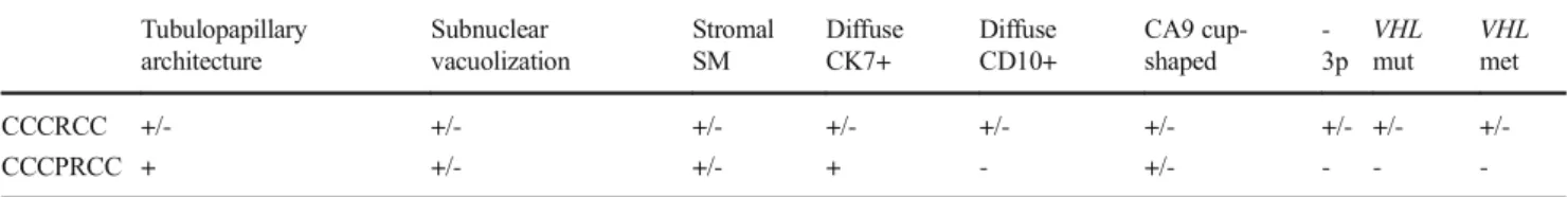
Table 3 Overlapping and discriminating features of CCRCCs and CCPRCCs. As we accepted the view of Hes et al. [18] thatVHLgene alteration is not compatible with the diagnosis of CCPRCC, alteredVHL
status was found as the most reliable discriminating feature between CCRCCs and CCPRCCs in our cohort
Tubulopapillary architecture
Subnuclear vacuolization
Stromal SM
Diffuse CK7+
Diffuse CD10+
CA9 cup- shaped
- 3p
VHL mut
VHL met
CCCRCC +/- +/- +/- +/- +/- +/- +/- +/- +/-
CCCPRCC + +/- +/- + - +/- - - -
SMsmooth muscle;mutmutation;methypermethylaiton
that hypermethylation may inactivate theVHL gene even when both wild-type alleles are retained [16]. In our analysis, hypermethylation was noted in seven cases; moreover coexistingVHLgene mutation and methylation was seen in Case 22. After a search for methylation data, only 2 tumors analyzed were found in the literature out of 400 or so CCPRCCs [5,14]. Methylation analyses performed by others in the future may validate our assumption that aVHLpromoter hypermethylation is definitely not compatible with the diag- nosis of CCPRCC. In Case 24 (and in Case 12 in the CCPRCC group) an SNP was observed in the 5’UTR region, a finding treated as insignificant, because the nucleotide change did not induce any amino acid change as well.
Interestingly, in 8 cases the histological and immunphenotypic data were entirely consistent with the histopathological diag- nosis of CCPRCC, but the presence ofVHLabnormalities led us to place these samples into the group of low-grade CCRCC with CK7 immunoreactivity and no 3p loss. Every case was in the pT1 stage, and there was no progression or recurrence.
In summary, in our study the immunophenotype and the genetic profile of 31 RCCs composed of clear cells, low-grade nuclei and a tubulopapillary architecture were investigated retrospectively. Twenty-one cases were classified as CCPRCC (CK7+, CA9+; -3p absent,VHLabnormality not present) and 10 as CCRCC with diffuse CK7-positivity (CK7+, CA9+; -3p absent,VHLabnormality present). Based on our findings, the following conclusions can be drawn. First, CCPRCCs rarely exhibit a predominant papillary architecture, hence their name is misleading. Second, a linear nuclear ar- rangement away from the basement membrane and cup-like CA9 positivity are not obligatory features. Third, the evidence for their malignant potential is still subject of debate. Fourth, RCCs with CCPRCC morphology, diffuse CK7 positivtiy, and with an alteredVHLstatus (mutation, or promoter hyper- methylation) do exist; and these tumors can be interpreted as CCRCC with diffuse CK7 positivity, and they can be definite- ly differentiated from CCPRCCs only by carrying out molec- ular tests for theVHLstatus. And last but, not least the bio- logical behavior of both CCPRCCs and CCRCCs with diffuse CK7 positivity seems to be indolent with a favorable clinical outcome. Overlapping and discriminating features of CCPRCCs and CCRCCs are summarized in Table3.
Funding Open access funding provided by Semmelweis University (SE).
This study was funded by internal sources of the institutions (2nd Department of Pathology and 1st Department of Pathology and Experimental Cancer Research, Semmelweis University, Budapest, Hungary; Department of Pathology, University of Szeged, Szeged, Hungary). No grants were used.
Compliance with Ethical Standards
Conflict of interest The authors declare that they have no conflict of interest.
Ethical approval The study was ethically approved by the Medical Research Council of Hungary (174894/2017/EKU). The renal carcinoma cases were retrospectively selected from the archives of the pathological institutions and informed consent was not obtained.
Open AccessThis article is distributed under the terms of the Creative C o m m o n s A t t r i b u t i o n 4 . 0 I n t e r n a t i o n a l L i c e n s e ( h t t p : / / creativecommons.org/licenses/by/4.0/), which permits unrestricted use, distribution, and reproduction in any medium, provided you give appropriate credit to the original author(s) and the source, provide a link to the Creative Commons license, and indicate if changes were made.
References
1. Srigley JR, Delahunt B, Eble JN et al (2013) The International Society of Urological Pathology (ISUP) Vancouver Classification of Renal Neoplasia. Am J Surg Pathol 37:1469–1489
2. Moch H, Humphrey PA, Ulbright TM, Reuter VE (2016) WHO Classification of Tumours of the Urinary System and Male Genital Organs. International Agency for Research on Cancer
3. Tickoo SK, dePeralta-Venturina MN, Harik LR et al (2006) Spectrum of epithelial neoplasms in end-stage renal disease: an experience from 66 tumor-bearing kidneys with emphasis on his- tologic patterns distinct from those in sporadic adult renal neopla- sia. Am J Surg Pathol 30:141–153
4. Gobbo S, Eble JN, Grignon DJ et al (2008) Clear cell papillary renal cell carcinoma: a distinct histopathologic and molecular genetic entity. Am J Surg Pathol 32:1239–1245
5. Aydin H, Chen L, Cheng L et al (2010) Clear cell tubulopapillary renal cell carcinoma: a study of 36 distinctive low-grade epithelial tumors of the kidney. Am J Surg Pathol 34:1608–1621
6. Rohan SM, Xiao Y, Liang Y et al (2011) Clear-cell papillary renal cell carcinoma: molecular and immunohistochemical analysis with emphasis on the von Hippel-Lindau gene and hypoxia-inducible factor pathway-related proteins. Mod Pathol 24:1207–1220 7. Ross H, Martignoni G, Argani P (2012) Renal cell carcinoma with
clear cell and papillary features. Arch Pathol Lab Med 136:391–399 8 . M i c h a l M , H e s O , H a v l i c e k F ( 2 0 0 0 ) B e n i g n r e n a l angiomyoadenomatous tumor: a previously unreported renal tu- mor. Ann Diagn Pathol 4:311–315
9. Michal M, Hes O, Kuroda N et al (2009) Difference between RAT and clear cell papillary renal cell carcinoma/clear renal cell carcino- ma. Virchows Arch.https://doi.org/10.1007/s00428-009-0788-9 10. Deml KF, Schildhaus HU, Compérat E et al (2015) Clear cell pap-
illary renal cell carcinoma and renal angiomyoadenomatous tumor:
two variants of a morphologic, immunohistochemical, and genetic distinct entity of renal cell carcinoma. Am J Surg Pathol 39:889– 901
11. Tickoo SK, Reuter VE (2011) Differential diagnosis of renal tumors with papillary architecture. Adv Anat Pathol 18:120–132 12. Kuroda N, Ohe C, Kawakami F et al (2014) Clear cell papillary
renal cell carcinoma: a review. Int J Clin Exp Pathol 7:7312–7318 13. Gobbo S, Eble JN, Maclennan GT et al (2008) Renal cell carcino- mas with papillary architecture and clear cell components: the util- ity of immunohistochemical and cytogenetical analyses in differen- tial diagnosis. Am J Surg Pathol 32:1780–1786
14. Diolombi ML, Cheng L, Argani P et al (2015) Do Clear Cell Papillary Renal Cell Carcinomas Have Malignant Potential? Am J Surg Pathol 39:1621–1634
15. Fallon KB, Palmer CA, Roth KA et al (2004) Prognostic value of 1p, 19q, 9p, 10q, and EGFR-FISH analyses in recurrent oligodendrogliomas. J Neuropathol Exp Neurol 63:314–322
16. Akanni OE, Ferrari M (2006) Sequencing of Von Hippel-Lindau (VHL) Gene from Genomic DNA for Mutation Detection in Italian Patients. EJIFCC 17:12–16
17. Herman JG, Graff JR, Myohanen S et al (1996) Methylation- specific PCR: a novel PCR assay for methylation status of CpG islands. Proc Natl Acad Sci USA 93:9821–9826
18. Hakimi AA, Tickoo SK, Jacobsen A et al (2015) TCEB1-mutated renal cell carcinoma: a distinct genomic and morphological sub- type. Mod Pathol 28:845–853
19. Lan TT, Keller-Ramey J, Fitzpatrick C et al (2016) Unclassified renal cell carcinoma with tubulopapillary architecture, clear cell phenotype, and chromosome 8 monosomy: a new kid on the block.
Virchows Arch 469:81–91
20. Canzonieri V, Volpe R, Gloghini A et al (1993) Mixed renal tumor with carcinomatous and fibroleiomyomatous components, associ- ated with angiomyolipoma in the same kidney. Pathol Res Pract 189:951–956
21. Martignoni G, Brunelli M, Segala D et al (2014) Renal cell carci- noma with smooth muscle stroma lacks chromosome 3p and VHL alterations. Mod Pathol 27:765–774
22. Petersson F, Branzovsky J, Martinek P et al (2014) The leiomyomatous stroma in renal cell carcinomas is polyclonal and not part of the neoplastic process. Virchows Arch 465:89–96 23. Kuhn E, De Anda J, Manoni S et al (2006) Renal cell carcinoma
associated with prominent angioleiomyoma-like proliferation:
Report of 5 cases and review of the literature. Am J Surg Pathol 30:1372–1381
24. Shannon BA, Cohen RJ, Segal A et al (2009) Clear cell renal cell carcinoma with smooth muscle stroma. Hum Pathol 40:425–429 25. Petersson F, Martinek P, Vanecek T et al (2018) Renal Cell
Carcinoma With Leiomyomatous Stroma: A Group of Tumors With Indistinguishable Histopathologic Features, But 2 Distinct Genetic Profiles: Next-Generation Sequencing Analysis of 6 Cases Negative for Aberrations Related to the VHL gene. Appl Immunohistochem Mol Morphol 26:192–197
26. Williamson SR, Zhang S, Eble JN et al (2013) Clear cell papillary renal cell carcinoma–like tumors in patients with von Hippel- Lindau disease are unrelated to sporadic clear cell papillary renal cell carcinoma. Am J Surg Pathol 37:1131–1139
27. Rao P, Monzon F, Jonasch E et al (2014) Clear cell papillary renal cell carcinoma in patients with von Hippel-Lindau syndrome– clinicopathological features and comparative genomic analysis of 3 cases. Hum Pathol 45:1966–1972
28. Hes O, Compérat EM, Rioux-Leclercq N (2016) Clear cell papillary renal cell carcinoma, renal angiomyoadenomatous tumor, and renal cell carcinoma with leiomyomatous stroma relationship of 3 types of renal tumors: a review. Ann Diagn Pathol 21:59–64
29. Williamson SR, Eble JN, Cheng L et al (2013) Clear cell papillary renal cell carcinoma: differential diagnosis and extended immuno- histochemical profile. Mod Pathol 26:697–708
30. Zhou H, Zheng S, Truong LD et al (2014) Clear cell papillary renal cell carcinoma is the fourth most common histologic type of renal cell carcinoma in 290 consecutive nephrectomies for renal cell car- cinoma. Hum Pathol 45:59–64
31. Dhakal HP, McKenney JK, Khor LY et al (2016) Renal Neoplasms With Overlapping Features of Clear Cell Renal Cell Carcinoma and Clear Cell Papillary Renal Cell Carcinoma: A Clinicopathologic Study of 37 Cases From a Single Institution. Am J Surg Pathol 40:141–154
32. Williamson SR, Gupta NS, Eble JN et al (2015) Clear Cell Renal Cell Carcinoma With Borderline Features of Clear Cell Papillary R e n a l C e l l C a r c i n o m a : C o m b i n e d M o r p h o l o g i c , Immunohistochemical, and Cytogenetic Analysis. Am J Surg Pathol 39:1502–1510
33. Aron M, Chang E, Herrera L et al (2015) Clear Cell-Papillary Renal Cell Carcinoma of the Kidney Not Associated With End-stage Renal Disease Clinicopathologic Correlation With Expanded Immunophenotypic and Molecular Characterization of a Large C o h o r t Wi t h E m p h a s i s o n R e l a t i o n s h i p Wi t h R e n a l Angiomyoadenomatous Tumor. Am J Surg Pathol 39:873–888 34. Alexiev BA, Drachenberg CB (2014) Clear cell papillary renal cell
carcinoma: Incidence, morphological features, immunohistochem- ical profile, and biologic behavior: A single institution study. Pathol Res and Pract 210:234–241
35. Petersson F, Grossmann P, Hora M et al (2013) Renal cell carcino- ma with areas mimicking renal angiomyoadenomatous tumor/clear cell papillary renal cell carcinoma. Hum Pathol 44:1412–1420 36. Dhakal P, Giri S, Siwakoti K et al (2017) Renal Cancer in
Recipients of Kidney Transplant. Rare Tumors 9:6550
37. Adam J, Couturier J, Molinié V et al (2011) Clear-cell papillary renal cell carcinoma: 24 cases of a distinct low-grade renal tumour and a comparative genomic hybridization array study of seven cases. Histopathology 58:1064–1071
38. Massari F, Ciccarese C, Hes O et al (2018) The Tumor Entity Denominated "clear cell-papillary renal cell carcinoma"
According to the WHO 2016 new Classification, have the Clinical Characters of a Renal Cell Adenoma as does Harbor a Benign Outcome. Pathol Oncol Res 24:447–456
39. Mai KT, Kohler DM, Belanger EC et al (2008) Sporadic clear cell renal cell carcinoma with diffuse cytokeratin 7 immunoreactivity.
Pathology 40:481–486
40. Mertz KD, Demichelis F, Sboner A et al (2008) Association of cytokeratin 7 and 19 expression with genomic stability and favor- able prognosis in clear cell renal cell cancer. Int J Cancer 123:569– 576
Publisher’s Note Springer Nature remains neutral with regard to juris- dictional claims in published maps and institutional affiliations.