Predicting the risk to develop preeclampsia in the first trimester combining promoter variant −98A/C of LGALS13 (placental
protein 13), black ethnicity, previous preeclampsia, obesity, and maternal age
L Madar-Shapiro1, I Karady1, A Trahtenherts1, A Syngelaki2, R Akolekar2, L Poon2, R Cohen2, A Sharabi-Nov3, B Huppertz4, M Sammar5, K Juhasz6, NG Than6,7, Z Papp7, R Romero8, KH Nicolaides2, and H Meiri1
1Hy Laboratories, Rehovot, Israel
2Harris Birth Rights, King’s College Hospital and Fetal Medicine Foundation, London, UK
3Ziv Medical Center, Safed and Tel-Hai Academic College, Tel Hai, Israel
4Biobank Graz and Institute of Cell Biology, Histology and Embryology, Medical University of Graz, Graz, Austria
5Prof. Ephraim Katzir Department of Biotechnology Engineering, ORT Braude College, Karmiel, Israel
6Systems Biology of Reproduction Momentum Research Group, Institute of Enzymology, Research Centre for Natural Sciences, Hungarian Academy of Sciences, Budapest, Hungary
7Maternity Private Department, Kutvolgyi Clinical Block, Semmelweis University, Budapest, Hungary
8Perinatology Research Branch, Eunice Kennedy Shriver National Institute of Child Health and Human Development, National Institutes of Health, U.S. Department of Health and Human Services (NICHD/NIH/DHHS), Bethesda, MD, and Detroit, MI
Abstract
Background—LGALS13 [placental protein 13 (PP13)] promoter DNA polymorphisms was evaluated in predicting preeclampsia (PE), given PP13’s effects on hypotension, angiogenesis, and immune tolerance.
Methods—First-trimester plasma samples (49 term and 18 intermediate) of PE cases matched with 196 controls were collected from King’s College Hospital, London, repository. Cell-free DNA was extracted and the LGALS13 exons were sequenced after PCR amplification. Expression of LGALS13 promoter reporter constructs was determined in BeWo trophoblast-like cells with
Corresponding Author: Dr. Hamutal Meiri, Hy Laboratories, 6 Plaut St., 7670606, Rehovot, Israel, Tel: +972-45-7774762, hamutal62@hotmail.com.
HHS Public Access
Author manuscript
Fetal Diagn Ther. Author manuscript; available in PMC 2018 May 30.
Published in final edited form as:
Fetal Diagn Ther. 2018 ; 43(4): 250–265. doi:10.1159/000477933.
A uthor Man uscr ipt A uthor Man uscr ipt A uthor Man uscr ipt A uthor Man uscr ipt
luciferase assays. Adjusted odds ratio (OR) was calculated for the A/A genotype combined with maternal risk factors.
Results—The A/A, A/C, and C/C genotypes in the −98 promoter position were in Hardy- Weinberg equilibrium in the control but not in the PE group (p < 0.036). The dominant A/A genotype had higher frequency in the PE group (p < 0.001). The A/C and C/C genotypes protected from PE (p < 0.032). The ORs to develop term and all PE, calculated for the A/A genotype, previous PE, body mass index (BMI) >35, black ethnicity, and maternal age >40 were 15.6 and 11.0, respectively (p < 0.001). In luciferase assays, the “−98A” promoter variant had lower expression than the “−98C” variant in non-differentiated (−13%, p = 0.04) and differentiated (−26%, p < 0.001) BeWo cells. Forskolin-induced differentiation led to a larger expression increase in the “−98C” variant than in the “−98A” variant (4.55-fold versus 3.85-fold, p < 0.001).
Conclusion—Lower LGALS13 (PP13) expression with the “A” nucleotide in the −98 promoter region position (compared to “C”) and high OR calculated for the A/A genotype in the −98A/C promoter region position, history of previous PE, BMI >35, advanced maternal age >40, and black ethnicity could serve to aid in PE prediction in the first trimester.
Keywords
LGALS13; galectins; gene expression; PCR; placenta; preeclampsia; pregnancy disorders; single nucleotide polymorphism
Introduction
Preeclampsia (PE) is an obstetrical syndrome affecting 2%–7% of all pregnancies and is a main cause of maternal and perinatal morbidity and mortality. Characterized by
hypertension and proteinuria after 20 weeks of gestation in previously normotensive women [1–3], PE also affects the liver, kidneys, heart, and the circulation system [4,5] and can be exacerbated to brain stroke and convulsion (eclampsia) risking maternal and fetal life [1–5].
Previous PE of the pregnant woman and her mother are major risk factors for PE along with black ethnicity [6–8]. Single nucleotide polymorphisms (SNPs) have been identified as risk factors for PE leading to gestational hypertension [9], vasodilatation [10], angiogenesis regulation [11], interleukin-related inflammation [12], and human placental diseases [13, 14]. Affected pathways include: impaired placentation [15], immune-fetal rejection [16], anti-angiogenic state [17], and various cardiovascular disorders [18, 19].
Placental protein 13 (PP13) and its encoding gene located on chromosome 19 (LGALS13, NM_013268.2) are implicated in the risk to develop PE [16, 20]. PP13 protein is a placental- specific member of the galectin family, which has high affinity for sugar residues of
glycoproteins. PP13 is implicated in placentation [16, 20, 21], and is involved in
inflammation and immune defense required for trophoblast invasion during uterine artery remodeling in early pregnancy [16, 21–24]. PP13 decreases blood pressure, causes blood vessel dilation and diameter expansion, and stimulates the development of larger placenta and pups in pregnant rats [25]. Lower LGALS13 expression [26, 27] and reduced serum PP13 levels [22] are correlated with high risk to develop PE in the first trimester. The
A uthor Man uscr ipt A uthor Man uscr ipt A uthor Man uscr ipt A uthor Man uscr ipt
position 221 of the open reading frame [14] were identified in South Africa (SA) and were implicated as PP13-related DNA markers of the prediction of PE. The purpose of this study was to investigate polymorphisms in the LGALS13 gene using first-trimester plasma samples and to assess how circulating free DNA (cfDNA) can be involved in the risk to develop PE.
Materials and Methods
Study population
We used a prospective cohort of women attending their routine first hospital visit at King’s College Hospital, London, at gestational weeks 11+0 to 13+6 between March 2006 and September 2009. The study was approved by the Ethics Committee of King’s College Hospital (REC reference: 02-03-033). Women agreeing to participate provided written informed consent. Pregnancy age was determined by measurement of the fetal crown-rump length (CRL) [29].
We included pregnant women with viable singleton pregnancies who delivered live or a phenotypically normal stillbirth at or after 24 weeks of gestation. We excluded pregnancies with major fetal abnormalities and those ending in termination, miscarriage, or fetal death before 24 weeks.
Samples of serum and plasma were drawn in the first trimester and stored at −80° C for subsequent analysis. The samples were tested for a large diversity of biochemical markers as detailed by Akolekar et al. [8], including serum PP13.
Maternal history and characteristics
Patients were asked to complete a questionnaire on maternal age, racial origin (Caucasian, African, South Asian, East Asian, and mixed), method of conception (spontaneous or assisted conception requiring the use of ovulation drugs), cigarette smoking and substance abuse during pregnancy (each as yes/no), history of chronic hypertension (yes/no), history of type 1 or type 2 diabetes mellitus (yes/no), family history of PE in the mother of the patient (yes/no), and obstetric history including parity (parous/nulliparous if no previous
pregnancies at or after 24 weeks) and a history of PE in previous pregnancy (yes/no). The questionnaire was then reviewed by a doctor together with the patient, and maternal weight and height were measured [8]. Maternal mean arterial blood pressure was measured by automated devices [30]. Trans-abdominal color Doppler ultrasound was used to visualize the left and right uterine artery and to measure the pulsatility index (PI) of the uterine arteries (UTPI) of each vessel and calculate the mean UTPI [31–32].
Outcome measures
The definition of PE was according to the International Society for the Study of
Hypertension in Pregnancy [33]. The systolic blood pressure should be 140 mm Hg or more and/or the diastolic blood pressure should be 90 mm Hg or more on at least two occasions 4 hours apart, developing after 20 weeks of gestation in previously normotensive women.
There should also be proteinuria of 300 mg or more in 24-hour urine collection or two
A uthor Man uscr ipt A uthor Man uscr ipt A uthor Man uscr ipt A uthor Man uscr ipt
readings of at least 2+ on dipstick analysis of midstream or catheter urine specimens, if no 24-hour urine collection is available. PE cases superimposed on chronic hypertension were excluded [33]. Data on pregnancy outcome were collected from the hospital maternity records or the women’s general medical practitioners. The obstetric records of all women with hypertension were examined to differentiate between gestational hypertension and chronic hypertension. For this study, we used only cases of term PE (n=49; delivery at >37 weeks) and preterm PE (n=18; delivery at 34–37 weeks), but not early PE (delivery at <34 weeks) cases due to shortage of cases.
Nested case-control study for biochemical markers
In the nested case-control study, the cases were drawn from the study population as described above on the basis of availability of stored samples. The controls (n=196) were selected from pregnancies with no complications and normal outcome, and were matched to the cases according to gestational week and storage time. None of the samples were previously thawed and refrozen.
Serum PP13 testing
Serum PP13 was measured by DELFIA (Dissociation-Enhanced Lanthanide Fluorescent Immunoassay) using research reagents (PerkinElmer Life and Analytical Sciences, Turku, Finland) [8].
Blood processing and DNA extraction
To study PP13 LGALS13 DNA polymorphisms, we used maternal plasma that was drawn from first-trimester (gestational weeks 10–13) maternal peripheral veins and stored in ethylene-diamine-tetra-acetic acid (EDTA) tubes. Blood was centrifuged at 1,600 × g for 10 minutes at 4° C, and the supernatant plasma was aspired and re-centrifuged again at 13,000
× g for 10 minutes at 4° C to remove residual cells. Cell-free DNA (cfDNA) was extracted from 0.5 mL of plasma using AccuPrep Genomic DNA extraction kit (Bioneer Corporation, Daejeon, South Korea), according to the manufacturer’s instructions. DNA was eluted in 50 μL double-distilled water.
We used Hylabs’ recombinant clone of LGALS13 that was constructed according to the website of the National Center for Biotechnology Information (NCBI) (http://
www.ncbi.nlm.nih.gov/) to sequence the LGALS13 (NM 013268.2) gene. Primer pairs for PCR amplification were designed to encompass the promoter and intronic sequences flanking the PP13 encoding regions of each LGALS13 exon under investigation.
Primers were synthesized by the Hylabs’ company oligo service unit using a BH5 oligo making devise (Metabion, Planegg, Germany) and the solid-phase oligonucleotide synthesis method (ATDBio, University of South Hampton, UK).
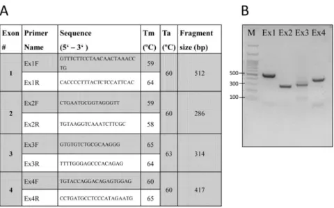
Primer sequences are shown in Figure 1A; they were verified by the Multiple Primer Analyzer (ThermoFisher, Waltham, MA USA) and sequence specificity using NCBI Basic Local Alignment Search Tool (http://www.ncbi.nlm.nih.gov/BLAST/).
A uthor Man uscr ipt A uthor Man uscr ipt A uthor Man uscr ipt A uthor Man uscr ipt
PCR Reaction
PCR products were amplified according to LGALS13 exons and the flanking introns using Takara Ex Taq Hot Start kit (Cat. #R006A, Takara Bio, Kusatsu, Shiga, Japan). Each reaction was prepared to a final volume of 50 μL containing 10 μL of pregnant women’s plasma extracted genomic DNA (gDNA), and the Takara kit reagents including: 5 μL of 10×
Taq buffer, 4 μL of dNTPs mixture (2.5 mM each), 1 μL primer pairs at 10 mM concentration, 0.25 μL of Taq DNA polymerase (5 U/μL) and 30 μL of double-distilled water. Amplifications were performed with the following sequential steps: denaturing at 98°
C for 10 seconds, followed by 40 cycles of amplification with initial denaturing at 94° C for 30 seconds, annealing of primers at corresponding temperature for 30 seconds and extension at 72° C for 60 seconds, and a final extension at 72° C for 10 minutes.
The PCR products (9 μL each) were then mixed with 1 μL of 10× loading buffer (Takara, Japan) and resolved by electrophoresis on 2% agarose gels in 1× TAE buffer (90 mM Tris- HCl, 90 mM boric acid, and 1mM EDTA, pH 8.0) for 20 minutes to ensure a single PCR product (Figure 1B). The gels were then photographed under ultraviolet light (260 nm) using a Bio-Rad Universal Hood and Bio-Rad Quantity One software (Bio-Rad, Hercules, CA, USA).
Sequencing reactions were carried out using the BigDye® Terminator v1.1 Cycle Sequencing Kit (Applied Biosystems, Foster City, CA, USA) and the products were analyzed using an ABI 3730XL Genetic Analyzer (Applied Biosystems). Sequencing was bi-directional to verify accuracy.
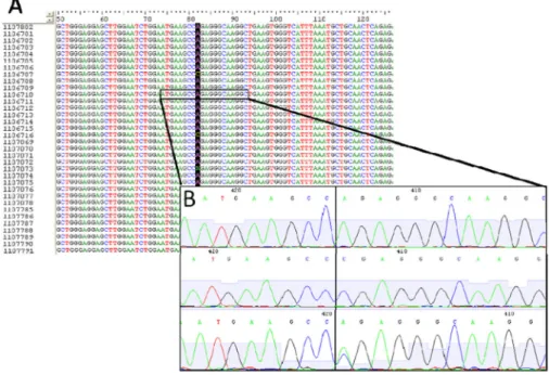
Mutational analysis of the LGALS13 gene was performed using the BioEdit Sequence Alignment Editor version 7.2.5 (Ionis Pharmaceuticals, Carlsbad, CA, USA) (Figure 2A).
SNPs were verified by repeating the sequence and PCR procedures (Figure 2B). The sequence analyzed was maternal, and there was no background signal at the polymorphic region to imply any detection of fetal cfDNA, most likely because of its very low level.
Transcription factor binding site analysis
The Transfac Database of the BIOBASE Biological Databases (www.biobase-
international.com) was used to predict putative transcription factor binding sites in the promoter of LGALS13 gene. The positional probability matrix and the positional weight matrix of the canonical TFAP2A (transcription factor AP-2 alpha, activating enhancer binding protein 2 alpha) binding site (MA0003.1) were downloaded from the Jaspar database (http://jaspar.genereg.net/).
Promoter luciferase assays
To test the effect of the A/C variation in −98 position of the LGALS13 promoter on gene expression, luciferase assays coupled with trophoblast differentiation experiments were completed following published protocols [34]. Briefly, the LGALS13 promoter reporter construct, which contains Gaussia luciferase reporter gene linked to the LGALS13 promoter, was designed at the NIH Perinatology Research Branch and generated by GeneCopoeia (Rockville, MD, USA). The “−98C” clone contains a cytosine in the −98 position. The
A uthor Man uscr ipt A uthor Man uscr ipt A uthor Man uscr ipt A uthor Man uscr ipt
“−98A” reporter construct was generated at the Hungarian Academy of Sciences Research Centre for Natural Sciences by replacing cytosine for adenine in −98 position by site- directed mutagenesis using the QuikChange Lightning kit (Agilent Technologies, Santa Clara, CA, USA), following the manufacturer’s protocol.
The “−98C” and “−98A” reporter constructs (1 μg/well in a 24-well plate) were transfected into BeWo cells (American Type Culture Collection, Manassas, VA, USA) cultured in F12K medium (Sigma-Aldrich, St. Louis, MO, USA) supplemented with 10% fetal bovine serum (Gibco-ThermoFisher Scientific, Waltham, MA, USA) using Lipofectamine-2000 reagent (Invitrogen-ThermoFisher Scientific), according to the manufacturer’s protocol. Twenty-four hours after transfection, BeWo cells were treated with either Forskolin (25 μM in DMSO;
Sigma-Aldrich) or DMSO control, and incubated at 37° C for 48 hours. In all experiments, supernatants were collected, and secreted Gaussia luciferase activity was determined by Secrete-Pair Gaussia Luciferase Assay (GeneCopoeia), according to the manufacturer’s protocol. This assay measured the Gaussia luciferase reporter gene’s expression linked to the LGALS13 promoter by measuring the luciferase activity of the secreted luciferase reporter protein, since secreted luciferase activity correlates with LGALS13 promoter activity and luciferase gene expression. The luminescence was immediately measured with a Victor X3 microplate reader (PerkinElmer, Inc., Waltham, MA, USA).
Statistical analysis
For categorical variables, comparisons between each outcome group and unaffected controls were made by Fisher’s exact test. Kruskal-Wallis or Mann-Whitney non-parametric tests were used for continuous variables. P values of 0.05 or less were considered as significantly different. The data were analyzed using the SPSS version 24 (SPSS Inc., Chicago, IL, USA).
Hardy-Weinberg equilibrium and χ2 tests were used to compare the genotype and allelic frequency distribution in the study groups. Genotypes and alleles were considered to be in Hardy-Weinberg equilibrium if the observed frequencies did not differ significantly from the expected (p > 0.05). The 95% confidence intervals (CI) were determined to verify the pattern of population distribution and overlap.
Data generated by luciferase assays were analyzed using the t-test for comparison between the constructs. Odds ratios (OR) for individual maternal risk to develop PE were calculated first on their own and then adjusted given having the other ones. For BMI, we used a continuous assessment and a categorical one, dividing the patients into higher BMI (>35) and lower BMI groups. Maternal age was also assessed as a continuous parameter and after defining the group age >40 as advanced maternal age (AMA) in pregnancy versus younger groups. Multiple regression was then calculated according to all adjusted risk factors from the observed measures and conducted once for the continuous BMI and maternal age and once for BMI >35 and AMA >40 versus the respective lower others.
Positive likelihood ratio (LR) was calculated according to sensitivity/(1-specificity), and negative LR was calculated according to (1-sensitivity)/specificity. Overall, LR was the division of the positive and negative LRs.
A uthor Man uscr ipt A uthor Man uscr ipt A uthor Man uscr ipt A uthor Man uscr ipt
Power analysis was conducted assuming the detection accuracy of promoter genotype polymorphism is 100%. Accordingly, we evaluated the frequency of the “A” allele in the control and PE groups. To yield the power of the study given 5% type 1 error, we entered these values into the PS Power and Sample Size Program (version 3.1.2) (http://ps-power- and-sample-size-calculation.software.informer.com).
Results
We have identified 20 intermediate PE cases (delivered at gestational age between 34+0 weeks and 36+6 weeks) and 50 cases of late PE (delivery >37 weeks). These were matched with 200 unaffected controls, according to the time of enrolment (+1 week) and gestational age at enrolment (+1 week). The amount of cfDNA extracted from the samples was sufficient to run the analysis for 196 controls, 18 intermediate PE cases, and 49 term PE cases. In the case of other samples, we could not isolate a suitable amount of cfDNA to conduct the analysis.
Cases and controls’ demographic and pregnancy information is summarized in Table 1, showing that gestational week (crown-rump length) at enrolment was not different between the groups. Cases that developed PE had higher maternal BMI at enrolment, and lower frequency of spontaneous conception (e.g., more conceived by assisted reproduction technology. In the PE group, there were more women with a history of previous PE and black ethnicity.
The PP13 serum level
The PP13 serum level was previously reported by Akolekar et al. [8]. Accordingly, first- trimester PP13 converted to multiple of the gestation specific medians in the unaffected patients was 1.00 (0.76–1.33) (median and 95% CI), compared to 0.93 (0.7–1.3) and 1.11 (0.89–1.49) for intermediate PE and term PE, respectively. The detection rate for 10% false positive rate was 41.4% (32.2–51.2%) and 37.8% (28.9–47.6%) for intermediate PE and term PE, respectively.
Combining maternal prior risk factors, biochemical and biophysical markers, the prediction of PE was 79.5% and 64.2% for intermediate PE and term PE, respectively. Adding PP13 to the prediction of PE increased the prediction accuracy to 85.9% and 71.2%, respectively.
Polymorphisms
As depicted in Table 2, the respective primers revealed the presence of several polymorphic variants previously reported by Gebhardt et al. [14] and a few new ones, not previously listed in the NCBI SNP database for LGALS13. In this cohort, the DelT221 variant of Gebhardt et al. [14] was not found. This is most likely related to the relative small study size. For 0.8 power, a sample size of 168 cases of early severe PE versus 672 controls is required to identify the DelT221 mutation. Such large number of early cases is not available to us today.
There were additional SNPs that were reported in SA and not found in the London cohort (Table 2). Those that were detected (new and old ones) did not appear in a significant
A uthor Man uscr ipt A uthor Man uscr ipt A uthor Man uscr ipt A uthor Man uscr ipt
number of samples to run analysis for elucidating any specific conjunction with PE except
−98A/C [28].
The −98A/C genotype variation (dbSNP: rs3764843)
The −98 A/C genotype polymorphism was detected using the pair of the first two primers (Fig. 2). Table 3 depicts the genotype distribution in PE and unaffected controls. The A/A variant at the −98 position [28] was found to be the dominant genotype in all groups but was above 80% in the PE cases and below 70% in the controls. The C/C genotype has the lowest frequency and A/C was inbetween. The presence of C in either the A/C or C/C genotype was of higher frequency in control compared to PE cases.
The pattern of genotype distribution was more significantly different when comparing term PE cases to controls (p=0.032) than when all PE cases were compared to controls (p=0.068).
There was no difference (p=0.730) in genotype pattern distribution for intermediate PE compared to controls (Table 3), presumably due to the very low number of intermediate PE cases.
Altogether, our sample had approximately 4 controls to 1 term PE case. Thus, among the 196 unaffected controls, the “A” allele appears 312 times, corresponding to a probability of 0.8. Among the 49 term PE cases, the “A” allele appears 80 times, corresponding to a probability of 0.92. Accordingly, the study power for this polymorphism was 0.9 given α=0.05. The sample size of intermediate PE was underpowered for an accurate probability assessment in this study.
Cohort comparison between London and South Africa (SA)
• In both the SA and the London cohorts, the A/A genotype was the dominant allele, but its frequency was below 70% in the control group and above 80% in the PE group in both London and SA (p < 0.001) (Table 4). For the A/A genotype, the ORs for term PE was 2.91 (1.06–5.32) for the London cohort and 1.84 (0.85–2.73) for the SA population.
• The presence of C (A/C and C/C genotypes) was in higher frequency in the controls compared to the PE group in both the SA and London cohorts.
In summary, while there were some differences in the genotype distributions between SA and London, the presence of cytosine nucleotide in the −98 promoter position (either as a heterozygous (A/C) or homozygous (C/C) genotype) was higher in unaffected controls versus all PE cases and particularly in term PE cases (p<0.001).
The comparison of genotype distribution according to racial origin is depicted in Table 5.
For the population at large, both black and non-black had a higher frequency of the A/A genotype and a lower frequency of the A/C and C/C genotypes (p=0.958). However, there was a significant correlation between black ethnicity and PE. Term PE was more frequent in blacks compared to the non-blacks (31.5% vs. 12.1%, p<0.001).
A uthor Man uscr ipt A uthor Man uscr ipt A uthor Man uscr ipt A uthor Man uscr ipt
Genotype, ethnicity and outcome
The C allele (in either C/C or A/C genotype) appears to confer protection from developing term PE for all ethnic groups (Table 6). The A/A genotype was more common in black women with term PE. Accordingly, there was a significant correlation between term PE outcome, A/A genotype, and black ethnicity.
Hardy-Weinberg analysis
Applying the Hardy-Weinberg equation to the genotype distribution as summarized in Table 3 yielded the following:
• The A/A genotype was detected in 82.1% of cases and 67.3% of controls.
• Heterozygosity (A/C) was detected in 13.4% of cases and 24.5% of controls.
• The C/C genotype was detected in 4.5% of the cases and 8.2% of controls.
Accordingly, the genotype frequencies in the control individuals were in Hardy-Weinberg equilibrium. However, all PE cases deviated significantly from equilibrium (p=0.036).
Odds ratios
Calculation of the OR was based on a recessive homozygosity model for PE development. It was conducted by comparing the A/A genotype to other genotypes and yielded an OR of 2.22 (0.61–3.9) for all PE, 2.91 (0.83–4.5) for term PE, and 1.26 (0.29–1.83) for
intermediate (values not shown). This was consistent with the previous report from SA performed at delivery [28] that has demonstrated that the A/A genotype is associated with high risk to develop PE, while the C/C and A/C genotypes were protective against the development of PE. However, on its own the promoter variant genotype was not strong enough as a stand-alone marker for predicting PE.
The first model
A multivariate analysis was subsequently conducted to combine the A/A genotype in the
−98 promoter position with black ethnicity, previous PE, BMI, and maternal age. While ethnicity and previous PE were used as categorical confounders (yes/no), BMI and maternal age were used as continuous ones. The individual ORs were first calculated as “stand alone”
(not shown) and then recalculated to account for having the other risk factors to yield the adjusted OR (upper part of Table 7).
The first model (top part of Table 7) yields the following values:
• The overall multiple marker assessment yielded 77.9% accuracy for all PE and 83.7% accuracy for term PE with 6% and 3% false positive rates, respectively.
• The combined OR for all PE had R2=0.24, χ2(5)= 47.35, p<0.001, and an adjusted OR=7. For the term PE, the values were R2=0.27, χ2(5)=44.99, p<0.001 and the adjusted OR=14.
• The positive LR [sensitivity/(1-specificity)] was 5.21 and 10.2 for all PE and term PE, respectively.
A uthor Man uscr ipt A uthor Man uscr ipt A uthor Man uscr ipt A uthor Man uscr ipt
• The negative LR [(1-sensitivity)/specificity] was 0.73 and 0.72 for all PE and term PE, respectively.
• The overall LR = positive LR/negative LR was 7.13 for all PE and 14.17 for the term PE group, respectively.
The second model
OR was subsequently recalculated with all confounders considered as categorical variables (BMI >35 vs. the others and AMA >40 vs. younger women). Using this model, the adjusted ORs for each confounder were improved, particularly the OR for BMI that was dramatically increased from 1.12 (1.06–1.19) for either all PE or term PE (Table 7, upper part) to 4.36 (1.62–11.78) and 3.68 (1.24–10.95) for all PE and term PE, respectively (Table 7, lower part).
• The overall accuracy slightly increased to a 78.3% detection rate for all PE and 83.3% for term PE with similar 6% and 3% false positive rates, respectively.
• The combined adjusted OR increased to 11 for all PE and 15.6 for term PE.
• The overall LR slightly increased to 11.14 and 15.8 for all PE and term PE, respectively.
The effect of −98 polymorphism on LGALS13 expression
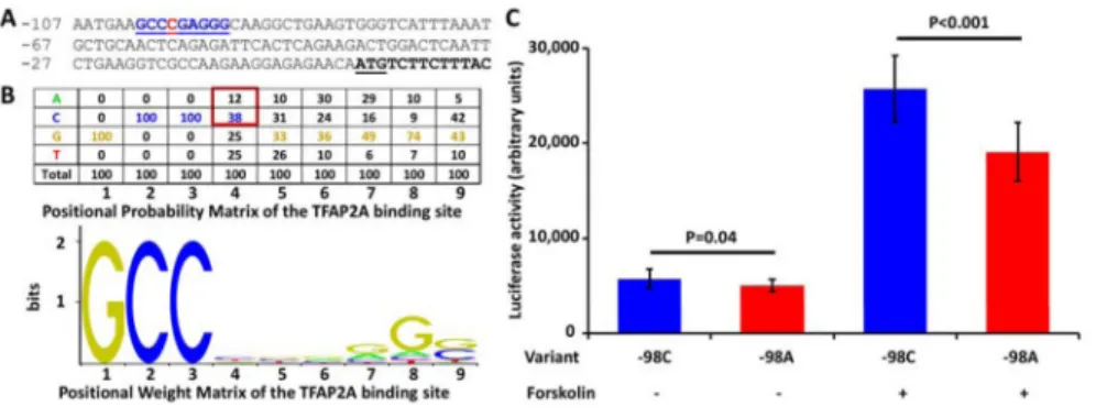
In order to study the functional effect of this polymorphism, first we performed a transcription factor binding site search using the Transfac database, which revealed a TFAP2A binding site in the LGALS13 gene promoter at −101 to −93 positions (Figure 3A).
Of interest, the comparison of this binding site nucleotide sequence with the positional probability matrix and positional weight matrix of the canonical TFAP2A binding site (MA0003.1) in the Jaspar database showed that: 1) 7 out of the 9 bases in the LGALS13 promoter binding site match the most frequent bases in the canonical binding site; and 2) the
“C” in the −98 position has a higher occurrence and, thus, binding affinity than “A” in the canonical binding site (Figure 3B). This suggested that having “A” in the −98 position may lead to a weaker binding of TFAP2A than having “C” in the same position in the LGALS13 promoter and a consequent lower LGALS13 gene expression.
To investigate this hypothesis, next we generated “−98C” and “−98A” LGALS13 promoter clones and examined their expression in non-differentiated and differentiated BeWo cells.
BeWo cells have TFAP2A expression in the non-differentiated state, which increases during trophoblast differentiation. Therefore, we expected to find an increasing difference in luciferase activity with the progress of differentiation between the two clones. Indeed, promoter “−98A” variant had a 13% lower expression in non-differentiated BeWo cells (p=0.04), while it had a 26% lower expression in Forskolin-induced BeWo cells after 48 hours of differentiation (p<0.001) compared to the “−98C” variant. The expression of both promoter variants increased during the 48 hours of differentiation. However, the increase in the expression of “−98C” variant was by 4.55-fold, while the increase in the expression of the “−98A” variant was only by 3.85-fold increase (−15%, p<0.001).
A uthor Man uscr ipt A uthor Man uscr ipt A uthor Man uscr ipt A uthor Man uscr ipt
Discussion
Major findings of the study
1) The LGALS13 −98 A/C genotype was the lowest in term PE compared to controls (p<0.032), similar to the SA cohort; 2) control but not all PE allele frequencies were in Hardy-Weinberg equilibrium (p=0.036); 3) the OR for term PE calculated for previous PE, BMI, maternal age, black ethnicity, and the A/A genotype was 14 (p<0.001) and increased to 15.6 when BMI and AMA were used as categorical variables; 4) in luciferase assays, the LGALS13 promoter “−98A” variant had 13% (p=0.04) and 26% (p<0.001) lower expression than the “−98C” variant in non-differentiated and differentiated BeWo cells, respectively.
After 48 hours of differentiation, there was a 4.55-fold increase in the expression of the
“−98C” variant versus a 3.85-fold of “−98A” variant (p<0.001).
The relevance of −98A/C LGALS13 polymorphism for preeclampsia prediction
In the present study, we searched for first-trimester SNPs of the LGALS13 gene encoding for PP13 and the potential relevance of these SNPs to the prediction of PE. An A/C genotype for the promoter −98 position has been previously reported for an SA cohort collected at delivery. Our study revealed it in a cohort from London with samples collected before the development of PE symptoms during the first-trimester evaluation of pregnancy. The results show that such polymorphism analysis could add to the prediction accuracy of the risk to develop PE.
While the frequency of the A/A genotype was higher among patients who subsequently developed PE, the presence of cytosine (“C”) in the −98 position (either in its heterozygous A/C genotype variant or the homozygous C/C variant) seems to convey protection from the development of PE, as was previously reported for the cohort in SA [28]. This protection from PE is probably related to the higher expression of LGALS13 when “C” is in the −98 position compared to “A” in this position in the LGALS13 promoter, as was detected in BeWo trophoblast-like cells. Our study revealed that while the unaffected control group was in Hardy-Weinberg equilibrium, the PE group deviated from it. This was true for both the London and SA cohorts, and strongly emphasized that having the “C” variant in the −98 position is protecting from the development of PE.
PP13 was implicated in several functions of placental development, including immune tolerance to the paternal genes of the migrating trophoblasts [16, 23] as well as blood pressure and uteroplacental vasculature control [25]. Therefore, knowledge of
polymorphisms in the promoter region for this gene might facilitate our understanding of its potential role in regulating the normal progress of pregnancy. Regardless of the differences in the actual prevalence of the A/A, C/C, and A/C genotypes between the SA and London cohorts, the presence of cytosine, either in the C/C or A/C format, appeared to convey protection from PE, and black ethnicity added to the accuracy of PE prediction. The
discrepancy in the actual genotype distributions may be related to the difference in the origin of the analyzed black tribe, which were mainly Sub-Saharan and Caribbean origin in London compared to Zulu and additional South- and Central African tribes in SA [8, 15]. In this context, one may address the issue of ancestry information markers (AIMS). To the best
A uthor Man uscr ipt A uthor Man uscr ipt A uthor Man uscr ipt A uthor Man uscr ipt
of our knowledge, there are no AIMS for PE that can be used in the prediction of this disorder according to the ethnic origin, although such AIMS were described for certain different geographical regions in the context of other diseases [6,7]. Given the different frequency of PE even among the black ethnic groups, i.e. South African, Caribbean African, and African-American, in the future the polymorphism of LGALS13 may be developed as a potential AIM for PE.
The relevance of −98A/C polymorphism for LGALS13 expression
How may the cytosine nucleotide in the A/C or C/C configuration (versus A/A genotype) in the −98 position in the promoter region of LGALS13 convey resistance to PE? The
transcription factor binding site analysis has indicated that having “C” in the −98 position in the LGALS13 promoter compared to having “A” in this position predicts a higher binding of TFAP2A. This transcription factor is critical for the trophoblastic expression of several placental genes including LGALS13 and other galectin genes in its close vicinity on Chr19 [34]. Thus, this provides a molecular rationale for the higher expression of LGALS13 in individuals who have “C” in the −98 position and consequently more PP13 protein synthesis.
Indeed, the in vitro expression studies confirmed this binding and in silico analysis. We performed luciferase assays on the “−98C” and the “−98A” LGALS13 promoter clones. It was found that the “98A” variant had a lower expression in both non-differentiated and differentiated placenta-derived BeWo cells compared to the expression in the “−98C”
variant. The effect is strong during differentiation, where having the “−98C” variant in the promoter construct was accompanied by a higher fold increase in expression compared to having the “−98A” variant. Since luciferase assays measured only the expression of the promoter constructs transfected into BeWo cells but not the internal expression of LGALS13 gene copies in BeWo cells, the genotype of BeWo cells for the −98A/C LGALS13
polymorphisms as well as their aneuploidy [35] did not interfere with our assay.
The expression studies with the “−98A” and “−98C” promoter reporter variants emphasized the effect of this polymorphism on LGALS13 expression in the context of trophoblast differentiation. It is important, since induction of LGALS13 expression during the differentiation of the trophoblasts to generate placental villi correlates with the increased expression of TFAP2A among other transcription regulatory genes [34]. Thus, this
functional observation validates the in-silico prediction of the role of TFAP2A binding to the polymorphism-containing binding site. Moreover, Kliman et al. [23] have shown that PP13 is involved in the remodeling of the utero-placental vasculature. In addition, Kliman et al [23] and Than et al. [16] have shown that PP13 induces white blood cell apoptosis and confers maternal immune tolerance to the pregnancies. Thus, the results of this study point to the potential link between placental development, trophoblast differentiation, LGALS13 expression, maternal blood PP13 concentrations, and normal pregnancy maintenance, and prompts additional studies—functional and clinical—to uncover this unexplored area in detail.
A uthor Man uscr ipt A uthor Man uscr ipt A uthor Man uscr ipt A uthor Man uscr ipt
The relevance of LGALS13 polymorphisms for the development of preeclampsia
We determined LGALS13 on the level of cfDNA from maternal blood. Impairments in the LGALS13 nucleotide sequence and its RNA/protein expression have been found to affect the risk to develop PE [14, 22, 24, 28, 36]. In addition to the −98A/C promoter
polymorphism, the deletion of the nucleotide thymidine in position 221 (DelT221) also conveyed high risk to develop PE. Although the frequency of DelT221 is 1:50,000 pregnancies in the Western world, in SA it appears in 1:1,000 deliveries among black women [14], and conveyed an 89% positive predictive value for developing early-onset severe PE when present in its heterozygous form. The homozygous form was not associated with viable pregnancy. Sammar et al. [37] constructed the DelT221 variant and expressed both the wild type and the DelT221 variants of LGALS13 in Escherichia coli. The polypeptides were subsequently purified as recombinant proteins yielding DelT221 as a shorter variant compared to the wild type due to a premature stop codon in the open reading frame. When the wild type and truncated PP13 were applied, each was capable of reducing the blood pressure in gravid rats, and this effect lasted for the entire period of PP13 delivery from slow-releasing inter-peritoneal pumps [25]. However, unlike the wild-type, the truncated PP13 failed to cause expansion of the utero-placental vasculature and could not increase placental size and pup weight [25, 37]. These findings emphasize the role of the sugar-binding residues of the PP13 molecule that are partially missing in the truncated variant as was demonstrated previously [16, 34, 36]. To our disappointment, the 1:50,000 frequency of the DelT221 mutation was too low to enable the detection of this mutation in the London cohort.
This study adds an additional piece of information to the importance of the LGALS13 locus in the development of PE. Having now two variants of polymorphic LGALS13 (DelT221 and
−98A/C polymorphism) demonstrates the correlation between an impaired PP13 sequence and expression and the elevated risk to develop PE. In the case of the DelT221 mutation, a strong risk to develop early-onset and severe PE was discovered [14]. In the case of the −98 A/C promoter variants, the reduced expression appears to be related to term PE. On its own, the −98 A/C promoter variant was a low predictor but combined with the history of previous PE, black ethnicity, AMA, and high BMI, it added to the prediction accuracy. Accordingly, each variant conveyed a different risk level to develop PE and was associated with a different subtype of PE. Further studies in PE and animal models may reveal the physiological and morphological mediators involved in the risk to develop PE derived from each variant and for the two combined.
The relevance of gene polymorphisms for the development of preeclampsia Preeclampsia is a multifactorial disorder. Our study provides an additional tool for
identifying patients at risk to develop PE, in the context of the sequence and/or expression of the LGALS13 gene and the encoded PP13 protein. This tool may help direct us to develop a novel drug-target composite through identifying the sub-group of patients whose risk to develop PE is associated with impaired PP13 sequence/expression/function. We have identified here a potential link between a marker discovery, risk stratification, and identification of a targeted group for evaluating potential new methods of therapy, which opens a road to deliver better patient monitoring and management [38].
A uthor Man uscr ipt A uthor Man uscr ipt A uthor Man uscr ipt A uthor Man uscr ipt
Polymorphisms were already identified in various genes that are involved in different functions related to the PE disorder [9–14]. Among them are the polymorphisms in genes that are involved in blood pressure regulation through the renin-angiotensin-aldosterone system. Polymorphisms in this system are associated with hypertensive disorders in pregnancy as was reported for the white population of Poland [39], and the black Afro- Caribbean, but not the white Europeans in South and Central London [9]. However, this was not the case among Asian women of Taiwan [9]. More than one type of polymorphism in this system, including the variant related to hypertensive disorders in pregnancy, was identified among Tunisian Arabs [40]. At the same time, the latter two variants of polymorphisms have been shown to be closely related to other cardiovascular disorders, including congestive cardiomyopathy, supraventricular arrhythmias, reduced exercise capacity, and an increase in left ventricular systolic dimension. These disorders underline high susceptibility for cardiovascular morbidity among women who had experienced hypertensive disorders in pregnancy [41–42].
Polymorphisms were also identified in the endothelial nitric oxide synthase (eNOS) system that regulates vasodilation, one of the hallmarks of early stages of pregnancy development.
Linkage studies in affected sibling pairs have implicated the NOS3 gene (encoding eNOS) locus on chromosome 7q35 [10, 43] to be responsible for a higher risk to develop PE.
However, more studies are required because this gene candidate might differ between ethnic groups [9–10].
Analysis of the polymorphisms of IL27 in PE among Han Chinese women revealed a significantly reduced risk of PE in one genotype variant compared to the others in a dominant allele model. This was particularly true for the severe PE subgroup, implying that SNPs in IL27 may have an effect on individual susceptibility to PE [12].
Our findings are driving us to develop additional tools for PE prediction based on SNPs, such as quantitative real time multiplex PCR or DNA chips to evaluate the risk to develop PE with cfDNA tools. Preeclampsia has a significant inherited component, and it is likely that many genes are involved [44]. Considering that the etiology of PE remains complex, identification of genes that may predispose to developing PE could form an aiding tool for prevention and treatment. It might also assist in preventing cardiovascular disorders given that women with a history of PE are at increased risk of cardiovascular disease in later life [45].
Prediction of preeclampsia—the contribution of the PP13 promoter A/A genotype
This study adds additional information to the importance of the history of previous PE, black ethnicity, and high BMI in the attempt to stratify the risk to develop PE. In the ASPRE study, these parameters were integrated together with biochemical and biophysical markers to stratify the patients at high risk for developing intermediate PE, and its prevention by aspirin [46–48]. The ASPRE study was less successful in the prediction and prevention of term PE. Here, in an additional investigation within the ASPRE study, we have found an algorithm where BMI >35, history of previous PE, black ethnicity, and the genotype of LGALS13 in the −98 promoter region provides a very good prediction of term PE. Given
A uthor Man uscr ipt A uthor Man uscr ipt A uthor Man uscr ipt A uthor Man uscr ipt
has opened the door to new means of prevention of term PE among obese women using metformin. Adding the A/A genotype of the −98 promoter region of PP13 to obesity (BMI
>35) may help to narrow down the group to be treated by metformin and to explore if in this way the efficacy of metformin in reducing the frequency of PE may be increased.
Reducing inflammation is an expected pathway for the prevention of term PE associated with obesity [50]. However, in our study obesity was an independent confounder from the A/A genotype. Accordingly, we are unable to evaluate whether the improved prediction of term PE by LGALS13 genotype polymorphism together with high BMI could be related to any role of PP13 in damping maternal inflammation that is currently the suspected signaling pathway by which obesity increases the risk to develop PE [50].
In the past, members of our group have shown the value of PE risk adjustment to the blood groups with the B group and particularly the AB group identified as being at higher risk for generating low PP13 blood level given PP13’s high affinity to the sugar residues of this blood group’s antigens [51]. However, the B (~8%) and AB (~3%) blood groups are rare compared to the A and O groups (anticipating only two AB patients in our entire PE group).
Thus, there were too few carriers of the relevant blood groups to justify the adjustment to this confounder. However, it would be interesting to examine the impact of this confounder in larger cohorts.
Study Limitations
While our study was powered enough to predict term PE according to −98A/C
polymorphism, the study was underpowered to evaluate the value of this polymorphism for predicting intermediate PE. We have shown that lower PP13 expression with the A/A variant compared to A/C and C/C is associated with higher risk to develop all cases of PE, and the study was powered enough to certainly make such a claim in the context of term PE but underpowered to evaluate it in the context of intermediate PE. Thus, we are hesitating to make a strong statement regarding lower PP13 expression and intermediate PE. However, since all PE are less affected by the A/A genotype than term PE, it is reasonable to assume a lower correlation between the A/A genotype and the risk to develop intermediate PE. Could we use PP13 polymorphism in the context of identifying subtypes of PE? The evidence today has demonstrated PP13’s role in the immune tolerance of the maternal decidua during pregnancy, as was shown by Than et al. [34]. A part of it is related to maternal artery remodeling, as was described by Kliman et al. [23]. PP13 is involved in structural arterial expansion, which does require the molecular parts that are important to immune tolerance [37]. This set of evidence links PP13 to early and intermediate PE. The A/A genotype variation of reduced PP13 expression might be correlated with the hypotension induced by PP13, which occurs regardless of the molecular part responsible for immune tolerance [25].
Thus, PP13 impacts on main maternal artery expansion to enable the increase in cardiac output to coop with the increased burden of pregnancy and the systemic effect of PP13 on vasodilation, and the endothelial layer might be relevant to its importance in endothelial insufficiency in term PE. More studies are required to explore this point.
PP13 is a multifaceted molecule. Kliman et al. [23] have demonstrated the importance of PP13 in spiral artery remodeling associated with early and intermediate PE. Than et al. [16]
A uthor Man uscr ipt A uthor Man uscr ipt A uthor Man uscr ipt A uthor Man uscr ipt
have shown the role of PP13 in immune tolerance. PP13 is also implicated in regulating blood pressure, artery vasodilation, and the development of larger placentas and pups, relevant to the signaling pathway of the eNOS and prostaglandin 2 receptors in the endothelial layer [25, 37, 52] that are also relevant to term PE. Thus, it appears that the impact of the promoter variant polymorphism is certainly related to term PE but its potential relevance to early and intermediate PE needs further evaluation.
This is only the second study that explores the promoter variant and its relevance to the elevated risk to develop PE, following a study conducted in SA [28]. We demonstrated for the first time that this polymorphism is a risk factor deriving from impaired PP13
expression. More studies are required with additional cohorts of other ethnic origins to verify the use of this polymorphism in predicting PE and its subtypes. For identifying the DelT221 mutation, a larger sample size is required in view of the rare mutation frequency.
Is the LGALS13 DNA detected here in this study of maternal or fetal origin?
Studies have shown that approximately 5%–10% of the cfDNA in first-trimester maternal blood is of fetal/placental origin, while 90%–95% is of maternal origin [52]. Accordingly, 90%–95% of cell-free genomic DNA fragments carrying the LGALS13 gene originate from maternal tissues. Since total cfDNA was subjected to PCR amplification, it is expected that the maternal/fetal cfDNA ratio will remain the same after PCR and sequencing. As can be seen in Figure 2, the chromatograms at the −98 polymorphism loci are distinct, with no background, therefore reflecting the maternal genotype. Thus, we estimate that the fetal fraction of LGALS13 in this study was too low for detection.
Future multiplex PCR for the assessment of the LGALS13 locus
This study together with former reports [14, 26] has indicated the potential value of bringing to the market a multiplex PCR for the assessment of the LGALS13 locus in identifying the risk to develop PE. Such diagnostics may be useful in identifying a potential paternal contribution to the risk to develop PE and evaluating the pre-pregnancy carrier. In this context, it appears questionable to test the −98A/C polymorphism on its own. So far we have found that a history of previous PE, high BMI, and black ethnicity combined with the A/A genotype in the cfDNA are useful. A multiplex PCR for all polymorphic variants of the LGALS13 locus (including the −98A/C variants), DelT221, and potentially variants of other nearby galectin mutations, may be useful in identifying individual patient’s risk and could potentially be used for targeted therapy.
Conclusion
This is the first study conducted to identify LGALS13 DNA sequence variants in maternal blood in the first trimester. Our findings suggest that the −98A/C gene polymorphisms in the promoter region of the LGALS13 gene may be associated with PE development with a strong correlation to black ethnicity and high (>35) BMI. The −98 A/C genotype appears to provide protection from PE development. More studies are required to verify the use of the
−98A/C promoter variant for aiding PE prediction alone or in combination with genetic variants of other genes with possible diagnostic significance [53–56]. Further studies are
A uthor Man uscr ipt A uthor Man uscr ipt A uthor Man uscr ipt A uthor Man uscr ipt
also required in PE models to examine the impact of the polymorphism on the disease development and in order to find novel therapies.
Acknowledgments
Funding: This research was supported, in part, by ASPRE FP7 # 601852 grant (to LML, RC, KHN, and HM), by the Perinatology Research Branch, Division of Intramural Research, Eunice Kennedy Shriver National Institute of Child Health and Human Development (NICHD), National Institutes of Health (NIH), Department of Health and Human Services (DHHS), by Federal funds from the NICHD under Contract No. HSN275201300006C, and by the Hungarian Academy of Sciences Momentum Grant “LP2014-7/2014” (to NGT).
We thank Zsolt Gelencser (Hungarian Academy of Sciences) for technical assistance.
References
1. Walker JJ. Pre-eclampsia. Lancet. 2000; 356:1260–1265. [PubMed: 11072961]
2. Roberts JM, Cooper DW. Pathogenesis and genetics of pre-eclampsia. Lancet. 2001; 357:53–56.
[PubMed: 11197372]
3. Khan KS, Wojdyla D, Say L, Metin-Gülmezoglu AM, Van Look PA. WHO analysis of causes of maternal death: a systematic review. Lancet. 2006; 367:1066–1074. [PubMed: 16581405]
4. Duley L. The global impact of pre-eclampsia and eclampsia. Semin Perinatol. 2009; 33:130–137.
[PubMed: 19464502]
5. Redman CW, Sacks GP, Sargent IL. Preeclampsia: an excessive maternal inflammatory response to pregnancy. Am J Obstet Gynecol. 1999; 180(Pt 1):499–506. [PubMed: 9988826]
6. Nakimuli A, Chazara O, Byamugisha J, Elliott AM, Kaleebu P, Mirembe F, et al. Pregnancy, parturition and preeclampsia in women of African ancestry. Am J Obstet Gynecol. 2013; 210:510–
520. e1. [PubMed: 24184340]
7. Pattinson, R., editor. National Committee for the Confidential Enquiry into Maternal Deaths. Saving Mothers 2011–2013: Sixth Report on the Confidential Enquiries into Maternal Deaths in South Africa. Department of Health; South Africa: 2015. p. 1-93.http://www.kznhealth.gov.za/mcwh/
Maternal/Saving-Mothers-2011-2013-short-report.pdf
8. Akolekar R, Syngelaki A, Sarquis R, Zvanca M, Nicolaides KH. Prediction of early, intermediate and late pre-eclampsia from maternal factors, biophysical and biochemical markers at 11–13 weeks.
Prenat Diagn. 2011; 31:66–74. [PubMed: 21210481]
9. Yang J, Shang J, Zhang S, Li H, Huiong L. The role of the rennin-angiotensin-aldosterone system in preeclampsia: genetic polymorphism and micro RNA. J Mol Endocrinol. 2013; 50:R53–R66.
[PubMed: 23369849]
10. Yu CK, Casas JP, Savvidou MD, Sahemey MK, Nicolaides KH, Hingorani AD. Endothelial nitric oxide synthase gene polymorphism (Glu298Asp) and development of pre-eclampsia: a case- control study and a meta-analysis. BMC Pregnancy Childbirth. 2006; 16:1–9.
11. Papazoglou D, Galazios G, Koukourakis MI, Panagopoulos I, Kontomanolis EN, Papatheodorou K, et al. Vascular endothelial growth factor gene polymorphism and pre-eclampsia. Mol Hum Reprod.
2004; 10:321–324. [PubMed: 14997002]
12. Chen P, Gong Y, Pu Y, Wang Y, Zhou B, Song Y, et al. Association between polymorphisms in IL-27 gene and pre-eclampsia. Placenta. 2016; 37:61–64. [PubMed: 26654512]
13. Arias F, Rodriquez L, Rayne SC, Kraus FT. Maternal placental vasculopathy and infection: two distinct subgroups among patients with preterm labor and preterm ruptured membranes. Am J Obstet Gynecol. 1993; 168:585–591. [PubMed: 8438933]
14. Gebhardt S, Bruiners N, Hillerman R. A novel exonic variant (221DelT) in the LGALS13 gene encoding placental protein 13 (PP13) is associated with preterm labour in a low risk population. J Reprod Immunol. 2009; 82:166–173. [PubMed: 19818512]
15. Huppertz B. Placental origins of preeclampsia: challenging the current hypothesis. Hypertension.
2008; 51:970–975. [PubMed: 18259009]
A uthor Man uscr ipt A uthor Man uscr ipt A uthor Man uscr ipt A uthor Man uscr ipt
16. Than NG, Romero R, Goodman M, Weckle A, Xing J, Dong Z, et al. A primate subfamily of galectins expressed at the maternal-fetal interface that promote immune cell death. Proc Natl Acad Sci USA. 2009; 16:9731–9736.
17. Romero R, Nien JK, Espinoza J, Todem D, Fu W, Chung H, et al. A longitudinal study of angiogenic (placental growth factor) and anti-angiogenic (soluble endoglin and soluble vascular endothelial growth factor receptor-1) factors in normal pregnancy and patients destined to develop preeclampsia and deliver a small for gestational age neonate. J Matern Fetal Neonatal Med. 2008;
21:9–23. [PubMed: 18175241]
18. Thilaganathan B. Placental syndromes: getting to the heart of the matter. Ultrasound Obstet Gynecol. 2017; 49:7–9. [PubMed: 28067440]
19. Osol G, Bernstein I. Preeclampsia and maternal cardiovascular disease: consequence or predisposition? J Vasc Res. 2014; 51:290–304. [PubMed: 25300258]
20. Than NG, Sumegi B, Than GN, Berente Z, Bohn H. Isolation and, sequence analysis of a cDNA encoding human placental tissue protein 13 (PP-13), a new lysophospholipase, homologue of human eosinophil Charcot-Leyden Crystal protein. Placenta. 1999; 20:703–710. [PubMed:
10527825]
21. Balogh A, Pozsgay J, Matkó J, Dong Z, Kim CJ, Várkonyi T, et al. Placental protein 13 (PP13/
Galectin-13) undergoes lipid raft-associated subcellular redistribution in the syncytiotrophoblast in preterm preeclampsia and HELLP syndrome. Am J Obstet Gynecol. 2011; 205:156, e1–e14.
22. Huppertz B, Meiri H, Gizurarson S, Osol G, Sammar M. Placental protein 13 (PP13): a new biological target shifting individualized risk assessment to personalized drug design combating pre-eclampsia. Hum Reprod Update. 2013; 19:391–405. [PubMed: 23420029]
23. Kliman HJ, Sammar M, Grimpel Y-I, Lynch SK, Milano KM, Pick E, et al. Placental Protein 13 and decidual zones of necrosis: an immunologic diversion that may be linked to preeclampsia.
Reprod Sci. 2012; 19:16–30. [PubMed: 21989657]
24. Than NG, Romero R, Balogh A, Karpati E, Mastrolia SA, Staretz-Chacham O, et al. Galectins:
double edged swords in the cross-roads of pregnancy complications and female reproductive tract inflammation and neoplasia. J Pathol Transl Med. 2015; 49:181–208. [PubMed: 26018511]
25. Gizurarson S, Sigurdardottir ER, Meiri H, Huppertz B, Sammar M, Sharabi-Nov A, et al. Placental Protein 13 administration to pregnant rats lowers blood pressure and augments fetal growth and venous remodeling. Fetal Diagn Ther. 2016; 39:56–63. [PubMed: 26314825]
26. Sammar M, Nisemblat S, Fleischfarb Z, Golan A, Sadan O, Meiri H, et al. Placenta-bound and body fluid PP13 and its mRNA in normal pregnancy compared to preeclampsia, HELLP and preterm delivery. Placenta. 2011; 32(suppl):S30–S36. [PubMed: 21257080]
27. Farina A, Zucchini C, Sekizawa A, Purwosunu Y, de Sanctis P, Santarsiero G, et al. Performance of messenger RNAs circulating in maternal blood in the prediction of preeclampsia at 10–14 weeks.
Am J Obstet Gynecol. 2010; 203:e1–e6.
28. Bruiners, N., Bosman, M., Postma, A., Gebhardt, S., Rebello, G., Sammar, M., et al. Promoter variant −98A–C of the LGALS13 gene and pre-eclampsia. 8th World Congr Prenat Med Fetal Dev; Florence. September 2008;
29. Hadlock FP, Shah YP, Kanon DJ, Lindsey JV. Fetal crown-rump length: re-evaluation of relation to menstrual age (5–18 weeks) with high-resolution real-time US. Radiology. 1992; 182:501–505.
[PubMed: 1732970]
30. Poon LC, Stratieva V, Piras S, Piri S, Nicolaides KH. Hypertensive disorders in pregnancy:
combined screening by uterine artery Doppler, blood pressure and serum PAPP-A at 11–13 weeks.
Prenat Diagn. 2010; 30:216–223. [PubMed: 20108221]
31. Martin AM, Bindra R, Curcio P, Cicero S, Nicolaides KH. Screening for pre-eclampsia and fetal growth restriction by uterine artery Doppler at 11–14 weeks of gestation. Ultrasound Obstet Gynecol. 2001; 18:583–586. [PubMed: 11844193]
32. Poon LCY, Staboulidou I, Maiz N, Plasencia W, Nicolaides KH. Hypertensive disorders in pregnancy: screening by uterine artery Doppler at 11–13 weeks. Ultrasound Obstet Gynecol. 2009;
34:142–148. [PubMed: 19644947]
33. Lindheimer MD, Taler SJ, Cunningham FG. American Society of Hypertension position paper:
A uthor Man uscr ipt A uthor Man uscr ipt A uthor Man uscr ipt A uthor Man uscr ipt
34. Than NG, Romero R, Xu Y, Erez O, Xu Z, Bhatti G, et al. Evolutionary origins of the placental expression of chromosome 19 cluster galectins and their complex dysregulation in preeclampsia.
Placenta. 2014; 35:855–865. [PubMed: 25266889]
35. Long CA, Bauer GS, Lowe ME, Strauss AW, Gast MJ. Isolation and characterization of the gene from a human genome encoding 17β-estradiol dehydrogenase: a comparison of Jar and BeWo choriocarcinoma cell lines. Am J Obstet Gynecol. 2010; 163:1976–1981.
36. Than NG, Balogh A, Romero R, Karpati E, Erez O, Szilagyi A, et al. Placental Protein 13 (PP13) - a placental immunoregulatory galectin protecting pregnancy. Front Immunol. 2014; 5:1–25.
[PubMed: 24474949]
37. Sammar M, Nisamblatt S, Gonen R, Huppertz B, Gizurarson S, Osol G, et al. The role of the carbohydrate recognition domain of placental protein 13 (PP13) in pregnancy evaluated with recombinant PP13 and the DelT221 PP13 variant. PLoS One. 2014; 9:e102832. [PubMed:
25079598]
38. Hahn S. Preeclampsia - will orphan drug status facilitate innovative biological therapies? Front Surg. 2015; 2:1–4. [PubMed: 25674565]
39. Bogacz A, Bartkowiak-Wieczorek J, Procyk D, Seremak-Mrozikiewicz A, Majchrzycki M, Dziekan K, et al. Analysis of the gene polymorphism of aldosterone synthase (CYP11B2) and atrial natriuretic peptide (ANP) in women with preeclampsia. Eur J Obstet Gynecol Reprod Biol.
2016; 197:11–15. [PubMed: 26686590]
40. Saidi S, Mahjoub T, Almawi WY. Aldosterone synthase gene (CYP11B2) promoter polymorphism as a risk factor for ischaemic stroke on Tunisian Arabs. J Renin Angiotensin Aldosteron Syst.
2010; 11:180–186.
41. Lo HM, Lin FY, Lin JL, Tseng CD, Hsu KL, Chiang FT, et al. Electrophysiological properties in patients undergoing atrial compartment operation for chronic atrial fibrillation with mitral valve disease. Eur Heart J. 1997; 18:1805–1815. [PubMed: 9402456]
42. Ardizzone N, Cappello F, Di Felice V, Rappa F, Minervini F, Marasà S, et al. Atrial natriuretic peptide and CD34 overexpression in human idiopathic dilated cardiomyopathies. APMIS. 2007;
115:1227–1233. [PubMed: 18092954]
43. Lade JA, Moses EK, Guo G, Wilton AN, Grehan M, Cooper DW, et al. The eNOS gene: a candidate for the preeclampsia susceptibility locus? Hypertens Pregnancy. 1999; 18:81–93.
[PubMed: 10464002]
44. Pridjian G, Puschett JB. Preeclampsia. 2. Experimental and genetic considerations. Obstet Gynecol Surv. 2002; 57:619–640. [PubMed: 12218669]
45. Smith GC, Pell JP, Walsh D. Pregnancy complications and maternal risk of ischaemic heart disease: a retrospective cohort study of 129,290 births. Lancet. 2001; 357:2002–2006. [PubMed:
11438131]
46. O’Gorman N, Wright D, Poon LC, Rolnik DL, Syngelaki A, et al. Multicenter screening for preeclampsia by maternal factors and biomarkers at 11–13 weeks’ gestation: comparison to NICE guidelines and ACOG recommendations. Ultrasound Obstet Gynecol. 2017; 49:756–760.
[PubMed: 28295782]
47. O’Gorman N, Wright D, Syngelaki A, Akolekar R, Wright A, Poon LC, Nicolaides KH.
Competing risks model in screening for preeclampsia by maternal factors and biomarkers at 11–13 weeks gestation. Am J Obstet Gynecol. 2016; 214:103.e1–103.e12. [PubMed: 26297382]
48. Rolnik D, Wright D, Poon LC, O’Gorman N, Syngelaki A. Aspirin versus placebo in pregnancies at high risk for preterm preeclampsia. N Engl J Med. 2017; 377:613–622. [PubMed: 28657417]
49. Syngelaki A, Nicolaides KH, Balani J, Hyer S, Akolekar R, et al. Metformin versus placebo in obese pregnant women without diabetes mellitus. N Engl J Med. 2016; 374:434–443. [PubMed:
26840133]
50. Wolf M, Kettyle E, Sandler L, Ecker JL, Roberts J, Thadhani R. Obesity and preeclampsia: the potential role of inflammation. Obstet Gynecol. 2001; 98(Pt 1):757–762. [PubMed: 11704165]
51. Than NG, Romero R, Meiri H, Erez O, Xu Y, et al. PP13, maternal ABO blood groups and the risk assessment of pregnancy complications. PLoS One. 2011; 6:e21564. [PubMed: 21799738]
A uthor Man uscr ipt A uthor Man uscr ipt A uthor Man uscr ipt A uthor Man uscr ipt
52. Drobnjak T, Gizurarson S, Gokina NI, et al. Placental protein 13 (PP13)-induced vasodilation of resistance arteries from pregnant and nonpregnant rats occurs via endothelial signaling pathways.
Pregnancy Hypertens. 2017; 36:186–195.
53. Lo YMD, Chan KCA, Dun H, Chen EZ, Jiang P, Lun FMF, Zheng YW, Leung TY, Lau TK, Cantor CR, Chiu RWK. Maternal plasma DNA sequencing reveals the genome-wide genetic and
mutational profile of the fetus. Sci Transl Med. 2010; 2:61ra91.
54. Rigourd V, Chelbi S, Chauvet C, Rebourcet R, Barbaux S, Bessières B, et al. Re-evaluation of the role of STOX1 transcription factor in placental development and preeclampsia. J Reprod Immunol.
2009; 82:174–181. [PubMed: 19577309]
55. Fong FM, Sahemey MK, Hamedi G, Eyitayo R, Yates D, Kuan V, et al. Maternal genotype and severe preeclampsia: a HuGE review. Am J Epidemiol. 2014; 180:335–345. [PubMed: 25028703]
56. Haram K, Mortensen JH, Nagy B. Genetic aspects of preeclampsia and the HELLP syndrome. J Pregnancy. 2015; 2014:1–13.
A uthor Man uscr ipt A uthor Man uscr ipt A uthor Man uscr ipt A uthor Man uscr ipt
Figure 1. Primer details and PCR products
A, The primers used for each exon PCR (Ex1–Ex4) are detailed for their forward (F) and reverse (1R) order along with the respective melting temperature (Tm), annealing temperature (Ta), and the amplified DNA fragment size. B, PCR amplification of each LGALS13 exon (Ex1–Ex4) presented after separation on gel electrophoresis. M, DNA base pair marker at 100K unit.
A uthor Man uscr ipt A uthor Man uscr ipt A uthor Man uscr ipt A uthor Man uscr ipt
Figure 2. Sequencing of the −98 site
A, A multiple sequence alignment of the PCR products of exon 1 from several specimens, including LGALS13 promoter region and flanking intron. B, Enlarged vision of 20 nucleotides including the −98 position (peaks after the black vertical line). On top, the subject marked in section A that carries the A/A genotype. The two rows below show additional subjects with C/C genotype (middle) and A/C genotype (bottom).
A uthor Man uscr ipt A uthor Man uscr ipt A uthor Man uscr ipt A uthor Man uscr ipt
Figure 3. Promoter luciferase assays
A, Partial promoter sequence of LGALS13. Nucleotide positions from the translation initiator codon (underlined) are shown on the left. The first exon is shown in bold black, the predicted TFAP2A binding site is depicted in bold blue underlined letters, and the “C” in the
−98 position is highlighted in red. B, Positional probability matrix (PPM) and positional weight matrix (PWM) of the canonical TFAP2A binding site (MA0003.1) as downloaded from the Jaspar database (http://jaspar.genereg.net/). PPM values provide the normalized occurrence of each nucleotide at each position in the canonical TFAP2A binding site, while PWM values—log-LR derived from PPM—provide binding affinity scores for each nucleotide at each position. The most frequent nucleotides in each position in the PPM are depicted with values colored according to the coloring of nucleotides in the PWM. As shown with the red square, the “C” in the fourth position of the TFAP2A binding site has a higher occurrence and, thus, affinity than “A.” C, Luciferase activity of “−98C” and “−98A”
LGALS13 promoter clones. Promoter “−98A” variant had 13% lower expression in non- differentiated BeWo cells (p=0.04) and 26% lower expression in Forskolin-induced BeWo cells after 48 hours of differentiation (p<0.001) than the “−98C” variant. While the expression of both promoter variants increased during the 48 hours of differentiation, the
“−98C” variant had 4.55-fold increase, while the “−98A” variant had only a 3.85-fold increase in expression (−15%, p<0.001). All experiments were run in triplicate.