Journal of
Translational Medicine &
Epidemiology
Special Issue on
von Hippel Lindau Disease
Edited by:
Hiroshi Kanno
Professor, Department of Neurosurgery, Yokohama City University School of Medicine, Japan
Corresponding author
Hartmut P.H. Neumann, Section for Preventive Medicine, Department of Nephrology and General Medicine, University of Freiburg, Freiburg, Germany, E-mail:
Submitted: 06 February 2014 Accepted: 11 March 2014 Published: 13 March 2014 Copyright
© 2014 Neumann et al.
OPEN ACCESS Keywords
• von Hippel-Lindau disease
• Pancreatic neuroendocrine tumors
Review Article
von Hippel-Lindau Disease Associated Pancreatic
Neuroendocrine Tumors – Molecular Genetics and Clinical Aspects
Birke Bausch1, Ernst von Dobschuetz2, Xiao-Ping Qi3, Martin K. Walz4, Georges Weryha5, Attila Patocs6, Karoly Rácz7, Frederic Castinetti8, David Taieb9, Schu-Ren Yang10, Philipp T. Meyer11, Monika Engelhardt12, Kurt Werner Schmid13, Bahadir M. Güllüoglu14, Ozer Makay15, Laura von Duecker16, Angelica Malinoc16, Stefan Zschiedrich16, Giuseppe Opocher17 and Hartmut P. H. Neumann16*
12nd Department of Medicine, Albert-Ludwigs University Medical Center of Freiburg, Germany
2Clinic of General-, Visceral- and Thoracic Surgery, St. Adolf-Stift Reinbek, Academic Teaching Hospital of the University of Hamburg, Germany
3Departments of Oncologic and Urologic Surgery, The 117th PLA Hospital, PLA Hangzhou Clinical College, Anhui Medical University, China
4Department of Surgery and Center of Minimally Invasive Surgery, Kliniken Essen-Mitte, Germany
5Department of Endocrinology, University of Lorraine, France
6HAS-SE “Lendület” Hereditary Endocrine Tumor Research Group, Hungarian Academy of Sciences and Semmelweis University, Hungary
72nd Department of Internal Medicine, Semmelweis University, Hungary
8Aix-Marseille University, Department of Endocrinology, La Timone Hospital, France
9Department of Nuclear Medicine, La Timone University Hospital, CERIMED, Aix- Marseille University, Marseille, France
10Department of General Radiology, Albert-Ludwigs University Medical Center of Freiburg, Germany
11Department of Nuclear Medicine, Albert-Ludwigs University Medical Center of Freiburg, Germany
12Department of Hematology, Oncology and Stem Cell Transplantation, Albert-Ludwigs University Medical Center of Freiburg, Germany
13Institute of Pathology, University Hospital of Essen, University Duisburg-Essen, Germany
14Breast & Endocrine Surgery Unit, Marmara University Pendik Research and Training Hospital, Turkey
15Division of Endocrine Surgery, Department of General Surgery, Ege University Hospital, Turkey
16Department of Nephrology and General Medicine, Albert-Ludwigs University Medical Center of Freiburg, Germany
17Veneto Institute of Oncology, IRCCS and Department of Medicine-DIMED, University of Padova, Italy
INTRODUCTION
Pancreatic neuroendocrine tumors (PNETs) is the terminus suggested by the WHO for tumors deriving from the cells of the islets of Langerhans of the pancreas [1]. Other frequently used terms are pancreatic endocrine tumors or islet cell tumors. These tumors belong to the family of neuroendocrine tumors (NETs).
NETs can be located in many different sites of the body. NETs are often classified as foregut, midgut and hindgut tumors [2]. They occur as sporadic and syndromic entities. Best known are NETs as a component of multiple endocrine neoplasia type 1 which is mainly formed by parathyroid adenoma, NET and pituitary adenoma. Less known is that NETs are also a component of von Hippel-Lindau disease (VHL). In VHL disease, NETs are almost exclusively located in the pancreas. In addition to PNETs the classic lesions of VHL disease are multiple serous pancreatic cysts or cystadenomas, hemangioblastomas of the retina and the central nervous system (CNS), clear cell renal carcinomas and pheochromocytomas [3].
SYMPTOMS
PNETs may produce a variety of hormones or vasoactive peptides and can thus cause distinct endocrine syndromes.
These include gastric ulcer, abdominal pain and diarrhea due to gastrinoma and an over secretion of gastrin, episodes of hypoglycaemia due to insulinoma and an over secretion of insulin, and other syndromes such as Werner Morrison syndrome due to an over secretion of vasoactive intestinal peptide (VIP) and glucagonoma due to an over secretion of glucagon. Such clinical presentations are known from sporadic or MEN1 associated PNETs. In contrast, VHL associated PNETs present almost exclusively as non-secreting endocrine tumors, as space occupying masses or metastases. Therefore, PNETs are mostly diagnosed in early adulthood with a mean age of about 35 years. PNETs associated with VHL are mostly diagnosed in an advanced stage except in individuals subjected to a VHL specific surveillance program. This underlines the importance of specific diagnostic strategies.
BIOCHEMISTRY
Systematic analyses of serum concentrations of hormones in patients with VHL associated PNETs are pending, but secreting PNETs in VHL are extremely rare. Such analyses may include pancreatic polypeptide, gastrin, insulin, C peptide, VIP, glucagon, and somatostatin, and as an additional marker chromogranin A.
DIAGNOSIS OF PNETS
PNETs can cause symptoms as local masses or by excretion of
several hormones. Since introduction of serial imaging radiology techniques such as ultrasonography, computerized tomography (CT) and magnetic resonance imaging (MRI) the percentage of PNETs diagnosed as “incidentalomas”, as asymptomatic tumors, is rising. Finally the era of molecular genetics and molecular genetic diagnosis opened the doors to preventive medicine allowing the identification of particular tumors as specific manifestations of hereditary syndromic diseases. Once the diagnosis is established, the patients can be offered specific surveillance programs and protocols. Similarly, relatives can be tested for a given pathogenic mutation and can be clinically investigated. This is the way by which asymptomatic PNETs in VHL disease can be detected [4-8].
MOLECULAR GENETICS AND VHL PHENOTYPES
VHL disease is an autosomal dominant disorder. The penetrance is age dependent, but high in affected subjects older than 40 years of age. The susceptibility genes for hereditary PNETs are the VHL gene located on chromosome 3p25-26 and the MEN1 gene predisposing for multiple endocrine neoplasia type 1 located on chromosome 11q13. The VHL gene has 3 exons and encodes the VHL protein with 213 amino acids. The MEN1 gene consists of 10 exons and encodes the Menin protein with 615 amino acids. Germline mutations are distributed over all exons of both genes, and in addition large deletions encompassing one to all exons of both genes have been described [6,9-15]. In VHL patients with PNETs all types of mutations have been described including missense mutations, stop codon mutations, intraexonic insertions and deletions, splice mutations and large deletions/
rearrangements [5,10,12,14,16] (Table 1).
We have conducted a study to evaluate the frequency of germline mutations in unselected patients with NETs. Our registry-based approach used the German-NET-Registry with 259 patients with the primary diagnosis of a NET. All patients provided blood DNA. All 10 exons of the MEN1 gene and all 3 exons of the VHL genes were analysed for intra-genic mutations and large deletions. In the NET-Registry, 9% of the patients with PNETs had germline mutations, 8 in MEN1 and 1 in VHL [16].
In addition, we evaluated the spectrum of VHL germline mutations and the corresponding phenotypes of all patients registered in the German VHL-Registry. The registry contained 487 molecular genetically confirmed patients. All patients had magnetic resonance imaging or computed tomography of the abdomen. The prevalence of NETs was 53/487 (11%).
Remarkably there were striking differences of occurrence of PNETs in patients with different mutations. Among patients with the mutation VHL p.R167W, 47% developed PNETs, compared to Abstract
Pancreatic neuroendocrine tumors (PNETs) occur in about 10% of patients with von Hippel-Lindau disease (VHL). Females are more frequently affected than men. VHL associated PNETs are virtually always endocrine inactive. PNETs are mostly detected in patients with already known VHL disease. VHL associated PNETs occur as single or multiple tumors. Magnetic resonance imaging (MRI) is the method of choice to detect PNETs. Imaging in the early arterial phase is of utmost importance. Nuclear medicine imaging with newly introduced and promising agents such as [68Ga]-SST receptor analogs or [18F]-DOPA-PET CT, is recommended preoperatively to confirm the diagnosis, to exclude multifocal tumors and to identify potential metastases. Surgery should be performed for PNETs measuring 3 or more cm in diameter. PNETs of the tail or body of the pancreas can be resected by endoscopic technique. Treatment options for malignant tumors include tumor debulking, nuclear radiation by [90Y] or [177Lu]-labelled DOTA-TATE or DOTA-TOC, somatostatin analogs, and tyrosine kinase inhibitors. Regular follow-up investigations with MRI of the abdomen are recommended for all VHL patients in order to detect and remove these tumors before reaching 3 cm in diameter. Once PNETs are excluded in VHL patients, controls every 2-3 years are adequate.
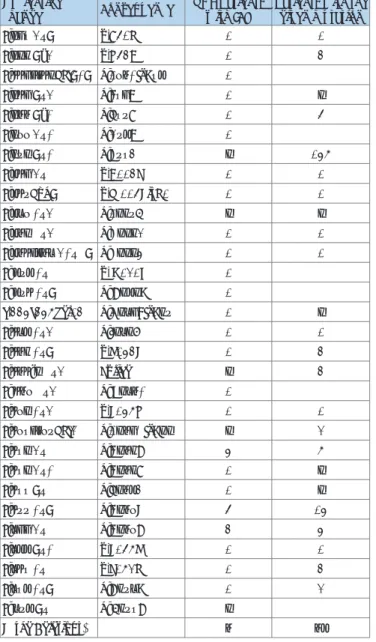
Nucleotide
change Consequence VHL Patients
with ICT Patients with the given mutation
c.208 G>T p.E70X 1 1
c.221 T/G p.V74G 1 4
c.440_442delTCT p.F76CfsX83 1
c.240 T>G p.S80R 1 2
c.266 T/C p.L89P 1 7
c.277 G>C p.G93R 1
c.292 T>C p.Y98H 2 158
c.340 G>A p.G114S 1 1
c.349dupT p.W117LfsX1 1 1
c.357 C>G p.F119L 2 2
c.362 A>G p.D121G 1 1
c.364_365 GC>AT p.A122I 1 1
c. 393 C>A p. N131K 1
c. 394 C>T p. Q132X 1
c.445_458del14 p.N150SfsX19 1 2
c.453 C>G p.I151M 1 1
c.461 C>T p.P154L 1 4
c.464-2 A>G Splice 2 4
c. 467 A>G p. Y156C 1
c.472 C>G p.L158V 1 1
c.478_479delGA p.E160AfsX12 2 3
c.482 G>A p.R161Q 5 8
c.482 G>C p.R161P 1 2
c.488 T>A p.L163H 1 2
c.499 C>T p.R167W 7 15
c.500 G>A p.R167Q 4 5
c.533 T>C p.L178P 1 1
c.548 C>A p.S183X 1 4
c.583 C>T p.Q195X 1 3
c. 593 T>A p. L198Q 2
Large Deletions* 6 63
Table 1: VHL germline mutations in VHL patients with PNETs [16].
* indicates deletions of 1, 2 or 3 exons of the VHL gene.
Variables VHL Sporadic p-value
Age (years) mean 36.03 57.07 <0.001
SD 11.13 11.98
Sex female 34 37 0.010
male 20 55
Tumor Number single 41 64 0.011
multiple 13 5
Tumor Biology benign 39 24 <0.001
malignant 15 68
Pancreatic Tumor as First
Presentation yes 2 92 <0.001
no 52 0
Family History of VHL positive 32 1 <0.001
negative 22 91
Table 2: Comparison between VHL-related and sporadic PNETs [16].
p-values of Chi-square or Fisher test considered to be significant are represented in bold.
Abbreviations: SD for standard deviations, VHL for von Hippel-Lindau disease.
Figure 1 A neuroendocrine tumor of the pancreatic head (arrows), CT scan, early arterial phase.
Figure 2 Two pancreatic neuroendocrine tumors (arrows), CT scan, early arterial phase.
Figure 3 A neuroendocrine tumor of the distal part of the pancreas (tail) (arrows), MRI, early arterial phase.
Figure 4 Very small pancreatic neuroendocrine tumor (arrows), MRI.
Note that the early arterial phase was instrumental to detect this small tumor.
only 2% of those with the mutation p.Y98H.
In total, there were 92 truly sporadic, i.e. mutation-negative PNET patients. Comparing these 92 patients to 54 VHL patients, statistically significant differences were predominance of female gender, multifocal PNETs and lower malignancy rate in VHL compared to sporadic cases [16] (Table 2).
In summary, PNETs are rarely the first presentation of VHL disease. Therefore, molecular genetic testing for germline mutations of the VHL gene is not generally recommended for all patients with PNETs, unless they have multifocal tumors, associated, VHL-specific tumors and/or a family history for VHL disease.
COMPUTERIZED TOMOGRAPHY AND MAGNETIC RESONANCE IMAGING
PNETs can be accurately diagnosed by computerized tomography (CT) and magnetic resonance imaging (MRI) (Figures 1 – 4) [17,18]. MRI is the method of choice because of the absence of radiation and no contrast medium side effects in patients with normal renal function. Imaging in the early arterial phase is essential. It must be performed 1st 20 seconds, 2nd between 20 and 40 seconds and 3rd within 2 minutes after i.v. contrast application. All tumors need to be exactly measured in 2, better in 3 dimensions. Actual and former images have to be compared.
Newly identified tumors may be visible retrospectively in former images due to a small size or lack of contrast uptake. It takes much of the burden from a patient, if the information can be provided that the tumor is not new and the growth rate can be defined.
NUCLEAR MEDICINE IMAGING
Several radiopharmaceuticals are used in the diagnostic of PNETs (Figure 5). The somatostatin receptor (SSTR) ligand
111In-DTPA-octreotide (OctreoScan®) has been extensively used for SSTR scintigraphy and single-photon emission tomography (SPECT; or hybrid SPECT/CT). However, 111In-DTPA-octreotide imaging has been replaced whenever available by positron emission tomography (PET; commonly performed as hybrid PET/CT) using SSTR-ligands. Currently, three DOTA-coupled peptides: DOTATOC (Tyr3-octreotide), DOTATATE (Tyr3- octreotate), and DOTANOC (Nal3-octreotide) have excellent
affinity for SST2 receptors (IC50: 2.5 nM; 0.2 nM; and 1.9 nM respectively). DOTA-NOC also binds specifically to SST3, SST4 and SST5 receptors. Physiological distribution is similar to 111In- DTPA-octreotide. Additionally, tracer uptake is frequently found in the pancreas particularly in the processus uncinatus mimicking a focal tumor uptake.
SSTR PET/CT offers superior image resolution, lesion-to- background contrast and thus overall diagnostic accuracy.
Sufficient SSTR expression as imaged by SSTR imaging is also a prerequisite for possible peptide receptor radionuclide therapy (PRRT) in advanced, inoperable cases using beta-particle emitting SSTR ligands like 90Y- or 177Lu-labelled DOTA-TATE or DOTA- TOC (for a recent review see [26]). In addition, PET scanning with [18F] fluorodopa ([18F]-FDOPA) or [18F] fluorodeoxyglucose (FDG; especially in higher proliferating tumors) may be helpful in some cases. Pancreatic NETs exhibit variable [18F]-FDOPA uptake patterns. The main drawback of the technique is related to the intense and prolonged [18F]-FDOPA uptake by the exocrine pancreas, resulting in a low tumor-to-background uptake ratio. The optimal timing for acquisition and the use of oral premedication with carbidopa, a peripheral aromatic amino acid decarboxylase (AADC) inhibitor, remains to be evaluated.
DIFFERENTIAL DIAGNOSIS
The most relevant differential diagnosis of PNETs in VHL are pancreatic serous cystadenomas. Common in VHL are multiple pancreatic cysts. Cystadenomas consist of micro cystic structures and are related to benign pancreatic cysts. Whenever, seemingly solid lesions of the pancreas have a cystic component, cystadenomas are very likely. In contrast to PNETs, metastases due to VHL associated cystadenomas have not been described.
Therefore, surgical removal is rarely indicated. This is of outstanding importance for lesions which cannot be removed by endoscopic organ sparing techniques.
In VHL, NETs outside the pancreas are extremely rare, but a patient with a carcinoid has been described [19].
SURGERY FOR PNETS IN VHL
Pancreatic surgery should only be performed in specialized centers, and experience with VHL patients is extremely important.
Excellent imaging is the fundamental platform for successful surgery. Indication for surgery depends on the type of pancreatic lesion. Pancreatic cysts and cystadenomas do not need surgical treatment. Two different surgical methods must be considered for solid lesions depending on the size and number of the tumors.
In smaller lesions, where malignancy is very unlikely an organ preserving tumor removal is indicated. In cases of evidence or suspect of malignant PNETs more extended resections should be considered [4,20].
Classical surgery with laparotomy is the method of choice for PNETs of the head, the proximal corpus and the processus uncinatus of the pancreas.
It is important to operate as less invasive as possible.
Especially for small tumors enucleation is the procedure of choice. Intraoperative ultrasonography is excellent to screen for undetected, potential multifocal disease and to plan resections close to the pancreatic duct to lower the risk of pancreatic fistula.
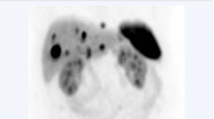
Figure 5 Malignant pancreatic neuroendocrine tumor. The pancreatic tumor was removed months before. The 68Gallium DOTATATE PET shows multiple liver metastases. Normal uptake of the tracer in the spleen and both kidneys.
Partial pancreaticoduodenectomy is the surgical strategy for large tumors. In the classic Whipple´s procedure duodenum, regional lymph nodes, gastric antrum, gallbladder, and distal bile tract are removed together with the resected pancreatic head. The modification by Traveso and Longmire preserves the pylorus and represents the procedure of choice in most oncologic resections. Removal of tumors infiltrating the portal vein is not contraindicated. For reconstruction of the biliary tract, intestinal passage, and drainage of the pancreatic tail a wide variety of safe surgical techniques are available. Postoperative complications include pancreatic fistula, hemorrhage, delayed gastric emptying, diabetes mellitus and malnutrition; they have been widely avoided by modification of the surgical technique, improved postoperative care and interventional endoscopic and radiologic treatment of complications [21].
Minimally invasive surgery is preferred in selected PNETs of the pancreatic tail and the distal pancreatic body. Best candidates are patients with tumors less than 3 cm in diameter and without lymph node metastases. The approach will be laparoscopic or retroperitoneoscopic. Preservation of the spleen should be intended [22]. In a first series 3 patients were operated by the laparoscopic approach. Operating time ranged between 215 – 360 min. Due to infiltrations and/or adhesions of the splenic vein, the spleen could be preserved only in one case. One patient turned out to have a malignant PNET with lymph node metastases [21].
HISTOPATHOLOGY
Immunohistochemical demonstration of the neuroendocrine nature of the tumor is mandatory for the diagnosis of PNET [23]. PNETs are usually positive for the general neuroendocrine markers synaptophysin and chromogranin; additionally immunoreactivity for cytokeratin, insulin, gastrin, glucagon, somatostatin and/or neurospecific enolase (NSE) may be encountered. PNETs may show a trabecular, solid, and/or glandular growth pattern. The cells are often uniform, the finely granulated cytoplasm is usually eosinophilic [24,25]. It is important to emphasize that there is a lack of clearly defined histological criteria for malignancy. Textbooks such as the WHO classification of tumors compare clinical features and histopathological findings, but neither a single morphological criterion nor a combination of criteria, e. g. the “classical” stigmata of malignancy such as cell atypia, mitoses and vascular invasion, can be applied for precise prediction of clinical behaviour. Even the demonstration of tumor infiltration of adjacent tissue is not generally accepted as a hallmark of malignancy. Cellular grading as G2, in contrast to G1, a Ki-67 proliferation index, which is also mandatory for reporting PNETs, of >20% and more than 20 mitoses per 10 high power vision fields are often the basis for reports of malignant neuroendocrine tumors. However, the only proof for malignancy are lymph node or distant metastases [1,24].
TREATMENT OF MALIGNANT PNETS AND METASTASES
Treatment of malignant PNETs is a challenge on its own. In contrast to adenocarcinoma of the pancreas, PNETs are slowly growing tumors, and all activities are justified. The primary goal is complete removal of the tumor and potential metastases. If not
possible, as much tumor tissue as possible should be resected (debulking). After surgical treatment, MRI and nuclear medicine imaging (preferably SSTR PET/CT) should be repeated. In case of advanced, inoperable tumors or tumor remnants, PRRT with 90Y- or 177Lu-labelled DOTA-TOC or DOTA-TATE is the next option.
PRRT is commonly performed in 3 to 6 cycles in 2-3 months intervals [26]. In parallel or after this treatment long acting somatostatin should be started. Systemic treatment on malignant PNET involves multitarget tyrosine kinase inhibitors (semaxanib, sunitinib and vatalanib), thalidomide and interferon alpha-2a which are widely studied to prolong disease stability. Salvage therapy with anti-angiogenesis drugs has also been shown to be of benefit in some patients not suitable for surgery [27-29].
FOLLOW UP STRATEGIES
The method of choice for follow up investigations is MRI with early arterial phase imaging. Patients with PNETs need a strict follow up. This is true for patients with small PNETs so far not requiring surgery and for patients who underwent surgery. Since only about 10% of all VHL patients have a risk for PNETs it is of debate how long follow up intervals should be, if an actual MRI gives no evidence for such tumors. There is no general international agreement, but being aware that PNETs are indolent, slowly growing tumors, intervals of 2 or even 3 years seem adequate.
REFERENCES
1. Klimstra D, Arnold R, Capella C, et al. Neuroendocrine neoplasms of the pancreas. In WHO Classification of Tumours of the Digestive System. Edited by Bosman F, Carneiro F, Hruban RH, Theise N Lyon, France: IARC Press; 2010: 322-326.
2. Jensen R, Jeffrey A. Norton. Endocrine Tumors of the Gastrotintestinal Tract and Pancreas. In Harrison’s Endocrinology. Volume 22. 2 edition. Edited by Jameson J: McGraw-Hill Companies, Columbus, OH;
2010: 348-366.
3. Lonser RR, Glenn GM, Walther M, Chew EY, Libutti SK, Linehan WM, et al. von Hippel-Lindau disease. Lancet. 2003; 361: 2059-2067.
4. Corcos O, Couvelard A, Giraud S, Vullierme MP, Dermot O’Toole, Rebours V, et al. Endocrine pancreatic tumors in von Hippel-Lindau disease: clinical, histological, and genetic features. Pancreas. 2008; 37:
85-93.
5. Hammel PR, Vilgrain V, Terris B, Penfornis A, Sauvanet A, Correas JM, et al. Pancreatic involvement in von Hippel-Lindau disease. The Groupe Francophone d’Etude de la Maladie de von Hippel-Lindau.
Gastroenterology. 2000; 119: 1087-1095.
6. Libutti SK, Choyke PL, Alexander HR, Glenn G, Bartlett DL, Zbar B, et al. Clinical and genetic analysis of patients with pancreatic neuroendocrine tumors associated with von Hippel-Lindau disease.
Surgery. 2000; 128: 1022-1027.
7. Binkovitz LA, Johnson CD, Stephens DH. Islet cell tumors in von Hippel- Lindau disease: increased prevalence and relationship to the multiple endocrine neoplasias. AJR Am J Roentgenol. 1990; 155: 501-505.
8. Yamasaki I, Nishimori I, Ashida S, Kohsaki T, Onishi S, Shuin T. Clinical characteristics of pancreatic neuroendocrine tumors in Japanese patients with von Hippel-Lindau disease. Pancreas. 2006; 33: 382- 385.
9. Chandrasekharappa SC, Guru SC, Manickam P, Olufemi SE, Collins FS,
Emmert-Buck MR, et al. Positional cloning of the gene for multiple endocrine neoplasia-type 1. Science. 1997; 276: 404-407.
10. Crossey PA, Richards FM, Foster K, Green JS, Prowse A, Latif F, et al.
Identification of intragenic mutations in the von Hippel-Lindau disease tumour suppressor gene and correlation with disease phenotype.
Hum Mol Genet. 1994; 3: 1303-1308.
11. Giraud S, Zhang CX, Serova-Sinilnikova O, Wautot V, Salandre J, Buisson N, et al. Germ-line mutation analysis in patients with multiple endocrine neoplasia type 1 and related disorders. Am J Hum Genet.
1998; 63: 455-467.
12. Glavac D, Neumann HP, Wittke C, Jaenig H, Masek O, Streicher T, et al.
Mutations in the VHL tumor suppressor gene and associated lesions in families with von Hippel-Lindau disease from central Europe. Hum Genet. 1996; 98: 271-280.
13. Delman KA, Shapiro SE, Jonasch EW, Lee JE, Curley SA, Evans DB, et al. Abdominal visceral lesions in von Hippel-Lindau disease: incidence and clinical behavior of pancreatic and adrenal lesions at a single center. World J Surg. 2006; 30: 665-669.
14. Zbar B, Kishida T, Chen F, Schmidt L, Maher ER, Richards FM, et al.
Germline mutations in the Von Hippel-Lindau disease (VHL) gene in families from North America, Europe, and Japan. Hum Mutat. 1996;
8: 348-357.
15. Lemmens I, Van de Ven WJ, Kas K, Zhang CX, Giraud S, Wautot V, et al. Identification of the multiple endocrine neoplasia type 1 (MEN1) gene. The European Consortium on MEN1. Hum Mol Genet. 1997; 6:
1177-1183.
16. Erlic Z, Ploeckinger U, Cascon A, Hoffmann MM, von Duecker L, Winter A, et al. Systematic comparison of sporadic and syndromic pancreatic islet cell tumors. Endocr Relat Cancer. 2010; 17: 875-883.
17. Ichikawa T, Peterson MS, Federle MP, Baron RL, Haradome H, Kawamori Y, et al. Islet cell tumor of the pancreas: biphasic CT versus MR imaging in tumor detection. Radiology. 2000; 216: 163-171.
18. Viola KV, Sosa JA. Current advances in the diagnosis and treatment of pancreatic endocrine tumors. Curr Opin Oncol. 2005; 17: 24-27.
19. Kees A. [Malignant carcinoid and phaeochromocytoma in von- Hippel-Lindau’s disease--a case report (author’s transl)]. Wien Klin
Wochenschr. 1980; 92: 218-221.
20. Blansfield JA, Choyke L, Morita SY, Choyke PL, Pingpank JF, Alexander HR, et al. Clinical, genetic and radiographic analysis of 108 patients with von Hippel-Lindau disease (VHL) manifested by pancreatic neuroendocrine neoplasms (PNETs). Surgery. 2007; 142: 814-818.
21. von Dücker L, Walz MK, Voss C, Arnold G, Eng C, Neumann HP.
Laparoscopic organ-sparing resection of von Hippel-Lindau disease- associated pancreatic neuroendocrine tumors. World J Surg. 2011; 35:
563-567.
22. Engelhardt M, Eber SW, Germing U, Heimpel H, Kern W, Schmugge M.
Prävention von Infektionen und Thrombosen nach Splenektomie oder funktioneller Asplenie. 2013.
23. Lam KY, Lo CY. Pancreatic endocrine tumour: a 22-year clinico- pathological experience with morphological, immunohistochemical observation and a review of the literature. Eur J Surg Oncol. 1997; 23:
36-42.
24. Maher E, Nothanson K, Komminoth P, Neumann HP, Plate KH, et al:
Von Hippel-Lindau syndrome (VHL). In World Health Organization Classification of Tumours, Pathology and Genetics of Tumours of Endocrine Organs. Edited by DeLellis R, Lloyd RV, Heitz PU, Eng C.
Lyon: IARC Press; 2004: 230-237.
25. Lubensky IA, Pack S, Ault D, Vortmeyer AO, Libutti SK, Choyke PL, et al.
Multiple neuroendocrine tumors of the pancreas in von Hippel-Lindau disease patients: histopathological and molecular genetic analysis. Am J Pathol. 1998; 153: 223-231.
26. Ambrosini V, Fani M, Fanti S, Forrer F, Maecke HR. Radiopeptide imaging and therapy in Europe. J Nucl Med. 2011; 52: 42S-55S.
27. Raymond E, Dahan L, Raoul JL, Bang YJ, Borbath I, Lombard-Bohas C, et al. Sunitinib malate for the treatment of pancreatic neuroendocrine tumors. N Engl J Med. 2011; 364: 501-513.
28. Capitanio JF, Mazza E, Motta M, Mortini P, Reni M. Mechanisms, indications and results of salvage systemic therapy for sporadic and von Hippel-Lindau related hemangioblastomas of the central nervous system. Crit Rev Oncol Hematol. 2013; 86: 69-84.
29. Burns WR, Edil BH. Neuroendocrine pancreatic tumors: guidelines for management and update. Curr Treat Options Oncol. 2012; 13: 24-34.
Bausch B, von Dobschuetz E, Qi XP, Walz MK, Weryha G, et al. (2014) von Hippel-Lindau Disease Associated Pancreatic Neuroendocrine Tumors – Molecular Genetics and Clinical Aspects. J Transl Med Epidemiol 2(1): 1019.
Cite this article