Author Query Form
Journal Title : JCN
Article Number : 494476
Dear Author/Editor,
Greetings, and thank you for publishing with SAGE Publications. Your article has been copyedited, and we have a few queries for you. Please address these queries when you send your proof corrections to the production editor. Thank you for your time and effort.
Please assist us by clarifying the following queries:
No. Query Remarks
1 Please check that all authors are listed in the proper order; clarify which part of each author’s name is his or her surname; and verify that all author names are correctly spelled/
punctuated and are presented in a manner consistent with any prior publications: Anna Gajda, MD, Emese Horva´th, MD, Tibor Hortoba´gyi, MD, PhD, Gyurgyinka Gergev, MA, Hajnalka Szabo´, MD, PhD, Katalin Farkas, BSc, Nikoletta Nagy, MD, PhD, Ma´rta Sze´ll, PhD, DSc, and La´szlo´ Sztriha, MD, PhD, DSc
2 Please verify that the existing conflict of interest statement is accurate and correct: ‘‘The authors declared no potential conflicts of interest with respect to the research, authorship, and/or publication of this article.’’
3 Please verify that the existing funding disclosure is accurate and correct: ‘‘The authors disclosed receipt of the following financial support for the research, authorship, and/or publication of this article: Supported by TA´ MOP-4.2.2.A-11/1/KONV-2012-0035 grant and TA´ MOP-4.2.2/B-10/1/KONV 2010-0012 grant.’’
Nemaline Myopathy Type 2 (NEM2):
Two Novel Mutations in the Nebulin (NEB) Gene
Anna Gajda, MD
1, Emese Horva´th, MD
2, Tibor Hortoba´gyi, MD, PhD
3, Gyurgyinka Gergev, MA
1,4, Hajnalka Szabo´, MD, PhD
1, Katalin Farkas, BSc
5, Nikoletta
AQ 1
Nagy, MD, PhD
2,5, Ma´rta Sze´ll, PhD, DSc
2,5, and La´szlo´ Sztriha, MD, PhD, DSc
1Abstract
Nemaline myopathy is a type of the heterogeneous group of congenital myopathies. Generalized hypotonia, weakness, and delayed motor development are the main clinical features of the typical congenital form. Histopathology shows characteristic nemaline rods in the muscle biopsy. Mutations in at least 7 genes, including nebulin gene (NEB), proved to be responsible for this muscle disease. We present a boy with nemaline myopathy type 2 (NEM2) caused by compound heterozygosity for 2 novel mutations, a deletion and a duplication in theNEBgene. The deletion was inherited from the father and the duplication from the mother. Testing all family members supports genetic counseling.
Keywords
congenital myopathy, hypotonia, nebulin
Received April 9, 2013. Received revised April 26, 2013. Accepted for publication May 28, 2013.
Congenital myopathies are a group of genetic muscle disorders with clinical, pathologic, and molecular heterogeneity.1Nema- line myopathy is a pathologically well-defined subgroup of congenital myopathies characterized by the presence of rodlike structures in the muscle fibers.2The disease presents a wide range of clinical variability and severity, although generalized hypotonia, muscle weakness, feeding difficulties, and delay of motor milestones are almost always present.2All molecularly characterized forms of nemaline myopathy are autosomal, but inheritance can be recessive or dominant, and singleton cases could arise from de novo dominant mutations.2,3 To date, 7 genes have been linked to this condition: a-tropomyosin (TPM3), nebulin (NEB), skeletal musclea-actin (ACTA1),b- tropomyosin (TPM2), muscle troponin T1 (TNNT1), cofilin-2 (CFL2) and the recently identified KBTBD13 gene, whose function is still unknown.2-4
Disease resulting from mutations in theNEBgene is classi- fied as nemaline myopathy type 2 (NEM2; OMIM 256030), the most common form of nemaline myopathy.2NEM2 is a rare autosomal recessive condition characterized by early-onset muscle weakness, which is most pronounced in the axial mus- cles and proximal limb-girdles. Weakness in facial and bulbar muscles commonly results in dysarthria.5The course of the disease is nonprogressive or slowly progressive and life expec- tancy depends on the severity of respiratory muscle weakness.
Mechanical ventilation is necessary for some patients and orthopedic complications are frequent.6Mental development and intelligence are typically within the normal range.6In this report, we present the clinical and histologic findings in a patient suffering from NEM2 and describe compound hetero- zygosity for 2 novel mutations in theNEBgene.
Case Report Clinical Presentation
The patient, a boy, was born at term from a fourth uneventful pregnancy. Birth weight was 3450 g and Apgar scores were 9 and 10 at 5 and 10 minutes, respectively. Floppiness and weak
1Department of Pediatrics, University of Szeged, Szeged, Hungary
2Department of Medical Genetics, University of Szeged, Szeged, Hungary
3Department of Neuropathology, University of Debrecen, Debrecen, Hungary
42nd Department of Pediatrics, Semmelweis University, Budapest, Hungary
5Dermatological Research Group of the Hungarian Academy of Sciences, University of Szeged, Szeged, Hungary
Corresponding Author:
Anna Gajda, MD, Department of Pediatrics, University of Szeged, 14-15 Kor- a´nyi fasor, H-6720 Szeged, Hungary.
Email: annagajd@gmail.com
00(0) 1-4
ªThe Author(s) 2013 Reprints and permission:
sagepub.com/journalsPermissions.nav DOI: 10.1177/0883073813494476 jcn.sagepub.com
cry were noticed in the neonatal period. Generalized hypotonia, weakness, and delayed acquisition of motor milestones were observed in early childhood. He began to walk with support at the age of 2 years. Deep tendon reflexes were reduced. There was no fasciculation in the tongue. He suffered also from recur- rent respiratory tract infections. The serum creatine kinase activity was normal. The optic fundus and brain magnetic reso- nance imaging (MRI) were also normal. Tandem mass spectro- metry and urine gas chromatography screening showed no evidence of inborn error of metabolism. Nerve conduction velocity was normal, and electromyography did not show any abnormalities. There was no homozygous deletion in exons 7 and 8 of theSMN1 (Survival of Motor Neuron 1) gene. The possibility of a congenital myopathy emerged and muscle biopsy was performed at the age of 4 years. Severe respiratory tract infection and pneumonia led to respiratory failure, later requiring tracheostomy and mechanical ventilation. His condi- tion subsequently improved; however, he needs ventilatory support during sleep. Cardiologic examination did not show any abnormalities. The patient’s cognitive abilities were appro- priate for his age.
The patient’s parents and his 3 siblings (a brother and 2 sisters) are asymptomatic.
Informed consent was requested from the parents prior to the muscle biopsy and molecular genetic studies.
Muscle Biopsy
The histologic workup of the muscle biopsy tissue followed standard procedures. The majority of the muscle fibers were hypoplastic or atrophic with large variation in fiber size. Fiber necrosis, regeneration, phagocytosis, or inflammatory cell infi- ltration were not noted. Gomori’s trichrome technique detected prominent red-stained inclusion bodies in the fibers with varia- tion in number and distribution. Gomori trichrome stained
sections showed rod-shaped particles in the fibers (Figure 1A). Electron microscopy revealed that the rods appeared as electron-dense structures localized mainly along the thickened Z-lines (Figure 1B). These rod-shaped particles were identified as nemaline rods/bodies, and these findings were consistent with the diagnosis of nemaline myopathy.
Genetics
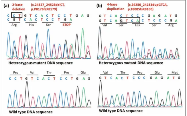
In view of the morphologic findings, a search was initiated in collaboration with the commercial diagnostic company Cento- gene GmbH (Rostock, Germany) to find the molecular genetic etiology of nemaline myopathy in this child. Tests forACTA1 andTPM3 were negative. Eventual testing of the NEB gene revealed 2 previously unreported heterozygous mutations: a deletion (c.24527_24528delCT p.P8176fsX8179; Figure 2A) in exon 174 and a duplication (c.24250_24253dupGTCA p.T8085fsX8100; Figure 2B) in exon 171. These mutations result in a frameshift and a premature termination codon, respectively, presumably leading to truncated nebulin protein.
Further testing of the parents revealed that the father carries the deletion and the mother has the heterozygous duplication (Figure 3). One of the 2 sisters of the patient carries wild- type alleles, whereas another sister and the brother are hetero- zygous for the duplication (Figure 3). Thus, the clinically unaffected family members carry either wild-type alleles or only 1 of the mutant alleles; either the duplication or the dele- tion. The patient, however, is a compound heterozygous carrier of both mutations.
Discussion
Nemaline myopathies are a clinically and molecularly hetero- geneous group of congenital myopathies.1-3 The combination of characteristic clinical and histopathologic features are Figure 1.Nemaline myopathy. (A) Gomori trichrome staining of plastic-embedded semithin sections shows numerous rod structures in the muscle fibers. (B) Electron microscopy reveals that the rods are electron-dense structures.
2 Journal of Child Neurology 00(0)
diagnostic for the disorder in most cases.1-3The presence of red inclusions detected with Gomori trichrome staining and of rod- shaped particles in toluidine blue–stained tissue from the patient strongly suggest nemaline myopathy.1-3Ultrastructural studies reveal nemaline bodies as electron-dense, rod-shaped structures appearing as thickened Z-disks.1-3Muscle imaging by magnetic resonance can be helpful to visualize the pattern of selective muscle involvement and guide in localizing the site of the biopsy.7,8
The workup of a case with nemaline myopathy is further complicated by its heterogeneous genetic background: 7 known causative genes have been linked to this condition and both autosomal dominant and recessive inheritance has been obser- ved.2,9Six of these genes encode proteins associated with sar- comeric thin filaments.2Recessive mutations in theNEBgene, located on chromosome 2q22-23, are the most commonly recognized cause of the disease.2,4This gene has 182 exons, and missense, nonsense, and frameshift mutations have been reported.10 Hotspots have not been found and many patients proved to be compound heterozygotes for 2 mutations within the gene.2,10The nebulin protein has a wide range of functions, including thin filament length specification and regulation of muscle contraction.4
The clinical, histologic, and molecular genetic findings in our patient are consistent with the typical congenital form of nemaline myopathy type 2, caused by mutations in theNEB gene.2 Compound heterozygosity for 2 novel mutations was Figure 2.Identification of 2 novel mutations (1 deletion and 1 duplication) in theNEBgene. Direct sequencing of DNA isolated from the affected patient (II/4) revealed (A) a 2-base deletion (c.24527_24528delCT) in exon 174 of theNEBgene resulting in a premature termination codon (p.P8176fsX8179) in the translated sequence and (B) a 4-base duplication (c.24250_24253dupGTCA) in exon 171 of theNEBgene, result- ing in frameshift (p.T8085fsX8100) in the translated sequence.
Figure 3.Genetic screening of the affected family. The patient (II/4) and his clinically unaffected father (I/1) carry the deletion in heterozy- gous form, whereas the other unaffected family members (I/2, II/1, II/2, II/3) carry the wild-type allele. The patient (II/4) and his clinically unaffected mother (I/2), brother (II/2), and sister (II/3) carry the dupli- cation in heterozygous form, whereas the other unaffected family members (I/1, II/1) carry the wild-type allele. These results suggest that the deletion is of paternal origin and the duplication is of maternal origin. The patient (II/4) is, thus, a compound heterozygote for these 2 novel mutations.
found. A 2-base deletion (c.24527_24528delCT, p.P8176fsX81- 79) was inherited from the father and a 4-base duplication (c.24250_24253dupGTCA, p.T8085fsX8100) from the mother.
These novel mutations led to a translational frameshift and a pre- mature termination codon in the respective translated sequences and thus, presumably, to truncated nebulin protein.
Nemaline myopathy is a debilitating condition and further research is warranted in order to explore the details of the mole- cular pathology of this disorder. These efforts are complicated by the heterogenic molecular background of the disease and the fact that certain genes encode very large proteins like nebulin.
Molecular diagnosis, however, becomes available for more and more patients supporting preimplantation or prenatal diagnosis for subsequent pregnancies.2 This case report extends the genetic profile of nemaline myopathy with 2 previously unre- ported mutations in theNEBgene.
Acknowledgments
The authors are grateful for Centogene GmbH (Rostock, Germany) for performing the mutation analysis of the ACTA1, TPM3, and NEB genes.
Author Contributions
AG and EH contributed to data collection and the first draft of the manuscript. TH performed the evaluation of the muscle biopsy. GG and HS cared for the patient and KF and NN carried out the mutation analysis of the parents and family members. MS and LS were mentors who contributed equally to this work. AG and EH also contributed equally to this work.
Declaration of Conflicting Interests
The authors declared no potential conflicts of interest with respect to the research, authorship, and/or publication of this article.
AQ 2
Funding
The authors disclosed receipt of the following financial support for the research, authorship, and/or publication of this article: Supported by
TA´ MOP-4.2.2.A-11/1/KONV-2012-0035 grant and TA´MOP-4.2.2/
B-10/1/KONV 2010-0012 grant. AQ 3
Ethical Approval
The writing/research of this article did not require ethical approval.
References
1. Nance JR, Dowling JJ, Gibbs EM, Bo¨nnemann JJ. Congenital myopathies: an update. Curr Neurol Neurosci Rep. 2012;12:
165-174.
2. Wallgren-Pettersson C, Sewry CA, Nowak KJ, Laing NG. Nema- line myopathies.Semin Pediatr Neurol. 2011;18:230-238.
3. North KN. Clinical approach to the diagnosis of congenital myo- pathies.Semin Pediatr Neurol. 2011;18:216-220.
4. Labeit S, Ottenheijm CA, Granzier H. Nebulin, a major player in muscle health and disease.FASEB J. 2011;25:822-829.
5. Wallgren-Pettersson C.Congenital nemaline myopathy. A clinical follow-up of twelve patients.J Neurol Sci. 1989;89:1-14.
6. Wallgren-Pettersson C, Laing NG. Report of the 70th ENMC International Workshop: nemaline myopathy, 11-13 June 1999, Naarden, The Netherlands. Neuromuscul Disord. 2000;10:
299-306.
7. Fischer D, Herasse M, Ferreiro A, et al. Muscle imaging in domi- nant core myopathies linked or unlinked to the ryanodine receptor 1 gene.Neurology. 2006;67:2217-2220.
8. Mercuri E, Pichiecchio A, Allsop J, et al. Muscle MRI in inherited neuromuscular disorders: past, present, and future.J Magn Reson Imaging. 2007;25:433-440.
9. Wallgren-Pettersson C, Pelin K, Hilpela P, et al. Clinical and genetic heterogeneity in autosomal recessive nemaline myopathy.
Neuromuscul Disord. 1999;9:564-572.
10. Wallgren-Pettersson C, Donner K, Sewry C, et al. Mutations in the nebulin gene can cause severe congenital nemaline myopathy.
Neuromuscul Disord. 2002;12:674-679.
4 Journal of Child Neurology 00(0)
