1
Nephrogenic Diabetes Insipidus
András Balla1,2, László Hunyady1,2
1 Department of Physiology, Semmelweis University, Faculty of Medicine, Budapest, Hungary
2 MTA-SE Laboratory of Molecular Physiology, Hungarian Academy of Sciences and Semmelweis University, Budapest, Hungary
Correspondence author: László Hunyady
PO Box 259, H-1444 Budapest, Hungary, hunyady.laszlo@med.semmelweis-univ.hu
Short title: Nephrogenic Diabetes Insipidus
Key words: AQP2 gene, AVPR2 gene, diabetes insipidus, G protein–coupled receptor (GPCR), nephrogenic diabetes insipidus, NDI, signal transduction, type 2 vasopressin receptor (V2R)
Conflicts of interest:
The authors declare that there is no conflict of interest.
Acknowledgements:
This work was supported by the Hungarian National Research, Development and Innovation Fund (NKFI K116954 and NVKP_16-1-2016-0039).
Abbreviations:
2
ADH, antidiuretic hormone; AQP, aquaporin water channel; AVP, vasopressin; cAMP, 3',5'- cyclic adenosine monophosphate; cNDI, congenital forms of NDI; CREB, cAMP response element-binding protein; ddAVP, desmopressin (1-deamino-8-D-arginine vasopressin); DI, diabetes insipidus; ENaC, epithelial sodium channel; GPCR, G protein–coupled receptor; NaPi- 2, type 2 sodium-phosphate cotransporter; NBC1, sodium-bicarbonate cotransporter; NDI, nephrogenic diabetes insipidus; NCC, Na-Cl cotransporter; NKCC, Na+-K+-2Cl- cotransporter;
NSIAD, nephrogenic syndrome of inappropriate diuresis; PGE2, prostaglandin E2; PKA, protein kinase A; ROMK, renal outer medullary potassium channel; SIADH, syndrome of inappropriate antidiuretic hormone; V2R, type 2 vasopressin receptor;
Abstract:
The body fluid homeostasis is essential for the normal life. In the maintenance of water balance, the most important factor and regulated process is the excretory function of the kidneys. The kidneys are capable to compensate not only the daily fluctuations of water intake but also the consequences of fluid loss (respiration, perspiration, sweating, hemorrhage). The final volume and osmolality of the excreted urine is set in the collecting duct via hormonal regulation. The hormone of water conservation is the vasopressin (AVP) and large volume of urine is produced and excreted in absence of AVP secretion or if AVP is ineffective in the kidneys. The aquaporin-2 water channel (AQP2) is expressed in the principal cells and it plays essential role in the reabsorption of water in the collecting ducts via type 2 vasopressin receptor (V2R) mediated mechanism. If neural or hormonal regulation fails to operate the normal function of AVP-V2R-AQP2 system, it can result in various diseases such as diabetes insipidus (DI) ornephrogenic syndrome of inappropriate diuresis (NSIAD). The DI is characterized by
3
excessive production of hyposmotic urine (“insipidus” means tasteless) due to the inability of the kidneys to concentrate urine. In this chapter, we focus and discuss the pathophysiology of nephrogenic DI (NDI) and the potential therapeutic interventions in the light of the current experimental data.
Introduction:
The maintenance of constant volume and composition of the body fluids requires that the daily water intake and water loss are matched and tightly regulated. The balance between the water input and output is mainly regulated through the kidneys, although water intake is also controlled by thirst. The urine formation by the kidneys is the key regulatory factor in the maintenance of water and electrolyte homeostasis of the human body. The kidneys respond rapidly and appropriately to body fluid osmolality and volume fluctuations, however the kidneys are able to fulfill these homeostatic tasks only under hormonal and neural regulatory mechanisms. The glomerular filtration by the kidneys produce 120 ml of fluid in a minute and the tubular system allows great variations in the rate of final fluid excretion. The volume of urine production can be greatly varied in antidiuresis (water deprivation) and in water diuresis (after excessive fluid load). Normally, about 1% of the filtered water is finally excreted as urine;
the average urine output is ~0.5-1.5 ml/minute. Some segments of the tubular system have permanent high water permeability because certain types of aquaporin water channels (AQP) reside constitutively on their apical and basolateral membranes. In contrary, other segments are virtually water impermeable due to the lack of aquaporin water channels in the plasma membrane compartments. In healthy humans approximately 85% of the filtered fluid is reabsorbed in the proximal tubule and in the loop of Henle, independently on the water homeostatic status of the body. Approximately 8-14.5% of the glomerular filtered fluid is reabsorbed in the collecting ducts and it is under tight hormonal control via vasopressin
4
(arginine-vasopressin in humans, AVP; also called as antidiuretic hormone, ADH) and atrial natriuretic peptide (ANP) actions (Robertson 2001; Saito 2010). The AVP is synthesized as prohormone which is later cleaved to nonapeptide AVP in the supraoptic and paraventricular nuclei of the hypothalamus (Fliers et al. 1985; Sklar and Schrier 1983). The produced AVP is transported by axonal transport via the supraopticohypophyseal into the neurohypophysis and secreted from here in response to stimulation. A very sensitive mechanism allows that even less than 1% increase in osmolality stimulates osmoreceptors in the hypothalamus, which in turn induce the release of AVP into the systemic circulation. The secreted AVP circulates in the blood and primarily acts in the kidneys via V2R. In addition to the AVP production and secretion, higher increase in extracellular osmolality leads to thirst perception, which drives drinking behavior helping to restore the normal osmolality and volume of the fluid compartments in the body. The vascular effects (vasoconstriction and total peripheral resistance increase) of AVP due to type 1 vasopressin receptor (V1R) activation are manifested only at greatly elevated AVP levels. This huge increase in AVP production and secretion can be seen only in response to baroreceptor activation reflex when blood volume changes reach at least 5- 10%.
The collecting tubules are divided into subsegments such as cortical, outer medullary, and inner medullary collecting ducts but in all subsegments the AVP regulates the reabsorption of water. (The water permeability of the tight junctions between the epithelial cells of the collecting tubule is negligible). The overall action of the AVP is the water conservation and formation of hyperosmotic urine. During antidiuresis, the AVP makes the luminal membrane of the principal cells in the collecting ducts water-permeable and water is passively reabsorbed via AQP channels due to the existing osmotic gradient between the tubular fluid and the hyperosmotic interstitium. The effect of AVP is induced by the binding to and activation of the basolaterally located V2R in the collecting ducts which is followed by consequent signaling
5
transduction steps leading to the translocation of AQP2 from endomembranes to the apical plasma membrane (Moeller et al. 2013). Under normal circumstances, the AQP2 is inserted in the luminal membrane of the principal cells as homotetrameric water channel.
The AVP also regulates the urea permeability of the principal cells since its effect is required for the normal expression and function of urea transporters in the inner medullary collecting duct, enhancing the urea recycling and via this mechanism contributes to the buildup of corticopapillary osmotic gradient. This osmotic gradient is important drive for the water reabsorption in the collecting tubule. It is important to note that the AVP on top of the above mentioned effects in the collecting ducts, it also increases the activity of the Na+-K+-2Cl- cotransporter (NKCC) in the thick ascending limb of the loop of Henle, hence augmenting the corticopapillary osmotic gradient (Fenton and Knepper 2007).. Moreover, the AVP increases sodium reabsorption in the collecting duct by increasing the activity of the amiloride-sensitive epithelial sodium channel (ENaC) (Bankir et al. 2005).
The constant physiological regulation greatly varies the rate of water reabsorption in the collecting ducts and finally 0.5-10% of the filtered water can be excreted. The rate of urine production is mainly regulated by water reabsorption through the aquaporin-2 channels (AQP2) presence and thus the water permeability in the collecting duct principal cells. The water permeability of the collecting ducts is AVP dependent and is under constant regulation. The huge variation of the water reabsorption in the collecting duct is the consequence of specific chain of events which modify the water permeability, the AQP2 presence in the luminal (apical) membrane of the collecting tubule principal cells. Under maximal AVP influence only ~0.3 ml fluid per minute is excreted, however in lack of AVP induced concentrating function of the kidneys it can raised as much as ~12 ml urine production per minute. The osmolality of urine changes reciprocally with the volume of urine output; the osmolality can be as high as ~1300 mosm/kg water in maximal antidiuretic condition and as low as ~30 mosm/kg water in maximal
6
diuresis in humans. After restoration of water balance, due to the termination of AVP secretion from the neurohypophysis and the elimination of circulating AVP molecules by kidney and liver vasopressinases, the AVP level decreases in a short time (10-30 min) (Czaczkes et al.
1964). Consequently, the V2R induced mechanisms in principal cells diminish resulting the relocation of AQP2 water channels into endomembrane compartments which in turn greatly reduces the water permeability and reabsorption capacity of the collecting duct. Under normal conditions, the presence of AQP2 water channels in the luminal surface of the principal cells is the permissive factor for the water reabsorption.
Impaired regulatory mechanisms in the AVP action or genetic defects in the participating proteins can lead to disturbed renal function and diseases such as diabetes insipidus (DI) or nephrogenic syndrome of inappropriate diuresis (NSIAD) depending on that loss-of-function or gain-of-function mutations are behind the restricted or exaggerated transport functions. Investigations of such disorders have contributed enormously to our understanding of the mechanisms of urinary concentration and identified the important proteins and revealed potential therapeutic interventions to cure or unburden the symptoms.
DI is a rare disease, the prevalence is approximately one per 30,000 and characterized by the inability to normally concentrate urine resulting in several symptoms such as polyuria (40-150 ml/kg/day, depending on the age), hyposthenuria (low specific gravity of urine, <290 mOsm/L), and compensatory polydipsia (Robertson 1995). This disease burdens normal life due to production and micturition of large volumes of urine all day long. Moreover, in severe forms of the disorder, the individuals must micturate and drink fluid even at night a few times.
The DI is frequently associated with urological complications such as impaired bladder function, hydronephrosis, chronic renal failure, and large dilations of the urinary tract (Shalev et al. 2004; Boyd et al. 1980). The patients can be held in water balance as long as the fluid intake compensates the excessive urine output. The thirst sensation and its regulation is essential
7
safety backup mechanism since the compensatory water intake reflexes can avoid the development of severe hyperosmolality in the blood plasma. Unfortunately, if the water intake is limited, the patient becomes dehydrated and hypovolumic by reason of excessive water loss.
This chapter gives an overview of the various DI types focusing to the NDI, including the pathophysiology and potential therapeutic interventions in such disorders.
Nephrogenic diabetes insipidus (NDI)
In the NDI disorder, albeit the AVP production and its secretion is normally regulated by the hyperosmotic stimuli, the principal cells in the collecting ducts do not respond normally to the hormone. The irresponsiveness can be caused by several factors such as genetic defects (familial or congenital nephrogenic diabetes insipidus due to mutations either in the AVPR2 or AQP2 genes) or can be acquired i.e. from drug therapies. In adults, the acquired forms are more
common, whereas children mostly present congenital forms of NDI (cNDI). In this chapter, we summarize the causes and the pathophysiological consequences of NDI, furthermore, we discuss the new treatment possibilities that have been proposed by the current research data in the literature to medicate or disburden the symptoms of NDI. The cited OMIM entries can be found at www.omim.org (Amberger and Hamosh 2017).
Types of NDI
Congenital forms of NDI (cNDI)
The hereditary NDI syndromes most commonly due to the mutations either in the AVPR2 or in the AQP2 gene (primary NDIs). Approximately 90% of the NDI cases are related
to the malfunction of the V2R and its signaling, and ~10% is due to the autosomal mutations in the AQP2 gene. Interestingly, the dysfunction of V2R or AQP2 cause indistinguishable clinical symptoms which are manifested immediately after birth. In top of the mutations in the AVPR2
8
or AQP2 gene, other genetic renal diseases can be in the background of the symptoms (secondary NDIs) (Bockenhauer and Bichet 2013). Most patients suffering from cNDI are diagnosed within the 3 years of life, with excessive vomiting, reduced growth, underweight, constipation, fever and dehydration.
Disease causing mutations of V2 vasopressin receptor (OMIM 304800)
The V2R belongs to the G protein–coupled receptor (GPCR) superfamily, composed by 371 amino acids that forms seven transmembrane domains. In the principal cells, the V2R acts mostly via coupling to Gs proteins thus regulating the intracellular 3',5'-cyclic adenosine monophosphate (cAMP) level in target cells. Members of the GPCR superfamily are the largest group of cell membrane receptors and their mutations are responsible for numerous human diseases given the fact that more than 600 loss of function mutations of GPCR’s have been identified (Schoneberg et al. 2004), including the majority of congenital NDI cases (Bichet 2009). The mutations in the V2R can cause failure in urine concentration due to several mechanisms including the inability to establish the corticopapillary osmotic gradient and the impaired water transport through collecting duct principal cells but the most palpable dysfunction that defective V2R function in the principal cells induces hyposmotic polyuria production. Under normal circumstances in healthy individuals, the AVP stimulation induced V2R activation triggers cAMP signaling cascades, most importantly the activation of protein kinase A (PKA) which in turn phosphorylates the AQP2 on several residues that triggers the vesicular transport of AQP2 containing endomembranes into the apical membrane allowing water reabsorption in the collecting ducts (Figure 1A) (Nielsen et al. 2002). Once the AVP secretion is diminished in response to the restored water balance, AQP2 abundance is also reduced due to its internalization through ubiquitin-mediated endocytosis and transport to storage vesicles and degradation (Kamsteeg et al. 2006). This regulation of AQP2 water channel
9
localization is crucial to increase urine osmolality and to reduce urine output in humans (Moeller et al. 2013). In addition to the classical PKA activation pathway, it is important to emphasize that the increased intracellular cAMP directly binds and activates other effector signaling protein such as Epac (exchange factor directly activated by cAMP) which functions as guanine-nucleotide exchange factor for the small GTPase. It was demonstrated that the AQP2 abundance on the luminal surface of the principal cells mainly depends on the Epac activation and was independent on the PKA activity (Kortenoeven et al. 2012). Interestingly, during the aforementioned signaling processes, the AVP-bound V2R active receptor conformation is phosphorylated by GRK molecules which leads to recognition and association with β-arrestin molecules and the receptor is desensitized (although the AVP is still bound to the receptor).
The arrestin recruitment initiates several important regulatory mechanisms. First, the arrestin molecules as scaffold proteins to organize the endocytosis of the V2R which results in the decreased cell surface receptor density and the degradation of the receptor in the lysosomes (the AVP is bound so tightly that it can be dissociated from the receptor only upon break-down of the receptor molecule). The β-arrestin mediated receptor down-regulation prevents from exaggerated reabsorption in the collecting ducts and allows the possibility to regulate smoothly and rapidly the rate of water reabsorption.
AVP induced V2R activation is necessary not just for the normal AQP2 trafficking pattern but also required for the normal AQP2 expression levels. It was demonstrated that although the intracellular cAMP level decays shortly after AVP stimulation, the AQP2 expression remains increased for days (DiGiovanni et al. 1994; Kortenoeven et al. 2012).
Interestingly, the upregulatory effect of AVP on the AQP2 expression level was dependent on extracellular signal-regulated kinase (ERK), Epac activation and cAMP response element- binding protein (CREB) but not on PKA activity (Umenishi et al. 2006; Kortenoeven et al.
2012). In addition to the above mentioned effects, the V2R normally contributes to the buildup
10
of corticopapillary interstitial osmotic gradient as its function is required for the normal expression and function of urea transporters in the collecting duct and Na+-K+-2Cl- cotransporter in the thick ascending limb of the loop of Henle (Fenton and Knepper 2007).
Moreover, the AVP increases sodium reabsorption in the collecting duct by increasing the activity of the amiloride-sensitive epithelial sodium channel (ENaC) (Bankir et al. 2005).
Several mutations have been identified causing congenital nephrogenic diabetes insipidus (cNDI) (Barak et al. 2001; Tao 2006; Wilbanks et al. 2002). The cNDI is a rare disease and it is caused due to the mutations in either AVPR2 or AQP2 gene. Among the cNDI cases approximately 90% of the patients are males with the X-linked recessive mutation in the AVPR2 gene resulting defective V2R in the kidney epithelial cells (Bichet 2009). The prevalence of this form (also called as type I cNDI or as XNDI) is ~1 in 150,000 male births although mild forms of X-linked cNDI can be present in females, as well. The AVPR2 gene is located on chromosome region Xq28, which contains three exons and two introns and after shortly its cloning it was proposed that it could be responsible for congenital NDI (Seibold et al. 1993;
Rosenthal et al. 1992; Birnbaumer et al. 1992). It was also demonstrated in early works that the patients with cNDI does not respond to desmopressin (dDAVP) treatment suggesting that the cAMP signaling is defective in these cases (Bichet et al. 1989; Bichet et al. 1988).
As it was mentioned above, few cNDI families were described in the literature, in which the female family members exhibit variable degrees but usually mild symptoms of diabetes insipidus (Carroll et al. 2006; van Lieburg et al. 1999; Nomura et al. 1997). These heterozygous female individuals possess both normal and mutated AVPR2 alleles. The studies on the skewed X-chromosome inactivation patterns of the female members suggested that the severity of symptoms were dependent on the rate of skewed methylation of X chromosome, and the NDI phenotype is caused by dominant methylation of the normal allele of AVPR2 gene (Nomura et al. 1997; Arthus et al. 2000).
11
The NDI causing various mutations in V2R evolve their dysfunctions with different mechanisms (Arthus et al. 2000; Tao 2006). More than 260 mutations are described in the Human Gene Mutation Database (http://www.hgmd.cf.ac.uk/ac/gene.php?gene=AVPR2), majority of them are missense mutations (~61%), which act by different mechanisms (Spanakis et al. 2008). In addition, approximately 25% of the mutations are frameshift mutations due to nucleotide deletions or insertions, ~10% is non-sense mutations, and ~9% of the cases are large deletions, but infrequently complex rearrangements, splicing mutations, in-frame deletions or insertions are also responsible for the X-linked NDI disorders (Bichet and Bockenhauer 2016).
The V2R is composed by 371 amino acids that forms several domains and mutations have been identified all over in the receptor but the most of the mutations are found in the transmembrane domains. The most frequent mutations are recurrent mutations which have been identified in 35 ancestrally independent families and mostly these occurred at potential mutational hot spots (a C-to-T or G-to-A nucleotide substitution occurred at a CpG dinucleotide) (Bichet and Bockenhauer 2016).
The mutation also can be classed by the nature of the impaired vasopressin receptor function. The classical classification of the mutations causing impaired V2R function is based on the pathophysiological and cellular consequences (Wesche et al. 2012). The AVP insensitivity can be the consequence of either decreased number of cell surface receptors or impaired receptor signal transduction (Figure 1B). Class I mutations of the AVPR2 gene can lead to impaired transcription, mRNA processing or translation of the receptor resulting truncated and usually rapidly degraded proteins. Taken together, the class I mutations prevent effective synthesis of the V2R. Class II mutations lead to the formation of misfolded full length proteins, which are recognized by the quality control system of the endoplasmic reticulum (ER), resulting defective intracellular trafficking of the V2R mutants, ER retention and often degradation of the trapped receptors (Robben et al. 2006). In fact, the majority of AVPR2 gene
12
mutations are missense/nonsense mutations which result class II mutant receptors. Class III mutants are another group of missense mutations, which reach the basolateral plasma membrane but they display reduced G protein binding and/or G protein activation ability in response to AVP stimulation (class IIIa subtype) or their AVP binding is defective due to their impaired ligand affinity (class IIIb subtype), leading to inappropriate intracellular cAMP production and AQP2 trafficking. Class IV mutants have normal ligand binding but their intracellular trafficking is altered causing impaired cAMP signal production mostly due to constitutive β-arrestin dependent internalization even in absence of ligands leading to decreased basolateral surface expression since the V2R accumulation in intracellular vesicles (Barak et al. 2001).
To complicate the picture further, the evaluation of the properties of a mutant V2R does not always lead to an unambiguous classification of the mutation. For example, the NDI causing R137H-V2R was classified as class IV mutation due to its constitutive β-arrestin dependent internalization (Barak et al. 2001). Moreover, the R137H-V2R has also impaired G-protein coupling (Rosenthal et al. 1993) and altered trafficking to the plasma membrane as well (Kocan et al. 2009). Interestingly, contrary to the symptoms of R137H, the R137C-V2R and R137L- V2R mutations lead to nephrogenic syndrome of inappropriate dieresis (NSIAD) due to the constitutive activity.
Gain-of function V2R mutations cause nephrogenic syndrome of inappropriate diuresis (NSIAD) (OMIM 300539)
Whereas the loss-of-function V2R mutations are responsible for the majority of the cNDI disorders, the gain-of-function mutations in the V2R lead to nephrogenic syndrome of inappropriate antidiuresis disease (NSIAD). This recently discovered syndrome may cause hyponatremia and especially in infants severe clinical symptoms such as convulsions. NSIAD
13
is a very rare, X-linked disorder with monogenic inheritance, and to date, mutations of Phe229, Arg137, Ile130 and Leu312 have been identified as gain-of-function mutations in V2R (Carpentier et al. 2012; Erdelyi et al. 2015; Feldman et al. 2005; Tiulpakov et al. 2016). The clinical manifestation of NSIAD such as hyponatremia and reduced urinary dilution is undistinguishable from the SIADH but very markedly the serum AVP level is low in NSIAD whereas it is elevated in SIADH (Decaux et al. 2007). It was demonstrated that NSIAD causing gain-of-function mutation can lead to selective G-protein activation and increased secondary messenger production in the cells. The increased activity of V2R can be blocked by an V2R inverse agonist tolvaptan which could be a possible treatment option for the patients with this type V2R mutation (Erdelyi et al. 2015).
Congenital NDIs due to mutated AQP2 (OMIM 125800)
In the other main form of cNDI which corresponds approximately ~10% of the inherited NDI cases, the patients suffers from autosomal mutation in the gene encoding the AQP2 water channel (Bichet 2009). The patients exhibit typical features of NDI similarly to V2R loss-in- function mutations caused symptoms, the disorder is vasopressin resistant, but it is associated with nonfunctional water channels. This form of hereditary NDI is also called as type II form of cNDI, and the characteristic difference from the type I form is that the intracellular cAMP level increases in response to AVP stimulation (Zimmerman and Green 1975; Deen et al. 1994;
Langley et al. 1991). The autosomal cNDI can be caused by heterozygous, homozygous, or compound heterozygous mutation in the AQP2 gene, and both autosomal-recessive or autosomal-dominant mode of inheritance are described (Morello and Bichet 2001; van Lieburg et al. 1999; Deen et al. 1994). The chromosomal location of AQP2 is 12q12-q13 and to date 61 mutations have been identified (http://www.hgmd.cf.ac.uk/ac/gene.php?gene=AQP2) (Bichet and Bockenhauer 2016). The majority of the mutations (48) are missense/nonsense mutations,
14
but few splicing, small deletion and insertion mutations are also presented in the databases. The consequences of AQP2 gene mutations can be different depending the nature of the mutation but most commonly result in impaired vesicular transport of AQP2 water channels. Under such condition the AQP2 molecules are retained in the endosomes and not translocated into the apical (luminal) surface of the principal cells (Tamarappoo and Verkman 1998). As it was discussed in case of the misfolded and misrouted V2R mutant proteins, theoretically, it is possible to intervene by the help of pharmacological chaperones which could facilitate the reverse of the intracellular retention of AQP2 mutant proteins. Most of the mutations are autosomal recessive mutations leading to misfolding, ER retention, and degradation of the produced AQP2 mutants (Moeller et al. 2013; Wesche et al. 2012; Frick et al. 2014). Although, the majority of the dominant mutations result in complete lack of functionality, some ER-retention mutants, in which the misfolding is not severe, display reduced degradation and partial responsiveness to AVP stimulation (Canfield et al. 1997; Frick et al. 2014; Marr et al. 2001; Marr et al. 2002;
Mulders et al. 1997). Interestingly, these dominant mutations cause less severe phenotype compared to patients with compound heterozygous or homozygous for recessive mutations (Mulders et al. 1998; Bichet and Bockenhauer 2016). The dominant mutations affect the carboxyl terminus of AQP2, their folding is correct and they are not retained in the ER, although due to the mutations they are not able to cooperate fully with the wild type AQP2 molecules. It was demonstrated that such mutants form mixed tetramer complex with the wild type AQP2 but they are misrouted and retained in the Golgi compartment (Mulders et al. 1998; de Mattia et al. 2004; Kamsteeg et al. 1999).
“Complex” NDI
The AVPR2 or AQP2 gene mutations caused symptoms are also called as “pure” NDI phenotypes since they display water loss but the electrolytes such as sodium, potassium,
15
chloride and calcium are normally conserved (Bockenhauer and Bichet 2017). The “complex”
NDI can be caused by mutant membrane proteins involved in sodium chloride reabsorption in the thick ascending limb of the loop of Henle leading to not just loss of water but also electrolyte loss. Such mutations can be found in the genes encoding the Na+-K+-2Cl- cotransporter, renal outer medullary potassium channel (ROMK), Barttin (Bartter syndrome, infantile, with sensorineural deafness), kidney-specific chloride channel ClC-Kb and chloride channel Ka causing polyuria, polydipsia and loss of sodium, chloride, calcium, magnesium and potassium (Bockenhauer and Bichet 2017).
Acquired forms of NDI
The majority of the NDI cases are acquired diseases caused be various factors including complications of drug treatments, pathophysiological alterations such as electrolyte disturbances including hypokalemia and hypercalcemia, and diseases (sickle cell disease, sarcoidosis, amyloidosis, multiple myeloma, Sjoergen's disease). Numerous data have been reported that NDI can be induced by various other drugs and medicine including lithium, demeclocycline, foscarnet, clozapine, cisplatin or methoxyflurane (Bendz and Aurell 1999).
Consumption and uptake of trace amounts of lithium is essential for the normal brain, immune and reproductive functions, and it is well known that lithium deficiency leads to serious health problems, including behavioral problems. The lithium treatment is still the most effective cure in bipolar disorders and has been used for decades despite the disadvantages such as renal side effects. The lithium caused DI is a serious, well-studied side effect in the treatment of bipolar mood disorders leading to polyuria (and consequent dehydration, thirst, and polydipsia), elevated renal sodium excretion and even chronic kidney disease (Nielsen et al. 2008). The lithium treatment induces vasopressin-resistant polyuria in 20-40% of the patients mostly due to the disruption of normal aquaporin function and increased sodium excretion as the result of
16
epithelial sodium channel (ENaC) dysfunction in the kidney collecting ducts (Marples et al.
1995). The renal lithium handling is similar of the sodium due to their similar size and charge.
The lithium enters the distal tubule segments through the amiloride-sensitive ENaC in the distal tubule segments and leaves the cells via Na+/H+ exchanger, NHE1 to the interstitium. It is demonstrated that clinically relevant lithium concentrations reduced the mRNA transcription and protein expression of AQP2 which can be reversed by administration of amiloride, moreover the lithium treatment disrupted the normal trafficking and distribution pattern of AQP2 (Kortenoeven et al. 2009; Li et al. 2006). In addition to the reduced AQP2 level in the apical surface the transcellular water transport is greatly diminished by the fact that lithium decreases the expression of the basolaterally located AQP3 level too in the collecting duct principal cells (Kwon et al. 2000). Chronic lithium treatment has been reported to inhibit the sodium transport through the amiloride-sensitive ENaC resulting in increased sodium excretion (Thomsen et al. 1999). Furthermore, it has been demonstrated that other sodium transport functions are also affected in response to lithium treatment. The distal convoluted tubule located thiazide-sensitive Na-Cl cotransporter (NCC), the proximal tubule located the type 2 sodium- phosphate cotransporter (NaPi-2) and electrogenic sodium-bicarbonate cotransporter (NBC1) are down-regulated during lithium treatment(Kwon et al. 2000). The lithium also reduces the water reabsorption ability by affecting the urea cycling that normally contributes to the buildup of corticopapillary interstitial osmotic gradient. It has been demonstrated that chronic lithium treatment induces decreased urea transporter abundance (Klein et al. 2002). Taken together, on top of the dysfunctional AQP2, sodium wasting due to the impaired sodium cotransporter factions and due to the reduced urine concentrating ability due to the urea transporter deficiency also play important role in the development of polyuria during chronic lithium treatment. The symptoms of the lithium-induced side effects can be alleviated by administration of thiazide and amiloride (Bedford et al. 2008a), angiotensin-converting enzyme inhibitors, AT1R
17
angiotensin II receptor blockers, or spironolactone mineralocorticoid receptor blocker (Kwon et al. 2009).
Demeclocycline is tetracycline derivative antibiotic, indicated for the treatment of various types of bacterial infections. It has been demonstrated that chronic demeclocycline treatment has several side effects including set up of NDI (Singer and Rotenberg 1973). It was revealed that demeclocycline-induced NDI is the consequence of the decreased AQP2 expression and its cell surface abundance in response to the treatment (Kortenoeven et al. 2013).
Interestingly, demeclocycline reduced the adenylate cyclase activity in kidney cells and its effect was independent on V2R signaling. The demeclocycline is not really used in the treatment of infections nowadays, but its renal side effects were utilized to alleviate the symptoms of syndrome of inappropriate antidiuretic hormone (SIADH). In SIADH the neurohypophysis secretes abnormally high levels of AVP, which in turn results in excessive water reabsorption in the collecting ducts. The too high level of water retention causes volume expansion in the fluid compartments whereas the osmolality of the extracellular and intracellular fluids become decreased compared with the normal levels. Moreover, the high level of AVP increases the Na+-reabsorption through NKCC cotransporter and epithelial sodium channel (ENaC) (Ecelbarger et al. 2001). The use of demeclocycline in SIADH is based on the fact that the drug reduces the AVP-evoked hyponatremia, although the development of V2 vasopressin receptor antagonists, the vaptans, supersedes its usage in the treatment of SIADH (Sherlock and Thompson 2010).
Treatment possibilities for NDI
In contrary to central DI where the desmopressin is widely and successfully used to mend the symptoms, the treatment of NDI patients is more difficult to achieve. In the past, several potential treatment possibilities of DI were described (reviewed recently by (Kalra et
18
al. 2016)) such as the usage of carbamazepine (Rado 1976; Meinders et al. 1974), clorpropamide (Wales and Fraser 1971), thiazide diuretics (Alon and Chan 1985; O'Doherty et al. 1962), amiloride (Bedford et al. 2008b; Kortenoeven et al. 2009), indapamide (Kocak et al.
1990), clofibrate (Moses et al. 1973), indomethacin (Weinstock and Moses 1990). The treatment of cNDI was tuned during the years. The combination of amiloride and hydrochlorothiazide was found superior than the combination of hydrochlorothiazide and a prostaglandin synthetase inhibitor (Alon and Chan 1985). The inhibitors of prostaglandin synthesis are also useful medicines to ameliorate the symptoms of cNDI, and among the tested inhibitors, the indomethacin was much more effective than ibuprofen (Libber et al. 1986). In addition to these drugs, the recent findings have suggested novel therapeutic possibilities.
The identification of the NDI causing mutations and the determination of the altered properties and cellular consequences altered could help to define or suggest therapeutic strategies for the treatment of NDI patients. The potential therapeutic approaches deal with either bypassing the V2R signal transduction processes or the improve of AQP2 function. It is proposed and highly recommended to identify the potential genetic defect as soon as possible in case of chance of congenital NDI. Both AVPR2 and AQP2 genes can be sequenced with ease due to their relatively small size. The genetic testing of infants whose families may be associated with congenital NDI can be performed even prenatal or perinatal periods from amniotic cells, chorionic villous samples or cord blood (Bichet and Bockenhauer 2016). Since the inherited NDI is manifested immediately after birth, it is fundamental to correctly recognize the disorder, and especially in case of infants, it is essential to treat the affected individuals with abundant fluid intake in order to avoid the repeated episodes of dehydration since these episodes are probably responsible for the generation of mental and physical retardation.
Possible interventions include direct stimulation of cAMP signal generation in the principal cells, bypassing the necessity of vasopressin activation, and different strategies to
19
rescue the impaired receptor function (Wesche et al. 2012). The most extensively investigated V2R mutants belong to the class II mutations, which cause ER retention. The ER has a quality- control system that prevents misfolded proteins to reach their final subcellular destinations (Ellgaard and Helenius 2003). Pharmacological chaperons or pharmacochaperons are small, cell permeable substances that meliorate the folding of misfolded mutated, otherwise functional proteins in the ER. By the result of pharmacochaperon action the misfolded proteins can bypass the ER quality-control system and rescue them from ER retention. This phenomenon proposed a therapeutic approach in which the chaperons can lead to increased plasma membrane expression of otherwise functional vasopressin receptor mutants. Numerous data have been presented the usage of pharmacological chaperons to mend the dysfunctional V2R mutants.
Pharmacological chaperones of the V2R can be antagonists (Bernier et al. 2004; Morello et al.
2000; Ranadive et al. 2009; Robben et al. 2007; Wuller et al. 2004) or agonists (Jean-Alphonse et al. 2009; Robben et al. 2009). The approach was also confirmed in clinical studies of X- linked NDI patients who suffer from ER retention V2R mutations. Administration of SR49059 V1a receptor nonpeptide antagonist significantly reduced the urine volume and water intake, and increased the urine osmolarity suggesting the pharmacochaperons may offer a new therapeutic approach to the treatment of the NDI patients who suffer from ER retention mutant V2R mutations. (Bernier et al. 2006).
The altered mutant V2 receptor conformations can result decreased potency for AVP and other ligands such as dDAVP. Theoretically the potency of other agonists can be affected differently and certain agonists may activate the mutant receptor despite of the conformational change. Screening and testing drug libraries can yield ligands which can be personal therapy in case of a certain mutation. In case of N321K-V2R mutation, which causeda conformation with altered agonist sensitivity, the mutant receptor can be stimulated and activated by dVDAVP proposing a therapeutic for patients with N321K mutation in the V2R (Erdelyi et al. 2014).
20
Taken together, the promising recent and future experimental data may provide personalized medicine that is specific for the individual patients with a certain mutations causing NDI.
It is also promising that some interventions may bypass the V2R activation in order to generate cAMP signal for the PKA mediated APQ2 translocation, thus urinary concentration can be independent of V2R. In the collecting duct, locally produced autocrine and paracrine factors including bradykinin, ATP, adenosine, endothelin, nitric oxide, and prostaglandin E2 (PGE2) may also regulate the water permeability (Pearce et al. 2015; Sands and Layton 2014).
It was demonstrated thatPGE2 receptor EP4 activation promotes AQP2 translocation via cAMP signal and PKA activation, moreover it also induces AQP2 expression in a cAMP response element-binding protein (CREB)-dependent manner (Gao et al. 2015). This finding makes the EP4 receptor agonists as potential therapeutic drugs for the treatment of DI, irrespectively of the type of the DI. It was also demonstrated that the agonist stimulation (pro)renin receptor, which is also expressed in the collecting duct and it is regulated by the PGE2 receptor EP4, induced AQP2 expression, whereas its inhibition partially prevented the decrease in urine volume and the increase in urine osmolality and AQP2 expression induced water deprivation in in vivo animal studies (Wang et al. 2016). It was discovered recently adenosine monophosphate kinase (AMPK) is capable to phosphorylate both AQP2 and urea transporter UT-A1in rat inner medullary collecting ducts in response to metformin stimulation (Klein et al. 2016). The authors used metformin as AMPK activator, which is approved to use in patients with type 2 diabetes mellitus and polycystic ovary syndrome. The metformin increased the apical membrane expression of AQP2, both osmotic water permeability and urea permeability in collecting ducts. Erlotinib, which is a receptor tyrosine kinase inhibitor selective for epidermal growth factor receptor (EGFR), is a medication in the treatment of some cancer including non-small cell lung cancer, pancreatic cancer. It is currently identified in a high–
throughput chemical screening assay that erlotinib was effective to stimulate AQP2
21
translocation and reduced urine output 45% in lithium-induced NDI model in cAMP- independent manner (Cheung et al. 2016). Since animal studies suggest metformin and erlotinib as novel therapeutic options for NDI and given the fact that both are approved medications, it was suggested to initiate clinical trials to determine the benefits of these drugs in the treatment of various forms of diabetes insipidus (Bockenhauer and Bichet 2017; Bech et al. 2018). It was demonstrated that activation of cGMP-mediated signaling pathway could result in AQP2 translocation and administration of cGMP phosphodiesterase type 5 inhibitor sildenafil significantly increased the AQP2 insertion into the luminal membrane compartments. Sildenafil induced elevated intracellular cGMP levels and the plasma membrane accumulation of AQP2 was not dependent on V2R activation (Bouley et al. 2005). Other possibility to bypass the V2R induced AQP2 translocation is the stimulation of β3 adrenergic receptor. The β3 adrenergic receptor is also expressed in the collecting duct and it was demonstrated that its stimulation with the selective agonist BRL37344 induced cAMP signal and translocation of AQP2 in the principal cells, and also increased the activity of Na+-K+-2Cl- cotransporter in the thick ascending limb of the loop of Henle. Taken together, administration of BRL37344 promoted a potent antidiuretic effect in AVPR2-deficient animals (Procino et al. 2016). Since the mirabegron, a β3 adrenergic receptor is already introduced in the clinical practice to treat overactive bladder, enticing possibility to carry out clinical investigations to test the potential ameliorating effects of this drug in NDI patients (Cernecka et al. 2014; Chapple et al. 2014).
The other possible therapeutic option can be the administration of simvastatin, a lipid-lowering medication since it was recently demonstrated that it was able to reduce urine output and increase urine osmolality in hypercholesterolemic patients (Procino et al. 2016). A recent clinical study investigated the effects of sildenafil, metformin, and simvastatin on AVP- independent urine concentration in healthy volunteers, and only simvastatin had an effect on urine osmolality in healthy volunteers, although further studies are necessary to address the
22
possible effects of these drugs in combination or in patients suffering from NDI (Bech et al.
2018).
Other major types of and differential diagnosis of diabetes insipidus
Early diagnosis and correct typing of the DI is critical, especially in infants and small children since the consecutive, repetitive dehydration episodes can lead to mental and physical retardation (Bichet 2009). The symptoms of untreated DI eventually may lead to death due to exsiccosis, electrolyte imbalances, hyperosmolality, and low blood volume caused circulatory failure. The clinical diagnosis of the DI is based on functional tests. The widely used tests are the water deprivation test and administration of the dDAVP (desmopressin) to distinguish between the various forms of DI (Di Iorgi et al. 2012).
The DI can be inherited (less than 10% of the cases) or acquired (Fujiwara and Bichet 2005). The symptoms of DI can be the consequence of four fundamentally different reasons which are used for the classification the various forms of DI (Fenske and Allolio 2012). In the clinical practice, it is essential to diagnose the type of the syndrome for the proper treatment of the individual, especially in less severe forms of the symptoms (Robertson 1988).
The central DI (CDI) (OMIM 125700) is due to the impaired AVP production or release from the central nervous system and can be acquired or congenitaland the syndromes are the consequence of deficient release of AVP from the neurohypophysis (Arima et al. 2016). The prevalence of CDI is about 1:25000, and in children, CDI is more frequent than the NDI mostly due to brain injuries. The CDI can be treated by using an analogue of vasopressin, the V2R selective agonist desmopressin (1-deamino-8-D-arginine vasopressin, ddAVP) that has several benefits over administration of AVP due to the lack of vascular side effects and longer half-life.
The desmopressin is now available not only as nasal spray but also as oral tablet (even in sublingual form) to ease the way of administration. Interestingly, dissimilarly to CDI, in the
23
syndrome of inappropriate antidiuretic hormone (SIADH) the neurohypophysis secretes abnormally high levels of AVP, which in turn results in excessive water reabsorption in the collecting ducts. The too high level of water retention causes volume expansion in the fluid compartments and hyponatremia whereas the osmolality of the extracellular and intracellular fluids become decreased compared with the normal levels (Wakil et al. 2011).
The gestational DI is a rare complication, the prevalence is approximately one per 30,000 pregnancies (Fenske and Allolio 2012). This form of the DI is the consequence of the rapid elimination (metabolism) of the circulating AVP by the placental vasopressinase, also called placental cysteine aminopeptidase, secreted by the trophoblasts. In rare occasions, the disorder can be due to exaggeration of pre-existing mild forms of CDI or NDI. The symptoms usually manifest in the third trimester of pregnancy, especially carrying multiple fetuses (Ichaliotis and Lambrinopoulos 1965). The rapid elimination of the circulating AVP causes transient DI and usually can be treated by the administration of desmopressin which is resistant to vasopressinase cleveage (Ananthakrishnan 2016). The transient GDI is usually resolved after delivery (within few weeks), although in subsequent pregnancies can cause even severe symptoms (Kalelioglu et al. 2007).
The dipsogenic or psychogenic DI can be caused by primary polydipsia which is excessive water intake due to impaired thirst perception (dipsogenic DI) or psychological function (psychogenic DI). Primary polydipsia can be induced by either defective thirst sensation or perception and frequently is associated with schizophrenia, anxiety disorder and depression, although an increasing number of cases demonstrate primary polydipsia in non- psychiatric patients sometimes induced by drugs causing sensation of dry mouth (Sailer et al.
2017).
Genetic counselling
24
Early detection and providing abundant water supply or treatment for DI patients is essential to allow normal mental and physical development, normal lifespan (Bichet and Bockenhauer 2016). In case of infants and small children and it is highly recommended to sequence of their DNAs to obtain correct diagnosis if hypernatremic dehydration episodes occur (Bockenhauer and Bichet 2017). Since both AVPR2 and AQP2 are small genes, their sequence can be easily sequenced, allowing not just perinatal and prenatal testing but the genetic testing of the carriers, as well (Bichet and Bockenhauer 2016).
Conclusion
Diabetes insipidus is a rare disorder with diversity of backgrounds which impairments affect either the V2 vasopressin receptor or the AQP2 water channel functions. It is essential to keep the patients in water balance either by the treatment of the disease (i.e. in case of central DI) or by providing necessary amount of fluid intake. It is also pivotal to identify the nature of the disorder as early as possible to reveal the cause behind the disorder and to treat the patient appropriately. Although numerous potential new treatment possibilities proposed currently, more translational research is necessary to offer personalized medicine for the patients who suffer from distinct mutations leading to congenital forms of NDI.
25
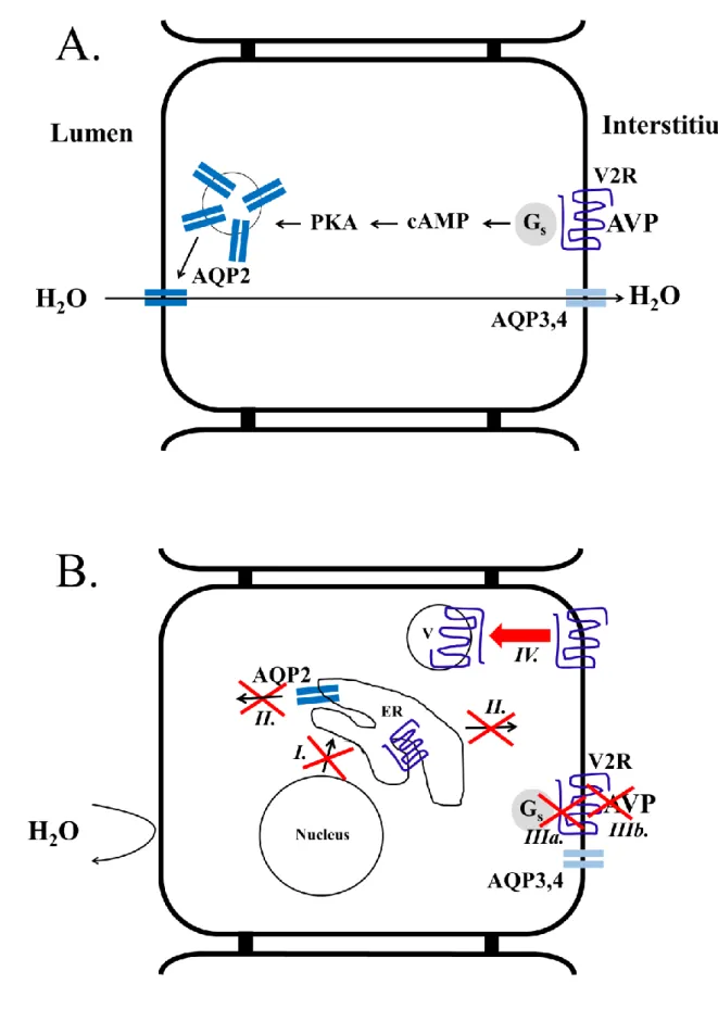
Figure 1: Schematic illustration of AVP-induced AQP2 insertion into the apical surface of collecting duct principal cells (A) and the possible causes behind the development of
26
NDI (B). Under normal circumstances (A), the AVP travels via bloodstream to the kidneys where it binds to the basolaterally located V2R. This receptor is a G protein–coupled receptor and initiates cAMP signaling within the principal cells. The activation of PKA induces the vesicular trafficking of AQP2 containing endomembranes. Finally, the AQP2 is translocated into the apical surface making this water permeable, and the water molecules are transported from the duct lumen to the kidney interstitium via the apically located AQP2 and the permanently located basolateral AQP3 and AQP4 water channels. (B) In cNDI, the impaired water transport is the consequence of mutations in the AVPR2 or AQP2 (labeled with roman numerals). Class I type of mutations can lead to impaired transcription, mRNA processing or translation, class II mutations lead to endoplasmic reticulum (ER) retention, class IIIa mutants display reduced G protein binding and/or G protein activation whereas class IIIb mutations cause impaired ligand affinity, class IV mutants display altered intracellular trafficking leading to receptor accumulation in intracellular vesicles.
References:
Alon U, Chan JC (1985) Hydrochlorothiazide-amiloride in the treatment of congenital nephrogenic diabetes insipidus. Am J Nephrol 5 (1):9-13. doi:10.1159/000166896
Amberger JS, Hamosh A (2017) Searching Online Mendelian Inheritance in Man (OMIM): A Knowledgebase of Human Genes and Genetic Phenotypes. Curr Protoc Bioinformatics 58:1 2 1-1 2 12. doi:10.1002/cpbi.27
Ananthakrishnan S (2016) Diabetes insipidus during pregnancy. Best Pract Res Clin Endocrinol Metab 30 (2):305-315. doi:10.1016/j.beem.2016.02.005
Arima H, Azuma Y, Morishita Y, Hagiwara D (2016) Central diabetes insipidus. Nagoya J Med Sci 78 (4):349-358. doi:10.18999/nagjms.78.4.349
Arthus MF, Lonergan M, Crumley MJ, Naumova AK, Morin D, De Marco LA, Kaplan BS, Robertson GL, Sasaki S, Morgan K, Bichet DG, Fujiwara TM (2000) Report of 33 novel AVPR2 mutations and analysis of 117 families with X-linked nephrogenic diabetes insipidus. J Am Soc Nephrol 11 (6):1044-1054
Bankir L, Fernandes S, Bardoux P, Bouby N, Bichet DG (2005) Vasopressin-V2 receptor stimulation reduces sodium excretion in healthy humans. J Am Soc Nephrol 16 (7):1920-1928.
doi:10.1681/ASN.2004121079
Barak LS, Oakley RH, Laporte SA, Caron MG (2001) Constitutive arrestin-mediated desensitization of a human vasopressin receptor mutant associated with nephrogenic diabetes insipidus. Proc Natl Acad Sci U S A 98 (1):93-98. doi:10.1073/pnas.011303698
27
Bech AP, Wetzels JFM, Nijenhuis T (2018) Effects of sildenafil, metformin, and simvastatin on ADH- independent urine concentration in healthy volunteers. Physiol Rep 6 (7):e13665.
doi:10.14814/phy2.13665
Bedford JJ, Leader JP, Jing R, Walker LJ, Klein JD, Sands JM, Walker RJ (2008a) Amiloride restores renal medullary osmolytes in lithium-induced nephrogenic diabetes insipidus. American journal of physiology Renal physiology 294 (4):F812-820. doi:10.1152/ajprenal.00554.2007
Bedford JJ, Weggery S, Ellis G, McDonald FJ, Joyce PR, Leader JP, Walker RJ (2008b) Lithium-induced nephrogenic diabetes insipidus: renal effects of amiloride. Clin J Am Soc Nephrol 3 (5):1324- 1331. doi:10.2215/CJN.01640408
Bendz H, Aurell M (1999) Drug-induced diabetes insipidus: incidence, prevention and management.
Drug Saf 21 (6):449-456. doi:10.2165/00002018-199921060-00002
Bernier V, Lagace M, Lonergan M, Arthus MF, Bichet DG, Bouvier M (2004) Functional rescue of the constitutively internalized V2 vasopressin receptor mutant R137H by the pharmacological chaperone action of SR49059. Molecular endocrinology 18 (8):2074-2084.
doi:10.1210/me.2004-0080
Bernier V, Morello JP, Zarruk A, Debrand N, Salahpour A, Lonergan M, Arthus MF, Laperriere A, Brouard R, Bouvier M, Bichet DG (2006) Pharmacologic chaperones as a potential treatment for X- linked nephrogenic diabetes insipidus. J Am Soc Nephrol 17 (1):232-243.
doi:10.1681/ASN.2005080854
Bichet DG (2009) V2R mutations and nephrogenic diabetes insipidus. Prog Mol Biol Transl Sci 89:15- 29. doi:10.1016/S1877-1173(09)89002-9
Bichet DG, Bockenhauer D (2016) Genetic forms of nephrogenic diabetes insipidus (NDI): Vasopressin receptor defect (X-linked) and aquaporin defect (autosomal recessive and dominant). Best Pract Res Clin Endocrinol Metab 30 (2):263-276. doi:10.1016/j.beem.2016.02.010
Bichet DG, Razi M, Arthus MF, Lonergan M, Tittley P, Smiley RK, Rock G, Hirsch DJ (1989) Epinephrine and dDAVP administration in patients with congenital nephrogenic diabetes insipidus.
Evidence for a pre-cyclic AMP V2 receptor defective mechanism. Kidney Int 36 (5):859-866 Bichet DG, Razi M, Lonergan M, Arthus MF, Papukna V, Kortas C, Barjon JN (1988) Hemodynamic and
coagulation responses to 1-desamino[8-D-arginine] vasopressin in patients with congenital nephrogenic diabetes insipidus. The New England journal of medicine 318 (14):881-887.
doi:10.1056/NEJM198804073181403
Birnbaumer M, Seibold A, Gilbert S, Ishido M, Barberis C, Antaramian A, Brabet P, Rosenthal W (1992) Molecular cloning of the receptor for human antidiuretic hormone. Nature 357 (6376):333- 335. doi:10.1038/357333a0
Bockenhauer D, Bichet DG (2013) Inherited secondary nephrogenic diabetes insipidus: concentrating on humans. American journal of physiology Renal physiology 304 (8):F1037-1042.
doi:10.1152/ajprenal.00639.2012
Bockenhauer D, Bichet DG (2017) Nephrogenic diabetes insipidus. Curr Opin Pediatr 29 (2):199-205.
doi:10.1097/MOP.0000000000000473
Bouley R, Pastor-Soler N, Cohen O, McLaughlin M, Breton S, Brown D (2005) Stimulation of AQP2 membrane insertion in renal epithelial cells in vitro and in vivo by the cGMP phosphodiesterase inhibitor sildenafil citrate (Viagra). American journal of physiology Renal physiology 288 (6):F1103-1112. doi:10.1152/ajprenal.00337.2004
Boyd SD, Raz S, Ehrlich RM (1980) Diabetes insipidus and nonobstructive dilation of urinary tract.
Urology 16 (3):266-269
Canfield MC, Tamarappoo BK, Moses AM, Verkman AS, Holtzman EJ (1997) Identification and characterization of aquaporin-2 water channel mutations causing nephrogenic diabetes insipidus with partial vasopressin response. Hum Mol Genet 6 (11):1865-1871
Carpentier E, Greenbaum LA, Rochdi D, Abrol R, Goddard WA, 3rd, Bichet DG, Bouvier M (2012) Identification and characterization of an activating F229V substitution in the V2 vasopressin receptor in an infant with NSIAD. J Am Soc Nephrol 23 (10):1635-1640.
doi:10.1681/ASN.2012010077
28
Carroll P, Al-Mojalli H, Al-Abbad A, Al-Hassoun I, Al-Hamed M, Al-Amr R, Butt AI, Meyer BF (2006) Novel mutations underlying nephrogenic diabetes insipidus in Arab families. Genet Med 8 (7):443- 447. doi:10.109701.gim.0000223554.46981.7a
Cernecka H, Sand C, Michel MC (2014) The odd sibling: features of beta3-adrenoceptor pharmacology.
Molecular pharmacology 86 (5):479-484. doi:10.1124/mol.114.092817
Chapple CR, Cardozo L, Nitti VW, Siddiqui E, Michel MC (2014) Mirabegron in overactive bladder: a review of efficacy, safety, and tolerability. Neurourol Urodyn 33 (1):17-30.
doi:10.1002/nau.22505
Cheung PW, Nomura N, Nair AV, Pathomthongtaweechai N, Ueberdiek L, Lu HA, Brown D, Bouley R (2016) EGF Receptor Inhibition by Erlotinib Increases Aquaporin 2-Mediated Renal Water Reabsorption. J Am Soc Nephrol 27 (10):3105-3116. doi:10.1681/ASN.2015080903
Czaczkes JW, Kleeman CR, Koenig M (1964) Physiologic Studies of Antidiuretic Hormone by Its Direct Measurement in Human Plasma. The Journal of clinical investigation 43:1625-1640.
doi:10.1172/JCI105038
de Mattia F, Savelkoul PJ, Bichet DG, Kamsteeg EJ, Konings IB, Marr N, Arthus MF, Lonergan M, van Os CH, van der Sluijs P, Robertson G, Deen PM (2004) A novel mechanism in recessive nephrogenic diabetes insipidus: wild-type aquaporin-2 rescues the apical membrane expression of intracellularly retained AQP2-P262L. Hum Mol Genet 13 (24):3045-3056.
doi:10.1093/hmg/ddh339
Decaux G, Vandergheynst F, Bouko Y, Parma J, Vassart G, Vilain C (2007) Nephrogenic syndrome of inappropriate antidiuresis in adults: high phenotypic variability in men and women from a large pedigree. J Am Soc Nephrol 18 (2):606-612. doi:10.1681/ASN.2006090987
Deen PM, Verdijk MA, Knoers NV, Wieringa B, Monnens LA, van Os CH, van Oost BA (1994) Requirement of human renal water channel aquaporin-2 for vasopressin-dependent concentration of urine. Science 264 (5155):92-95
Di Iorgi N, Napoli F, Allegri AE, Olivieri I, Bertelli E, Gallizia A, Rossi A, Maghnie M (2012) Diabetes insipidus--diagnosis and management. Horm Res Paediatr 77 (2):69-84.
doi:10.1159/000336333
DiGiovanni SR, Nielsen S, Christensen EI, Knepper MA (1994) Regulation of collecting duct water channel expression by vasopressin in Brattleboro rat. Proc Natl Acad Sci U S A 91 (19):8984- 8988
Ecelbarger CA, Kim GH, Wade JB, Knepper MA (2001) Regulation of the abundance of renal sodium transporters and channels by vasopressin. Exp Neurol 171 (2):227-234.
doi:10.1006/exnr.2001.7775
Ellgaard L, Helenius A (2003) Quality control in the endoplasmic reticulum. Nat Rev Mol Cell Biol 4 (3):181-191. doi:10.1038/nrm1052
Erdelyi LS, Balla A, Patocs A, Toth M, Varnai P, Hunyady L (2014) Altered agonist sensitivity of a mutant v2 receptor suggests a novel therapeutic strategy for nephrogenic diabetes insipidus.
Molecular endocrinology 28 (5):634-643. doi:10.1210/me.2013-1424
Erdelyi LS, Mann WA, Morris-Rosendahl DJ, Gross U, Nagel M, Varnai P, Balla A, Hunyady L (2015) Mutation in the V2 vasopressin receptor gene, AVPR2, causes nephrogenic syndrome of inappropriate diuresis. Kidney Int 88 (5):1070-1078. doi:10.1038/ki.2015.181
Feldman BJ, Rosenthal SM, Vargas GA, Fenwick RG, Huang EA, Matsuda-Abedini M, Lustig RH, Mathias RS, Portale AA, Miller WL, Gitelman SE (2005) Nephrogenic syndrome of inappropriate antidiuresis. The New England journal of medicine 352 (18):1884-1890.
doi:10.1056/NEJMoa042743
Fenske W, Allolio B (2012) Clinical review: Current state and future perspectives in the diagnosis of diabetes insipidus: a clinical review. The Journal of clinical endocrinology and metabolism 97 (10):3426-3437. doi:10.1210/jc.2012-1981
Fenton RA, Knepper MA (2007) Mouse models and the urinary concentrating mechanism in the new millennium. Physiol Rev 87 (4):1083-1112. doi:10.1152/physrev.00053.2006
29
Fliers E, Swaab DF, Pool CW, Verwer RW (1985) The vasopressin and oxytocin neurons in the human supraoptic and paraventricular nucleus; changes with aging and in senile dementia. Brain Res 342 (1):45-53
Frick A, Eriksson UK, de Mattia F, Oberg F, Hedfalk K, Neutze R, de Grip WJ, Deen PM, Tornroth- Horsefield S (2014) X-ray structure of human aquaporin 2 and its implications for nephrogenic diabetes insipidus and trafficking. Proc Natl Acad Sci U S A 111 (17):6305-6310.
doi:10.1073/pnas.1321406111
Fujiwara TM, Bichet DG (2005) Molecular biology of hereditary diabetes insipidus. J Am Soc Nephrol 16 (10):2836-2846. doi:10.1681/ASN.2005040371
Gao M, Cao R, Du S, Jia X, Zheng S, Huang S, Han Q, Liu J, Zhang X, Miao Y, Kang J, Gustafsson JA, Guan Y (2015) Disruption of prostaglandin E2 receptor EP4 impairs urinary concentration via decreasing aquaporin 2 in renal collecting ducts. Proc Natl Acad Sci U S A 112 (27):8397-8402.
doi:10.1073/pnas.1509565112
Ichaliotis SD, Lambrinopoulos TC (1965) Serum Oxytocinase in Twin Pregnancy. Obstet Gynecol 25:270- 272
Jean-Alphonse F, Perkovska S, Frantz MC, Durroux T, Mejean C, Morin D, Loison S, Bonnet D, Hibert M, Mouillac B, Mendre C (2009) Biased agonist pharmacochaperones of the AVP V2 receptor may treat congenital nephrogenic diabetes insipidus. J Am Soc Nephrol 20 (10):2190-2203.
doi:10.1681/ASN.2008121289
Kalelioglu I, Kubat Uzum A, Yildirim A, Ozkan T, Gungor F, Has R (2007) Transient gestational diabetes insipidus diagnosed in successive pregnancies: review of pathophysiology, diagnosis, treatment, and management of delivery. Pituitary 10 (1):87-93. doi:10.1007/s11102-007- 0006-1
Kalra S, Zargar AH, Jain SM, Sethi B, Chowdhury S, Singh AK, Thomas N, Unnikrishnan AG, Thakkar PB, Malve H (2016) Diabetes insipidus: The other diabetes. Indian J Endocrinol Metab 20 (1):9-21.
doi:10.4103/2230-8210.172273
Kamsteeg EJ, Hendriks G, Boone M, Konings IB, Oorschot V, van der Sluijs P, Klumperman J, Deen PM (2006) Short-chain ubiquitination mediates the regulated endocytosis of the aquaporin-2 water channel. Proc Natl Acad Sci U S A 103 (48):18344-18349. doi:10.1073/pnas.0604073103 Kamsteeg EJ, Wormhoudt TA, Rijss JP, van Os CH, Deen PM (1999) An impaired routing of wild-type aquaporin-2 after tetramerization with an aquaporin-2 mutant explains dominant nephrogenic diabetes insipidus. EMBO J 18 (9):2394-2400. doi:10.1093/emboj/18.9.2394 Klein JD, Gunn RB, Roberts BR, Sands JM (2002) Down-regulation of urea transporters in the renal inner
medulla of lithium-fed rats. Kidney Int 61 (3):995-1002. doi:10.1046/j.1523- 1755.2002.00210.x
Klein JD, Wang Y, Blount MA, Molina PA, LaRocque LM, Ruiz JA, Sands JM (2016) Metformin, an AMPK activator, stimulates the phosphorylation of aquaporin 2 and urea transporter A1 in inner medullary collecting ducts. American journal of physiology Renal physiology 310 (10):F1008- 1012. doi:10.1152/ajprenal.00102.2016
Kocak M, Karademir BM, Tetiker T (1990) Antidiuretic effect of indapamide in central diabetes insipidus. Acta Endocrinol (Copenh) 123 (6):657-660
Kocan M, See HB, Sampaio NG, Eidne KA, Feldman BJ, Pfleger KD (2009) Agonist-independent interactions between beta-arrestins and mutant vasopressin type II receptors associated with nephrogenic syndrome of inappropriate antidiuresis. Molecular endocrinology 23 (4):559-571.
doi:10.1210/me.2008-0321
Kortenoeven ML, Li Y, Shaw S, Gaeggeler HP, Rossier BC, Wetzels JF, Deen PM (2009) Amiloride blocks lithium entry through the sodium channel thereby attenuating the resultant nephrogenic diabetes insipidus. Kidney Int 76 (1):44-53. doi:10.1038/ki.2009.91
Kortenoeven ML, Sinke AP, Hadrup N, Trimpert C, Wetzels JF, Fenton RA, Deen PM (2013) Demeclocycline attenuates hyponatremia by reducing aquaporin-2 expression in the renal inner medulla. American journal of physiology Renal physiology 305 (12):F1705-1718.
doi:10.1152/ajprenal.00723.2012