Sandblasting reduces dental implant failure rate but not marginal bone level loss: A
systematic review and meta-analysis
La´szlo´ Ma´rk CzumbelID1, Bea´ta Kere´mi1, Noe´mi Gede2, Alexandra Miko´2, Barbara To´ th3,4, DezsőCsupor3,4, Andrea Szabo´5, Sa´ndor Farkasdi1, Ga´bor Gerber6, Ma´rta Balasko´2, Erika Pe´terva´riID2, Ro´ bert Sepp7, Pe´ter Hegyi2, Ga´bor VargaID1*
1 Department of Oral Biology, Faculty of Dentistry, Semmelweis University, Budapest, Hungary, 2 Institute for Translational Medicine, Medical School, University of Pe´cs, Pe´cs, Hungary, 3 Department of
Pharmacognosy, Faculty of Pharmacy, University of Szeged, Szeged, Hungary, 4 Interdisciplinary Centre of Natural Products, University of Szeged, Szeged, Hungary, 5 Department of Public Health, Faculty of Medicine, University of Szeged, Szeged, Hungary, 6 Department of Anatomy, Histology and Embriology, Faculty of Medicine, Semmelweis University, Budapest, Hungary, 7 Second Department of Internal Medicine and Cardiology Centre, University of Szeged, Szeged, Hungary
*varga.gabor@dent.semmelweis-univ.hu
Abstract
Introduction
Sandblasting is one of the oldest implant surface modifications to enhance osseointegration.
Regarding its superiority over machined surface controversies still exist. Our objective was to compare implant failures (IF) and marginal bone level (MBL) changes between sand- blasted and machined dental implants by a meta-analysis utilizing the available data. The PROSPERO registration number of the meta-analysis is CRD42018084190.
Methods
The systematic search was performed in Cochrane, Embase and Pubmed. Inclusion criteria included participants with neither systemic diseases, nor excessive alcohol consumption, nor heavy smoking. We calculated pooled Risk Ratio (RRs) with confidence intervals of 95% (CIs) for dichotomous outcomes (implant failure) and weighted mean difference (WMD) CIs of 95% for continuous outcomes (marginal bone level change). We applied the random effect model with DerSimonian-Laird estimation. I2and chi2tests were used to quantify statistical heterogeneity and gain probability-values, respectively.
Results
Literature search revealed 130 records without duplicates. Out of these, seven studies met the inclusion criteria and all were included in data synthesis, involving 362 sand-blasted and 360 machined implants. The results indicate that there is an 80% (RR = 0.2 95% CI:0.06–
0.67; I2= 0.0% p = 0.986) lower among sandblasted compared to machined implants after one year of use and 74% (RR = 0.26 95% CI:0.09–0.74; I2= 0.0% p = 0.968) five years of use, respectively. In contrast, there is no significant difference in MBL (WMD:-0.10mm, 95%
a1111111111 a1111111111 a1111111111 a1111111111 a1111111111
OPEN ACCESS
Citation: Czumbel LM, Kere´mi B, Gede N, Miko´ A, To´th B, Csupor D, et al. (2019) Sandblasting reduces dental implant failure rate but not marginal bone level loss: A systematic review and meta- analysis. PLoS ONE 14(5): e0216428.https://doi.
org/10.1371/journal.pone.0216428 Editor: Wisit Cheungpasitporn, University of Mississippi Medical Center, UNITED STATES
Received: January 24, 2019 Accepted: April 21, 2019 Published: May 3, 2019
Copyright:©2019 Czumbel et al. This is an open access article distributed under the terms of the Creative Commons Attribution License, which permits unrestricted use, distribution, and reproduction in any medium, provided the original author and source are credited.
Data Availability Statement: All relevant data are within the manuscript and its Supporting Information files.
Funding: This study was supported by an Economic Development and Innovation Operative Program Grant (GINOP 2.3.2-15- 2016-00048) and an Institutional Developments for Enhancing Intelligent Specialization Grant (EFOP-3.6.1-16- 2016-00022) of the National Research, Development and Innovation Office. Additional support was received by the Hungarian Human
CI:-0.20, 0.01; p>0.05; I2= 0.0%, p = 0.560 and WMD:-0.01mm, 95% CI:-0.12, 0.09;
p>0.05; I2= 26.2%, p = 0.258) between the two implant surfaces after one and five years of use.
Conclusions
This meta-analysis reveals that sandblasting is superior over machined surface in implant failure but not in marginal bone level in healthy subjects. It also points out the need for fur- ther randomized clinical trials with large sample size for objective determination of the clini- cal benefits of certain implant surface modifications.
Introduction
Since machined titanium dental implants were first used [1], enormous effort has been put into research to enhance osseointegration and increase the life span of implants. Many param- eters have been identified that influence the period of healing time and bone stability [2–4].
It has been suggested that surface roughness is one of the several key factors influencing the degree of biological integration and success rates of inserted implants [5–7]. As a result of extensive investigation, several surface modifications have emerged. These include sandblast- ing, acid-etching, anodization, plasma-spraying, coating with different bioactive surfaces and the combination of these [5,8]. Generally, implant surface roughness is modified by these pro- cesses. For roughness classification, four categories exist: smooth (Sa<0.5μm), minimally rough (Sa= 0.5–1μm), moderately rough (1μm<Sa<2μm) and rough implant surfaces (Sa
>2μm) [9].
Sandblasting was one of the first modifications invented, resulting in moderately rough or rough surfaces, and it is still used by many implant manufacturers [4,7]. During the blasting process, ceramic particles such as titanium oxide, aluminum oxide or silica [10] are blustered onto the implant surface at high velocity [11]. The size of sand particles and their speed when they reach the implant surface are the key parameters influencing surface roughness [8,12].
The size of the particles usually varies between 25–250μm [8,13]. As a result, the surface becomes irregular with depressions and pits, and roughness (Sa) is between 1.2–2.2μm [9,14].
In contrast, machined surfaces are smoother, having only shallow grooves on the surface [8].
The roughness of a machined surface is usually between 0.5–1μm [9].
Severalin vitrostudies have demonstrated the positive effects of sandblasted surfaces on osseointegration [7,15,16]. However, some preclinical and clinical investigations and reviews indicated that moderately rough surfaces may not perform better. These studies suggest that a rougher surface may modify the properties of biofilm formation and, therefore, bacteria could attach to the surface more easily [9,16,17]. Hence, the marginal bone around rough implants may be less stable [18] and more vulnerable to peri-implantitis [19,20].
Although the attention and utilization shifted from machined to sandblasted surface, the scientific reason behind is not well-founded. In other words, for clinical practice, no clear and strong evidence exists to support the use of sandblasted implants over machined ones.
The RCTs investigating the effect of sandblasted implants applied relatively small sample sizes providing weak evidence. Conducting meta-analysis could overcome the weaknesses of the individual RCTs by increasing sample size and the validity of the statistical analysis Several review papers have been published on this topic[9,20–22], which are, however, either not based on meta-analyses (because the authors, due to the great heterogeneity of the included
Resources Development Operational Program (EFOP-3.6.2-16-2017-00006), and also by the Higher Education Excellence Program of Hungarian Ministry of Human Capacities to Semmelweis University, Therapy Research Module. The funders had no role in study design, data collection and analysis, decision to publish, or preparation of the manuscript.
Competing interests: The authors have declared that no competing interests exist.
studies, did not perform any) or even if they are, the meta-analyses performed combine all kinds of moderately rough surfaces. As an outstanding example, the most recent systematic review pooled together extremely heterogeneous studies, in which there were great differences in the study design. Thus, in addition to the results of RCTs, also those of uncontrolled trials and retrospective studies were combined in a single statistical analysis [20], thereby represent- ing a very high level of bias. To our knowledge, no meta-analysis was performed involving exclusively RCTs, comparing the effect exerted on osseointegration by sandblasted implants with that exerted on it by machined implants. We assumed that identifying all relevant publi- cations and conducting a meta-analysis might overcome the weaknesses of small sample size and increase the value of evidence in the topic.
The objective of the present meta-analysis and systematic review was to test the hypothesis that there are significant differences in implant failure rates and marginal bone level changes between sand-blasted and machined dental implants.
Materials and methods Protocol and registration
This meta-analysis follows the PRISMA guideline [23]. The PRISMA checklist summarizing the content of this review is available in the supporting information (S1 Appendix).
The meta-analysis has been registered in Prospero (International Prospective Register of Systematic Reviews) database, 07/02/2018, registration number: CRD42018084190 (S2 Appendix).
Eligibility criteria
The PICO (patient characteristics, type of intervention, control and outcome) format was applied to the following clinical question: are there significant differences concerning implant failure rates and marginal bone level loss between machined and sandblasted dental implants among healthy patients?
For analysis, we considered records published in scientific journals compiling with our selected PICO. Patient characteristics: edentulous or partially edentulous participants who do not have any systemic diseases that would affect the osseointegration of implants. Type of intervention: treating tooth loss with endosteal dental implants, having undergone sandblast- ing surface modification. Control: treating tooth loss with endosteal dental implants, with machined surface (no surface modification). Outcome: the number of implants survived at each check-up, and changes in marginal bone level around the implants, which are measured using radiographic images.
Inclusion and exclusion criteria. Publications meeting the following eligibility criteria were included: 1) randomized controlled trials; 2) intervention: sandblasted implants; 3) con- trol group: machined implants; 4) healthy participants; 5) similar implant designs. Records written in English or available in English translations. Exclusion criteria: 1) any publication type other than randomized controlled trials; 2) application of growth factors; 3) bone aug- mentation; 4) surface modification only on the implant neck; 5) participants with systemic or local conditions affecting osseointegration; 6) gray or black literature.
Information sources
A systematic search in English language limited to randomized controlled clinical trials was performed in three different major electronic databases (Cochrane Central Library, Embase and PubMed) with records published up to 20 August 2018. Besides electronic databases, an
extensive hand search in the reference list of relevant articles and included records were also performed to find eligible records.
Search
The following research string, was used in the Cochrane database:“('machined':ti,ab,kw or 'turned':ti,ab,kw or 'blasted':ti,ab,kw or 'sandblasted':ti,ab,kw or 'sand-blasted':ti,ab,kw) and ('dental':ti,ab,kw or 'dentistry':ti,ab,kw) and 'implant':ti,ab,kw" with Cochrane Library publica- tion date to Aug 2018, in Trials.
The following search string was used for finding records in Embase: “('machined':ti,ab,kw OR 'turned':ti,ab,kw OR 'blasted':ti,ab,kw OR 'sandblasted':ti,ab,kw OR 'sand-blasted':ti,ab,kw) AND ('dental':ti,ab,kw OR 'dentistry':ti,ab,kw) AND 'implant':ti,ab,kw AND 'controlled clinical trial'/de AND [english]/lim”.
The following string was used to search on PubMed:„(machined[Title/Abstract] OR turned [Title/Abstract] OR blasted[Title/Abstract] OR sandblasted[Title/Abstract] OR sand-blasted [Title/Abstract] OR sand blasted[Title/Abstract]) AND (dental[Title/Abstract] OR dentistry [Title/Abstract]) AND implant[Title/Abstract] AND (Clinical Trial[ptyp] AND ("0001/01/
01"[PDAT]:"2018/08/20"[PDAT]) AND English[lang])”
Besides electronic databases, the reference lists of relevant articles were also searched.
Study selection
EndNote reference manger was used to organize and manage records. After removing dupli- cates, the remaining records were screened for suitability by two authors (L.M.Cz. and B.K.) based on the titles and abstracts of the published original papers. The eligibility of full texts of the remaining records was assessed by two reviewers independently (L.M.Cz. and B.K.). Dis- agreement between reviewers was resolved by discussion or, if it was necessary, by consulting with a third reviewer (G.V.).
Data collection process and data items
Data extraction was performed by two authors independently (L.M.C. and K.B.) using a pre- constructed standardized data extraction form. The following information was extracted: first author’s name, year of publication, sample size, population type (type of edentulism), average age of participants, gender distribution, design of the studies, implant system for intervention and control, outcome (implant failure rate, marginal bone loss), conclusion of each study. In case of disagreement, a third author (G.V.) was also involved.
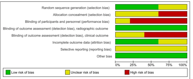
Risk of bias assessment
Quality and bias of the studies were evaluated according to the Cochrane Handbook [24], which is a broadly used guideline to assess randomized controlled trials. Studies were evalu- ated according to 8 domains. 1) Random sequence generation evaluates the strength of the method used for randomization. 2) Allocation concealment appraises the potential bias during allocation of the participants. 3) Blinding of participants and personnel assesses whether the patients and investigators were appropriately blinded to the treatment type. 4) Blinding of out- come assessment, radiographic outcome evaluates whether the personnel assessing x-ray images have been blinded. 5) Blinding of outcome assessment, clinical outcome appraises whether the clinical investigators evaluating the clinical outcome have been blinded. 6) Incom- plete outcome data evaluate the risk of attrition bias due to withdrawals, loss of participants during follow ups and other missing data. 7) Selective reporting assesses whether all pre-
determined outcomes have been measured and reported. 8) Other bias evaluates any other type of bias not falling into the previous 7 domains [24].
Summary measures and synthesis of results
Pooled Risk Ratio (RRs) with 95% confidence intervals (CIs) and weighted mean difference (WMD) with 95% CIs were calculated for dichotomous outcomes (IF) and for continuous out- comes (MBL change expressed in mm) respectively. Negative values in MBL change indicate a decrease in marginal bone level. Negative values of weighted mean differences indicate a greater decrease in MBL in sand-blasted implants compared to machined ones. Criteria for implant failure were defined according to Albrektsson et al. [25] Implant number was chosen as statistical unit. We only considered results credible if raw data for meta-analysis could be drawn from at least three records. We applied the random effect model with DerSimonian- Laird estimation. I2and chi-square tests were used to quantify statistical heterogeneity and gain probability-values, respectively; p<0.1 indicated a significant heterogeneity. [24] All sta- tistical analyses were performed using STATA 15.0.
Risk of bias across studies and additional analyses
Sensitivity analysis was performed by omitting studies (one by one) from the analyses and recalculating them in order to investigate the impact of the individual studies on the summary estimate. To check for publication bias, a visual inspection of funnel plots was performed.
Results Study selection
During the study selection process, a total of 188 records were identified, including one record found in the reference list of related articles. After removing duplicates, 130 items remained.
During the screening process, 114 records were excluded due to reasons such as other surface modification (n = 38) or different objectives (n = 76), investigating populations with systemic disease, evaluating surgical protocols, or comparing different macro designs of implants. For full-text evaluation 16 records were searched. Out of these publications nine records were excluded. The reasons for exclusion are explained below. Seven studies were eligible for quali- tative and quantitative analysis [19,26–31] (Fig 1).
Study characteristics
Description of excluded studies. Out of the nine excluded records, three records were not eligible because of evaluating other surface modifications than the ones investigated in this meta-analysis [32–34]. Two studies reported on previous results of ongoing studies that have been republished in updated records [35,36]. Two other records investigated different popula- tions (periodontitis-susceptible) [37,38]. One record was not RCT [39] and one other paper did not describe the surface modification used [40].
Description of the included studies. All involved studies were randomized controlled tri- als. A total of 722 implants (362 sandblasted and 360 machined) were included in the data syn- thesis. The populations represented in these studies were uniform, patients with alcohol and drug consumption or other medication abuse were excluded. Exclusion criteria also included bruxism, uncontrolled diabetes mellitus or any other significant medical condition that would affect the process of osseointegration. The mean age of participants in the studies varied between 50 and 58 years. In the five studies four different implant systems (Astra Tech, Bråne- mark, Steri-Oss and Southern Implants) were used. All implants in the control group had
minimally rough surface and all implants in the intervention group had moderately rough sur- face [9].
All study groups except one [26] followed the two-stage protocol [41]. However, even in the study using one-stage protocol, implants were only loaded 3 months following healing at the lower jaw, and 6 months of healing at the upper jaw. Out of the five studies two [19,26] treated edentulism with overdentures, another two [27,30] used fixed partial bridges. One study [42]
achieved rehabilitation with full arch bridges. The shortest follow-up was 2 years long, and the
Fig 1. PRISMA flow diagram of study selection process.
https://doi.org/10.1371/journal.pone.0216428.g001
longest lasted for 16 years [29]. Each study provided information on implant failure rate and marginal bone level change calculated from blinded radiographic measurements. In one study [26], only two out of four groups were included, two groups using immediate loading protocol were not included in the analysis. Additionally, MBL measurements of Astrand et al. [31] and Ravald et al. [28]were excluded since the reported patient-based data could not be converted to implant-based data to match the statistics of other studies. A detailed description of these studies is shown in Tables1and2.
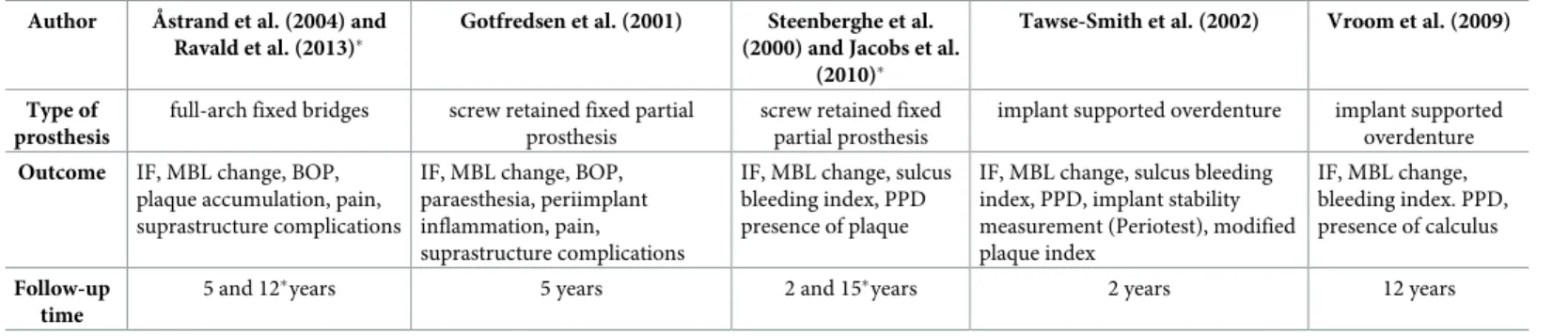
Table 1. Summary of study characteristics.
Author Åstrand et al. (2004)andRavald et al. (2013)�
Gotfredsen et al. (2001) Steenberghe et al. (2000) and Jacobs et al. (2010)�
Tawse-Smith et al.
(2002)
Vroom et al. (2009)
Study type block randomization separate for upper and lower jaw, with equal probability of receiving
either implant type
alternating implant placement split-mouth design random allocation to either implant system on a one-by-
one basis
alternating implant placement
Country Sweden 4 Scandinavian countries Belgium New Zealand not stated
Age �x= 61.5 �x= 53 �x= 59.7 55–80 �x= 53
Number of participants
males: 28, females: 38 males: 25, females: 25 males: 6, females: 12 total: 48 males: 7, females: 13
Extent of teeth loss
edentulous partially edentulous partially edentulous edentulous
(mandible only)
edentulous (mandible only) Sand-blasted
implant (intervention)
Astra Tech implants Astra Tech implants Astra Tech implants Southern Implants Astra Tech implants
Machined implants (control)
Branemark System MK II Astra Tech implants Branemark System MK II Sterioss Astra Tech implants
Surgical protocol two-stage technique (3 months and 6 months healing in the lower and upper jaw respectively
before abutment placement
two-stage technique (3–4 months and 6–7 months healing
in the lower and upper jaw respectively before abutment
placement
two-stage technique (3–4 months and 6–7 months healing in the lower and upper
jaw respectively before abutment placement
one-stage technique (3 months of healing before
loading)
two-stage technique (3–4 months healing before abutment
placement
�The publications of Ravald et al (2013) and Jacobs et al (2010) are the continuations of the studies published byÅstrand et al (2004); and Steenberghe et al (2000) respectively.
https://doi.org/10.1371/journal.pone.0216428.t001
Table 2. Summary of study characteristics.
Author Åstrand et al. (2004) and Ravald et al. (2013)�
Gotfredsen et al. (2001) Steenberghe et al.
(2000) and Jacobs et al.
(2010)�
Tawse-Smith et al. (2002) Vroom et al. (2009)
Type of prosthesis
full-arch fixed bridges screw retained fixed partial prosthesis
screw retained fixed partial prosthesis
implant supported overdenture implant supported overdenture Outcome IF, MBL change, BOP,
plaque accumulation, pain, suprastructure complications
IF, MBL change, BOP, paraesthesia, periimplant inflammation, pain, suprastructure complications
IF, MBL change, sulcus bleeding index, PPD presence of plaque
IF, MBL change, sulcus bleeding index, PPD, implant stability measurement (Periotest), modified plaque index
IF, MBL change, bleeding index. PPD, presence of calculus
Follow-up time
5 and 12�years 5 years 2 and 15�years 2 years 12 years
�The publications of Ravald et al (2013) and Jacobs et al (2010) are the continuations of the studies published byÅstrand et al (2004); and Steenberghe et al (2000) respectively.
IF: implant failure MBL: marginal bone level BOP: bleeding on probing PPD: probing pocket depth
https://doi.org/10.1371/journal.pone.0216428.t002
Risk of bias within studies
Bias in the studies was assessed according to the Cochrane Risk of Bias Tool. All seven included studies were included in the risk of bias assessment, however two pairs of studies [28, 31] and [27,29] were evaluated together because the study of Ravald et al [28] and Jacobs et al.
[29] are the continuation of previous studies ofÅstrand and coworkers [31] and those of Steenberghe and coinvestigators [27], respectively.
Two studies [26,27] had unclear random sequence generation, and other two [19,30] had a high risk of allocation concealment, due to the predictable sequence generation process used.
All studies performed blinding during the evaluation of x-ray images. However, due to its nature, no blinding could be carried out evaluating the implants clinically. Dropouts were iden- tified in four studies [19,26,30,31], two of these with unclear risk of bias.[19,26]. Access was not gained to study protocols or trial registers, however, no intext evidence of selective reporting was found.Fig 2,S1 TableandS3 Appendixcontain the summary of the risk of bias assessment.
Results of individual studies and synthesis of results
Sandblasted implants are better than machined implants concerning implant failure at 1, 2 and 5–6 years. Data for implant failure analysis after one year were pooled from five studies [19,26,27,30,31]. The results show that there is an 80% lower risk for sand-blasted implants to fail compared to machined implants after one year of use (RR = 0.20 95% CI: 0.06–
0.67; I2= 0.0% p = 0.986) (Fig 3andS1 File).
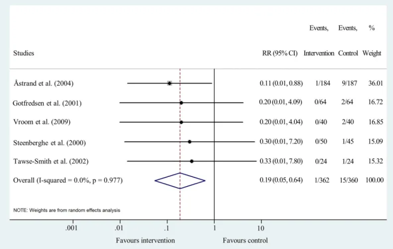
Data for cumulative implant failure after two years could be pooled from five studies [19, 26,27,30,31]. The meta-analysis revealed that the risk of sand-blasted implant failure is 81%
lower than that of machined implants (RR = 0.19 95% CI: 0.05–0.64; I2= 0.0% p = 0.977) (Fig 4andS1 File).
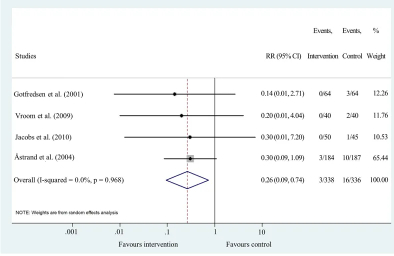
Data for analyzing the effect of sandblasting on implant failure after five or six years’ fol- low-up were pooled from four studies [19,29–31]. The results indicate that there is a 74%
lower risk of sandblasted implants to fail (RR = 0.26 95% CI: 0.09–0.74; I2= 0.0% p = 0.968) (Fig 5andS1 File).
Fig 2. Risk of bias graph. Percentage of each risk of bias item across included studies.
https://doi.org/10.1371/journal.pone.0216428.g002
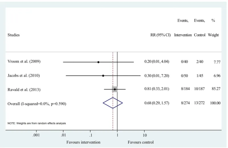
Results for cumulative IF after 12–15 years were synthesized from 3 studies [19,28,29].
Results show that there is no significant difference between the two treatment types (RR = 0.68 95% CI: 0.29–1.57; I2= 0.0% p = 0.590) (Fig 6andS1 File).
No detectable difference in MBL between sand-blasted and machined implants after 5 years of follow up
MBL change was analyzed one and five years after the delivery of the final prosthesis One-year data were pooled from three studies [19,26,27]. No significant difference was found between the two surface treatments, (weighted mean difference = -0.10, 95% CI: -0.20–0.01; p>0.05; I2
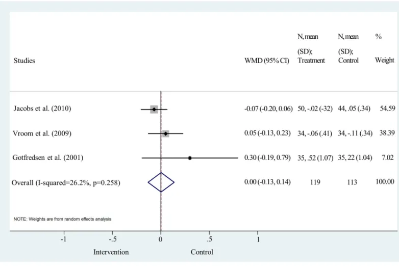
= 0.0%, p = 0.560) (Fig 7andS1 File). Data for 5-year analysis were pooled from three studies [19,29,30]. The statistical analysis clearly shows that the difference is not significant between the two implant surface types, the line of null effect falls within the range of the confidence interval (weighted mean difference = 0.00, 95% CI: -0.13–0.14; p>0.05; I2= 26.2%, p = 0.258) (Fig 8andS1 File).
Risk of bias across studies and additional analysis
Funnel plot analyses indicated a moderate level of publication bias (S4 Appendix). Statistical heterogeneity was not important in the results of IF at all time points. I2values were 0.0% and
Fig 3. Forest plot analysis of implant failure rate after one year.
https://doi.org/10.1371/journal.pone.0216428.g003
p values varied between 0.590 and 0.986 (Figs3,4,5and6). Heterogeneity was also negligible in the results of MBL change at 1 year (I2= 0.0%, p = 0.560) (Fig 7). I2(26.2%) and p (0.258) values indicated a slightly higher level of statistical heterogeneity for the results of MBL change at 5 years (Fig 8), however, this was still considered insignificant. [24].
Sensitivity analysis showed that the removal of the study of Astrand et al. [31] decreases the significance of the results of pooled risk ratio analysis at one, two and five/six years following implantation. This is most likely due to the large sample size of that study compared to the other RCTs.
Discussion
Summary of evidence
As a result of extensive investigations conducted in the past decades, several methods for implant surface modifications have emerged and numerous studies claimed superiority for one or other roughened surfaces. [43–45] However, no evidence supports a single decisive hypothesis. Therefore, the contradictory conclusions of the literature require further studies and careful re-analysis. As our objective stated, we re-evaluated the performance of sand- blasted implant surface over machined ones. To obtain the highest level of evidence, a meta- analysis was conducted including only RCTs available on the topic but excluding uncontrolled
Fig 4. Forest plot analysis of cumulative implant failure rate after two years.
https://doi.org/10.1371/journal.pone.0216428.g004
trials and retrospective studies. Implant stability was evaluated by measuring MBL changes and comparing cumulative implant failure rates. After appropriate selection, seven RCTs, 202 patients, having 362 sand-blasted and 360 machined implants could be included in our com- plex approach.
Our meta-analysis revealed that implant failure rates were significantly different between machined implants and sand-blasted ones. In contrast, the results of the individual RCTs could not reveal a significant difference between the two types of surfaces. This gained differ- ence reflects the increased number of samples and the high power of statistical methods of meta-analysis. Most implant failures happened during the first year after implantation. The reason for this could be that the surface modification of sandblasted implants creates a rougher surface which enhances the processes of bone formation on the implant itself. [46] Indepen- dent researchers published similar observations on other moderately rough surfaces, too. [47]
Our results also show that after one year, when osseointegration has already taken place, the difference between the two surfaces diminish and the significance of the difference between implant failures disappear. Additionally, a histological study, using small sample size, also con- firmed that, in the long term, both implant types maintained a decent level of osseointegration.
It was found that after 5 years the bone-implant contact level was 92.7% for machined and 81.2% for sand-blasted implants. [48].
Fig 5. Forest plot analysis of cumulative implant failure rate after 5/6 years.
https://doi.org/10.1371/journal.pone.0216428.g005
In the case of MBL change, our meta-analysis demonstrated no significant difference between the two implant types. The results of the individual RCTs included in our meta-analy- sis did not reveal any significant difference either. In contrast, a recent meta-analysis and also a novel review comparing machined implants to surface-modified implants concluded that rough implants may cause more bone loss [9,20]. The discrepancy between the meta-analysis reported by Doornewaard and coworkers [9] and our results could arise from the fact that we only examined sandblasted implants and excluded all other surface modifications in order to decrease heterogeneity. Additionally, they might have included patients with periodontitis, which was an exclusion factor in our case. Moreover, Wennerberg et al. [20] included not only RCTs, but also uncontrolled trials and retrospective studies in their analysis. Thus, they used a different statistical approach yielding high statistical heterogeneity, which might fundamen- tally influence the outcome [20]. Nevertheless, in all included studies, MBL measurements resulted in high standard deviations, which hinder any accurate statistical comparison. There- fore, the results have to be interpreted carefully.
However, some general trends are indicated by literature data.Åstrand and coworkers argued that the greatest loss in marginal bone occurred between implant placement and pros- thesis connection [31]. This change was found to be greater in machined than in sand-blasted implants [31]. Unfortunately, no published data on MBL change between implant placement and prosthesis connection are available for meta-analysis. Furthermore, based on data shown
Fig 6. Forest plot analysis of cumulative implant failure rate after 12/15 years.
https://doi.org/10.1371/journal.pone.0216428.g006
inFig 7, the mean bone change within the RCTs was between 0 and -0.10 mm for machined, -0.14 and -0.2 mm for sand-blasted implants from baseline to the first year, which is, in fact, very small [26,31]. According to Ravald and coinvestigators, the mean annual bone loss decreased gradually after five years. The annual mean bone attachment change was -0.02 and -0.04 mm for machined and sand-blasted implants, respectively, between five years and the end of the 12–15 years’ follow-up period [28]. In addition, there is evidence that bone gain can also occur around implants. The RCT ofÅstrand et al. reported more than 0.6 mm increase in MBL around 4 sandblasted and 2 machined implants over five years [31]. Vroom and cowork- ers also noted an increase in MBL around some implants with not much difference between the two surface types. The authors of this study argue that bone gain is a result of increased bone corticalization [19].
The different trends in MBL change and IF concerning the two implant types can be explained by the differences between the two measuring methods. MBL measurements can only detect bone changes when the implant is still stable at the annual checkup. If bone resorp- tion takes place so quickly that all the bone is resorbed within a year, the implants will be labeled to have failed and excluded from the MBL measurements, hence they no longer influ- ence MBL changes. Indeed, numerous studies show that the initial bone formation takes place at a faster rate around rough surface implants than around machined ones [7,49,50]. This may also explain why fewer sand-blasted implants failed compared to machined ones in the
Fig 7. Forest plot analysis of marginal bone level change after one year.
https://doi.org/10.1371/journal.pone.0216428.g007
first year after implantation. In addition, fast healing remains a key attribute of rough implants since healing time is a key element in modern implantology and implants with faster healing are prioritized [51,52].
The present study has a clear message for clinicians. We hypothesized that, concerning implant failure rates and marginal bone level loss, there are significant differences between sandblasted and machined dental implants. Our meta-analysis provided evidence that sand- blasting, indeed, significantly lowers implant failure rates although does not significantly affect marginal bone level changes. Thus, we recommend the use of sandblasted, moderately rough implants for patients with no systemic diseases as such implants support the osseointegration process with fewer complications than machined implants.
Limitations
A major limitation of the present paper is the relatively small number of randomized con- trolled trials available regarding this topic. Despite the large number of records found by the systematic search, only seven could be included. The limited number of reported data makes it impossible to perform sub-group analyses and to thoroughly investigate the causes behind cer- tain trends. Another issue that hinders in-depth analysis is the inhomogeneous reporting of outcome parameters. Some studies report the data separately for the lower and upper jaws, whereas others only report combined data. All studies reported one or two clinical parameters
Fig 8. Forest plot analysis of marginal bone level change after 5 years.
https://doi.org/10.1371/journal.pone.0216428.g008
such as bleeding on probing. However, the use of different reporting schemes made compari- son impossible for bleeding on probing tests, among others. Another limitation of the present work is that its conclusions apply only to healthy populations. There are several confounding factors which might create unfavorable conditions for moderately rough implants, such as patients with severe periodontitis [53]. However, these conditions were excluded from our analysis. Uncontrolled or unknown confounding factors not evenly affecting intervention and control groups may also contribute to differences in the outcomes. Finally, limitations of this meta-analysis include the heterogeneity of the implants used. Although implants with identical macro designs would be preferred, this was not really possible.
In conclusion, within the limitations of this meta-analysis, the results reveal that sandblast- ing is superior over machined surface concerning implant failure. On the other hand, no sig- nificant difference was found regarding marginal bone level changes between the two implant types. Our in-depth analysis of the literature also highlights that results are highly sensitive to heterogeneity and study design, which may lead to contradictory conclusions. In the future, consistent reporting on more clinical outcomes such as bleeding on probing, pocket probing depth and implant success rates are needed. Evaluation could be more meaningful if implant success is evaluated by RCTs rather than case-based implant failure studies. Therefore, a com- prehensive protocol should be compiled to guide clinicians conducting valuable RCTs evaluat- ing implant performance in order to decrease heterogeneity of papers and to increase clinical applicability.
Supporting information S1 Appendix. PRISMA checklist.
(PDF)
S2 Appendix. PROSPERO registration.
(PDF)
S3 Appendix. Risk of bias summary: Review authors’ judgements about each risk of bias item for each included study.
(TIFF)
S4 Appendix. Publication bias (funnel plots).
(PDF)
S1 Table. Detailed evaluation of risk of bias of individual studies.
(PDF)
S1 File. Data included in meta-analysis.
(XLSX)
Author Contributions
Conceptualization: La´szlo´ Ma´rk Czumbel, Bea´ta Kere´mi, Noe´mi Gede, Alexandra Miko´, Bar- bara To´th, DezsőCsupor, Sa´ndor Farkasdi, Ga´bor Gerber, Ma´rta Balasko´, Erika Pe´terva´ri, Pe´ter Hegyi, Ga´bor Varga.
Data curation: La´szlo´ Ma´rk Czumbel, Bea´ta Kere´mi, Noe´mi Gede, Alexandra Miko´, Dezső Csupor, Andrea Szabo´, Sa´ndor Farkasdi, Erika Pe´terva´ri, Ro´bert Sepp, Pe´ter Hegyi, Ga´bor Varga.
Formal analysis: Noe´mi Gede.
Funding acquisition: Pe´ter Hegyi, Ga´bor Varga.
Investigation: La´szlo´ Ma´rk Czumbel, Bea´ta Kere´mi, Ga´bor Varga.
Methodology: La´szlo´ Ma´rk Czumbel, Bea´ta Kere´mi, Noe´mi Gede, Alexandra Miko´, Barbara To´th, DezsőCsupor, Sa´ndor Farkasdi, Ma´rta Balasko´, Erika Pe´terva´ri, Ro´bert Sepp, Pe´ter Hegyi, Ga´bor Varga.
Project administration: La´szlo´ Ma´rk Czumbel, Bea´ta Kere´mi, Pe´ter Hegyi, Ga´bor Varga.
Resources: Pe´ter Hegyi, Ga´bor Varga.
Software: Noe´mi Gede.
Supervision: Bea´ta Kere´mi, Andrea Szabo´, Sa´ndor Farkasdi, Ma´rta Balasko´, Pe´ter Hegyi, Ga´bor Varga.
Validation: Bea´ta Kere´mi, Noe´mi Gede, Alexandra Miko´, Barbara To´th, DezsőCsupor, Andrea Szabo´, Sa´ndor Farkasdi, Ga´bor Gerber, Ma´rta Balasko´, Erika Pe´terva´ri, Ro´bert Sepp, Pe´ter Hegyi, Ga´bor Varga.
Visualization: La´szlo´ Ma´rk Czumbel, Bea´ta Kere´mi, Noe´mi Gede, Ga´bor Varga.
Writing – original draft: La´szlo´ Ma´rk Czumbel, Bea´ta Kere´mi, Noe´mi Gede, Ga´bor Varga.
Writing – review & editing: La´szlo´ Ma´rk Czumbel, Bea´ta Kere´mi, Noe´mi Gede, Alexandra Miko´, Barbara To´th, DezsőCsupor, Andrea Szabo´, Sa´ndor Farkasdi, Ga´bor Gerber, Ma´rta Balasko´, Erika Pe´terva´ri, Ro´bert Sepp, Pe´ter Hegyi, Ga´bor Varga.
References
1. Branemark PI, Hansson BO, Adell R, Breine U, Lindstrom J, Hallen O, et al. Osseointegrated implants in the treatment of the edentulous jaw. Experience from a 10-year period. Scandinavian journal of plas- tic and reconstructive surgery Supplementum. 1977; 16:1–132. Epub 1977/01/01. PMID:356184.
2. Gaviria L, Salcido JP, Guda T, Ong JL. Current trends in dental implants. Journal of the Korean Associ- ation of Oral and Maxillofacial Surgeons. 2014; 40(2):50–60.https://doi.org/10.5125/jkaoms.2014.40.2.
50PMC4028797. PMID:24868501
3. Reeta J, Gyanchand R, Surbhi G. Implant surface design: An Overview. Annals of Prosthodontics and Restorative Dentistry. 2016; 2(1):17–20.
4. Vootla N, Reddy K. Osseointegration- Key Factors Affecting Its Success-An Overview. IOSR Journal of Dental and Medical Sciences. 2017; 16(04):62–8.https://doi.org/10.9790/0853-1604056268
5. Barfeie A, Wilson J, Rees J. Implant surface characteristics and their effect on osseointegration. Br Dent J. 2015; 218(5):E9–E.https://doi.org/10.1038/sj.bdj.2015.171PMID:25766196
6. Karl M, Albrektsson T. Clinical Performance of Dental Implants with a Moderately Rough (TiUnite) Sur- face: A Meta-Analysis of Prospective Clinical Studies. Int J Oral Maxillofac Implants. 2017; 32(4):717–
34. Epub 2017/07/15.https://doi.org/10.11607/jomi.5699PMID:28708905.
7. Velasco-Ortega E, Alfonso-Rodrı´guez CA, Monsalve-Guil L, España-Lo´ pez A, Jime´nez-Guerra A, Gar- zo´n I, et al. Relevant aspects in the surface properties in titanium dental implants for the cellular viability.
Materials Science and Engineering: C. 2016; 64:1–10.https://doi.org/10.1016/j.msec.2016.03.049.
8. Sykaras N, Iacopino AM, Marker VA, Triplett RG, Woody RD. Implant materials, designs, and surface topographies: their effect on osseointegration. A literature review. Int J Oral Maxillofac Implants. 2000;
15(5):675–90. Epub 2000/10/31. PMID:11055135.
9. Doornewaard R, Christiaens V, De Bruyn H, Jacobsson M, Cosyn J, Vervaeke S, et al. Long-Term Effect of Surface Roughness and Patients’ Factors on Crestal Bone Loss at Dental Implants. A System- atic Review and Meta-Analysis. Clin Implant Dent Relat Res. 2017; 19(2):372–99. Epub 2016/11/20.
https://doi.org/10.1111/cid.12457PMID:27860171.
10. Novaes AB Jr, Souza SLSd, Barros RRMd, Pereira KKY, Iezzi G, Piattelli A. Influence of implant sur- faces on osseointegration. Brazilian Dental Journal. 2010; 21:471–81. PMID:21271036
11. Smeets R, Stadlinger B, Schwarz F, Beck-Broichsitter B, Jung O, Precht C, et al. Impact of Dental Implant Surface Modifications on Osseointegration. BioMed Research International. 2016;
2016:6285620.https://doi.org/10.1155/2016/6285620PMC4958483. PMID:27478833
12. Jemat A, Ghazali MJ, Razali M, Otsuka Y. Surface Modifications and Their Effects on Titanium Dental Implants. BioMed Research International. 2015; 2015:791725.https://doi.org/10.1155/2015/791725 PMC4575991. PMID:26436097
13. Li S, Ni J, Liu X, Lu H, Yin S, Rong M, et al. Surface Characteristic of Pure Titanium Sandblasted with Irregular Zirconia Particles and Acid-Etched. Materials Transactions. 2012; 53(5):913–9.https://doi.org/
10.2320/matertrans.M2011291
14. Wennerberg A, Hallgren C, Johansson C, Danelli S. A histomorphometric evaluation of screw-shaped implants each prepared with two surface roughnesses. Clin Oral Implants Res. 1998; 9(1):11–9. Epub 1998/05/20. PMID:9590940.
15. Morita Y, Yamasaki K, Hattori K. A feasibility study for in vitro evaluation of fixation between prosthesis and bone with bone marrow-derived mesenchymal stem cells. Clinical Biomechanics. 2010; 25(8):829–
34.https://doi.org/10.1016/j.clinbiomech.2010.05.007PMID:20646804
16. Farkasdi S, Pammer D, Racz R, Hriczo-Koperdak G, Szabo BT, Dobo-Nagy C, et al. Development of a quantitative preclinical screening model for implant osseointegration in rat tail vertebra. Clin Oral Inves- tig. 2018. Epub 2018/10/31.https://doi.org/10.1007/s00784-018-2661-1PMID:30374828.
17. Teughels W, Van Assche N, Sliepen I, Quirynen M. Effect of material characteristics and/or surface topography on biofilm development. Clin Oral Implants Res. 2006; 17 Suppl 2:68–81. Epub 2006/09/14.
https://doi.org/10.1111/j.1600-0501.2006.01353.xPMID:16968383.
18. Quirynen M, Abarca M, Assche NV, Nevins M, Steenberghe DV. Impact of supportive periodontal ther- apy and implant surface roughness on implant outcome in patients with a history of periodontitis. Jour- nal of Clinical Periodontology. 2007; 34(9):805–15.https://doi.org/10.1111/j.1600-051X.2007.01106.x PMID:17716316
19. Vroom MG, Sipos P, de Lange GL, Grundemann LJ, Timmerman MF, Loos BG, et al. Effect of surface topography of screw-shaped titanium implants in humans on clinical and radiographic parameters: a 12-year prospective study. Clin Oral Implants Res. 2009; 20(11):1231–9. Epub 2009/08/28.https://doi.
org/10.1111/j.1600-0501.2009.01768.xPMID:19709060.
20. Wennerberg A, Albrektsson T, Chrcanovic B. Long-term clinical outcome of implants with different sur- face modifications. Eur J Oral Implantol. 2018; 11 Suppl 1:S123–s36. Epub 2018/08/16. PMID:
30109304.
21. Papaspyridakos P, Mokti M, Chen CJ, Benic GI, Gallucci GO, Chronopoulos V. Implant and prostho- dontic survival rates with implant fixed complete dental prostheses in the edentulous mandible after at least 5 years: a systematic review. Clin Implant Dent Relat Res. 2014; 16(5):705–17. Epub 2013/01/15.
https://doi.org/10.1111/cid.12036PMID:23311617.
22. Esposito M, Ardebili Y, Worthington HV. Interventions for replacing missing teeth: different types of den- tal implants. The Cochrane database of systematic reviews. 2014;(7):Cd003815. Epub 2014/07/23.
https://doi.org/10.1002/14651858.CD003815.pub4PMID:25048469.
23. Moher D, Shamseer L, Clarke M, Ghersi D, Liberati A, Petticrew M, et al. Preferred reporting items for systematic review and meta-analysis protocols (PRISMA-P) 2015 statement. Systematic reviews.
2015; 4:1. Epub 2015/01/03.https://doi.org/10.1186/2046-4053-4-1PMID:25554246; PubMed Central PMCID: PMCPMC4320440.
24. Higgins JP, Altman DG, Gotzsche PC, Juni P, Moher D, Oxman AD, et al. The Cochrane Collabora- tion’s tool for assessing risk of bias in randomised trials. BMJ (Clinical research ed). 2011; 343:d5928.
Epub 2011/10/20.https://doi.org/10.1136/bmj.d5928PMID:22008217; PubMed Central PMCID:
PMCPMC3196245.
25. Albrektsson T, Zarb G, Worthington P, Eriksson AR. The long-term efficacy of currently used dental implants: a review and proposed criteria of success. Int J Oral Maxillofac Implants. 1986; 1(1):11–25.
Epub 1986/01/01. PMID:3527955.
26. Tawse-Smith A, Payne AG, Kumara R, Thomson WM. Early loading of unsplinted implants supporting mandibular overdentures using a one-stage operative procedure with two different implant systems: a 2-year report. Clin Implant Dent Relat Res. 2002; 4(1):33–42. Epub 2002/04/10. PMID:11938636.
27. Steenberghe D, Mars G, Quirynen M, Jacobs R, Naert I. A prospective split-mouth comparative study of two screw-shaped self-tapping pure titanium implant systems. Clinical oral implants research [Inter- net]. 2000; 11(3):[202–9 pp.]. Available from:http://onlinelibrary.wiley.com/o/cochrane/clcentral/
articles/847/CN-00327847/frame.html.
28. Ravald N, Dahlgren S, Teiwik A, Gro¨ ndahl K. Long-term evaluation of Astra Tech and Brånemark implants in patients treated with full-arch bridges. Results after 12–15 years. Clinical oral implants
research [Internet]. 2013; 24(10):[1144–51 pp.]. Available from:http://cochranelibrary-wiley.com/o/
cochrane/clcentral/articles/991/CN-01001991/frame.html.
29. Jacobs R, Pittayapat P, van Steenberghe D, De Mars G, Gijbels F, Van Der Donck A, et al. A split- mouth comparative study up to 16 years of two screw-shaped titanium implant systems. J Clin Period- ontol. 2010; 37(12):1119–27. Epub 2010/09/30.https://doi.org/10.1111/j.1600-051X.2010.01626.x PMID:20874829.
30. Gotfredsen K, Karlsson U. A prospective 5-year study of fixed partial prostheses supported by implants with machined and TiO2-blasted surface. J Prosthodont. 2001; 10(1):2–7. Epub 2001/06/15. PMID:
11406789.
31. Åstrand P, Engquist B, Dahlgren S, Gro¨ndahl K, Engquist E, Feldmann H. Astra Tech and Brånemark system implants: a 5-year prospective study of marginal bone reactions. Clinical oral implants research [Internet]. 2004; 15(4):[413–20 pp.]. Available from:http://cochranelibrary-wiley.com/o/cochrane/
clcentral/articles/196/CN-00490196/frame.html.
32. Esposito M, Felice P, Barausse C, Pistilli R, Grandi G, Simion M. Immediately loaded machined versus rough surface dental implants in edentulous jaws: One-year postloading results of a pilot randomised controlled trial. European journal of oral implantology. 2015; 8(4):387–96. PMID:26669548
33. Burtscher D, Norer B, Dalla D, Beier U, Schubert K, Grunert I. A 7-year prospective radiographic evalua- tion of marginal bone level around two different implant systems: a randomized clinical trial. Clinical oral implants research [Internet]. 2015; 26(11):[1244–9 pp.]. Available from:http://cochranelibrary-wiley.
com/o/cochrane/clcentral/articles/846/CN-01411846/frame.html.
34. Cannizzaro G, Gastaldi G, Gherlone E, Vinci R, Loi I, Trullenque-Eriksson A, et al. Two or three machined vs roughened surface dental implants loaded immediately supporting total fixed prostheses:
1-year results from a randomised controlled trial. European journal of oral implantology [Internet]. 2017;
10(3):[279–91 pp.]. Available from:http://cochranelibrary-wiley.com/o/cochrane/clcentral/articles/104/
CN-01572104/frame.html.
35. Tawse-Smith A, Perio C, Payne AG, Kumara R, Thomson WM. One-stage operative procedure using two different implant systems: a prospective study on implant overdentures in the edentulous mandible.
Clin Implant Dent Relat Res. 2001; 3(4):185–93. Epub 2002/03/13. PMID:11887655.
36. Karlsson U, Gotfredsen K, Olsson C. A 2-year report on maxillary and mandibular fixed partial dentures supported by Astra Tech dental implants. A comparison of 2 implants with different surface textures.
Clin Oral Implants Res. 1998; 9(4):235–42. Epub 1998/10/07. PMID:9760898.
37. Donati M, Ekestubbe A, Lindhe J, Wennstrom JL. Marginal bone loss at implants with different surface characteristics—A 20-year follow-up of a randomized controlled clinical trial. Clin Oral Implants Res.
2018; 29(5):480–7. Epub 2018/03/24.https://doi.org/10.1111/clr.13145PMID:29569767.
38. Wennstro¨m J, Ekestubbe A, Gro¨ndahl K, Karlsson S, Lindhe J. Oral rehabilitation with implant-sup- ported fixed partial dentures in periodontitis-susceptible subjects. A 5-year prospective study. Journal of clinical periodontology [Internet]. 2004; 31(9):[713–24 pp.]. Available from:http://cochranelibrary-wiley.
com/o/cochrane/clcentral/articles/770/CN-00490770/frame.html.
39. Rasmusson L, Roos J, Bystedt H. A 10-year follow-up study of titanium dioxide-blasted implants. Clin Implant Dent Relat Res. 2005; 7(1):36–42. Epub 2005/05/21. PMID:15903173.
40. Schincaglia GP, Marzola R, Scapoli C, Scotti R. Immediate loading of dental implants supporting fixed partial dentures in the posterior mandible: A randomized controlled split-mouth study—Machined ver- sus titanium oxide implant surface. International Journal of Oral and Maxillofacial Implants. 2007; 22 (1):35–46. PMID:17340895
41. Branemark PI, Zarb GA, Albrektsson T, Rosen HM. Tissue-Integrated Prostheses. Osseointegration in Clinical Dentistry. Plastic and Reconstructive Surgery. 1986; 77(3):496–7. 00006534-198603000- 00037.
42. Astrand P, Engquist B, Dahlgren S, Gro¨ndahl K, Engquist E, Feldmann H. Astra Tech and Brånemark system implants: a 5-year prospective study of marginal bone reactions. Clinical oral implants research.
2004; 15(4):413-20.https://doi.org/10.1111/j.1600-0501.2004.01028.xCN-00490196. PMID:
15248875
43. Rocci M, Rocci A, Martignoni M, Albrektsson T, Barlattani A, Gargari M. Comparing the TiOblast and Osseospeed surfaces. Histomorphometric and histological analysis in humans. ORAL & implantology.
2008; 1(1):34–42. Epub 2008/04/01. PMID:23285334; PubMed Central PMCID: PMCPMC3476499.
44. Bornstein MM, Harnisch H, Lussi A, Buser D. Clinical performance of wide-body implants with a sand- blasted and acid-etched (SLA) surface: results of a 3-year follow-up study in a referral clinic. Int J Oral Maxillofac Implants. 2007; 22(4):631–8. Epub 2007/10/13. PMID:17929525.
45. Degidi M, Nardi D, Piattelli A. 10-year follow-up of immediately loaded implants with TiUnite porous anodized surface. Clin Implant Dent Relat Res. 2012; 14(6):828–38. Epub 2012/03/02.https://doi.org/
10.1111/j.1708-8208.2012.00446.xPMID:22376174.
46. Andrukhov O, Huber R, Shi B, Berner S, Rausch-Fan X, Moritz A, et al. Proliferation, behavior, and dif- ferentiation of osteoblasts on surfaces of different microroughness. Dent Mater. 2016; 32(11):1374–84.
https://doi.org/10.1016/j.dental.2016.08.217PMID:27637551.
47. Chrcanovic BR, Albrektsson T, Wennerberg A. Turned versus anodised dental implants: a meta-analy- sis. J Oral Rehabil. 2016; 43(9):716–28. Epub 2016/06/14.https://doi.org/10.1111/joor.12415PMID:
27295394.
48. Iezzi G, Vantaggiato G, Shibli JA, Fiera E, Falco A, Piattelli A, et al. Machined and sandblasted human dental implants retrieved after 5 years: a histologic and histomorphometric analysis of three cases.
Quintessence international (Berlin, Germany: 1985). 2012; 43(4):287–92. Epub 2012/04/26. PMID:
22532942.
49. Bruyn HD, Christiaens V, Doornewaard R, Jacobsson M, Cosyn J, Jacquet W, et al. Implant surface roughness and patient factors on long-term peri-implant bone loss. Periodontology 2000. 2017; 73 (1):218–27.https://doi.org/10.1111/prd.12177PMID:28000269
50. Piattelli A, Scarano A, Piattelli M, Calabrese L. Direct bone formation on sand-blasted titanium implants:
an experimental study. Biomaterials. 1996; 17(10):1015–8.https://doi.org/10.1016/0142-9612(96) 84677-1. PMID:8736737
51. Vandeweghe S, Ferreira D, Vermeersch L, Marien M, De Bruyn H. Long-term retrospective follow-up of turned and moderately rough implants in the edentulous jaw. Clin Oral Implants Res. 2016; 27(4):421–
6. Epub 2015/04/10.https://doi.org/10.1111/clr.12602PMID:25856319.
52. Vervaeke S, Collaert B, Cosyn J, De Bruyn H. A 9-Year Prospective Case Series Using Multivariate Analyses to Identify Predictors of Early and Late Peri-Implant Bone Loss. Clin Implant Dent Relat Res.
2016; 18(1):30–9. Epub 2014/07/06.https://doi.org/10.1111/cid.12255PMID:24995626.
53. Raes M, D’Hondt R, Teughels W, Coucke W, Quirynen M. A 5-year randomized clinical trial comparing minimally with moderately rough implants in patients with severe periodontitis. Journal of Clinical Peri- odontology. 2018; 45(6):711–20.https://doi.org/10.1111/jcpe.12901PMID:29611221