Research Article
Clinical Use of Next-Generation Sequencing in the Diagnosis of Wilson’s Disease
Dániel Németh,
1Kristóf Árvai,
2Péter Horváth,
1János Pál Kósa,
1,2Bálint Tobiás,
2Bernadett Balla,
2Anikó Folhoffer,
1Anna Krolopp,
1Péter András Lakatos,
1and Ferenc Szalay
111st Department of Internal Medicine, Semmelweis University, Koranyi Sandor Street 2/a, Budapest 1083, Hungary
2PentaCore Lab, Koranyi Sandor Street 2/a, Budapest 1083, Hungary
Correspondence should be addressed to D´aniel N´emeth; neemethd@gmail.com Received 8 July 2015; Revised 15 September 2015; Accepted 20 September 2015 Academic Editor: Alfred Gangl
Copyright © 2016 D´aniel N´emeth et al. This is an open access article distributed under the Creative Commons Attribution License, which permits unrestricted use, distribution, and reproduction in any medium, provided the original work is properly cited.
Objective. Wilson’s disease is a disorder of copper metabolism which is fatal without treatment. The great number of disease-causing ATP7Bgene mutations and the variable clinical presentation of WD may cause a real diagnostic challenge. The emergence of next- generation sequencing provides a time-saving, cost-effective method for full sequencing of the wholeATP7Bgene compared to the traditional Sanger sequencing. This is the first report on the clinical use of NGS to examineATP7Bgene.Materials and Methods. We used Ion Torrent Personal Genome Machine in four heterozygous patients for the identification of the other mutations and also in two patients with no known mutation. One patient with acute on chronic liver failure was a candidate for acute liver transplantation.
The results were validated by Sanger sequencing.Results. In each case, the diagnosis of Wilson’s disease was confirmed by identifying the mutations in both alleles within 48 hours. One novel mutation (p.Ala1270Ile) was found beyond the eight other known ones.
The rapid detection of the mutations made possible the prompt diagnosis of WD in a patient with acute liver failure.Conclusions.
According to our results we found next-generation sequencing a very useful, reliable, time-saving, and cost-effective method for diagnosing Wilson’s disease in selected cases.
1. Introduction
Wilson’s disease (WD) is a rare autosomal recessive disorder of copper metabolism.ATP7Bgene mutation is in the back- ground of the excessive copper accumulation which is fatal without treatment. More than 550 disease-causing mutations of the gene located at chromosome 13q14.3-q21.1 consisting of 21 exons have been identified [1].
The geographical distribution of the mutations of the ATP7B gene is inhomogeneous [2–5]. In Hungary the p.His1069Gln mutation is the most frequent one with 71%
prevalence among the patients [6].
The variable clinical presentation of WD may cause a real diagnostic challenge. The suspicion of the disease usually arises when hepatic or neurologic-psychiatric symptoms appear. Low ceruloplasmin level and presence of Kayser- Fleischer ring could support the diagnosis, but in many cases
only genetic testing could confirm it. Genetic investigation of asymptomatic siblings has an extreme importance, since the early treatment could prevent the manifestation of the disease [7]. In acute liver failure urgent genetic testing of all known mutations may strengthen the diagnosis of Wilson’s disease.
The emergence of next-generation sequencing (NGS) provides a time-saving, cost-effective method for full sequen- cing of the wholeATP7Bcoding sequence compared to the traditional Sanger sequencing. The NGS technology is based on the detection of a signal during the synthesis of the DNA strand, and therefore the synthesis does not need to be terminated for the perception. On the other hand, several DNA strands can be examined simultaneously [8].
This is the first report on the clinical use of NGS to examine ATP7B gene in WD patients including doubtful cases. We used Ion Torrent Personal Genome Machine in heterozygous patients for the identification of the other
Volume 2016, Article ID 4548039, 6 pages http://dx.doi.org/10.1155/2016/4548039
mutations and also in patients with no known mutation including one with acute on chronic liver failure.
2. Materials and Methods
The method we used for the genetic testing has been pre- viously published by our group for screening of neurofibro- matosis type 1 gene [9].
2.1. Biological Samples and DNA Isolation. Six (five male and one female) WD patients, four heterozygous forATP7B p.His1069Gln mutation identified by fast PCR test and two with unknown mutation, were selected for this study. The patients were diagnosed and treated at the 1st Department of Internal Medicine, Semmelweis University, Budapest. The diagnosis was based on the international WD score system published in 2003 [10], and each patient had 4 or more scores. The study was approved by the Semmelweis Univer- sity’s Committee of Research Ethics and was conducted in accordance with the Helsinki Declaration. All patients gave written informed consent.
Genomic DNA was isolated from 200𝜇L of periph- eral blood using ReliaPrep Blood gDNA Miniprep System (Promega, Madison, WI). Briefly, the blood samples were digested with Proteinase K solution in the presence of Cell Lysis Buffer, and, after 10 min of incubation at 56∘C, DNA was bound to ReliaPrep Binding Column. After three washes, DNA was eluted into 50𝜇L of nuclease-free water. The concentration of the isolated DNA was determined with Qubit dsDNA HS Assay Kit (Life Technologies, Carlsbad, CA).
2.2. Ion Torrent Sequencing. ATP7B(21 coding exons) ampli- cons were designed using the AmpliSeq Designer software (Life Technologies, CA, USA), targeting the complete coding sequence ofATP7Bgene, resulting in a total of 55 amplicons.
To gain a higher coverage of the coding exons, we designed the primers to also flank some parts of the introns. Amplicon library was prepared using the Ion AmpliSeq Library Kit 2.0 (Life Technologies, CA, USA); briefly, multiplex primer pools were added to 10 ng of genomic DNA and amplified with the following PCR cycles: at 99∘C for 2 min, at 99∘C for 15 s, and at 60∘C for 4 min (18 cycles), and holding on at 10∘C. Primers were partially digested using a FuPa reagent, and then sequencing adapters were ligated to the amplicons. The library was purified in multiple times using the Agencourt AMPure XP Reagent (Beckmann Coulter, CA, USA). The concentration of the final library was deter- mined by fluorescent measurement on Qubit 2.0 instrument (Life Technologies, CA, USA). Template preparation was performed with Ion OneTouch kit (Life Technologies, CA, USA) on semiautomatic Ion OneTouch instrument using an emPCR method. After breaking the emulsion, the non- templated beads were removed from the solution during the semiautomatic enrichment process on Ion OneTouch ES (Life Technologies, CA, USA) instrument. After adding the sequencing primer and polymerase, the fully prepared Ion Sphere Particle (ISP) beads were loaded into an Ion 314 v2
sequencing chip, and the sequencing runs were performed using the Ion PGM 200 Sequencing kit v2 (Life Technologies, CA, USA) with 500 flows.
2.3. Sanger Sequencing Validation. The PCR primers were designed using Primer3Plus (http://primer3plus.com/) soft- ware. Roche FastStart TaqMan Probe Master (Roche) kit was used to amplify the target regions and the PCR program was as follows: 95∘C for 10 min, 40 cycles of 95∘C for 30 s, 60∘C for 30 s, and 72∘C for 45 s, and the final step was 72∘C for 5 min. PCR products were enzymatically cleaned using ExoSAP IT (Affymetrix, Santa Clara, CA) according to the manufacturer’s instructions. Sanger sequencing was performed using BigDye Terminator v3.1 Cycle Sequencing Kit (Life Technologies) using an ABI 3130 instrument (Life Technologies).
2.4. Data Analysis. Data from the Ion Torrent runs were analyzed using the platform-specific pipeline software Tor- rent Suite v3.6 for base calling, trim adapter and primer sequences, filter out poor quality reads, and demultiplex the reads according to the barcode sequences. Briefly, TMAP (https://github.com/iontorrent/TMAP) algorithm was used to align the reads to the hg19 human reference genome, and then the variant caller plug-in was selected to run to search for germ line variants in the targeted regions. The variant caller algorithm parameters were more relaxed to avoid false negative cases. Integrative Genomics Viewer was used for visualization of the mapped reads. Variants were reviewed and annotated using dsSNP (http://www.ncbi.nlm.nih.gov /projects/SNP/) and Wilson Disease Mutation Database (http://www.wilsondisease.med.ualberta.ca/index.asp). For variant interpretation, Ingenuity Variant Analysis Pipeline (Ingenuity, Rewood City, CA) was also used. Pathogenic status of the variant was stated if it was a missense variant with<1% minor allele frequency and/or the variant was listed in the literature or in the databases as a pathogenic alteration.
All of the deleterious variants were confirmed by Sanger sequencing. The Sanger sequence data were investigated using ABI Sequence Scanner 1.0 (Life Technologies) and BioEdit (http://www.mbio.ncsu.edu/bioedit/bioedit.html) software.
3. Results
The demographic and clinical characteristics of the patients are shown in Table 1. One patient without known mutation was critically ill with acute on chronic liver disease. The typical laboratory findings (ALT 90, AST 178, ALP 88, and bilirubin 247) proposed Wilson’s disease. The diagnosis was strengthened by genetic testing making possible the liver transplantation via Eurotransplant program inPatient 5with acute on chronic liver failure.
In each case, the diagnosis of Wilson’s disease was confirmed by identifying the mutations in both alleles. The results were available within 48 hours.
The average read number per sample was 134386, with an average 1X on-target coverage of 99.46%. The mean raw
Table 1: Demographic and clinical characteristics of the patients.
Gender Age at onset
(year) KFR Neu HA Urin Cu Biopsy Cerul (g/L) ATP7Bstatus WD scorea Phenotype
Patient 1 Female 12 P A A ++ ND 0.18 p.Met769-fs/p.His1069Gln 6 S
Patient 2 Male 17 A P A + ND 0.05 p.Ala1063Val/p.His1069Gln 6 N1
Patient 3 Male 8 P A A ++ +b 0.06 p.His1069Gln/p.Gln1351Stop 8 H2
Patient 4 Male 17 P A A + ND 0.03 p.Ala1135-fs/p.Leu1305Pro 5 H2
Patient 5 Male 44 A A A ++ ND 0.08 p.Ala1270Ile/c.1707+2dupT 4 H1
Patient 6 Male 14 P P A ND ND 0.04 p.Arg969Gln/p.His1069Gln 7 N2
KFR: Kayser-Fleischer ring; Neu: neurological signs and/or CT/MRI alterations; HA: hemolytic anemia; Urin Cu: urinary copper, 1-2X ULN: +,>2x ULN or positive D-penicillamine challenge: ++; Cerul: ceruloplasmin, P: present; A: absent; ND: not done; S: sibling; H1: acute liver failure; H2: chronic liver disease;
N1: neurological symptoms with liver disease; N2: only neurological symptoms.
aAccording to the international score system, 4 or more scores, diagnosis of WD is highly likely.bRhodanine positivity.
T
(a1) (a2)
(b1) (b2)
CGGGGA340GTCAWTGACCCC350 ATCCGGGATGGGGTC90 AWTGAC100TCCCCG GCACCC90ACAGTACTTAC100TGGCCG TGA240GCTGACAGTW250ARKAAYGG
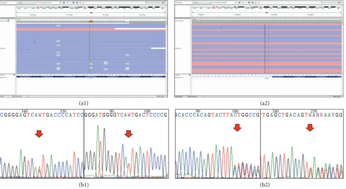
Figure 1: The identified mutations of patient 5. Both c.3809A>T (causing amino acid change p.Ala1270Ile) and c.1707+2dupT mutations are confirmed by Sanger-sequencing. The 3809A>C and A>G mutations are known, but the A>T substitution is a novel alteration at this position. (a1) Visualizing the alignment of the sequencing reads covering theATP7Bc.3809A>T heterozygous point mutation. The coverage was 400-fold (211-fold reference and 189-fold variant coverage). (b1) Validating our finding with Sanger sequencing, red arrow indicates the position of the point mutation. The mutation is present in both directions. (a2) Visualizing the alignment of the sequencing reads covering theATP7Bc.1707+2dupT heterozygous insertion mutation. The coverage was 399-fold (188-fold reference and 211-fold variant coverage). (b2) Validating our finding with Sanger-sequencing, red arrow indicates the position of the insertion. The mutation is present in both directions.
accuracy was 99.2%. The average base coverage depth was 1883 (Table 2). The number of identified variants per sample was between 8 and 13; however most of them were known as non-disease-causing variants.
Overall, we found nine disease-causing variants. The most frequent mutation was p.His1069Gln (exon 14, ATP loop) detected in four patients. One novel missense muta- tion (p.Ala1270Ile, exon 18, ATP hinge vide Figure 1) and three well-known missense mutations (p.Arg969Gln, exon 13, TM6; p.Ala1063Val, exon 14, ATP loop, and p.Leu1305Pro, exon 19 bet ATP hinge/TM7), three frame-shift mutations
(c1707+2dupT, exon 4, Cu6; p.Met769-fs exon 8 TM4, and p.Ala1135-fs exon 15 ATP loop), and one nonsense mutation (p.Gln1351Stop, exon 20, TM8) were detected. All of these variants had been validated by Sanger sequencing.
4. Discussion
Although there is an international diagnostic score system for WD [10], the set-up of the diagnosis remains a great challenge in many cases. The signs and symptoms are very colorful, and most of the criteria have relatively low sensitivity and/or
Table 2: Per sample and per amplicon coverage data.
ChromosomeAmplicon start
Amplicon
end Amplicon ID Gene ID Patient 1 Patient 2 Patient 3 Patient 4 Patient 5 Patient 6
chr13 52520358 52520440 AMPL1102520485 ATP7B 812 484 378 9467 3557 3275
chr13 52542530 52542653 AMPL1102672008 ATP7B 951 390 145 3149 5510 3183
chr13 52538909 52539030 AMPL1404436522 ATP7B 9 2 2 2 4 46
chr13 52523735 52523838 AMPL561308261 ATP7B 417 285 89 245 683 2611
chr13 52518244 52518356 AMPL561308388 ATP7B 1140 520 339 7934 6166 3595
chr13 52516492 52516613 AMPL561312436 ATP7B 600 309 70 1510 3218 2152
chr13 52515134 52515253 AMPL561312709 ATP7B 799 287 74 1340 3258 2733
chr13 52520435 52520563 AMPL561312859 ATP7B 622 308 149 409 906 2130
chr13 52511616 52511742 AMPL561313522 ATP7B 343 233 58 1462 2168 1069
chr13 52548475 52548562 AMPL561315457 ATP7B 740 557 248 660 1251 3869
chr13 52548014 52548135 AMPL561316164 ATP7B 506 109 93 244 614 3077
chr13 52535952 52536073 AMPL561319098 ATP7B 345 332 62 1845 3919 1350
chr13 52532445 52532575 AMPL561319439 ATP7B 495 382 75 260 477 2421
chr13 52518332 52518433 AMPL561320185 ATP7B 445 574 270 852 1305 2147
chr13 52511401 52511536 AMPL561321003 ATP7B 899 422 181 3036 4216 2756
chr13 52523839 52523934 AMPL561322321 ATP7B 802 389 181 3872 3781 2867
chr13 52520556 52520638 AMPL561322791 ATP7B 836 630 362 9187 4465 3540
chr13 52534277 52534394 AMPL561324803 ATP7B 670 295 126 3402 3937 2458
chr13 52542654 52542747 AMPL561326888 ATP7B 755 503 272 811 1791 3775
chr13 52524407 52524535 AMPL561327019 ATP7B 215 139 38 126 250 1023
chr13 52516614 52516708 AMPL561328057 ATP7B 546 401 206 591 1259 2779
chr13 52515254 52515365 AMPL561328062 ATP7B 768 466 191 508 1239 3144
chr13 52513223 52513345 AMPL561328524 ATP7B 454 382 113 239 732 1771
chr13 52548563 52548672 AMPL561329883 ATP7B 836 384 149 3334 4618 3056
chr13 52511743 52511824 AMPL561330934 ATP7B 718 556 323 946 1461 3040
chr13 52532576 52532683 AMPL561335846 ATP7B 671 433 227 5882 4370 2749
chr13 52511497 52511615 AMPL561335849 ATP7B 813 494 353 857 1761 3297
chr13 52549016 52549114 AMPL561337443 ATP7B 453 347 167 3818 3121 2050
chr13 52534395 52534476 AMPL561338245 ATP7B 734 542 476 1282 2378 2732
chr13 52539048 52539119 AMPL561339230 ATP7B 532 448 394 9393 3563 2527
chr13 52544567 52544690 AMPL561342268 ATP7B 806 449 130 2523 3519 2674
chr13 52548673 52548782 AMPL561343055 ATP7B 512 446 158 513 1052 3022
chr13 52548136 52548260 AMPL561345064 ATP7B 826 442 128 2692 4578 2624
chr13 52549115 52549227 AMPL561347059 ATP7B 469 369 144 398 771 2612
chr13 52539120 52539203 AMPL561347740 ATP7B 787 737 877 3031 2553 4243
chr13 52509711 52509847 AMPL561353128 ATP7B 329 280 70 208 669 2063
chr13 52549228 52549346 AMPL561354995 ATP7B 720 232 59 900 2717 2418
chr13 52508853 52508964 AMPL561358361 ATP7B 660 455 181 4930 3832 2325
chr13 52548783 52548894 AMPL561361353 ATP7B 1286 639 267 7815 7277 3588
chr13 52544691 52544813 AMPL561365512 ATP7B 542 354 150 336 898 2919
chr13 52508959 52509084 AMPL561366430 ATP7B 694 574 217 630 1344 2208
chr13 52524093 52524178 AMPL561367088 ATP7B 368 315 5 31 82 2778
chr13 52524179 52524298 AMPL561373391 ATP7B 725 390 108 2697 4100 2724
chr13 52544814 52544931 AMPL561373418 ATP7B 235 52 34 618 1093 1651
chr13 52548893 52549018 AMPL561375011 ATP7B 498 471 103 239 550 2166
chr13 52509084 52509181 AMPL561375394 ATP7B 719 461 144 5037 3879 2163
chr13 52531644 52531756 AMPL561379526 ATP7B 512 234 80 1601 2656 2155
chr13 52548255 52548381 AMPL561399016 ATP7B 467 446 98 286 676 2965
chr13 52548382 52548474 AMPL561401395 ATP7B 247 124 69 1000 357 2414
chr13 52513106 52513229 AMPL561308165 c.3699+27T>C,
ATP7B 560 372 88 1778 3799 2176
chr13 52585387 52585514 AMPL561308108 c.-36C>T, c.-75A>C,
ATP7B 294 287 110 313 616 2859
chr13 52585831 52585931 AMPL1275480480 ATP7B 493 397 129 807 780 1389
chr13 52585851 52585971 AMPL1275480698 ATP7B 458 140 55 1205 1642 811
chr13 52534093 52534223 AMPL1275484758 ATP7B 140 230 35 69 187 2149
chr13 52585478 52585613 AMPL561317674 ATP7B 560 186 58 997 1619 1979
specificity. Although genetic testing in itself can ascertain the diagnosis, it is limited by the great variety of the mutations.
It is also difficult to screen the siblings of a WD patient, especially of those who do not have identified mutations, since the abnormal laboratory results of copper metabolism may occur in heterozygous carriers. The tight observation of these siblings and the doubt if they are affected can make their life very stressful and uncomfortable. The detection of the mutations in the index patient and searching for the same in the siblings can resolve this problem.
The whole gene analysis ofATP7Bby PCR and capillary sequencing in a large cohort of WD patients has been recently published [11]. According to our knowledge based on PubMed data this is the first report on next-generation sequencing of the ATP7B gene for genetic diagnosis of Wilson’s disease in a clinical setting. Since the disease-causing mutations may occur in the whole length of the gene and every exon could be affected, the genetic examination by classical methods is ponderous and time-consuming.
Our study clearly shows the great benefit of NGS. The compound heterozygosity has been proved in each patient within a very short examination time. Previously, we pub- lished that p.His1069Gln mutation is most common one in Hungary (71%) similar to other Central and Eastern European countries [6, 12–14]. Results of this study are in concordance with the former epidemiological data, since this mutation was confirmed in the majority of the cases, in 4 out of 6. Among the eight other mutations we found, there is one novel mutation in exon 18 which is a missense mutation causing an asparagine-isoleucine change in the transporter.
Interestingly, the mutations beyond p.His1069Gln occurred only in one allele of a single patient.
However, it is already well known that p.His1069Gln homozygous mutation tends to relate with neurological symptoms; the effect of other infrequent mutations on the phenotype is hard to be examined due to the low number of cases (vide Table 1) [15].
p.Ala1063Val mutation detected in one patient who has been diagnosed with WD prior to genetic testing is thought to be a non-disease-causing variant according to the Wilson Disease Mutation Database, although only one publication suggested that it might be a polymorphism [4]. On the other hand, subsequent data show that it might be a variant of unknown significance (VUS) [16]. Furthermore it was the one and only nucleotide change in a WD family analyzed by Loudianos et al. [17]. Overall it seems that p.Ala1063Val mutation still might be associated with Wilson’s disease.
NGS gave a tremendous benefit for a 47-year-old patient with acute on chronic liver failure. Although nearly all patients with ALF due to Wilson’s disease are potentially diagnosed (or suspicion is very high) with use of simple biochemical and laboratory criteria (ratio of alkaline phos- phatase to bilirubin, ratio of AST to ALT, and Coombs negative hemolytic anemia) [18], the diagnosis may require an urgent genetic testing of all mutations. In some patients the laboratory data alone cannot give enough scores in the international score system [10], which is required by Eurotransplant program for donor liver allocation. In our case, the results of D-penicillamine test and the NGS arrived
simultaneously, confirming Wilson’s disease. According to the actual regulation of Eurotransplant Organization in case of acute on chronic liver failure only WD and Budd-Chiari syndrome are accepted as indication for urgent transplan- tation. Identifying mutations in both alleles gave a clear-cut evidence of the disease despite lack of Kayser-Fleischer ring, lack of neurological symptoms, and p.His1069Gln mutation.
Thanks to the quick diagnosis the patient has been trans- planted within two days and survived, and he is still in good condition one year later.
5. Conclusion
According to our results we found next-generation sequenc- ing to be a very useful, reliable, time-saving, and cost effective method for diagnosing Wilson’s disease in selected cases.
Conflict of Interests
The authors declare that there is no conflict of interests regarding the publication of this paper.
Authors’ Contribution
D´aniel N´emeth and Krist´of ´Arvai have made an equal contribution.
References
[1] K. Petrukhin, S. G. Fischer, M. Pirastu et al., “Mapping, cloning and genetic characterization of the region containing the Wilson disease gene,”Nature Genetics, vol. 5, no. 4, pp. 338–
343, 1993.
[2] P. Ferenci, “Regional distribution of mutations of the ATP7B gene in patients with Wilson disease: impact on genetic testing,”
Human Genetics, vol. 120, no. 2, pp. 151–159, 2006.
[3] G. Loudianos, V. Dessi, M. Lovicu et al., “Molecular characteri- zation of Wilson disease in the Sardinian population—evidence of a founder effect,”Human Mutation, vol. 14, no. 4, pp. 294–303, 1999.
[4] M. S. Nanji, V. T. T. Nguyen, J. H. Kawasoe et al., “Haplotype and mutation analysis in Japanese patients with Wilson disease,”
American Journal of Human Genetics, vol. 60, no. 6, pp. 1423–
1429, 1997.
[5] E. K. Kim, O. J. Yoo, K. Y. Song et al., “Identification of three novel mutations and a high frequency of the Arg778Leu mutation in Korean patients with Wilson disease,” Human Mutation, vol. 11, no. 4, pp. 275–278, 1998.
[6] A. Folhoffer, P. Ferenci, T. Csak et al., “Novel mutations of the ATP7B gene among 109 Hungarian patients with Wilson’s disease,”European Journal of Gastroenterology and Hepatology, vol. 19, no. 2, pp. 105–111, 2007.
[7] J. M. Walshe, “Diagnosis and treatment of presymptomatic Wilson’s disease,”The Lancet, vol. 332, no. 8608, pp. 435–437, 1988.
[8] A. von Bubnoff, “Next-generation sequencing: the race is on,”
Cell, vol. 132, no. 5, pp. 721–723, 2008.
[9] B. Balla, K. ´Arvai, P. Horv´ath et al., “Fast and robust next- generation sequencing technique using ion torrent personal
genome machine for the screening of neurofibromatosis type 1 (NF1) gene,”Journal of Molecular Neuroscience, vol. 53, no. 2, pp. 204–210, 2014.
[10] P. Ferenci, K. Caca, G. Loudianos et al., “Diagnosis and phenotypic classification of Wilson disease,”Liver International, vol. 23, no. 3, pp. 139–142, 2003.
[11] A. J. Coffey, M. Durkie, S. Hague et al., “A genetic study of Wilson’s disease in the United Kingdom,”Brain, vol. 136, no. 5, pp. 1476–1487, 2013.
[12] B. Tarnacka, G. Gromadzka, M. Rodo, P. Mierzejewski, and A. Czlonkowska, “Frequency of His1069Gln and Gly1267Lys mutations in Polish Wilson’s disease population,” European Journal of Neurology, vol. 7, no. 5, pp. 495–498, 2000.
[13] K. Caca, P. Ferenci, H. J. K¨uhn et al., “High prevalence of the H1069Q mutation in east german patients with wilson disease: rapid detection of mutations by limited sequencing and phenotype-genotype analysis,”Journal of Hepatology, vol. 35, no. 5, pp. 575–581, 2001.
[14] S. Vrabelova, O. Letocha, M. Borsky, and L. Kozak, “Mutation analysis of the ATP7B gene and genotype/phenotype correla- tion in 227 patients with Wilson disease,”Molecular Genetics and Metabolism, vol. 86, no. 1-2, pp. 277–285, 2005.
[15] P. Ferenci, “Phenotype-genotype correlations in patients with Wilson’s disease,”Annals of the New York Academy of Sciences, vol. 1315, no. 1, pp. 1–5, 2014.
[16] D. Curtis, M. Durkie, P. Balac (Morris) et al., “A study of Wilson disease mutations in Britain,”Human Mutation, vol. 14, no. 4, pp. 304–311, 1999.
[17] G. Loudianos, V. Dessi, M. Lovicu et al., “Mutation analysis in patients of Mediterranean descent with Wilson disease: identi- fication of 19 novel mutations,”Journal of Medical Genetics, vol.
36, no. 11, pp. 833–836, 1999.
[18] J. D. Korman, I. Volenberg, J. Balko et al., “Screening for Wilson disease in acute liver failure: a comparison of currently available diagnostic tests,”Hepatology, vol. 48, no. 4, pp. 1167–1174, 2008.
Submit your manuscripts at http://www.hindawi.com
Stem Cells International
Hindawi Publishing Corporation
http://www.hindawi.com Volume 2014
Hindawi Publishing Corporation
http://www.hindawi.com Volume 2014
INFLAMMATION
Hindawi Publishing Corporation
http://www.hindawi.com Volume 2014
Behavioural Neurology
Endocrinology
International Journal ofHindawi Publishing Corporation
http://www.hindawi.com Volume 2014
Hindawi Publishing Corporation
http://www.hindawi.com Volume 2014
Disease Markers
Hindawi Publishing Corporation
http://www.hindawi.com Volume 2014
BioMed
Research International
Oncology
Journal ofHindawi Publishing Corporation
http://www.hindawi.com Volume 2014
Hindawi Publishing Corporation
http://www.hindawi.com Volume 2014
Oxidative Medicine and Cellular Longevity
Hindawi Publishing Corporation
http://www.hindawi.com Volume 2014
PPAR Research The Scientific World Journal
Hindawi Publishing Corporation
http://www.hindawi.com Volume 2014
Immunology Research
Hindawi Publishing Corporation
http://www.hindawi.com Volume 2014
Journal of
Obesity
Journal ofHindawi Publishing Corporation
http://www.hindawi.com Volume 2014
Hindawi Publishing Corporation
http://www.hindawi.com Volume 2014
Computational and Mathematical Methods in Medicine
Ophthalmology
Journal ofHindawi Publishing Corporation
http://www.hindawi.com Volume 2014
Diabetes Research
Journal ofHindawi Publishing Corporation
http://www.hindawi.com Volume 2014
Hindawi Publishing Corporation
http://www.hindawi.com Volume 2014
Research and Treatment
AIDS
Hindawi Publishing Corporation
http://www.hindawi.com Volume 2014
Gastroenterology Research and Practice
Hindawi Publishing Corporation
http://www.hindawi.com Volume 2014
Parkinson’s Disease
Evidence-Based Complementary and Alternative Medicine
Volume 2014 Hindawi Publishing Corporation
http://www.hindawi.com