R E V I E W Open Access
How bioinformatics influences health informatics:
usage of biomolecular sequences, expression profiles and automated microscopic image analyses for clinical needs and public health
Vladimir Kuznetsov1,2, Hwee Kuan Lee1, Sebastian Maurer-Stroh1,3, Maria Judit Molnár4, Sandor Pongor5, Birgit Eisenhaber1and Frank Eisenhaber1,2,6*
Abstract:The currently hyped expectation of personalized medicine is often associated with just achieving the information technology led integration of biomolecular sequencing, expression and histopathological bioimaging data with clinical records at the individual patients’level as if the significant biomedical conclusions would be its more or less mandatory result. It remains a sad fact that many, if not most biomolecular mechanisms that translate the human genomic information into phenotypes are not known and, thus, most of the molecular and cellular data cannot be interpreted in terms of biomedically relevant conclusions. Whereas the historical trend will certainly be into the general direction of personalized diagnostics and cures, the temperate view suggests that biomedical applications that rely either on the comparison of biomolecular sequences and/or on the already known
biomolecular mechanisms have much greater chances to enter clinical practice soon. In addition to considering the general trends, we exemplarily review advances in the area of cancer biomarker discovery, in the clinically relevant characterization of patient-specific viral and bacterial pathogens (with emphasis on drug selection for influenza and enterohemorrhagicE. coli) as well as progress in the automated assessment of histopathological images. As molecular and cellular data analysis will become instrumental for achieving desirable clinical outcomes, the role of bioinformatics and computational biology approaches will dramatically grow.
Author summary:With DNA sequencing and computers becoming increasingly cheap and accessible to the layman, the idea of integrating biomolecular and clinical patient data seems to become a realistic, short-term option that will lead to patient-specific diagnostics and treatment design for many diseases such as cancer, metabolic disorders, inherited conditions, etc. These hyped expectations will fail since many, if not most
biomolecular mechanisms that translate the human genomic information into phenotypes are not known yet and, thus, most of the molecular and cellular data collected will not lead to biomedically relevant conclusions. At the same time, less spectacular biomedical applications based on biomolecular sequence comparison and/or known biomolecular mechanisms have the potential to unfold enormous potential for healthcare and public health. Since the analysis of heterogeneous biomolecular data in context with clinical data will be increasingly critical, the role of bioinformatics and computational biology will grow correspondingly in this process.
Keywords:Genome sequencing, Expression profiling, Histopathological bioimaging, Bioinformatics, Cancer
mutation, Cancer biomarker, AIDS, HIV, Influenza, H1N1, EnterohemorrhagicEscherichia coli, Quorum sensing, Digital pathology, Glaucoma, Dry eye, Tumor segmentation
* Correspondence:franke@bii.a-star.edu.sg
1Bioinformatics Institute (BII), Agency for Science, Technology and Research (A*STAR), 30 Biopolis Street, #07-01, Matrix 138671, Singapore
2School of Computer Engineering (SCE), Nanyang Technological University (NTU), 50 Nanyang Drive, Singapore 637553, Singapore
Full list of author information is available at the end of the article
© 2012 Kuznetsov et al.; licensee Biomed Central Ltd. This is an Open Access article distributed under the terms of the Creative Commons Attribution License (http://creativecommons.org/licenses/by/2.0), which permits unrestricted use, distribution, and reproduction in any medium, provided the original work is properly cited.
When will genome sequences, expression profiles and computer vision for bioimage interpretation be routinely used in clinical medicine?
There is apparently no doubt for anyone that modern life science research based on the new high-throughput tech- nologies most prominently represented by genomic se- quencing together with the increasingly powerful and, at the same time, affordable information technology products will dramatically change healthcare. The main idea behind these expectations is that the new availability of data char- acterizing the patients’individuality at the level of genome, biomolecules and gene/protein networks together with evermore powerful diagnostic, mainly imaging tools at the histological, anatomical and physiological levels allow ever finer stratification of the patients’conditions once the mo- lecular data is integrated with clinical data and, finally, it will lead to the design of personalized treatment regimes.
Unfortunately, the discussion in the media has become hyped with expectations increasingly getting out of touch with the progress that both biomedical science [1] and healthcare at the ground can deliver in the short and medium term. In this discussion and, to some extent, re- view article, we try to analyze what are major trends in computational biology and bioinformatics that support the advance towards stratified and personalized medicine and what are the fundamental and some of the proced- ural barriers on the path towards the solution of major healthcare problems such as infections, cancer, metabolic and neurodegenerative diseases, familial disorders, etc.
The article is structured as follows: In the section The hype around genomics and proteomics technologies in the healthcare context and fundamental reasons calling for a temperate view, we look into the general develop- ments that fuel the expectations of revolutionary change in health care and public health; we talk about several roadblocks that have been removed on the path towards personalized/stratified medicine and the possible role of bioinformatics and computational biology in this process.
We also emphasize what are the reasons why many of the expectations will not materialize in the short- to medium-term time frame. Section Management of innovation cycles of high-throughput technologies and the role of bioinformatics in this process is dedicated to issues that arise when bioinformaticians/computational biologist actually penetrate into the actual health care provision system under the condition when the applica- tion of new computational analysis methods and evalu- ation protocols is not really routine.
In sections Bioinformatics moving towards clinical on- cology: biomarkers for cancer classification, early diag- nostics, prognosis and personalized therapy (cancer biomarkers), Sequence-structure-function relationships for pathogenic viruses and bacteria and their role in com- bating infections (infectious diseases) and Impact of
Bioimage Informatics on Healthcare (computerized histopathology), we exemplarily discuss and partially re- view the progress in application areas that have already or will likely benefit in the near future from interaction with bioinformatics/computational biology approaches.
Although often histologically similar, increasingly more cancer subtypes are getting characterized at the level of the specific, individual biomolecular mechanisms that drive the growth of the tumor cell population and, thus, are essentially understood as different diseases. Cancer biomarkers are critical for diagnosis, classification, prog- nosis and therapy progress evaluation in this concept (section Bioinformatics moving towards clinical oncol- ogy: biomarkers for cancer classification, early diagnos- tics, prognosis and personalized therapy).
Due to their small genome and the possibility to suc- cessfully deduce phenotype properties from mutations, viral and bacterial pathogens are thankful objects for com- putational biology analysis in the clinical context (in con- trast to the situation with higher eukaryotes such as human; section Sequence-structure-function relationships for pathogenic viruses and bacteria and their role in com- bating infections). As example, we review in depth the clinically relevant characterization of patient-specific influ- enza viral infections. We also show that genome analysis of enterohemorrhagicE.coliallows selecting existing FDA approved drugs for treatment.
In section Impact of Bioimage Informatics on Health- care, we review advances in the automated assessment of histopathological and, to a minor extent, other med- ical images. Possibly, these developments in this area might have a non-spectacular but a very profound im- pact on health care delivery very soon since the pro- blems to overcome are more of the engineering type and not of fundamentally scientific origin.
The hype around genomics and proteomics technologies in the healthcare context and fundamental reasons calling for a temperate view
Several roadblocks towards the goal of stratified/perso- nalized medicine have disappeared very recently. The spectacular improvement of nucleic acid sequencing technologies lead to a reduction in costs, both in time and money, at a scale that can only be described as jaw- dropping for the observer. Whereas the first full human genome sequencing absorbed about 3 billion USD in the USA alone and it took about a decade to be accom- plished [2], recently offered machines such as Ion Pro- ton™ Sequencer (Life Technology) or HiSeq™ 2500 (Illumina) [3] move these numbers rather close towards 1000 USD and a single day. And this appears not to be the endpoint of the technology development with more progress to be expected in the medium-term future.
Naturally, dreams about all kinds of sequencing
applications, especially, in clinical contexts and with af- fluent patients start sprouting. To note, the progress of nucleic acid sequencing is just the most eye-catching; es- sentially, it hides dramatic progress also in many other areas and high-throughput technologies such as expres- sion profiling, histopathological image processing, etc.
We need to acknowledge, that for life sciences, where, historically, getting at least some verifiable, quantified data for their biological system of study was a major dif- ficulty and the setup of experiments and not the analysis of the measurement absorbed most of the intellectual capacity [4], the current deluge of quantified data is really a game changer and puts theoretical analysis detached from experimentation into general importance for the field for the first time.
The second major change is in IT itself. The older among the list of authors still remember their times as PhD students when the access to mainframe machines was cumbersome and heavily restricted and a good desk- top computer with graphical interface in the late eight- ies/early nineties had the price of a luxury sports car.
Today, for nominally the same money, one can equip several research teams if not a small institute with com- puter clusters (e.g., a 64 core computer trades for just about 10000 USD), storage systems and network tools that are more powerful than necessary for about 90% of the tasks in computational biology. Thus, computing and storage opportunities are essentially no longer the limiting factor for life science research compared with just a decade or even a few years ago.
The hype currently accumulating around the new op- portunities with sequencing and other high-throughput technologies, maybe, is sensed most directly in the entrepreneurs’ and scientists’ comments compiled by Bio-IT World at its WWW page dedicated to the 10th anniversary of its own launch [5]. Although there are some minority cautionary notes, one cannot get away with the general impression that concluding from mo- lecular data to clinically important statements is mainly seen as a problem of the scale of data generation. It is expected that the IT-centric efforts of integrating patient-specific sequencing, expression, tissue imaging data with clinical information (whatever might be the exact meaning of this “data integration”; just putting everything into one electronic database) will inevitably lead to significant healthcare outcomes in terms of per- sonalized medicine.
This surprisingly optimistic view remembers the eu- phoria that, ten years ago, accompanied the presentation of the first draft of the human genome caused by the an- ticipation that “Genetic prediction of individual risks of disease and responsiveness to drugs will reach the med- ical mainstream in the next decade or so. The develop- ment of designer drugs, based on a genomic approach to
targeting molecular pathways that are disrupted in dis- ease, will follow soon after” [6]. With hindsight, we know that the progress in the last decade has not reached the promises, not even nearly [1,7]. The hype in the media is also in suspicious contrast to the recent at- tempt of certain pharmaceutical companies to slash down their own research force and to promote the idea of open innovation, i.e., essentially unloading research efforts, costs and research risks into the public sphere.
Whereas the general developmental trend appears cor- rectly predicted, the devil is in the detail and the serious disagreement is about timescales and in which areas/
applications the healthcare breakthroughs from genom- ics and other technologies are more likely in the time closer to us. Moving from the scientific laboratory to ac- tual healthcare is also associated with a myriad of add- itional issues besides the scientific task itself. Apparently boring questions such as predictive power, robustness, standardization, availability and reliability of the new methods in conditions of routine application in regular hospitals, clinics and in the out-patient context by pos- sibly scientifically insufficiently trained personnel be- come urgent. This includes the comparison of the new methods with more traditional, tested approaches not only from the viewpoint of medical science but also cost-wise (in terms of money and working time for tests and data analyses). Since considerable economic interest is associated with the upcoming healthcare revolution not only from IT equipment and healthcare solution providers but also from charlatans who, for example, try to sell life style advice derived from the customers’own genome sequence already today, it is important to get the discussion away from the level of fairy tale and hyped promises and to assess the current state of the art realistically.
Besides the costs, the most important argument against having genome sequencing and expression profil- ing from every patient at present is the fact that the overwhelming part of this data cannot be interpreted into biologically and/or medically significant conclu- sions. Today, ever faster sequencing leads foremost to ever faster growing amounts of non-understood se- quence data. To note, we need to know about the bio- molecular mechanisms that translate the genome sequence into phenotypes when we wish to interfere ra- tionally at the molecular level. As elaborated elsewhere, the biological functions of about every second human gene are not well or even completely not known [1]. The whole mystery of non-coding RNA function is hardly scratched upon; yet, we know that many, also non- protein-coding regions of the genome are actively transcribed and this expression influences important biological processes [8,9]. Maybe, it was one of the most important insights from the whole human genome
sequencing project that we can estimate now how much human biology at the molecular level we do not know, namely most likely (much) more than 50% [1]. To just search for correlations between phenotypic, including clinical conditions and genomic changes will appear in- sufficient because of several reasons: 1) the path relating genome features and phenotype is extremely complex in many cases. 2) The statistical significance criteria will re- quire impossibly large cohorts. 3) Rationally designed therapy without mechanistic insight is problematic.
Given the pace of progress in the area of biomolecular mechanism discovery during the last decade, it is expected that it will take another century until we will understand our own genome. Presumably, scientific, technological and social factors will kick in that will ac- celerate the advance [1]; yet, it is clear that this is not a short term issue.
Most likely, biomedical applications that rely either on the comparison of DNA or, generally, nucleic acid sequences, without necessarily understanding their bio- logical meaning or on the biomolecular mechanisms that are already more or less known have the greatest likeli- hood to achieve importance for healthcare, public health and biotechnology. To the first class of applications be- long methods for the identification of the human indivi- dual’s origin and identity, be it in the forensic, genealogy or legal context, but also the diagnostics of hereditary diseases and the characterization of food items in terms of quality and origin. With regard to the latter class of applications, those diseases that require the investigation of less complex gene networks and biomolecular mechanisms will have better chances to benefit from se- quencing, expression profiling and histopathological imaging informatics than those with more complex mechanisms. In this light, the perspectives of fighting infections or cancer are more promising than, for ex- ample, those of battling obesity since energy metabolism appears to be one of the most complexly regulated sys- tems in humans.
In this context, does the sequencing of patients’DNA in a large scale make sense? In several countries, for ex- ample in Norway [10], programs are being implemented that aim exactly at realizing this vision, the sequencing of the patients’genomes and of their cancers. It appears to us that, at this stage, the move may be justified for small, rich countries that have the necessary capacity to finance an extensive follow-up fundamental research ef- fort to study the newly collected data since, in many cases, the clinical outcome for the specific patient might be negligible at present. Thus, sequencing, expression profiling, etc. make sense in a clinical setup where the data can enter into a research environment for proper, non-standard data analysis and where, beyond potential benefit for the specific patient, these expensive laboratory
investigations can have serendipitous consequences for the scientific knowledge gain that might benefit many other future patients.
Management of innovation cycles of high-throughput technologies and the role of bioinformatics in this process
In addition to fundamental scientific problems with bio- molecular mechanisms discovery, we need to emphasize that high-throughput technologies such as nucleic acid sequencing are far from mature. The renewal cycle involves maximally a couple of years and it might be already tomorrow that, due to some unexpected innovation, the equipment purchased yesterday is hope- lessly out of date even if the machines continue to look shiny. Since the new generation of sequencing, expres- sion profiling and other high-throughput technologies tend to generate the biological data at much lower costs and with higher accuracy than their predecessors, it does not make sense to produce more data than can be prop- erly analyzed within a reasonably short time frame; fu- ture researcher will rather look at regenerated data produced with newer technologies available then instead of reviving old data files.
Even for dedicated research institutions with rich bud- gets, it remains a financial problem to participate in every step of technology development. It is not just the purchase of new pieces of equipment, but also the estab- lishment of subsequent data analysis pipelines, software replacements and the training of the respective staff or even the hiring of new types of professionals. The latter issues might create more headache than the sequencer purchase itself.
Many clinical labs attached to research and other top- end hospitals around the world are thinking about how to prepare for a swift increase in genomics and proteo- mics analysis needs. Ever since their emergence in 2005, next-generation sequencing (NGS) technologies have proven revolutionary research tools in a variety of scien- tific disciplines of the life sciences. NGS technologies are now increasingly being applied in clinical environment, which is partly due to the emergence of novel and effi- cient sequencing protocols and partly to the appearance of smaller, less expensive sequencing platforms. The pos- sibilities of applying NGS in clinical research ranges from full human genome profiling [11], microbiome pro- filing [12] to biomarker discovery, stratification of patients for clinical trials, prediction of drug response and patient diagnosis. Such applications often involve targeted re-sequencing of genes of clinical relevance whereby not the entire genome is sequenced, only a few dozen PCR-amplified regions or known disease-related genes. These genes harbor diagnostic or causative muta- tions of diseases including indels and single nucleotide
polymorphisms. Individual genes have previously been interrogated in clinical testing using traditional techni- ques such as Sanger sequencing however NGS technolo- gies have already begun to supplant the previous tools of choice in these areas, offering increased speed and throughput with reduced running costs.
Targeted re-sequencing in the clinical context presents specific requirements and new challenges also for bio- informatics which is aggravated by new computational needs of fast changing sequencing platforms. Just to mention one problem, that of multiplexing: simultan- eous analyses of many patients for many diseases require accurate and unequivocal identification of many persons and many genes within an ensemble of many hundred thousand reads. Molecular bar-coding makes this pos- sible, but standard bioinformatics tools are not ready to handle bar-coding information [13,14].
Clinical labs seek the advice of bioinformaticians regarding what kind of software to use. The usual stand- ard answer is to use the current best of genomics soft- ware. Unfortunately, it is often found that these tools are not even always capable of doing the clinical application job, for example detecting specific mutation types. The reason is simple: Genome aligners were designed to map short reads to a whole genome, i.e., finding relatively strong similarities in a background of weak or minimal similarities. This scenario has called for specific speed- up solutions and approximations, many of which may not necessarily be true for amplicon sequencing proto- cols. So, clinicians usually face two problems: i) Buy an expensive hardware and non-transparent, and more often than not, very computer time-consuming commer- cial software from the platform vendor, or ii) seek advice from trained bioinformaticians who may point them to academic tools developed for genome analysis, but not necessarily suitable for amplicon sequencing. The solu- tion is not easy. Platform vendors cannot be blamed for proposing a technically sound solution which, for the moment, has no chances to follow the exponential growth of clinical analysis needs. So, it is the task of fu- ture bioinformatics projects to develop accurate and flexible solutions for clinical applications.
Bioinformatics moving towards clinical oncology:
biomarkers for cancer classification, early diagnostics, prognosis and personalized therapy Losses of human lives and sufferings as a result of can- cer remain one of the critical obstacles in prolonging ac- tive human life span. Worldwide, cancers are responsible for one in eight deaths [15]. In Singapore, cancers are the major causes of mortality and accounts for about 28.5% of all deaths [16]. In our present understanding, cancer is a disease involving genetic changes in certain cell populations that lead to cellular reprogramming and
uncontrolled cell division; in turn, the formation of a malignant mass can create a variety of clinical symp- toms. The huge individual genome variation and diver- sity of cellular phenotypes in cancers often complicates clinical detection, classification, prognosis and treatment of patients. In fact, histologically similar cancers do not necessarily represent the same disease due to differences in the biomolecular mechanisms leading finally to simi- lar clinical outcomes. Consequently, among the list of 10 most important human diseases, the pharmacotherapy efficacy of cancer is very low except for a few rare sub- types [17]. The progress in the early diagnostics/detec- tion and therapy of many cancers is very slow. For instance, for the past 30 years, ovarian cancers (OC) mortality rate has remained very high and unchanged, despite considerable efforts directed toward this disease.
Current clinical oncology needs (i) improvement of disease classification, (ii) increased specificity and sensi- tivity of early detection instruments/molecular diagnos- tics systems, (iii) improved disease risk profiling/
prediction, (iv) improvement of cancer therapeutic methods including next generation drugs with higher specificity and lowered toxicity (ideally, inhibitors of the exact biomolecular mechanisms that drive individual cancer growth) and generally more stratified or even personalized therapies, (v) understanding of the anti- cancer immune response, (vi) adequate monitoring and rehabilitation during post-treatment recovery period and (viii) patients’social adaptation.
At present, there are two main lines of support for clinical oncology from the side of computational biology fuelled by data generated by genomics and proteomics high-throughput technologies. On the one hand, genome and RNA sequencing as well as expression profiling of cancer biopsy samples opens the possibility to under- stand the biomolecular mechanisms that are behind the malignant transformation in the individual patient’s tumor case. On the other hand, the status of biomarkers can be measured and used to provide more accurate diagnostics of a specific cancer type, prognosis and se- lection of personalized therapy.
Hunting after cancer mutations in a clinical setup
The problems associated with large-scale sequencing and expression profiling of cancers need to be seen from two sides. Whereas the technical aspects of correct se- quence and expression profile determination from gen- erally miniscule biopsy amounts are considerable but manageable (see a recent review of some of the IT and bioinformatics aspects [18]), the evaluation of the data in terms of clinically relevant conclusions for the specific patient is presently impossible in most cases and the clinically relevant effort is centered more around the question whether the actual patient happens to carry a
cancer that belongs to one of the better understood sub- types. At the same time, sequencing and expression pro- filing of carefully selected cohorts of cancer patients are of immeasurable value for biomedical research aimed studying yet unknown biomolecular mechanisms.
Technically, analyzing somatic mutations in complex diseases such as cancer is particularly challenging since the mutant alleles can be easily diluted below detection thresholds due to the presence of wild type non-tumor DNA and the inherent genetic heterogeneity of the tumor itself. The problem is further aggravated by the limited amount of DNA (1-100 ng) available from biop- sies on the one hand, and the clinical sample prepar- ation, on the other: For example, clinical samples fixation in formalin randomly breaks DNA into 200-400 bp long fragments.
The current gold standard method tries to circumvent these problems by applying targeted PCR amplification to 100-200 bp long target sequences which is followed by Sanger sequencing of the PCR amplicons. Next gen- eration sequencing (NGS) platforms such as the 454 FLX Genome analyzer (Roche) or Ion Torrent Personal Genome Machine (Life Technology), offer important advantages due to their extremely high (1000-10000 fold) sequence coverage. Thus, sensitivity as compared to Sanger sequencing is increased. This is very important for detecting low frequency mutations, which makes NGS an attractive option for diagnostic sequencing.
For clinical analysis of the transcriptome, deep se- quencing technologies (e.g. RNA-seq, etc.) allow detect- ing low abundant RNA transcripts. Many classes of these transcripts (e.g., long non-coding RNAs) play es- sential regulatory roles in cancer development and can potentially be used for clinical sub-typing, detection, prognosis and therapy design of cancers. Detection of the rare genome aberrations and low-abundant tran- scripts in cancers and in human body fluids might be important. However, clinical studies of such data re- quire development of appropriated biomedical research infrastructure, collection of large patients’cohorts, man- agement of well-coordinated interdisciplinary research projects, dynamical and integrative databases, novel IT solutions and massive data analyses within a computa- tional biology research effort.
Another advantage of NGS technology is its ability to deal with parallel sequencing of multiple genes. The widely respected white paper of the American Society of Clinical Oncology [19] suggested that all targeted drugs should be registered based on the molecular profile in- dependently from the tumor type. Recently, researchers of the Massachusetts General Hospital argued that sim- ultaneous analysis of 12 genes is useful for the diagnosis of lung cancer [20]. Therefore, there is a clinical need for targeted re-sequencing of dozens of genes in each
cancer patient. There are several, commercially available multiplex re-sequencing assays in clinical use today. A typical analysis for cancer targets may require PCR- based re-sequencing of 10 to 1500, mainly exon-derived amplicons selected from 10 to 400 genes, and a mini- mum amount of 10 ng DNA [21].
Biomarkers for cancer classification: mutations in signaling proteins
A biomarker is a traceable biochemical substance that is informative about the status of a disease or medical con- dition. For practical purposes, it is sufficient to show a close correlation between the occurrence of the bio- marker and the cancer type and development in model systems and in clinical trials. Yet, the likelihood of the biomarker actually being associated with the cancer sub- type considered is dramatically increased if the bio- marker plays a role in the biomolecular mechanisms driving the cancer and not just in some secondary or tertiary effects of cancer growth. However, discovery of reliable diagnostic, prognostic and drug response cancer biomarkers faces big challenges due to patient hetero- geneity, small sample sizes, and high data noises.
A couple of cancer subtypes well-characterized mech- anistically have recently seen spectacularly successful treatment. Mutations in signaling proteins have been found to drive cells into the cancer state and the design of drugs that specifically bind to these mutated forms have been shown to suppress cancer development. For the drugs to be applied, a companion diagnostic test is necessary to verify whether the potential patient has in- deed a cancer driven by the target supposed. As a rule, this will dramatically shrink the number of patients but the selected ones have a high chance to receive benefits from the treatment. Three cases illuminating the trend towards mutation-specific targeting drugs are reviewed in some detail below.
Several forms of chronic myelogenous leukemia (CML) and gastrointestinal stromal tumors (GISTs) are characterized by the Philadelphia chromosome, a chromo- somal translocation, and the subsequent fusion of genes bcr and abl. As a result, the tyrosine kinase abl is locked in its active signaling state and affecting the down- stream pathways Ras/MapK (increased proliferation due to increased growth factor-independent cell growth), Src/Pax/Fak/Rac (increased cell motility and decreased adhesion), PI/PI3K/AKT/BCL-2 (suppression of apop- tosis) and JAK/STAT (driving proliferation). The inhibi- tor Imatinib (STI571, Gleevec) inhibits bcr-abl and, as a result, an originally fatal disease is transformed into a chronically manageable one [22]. The same inhibitor is also active for some sequence variants of c-kit and PDGF-R (platelet-derived growth factor receptor) and, thus, can be applied in a handful of other cancers. Since
application of the drug is essentially selectively killing sensitive cells, strains with resistant mutations survive and it might require the application of other batteries of drugs to bring these strains down, too [23].
Another case with some success are melanoma sub- types with the B-RAF mutation V600E that can be trea- ted with vemurafenib (PLX4032, RG7204) [24,25]. In melanomas with mutant B-RAF (V600E), the drug inhi- bits specifically B-RAF (V600E) monomers. Since the ERK signaling inhibition is tumor-specific, these RAF inhibitors have a broad therapeutic index and a remarkable clinical activity in patients with melanomas that harbor the respective B-RAF mutant (V600E). However, resist- ance invariably emerges, for example via alternative spli- cing. The version p61 B-RAF (V600E) shortened by exons 4-8 shows enhanced dimerization in cells with low levels of RAS activation and ERK signalling is resistant to the RAF inhibitor [25].
Certain EGFR (epidermal growth factor receptor, an- other tyrosine kinase) driven cancers of breast, lung, pancreas, etc. are sensitive to gefitinib (Iressa) or erloti- nib (Tarceva). The EGFR class includes Her1 (erb-B1), Her2 (erb-B2), and Her 3 (erb-B3). The EGFRs are hyper-activated due to a mutation in the tyrosine kinase domain and this leads to inappropriate activation of the anti-apoptotic Ras signalling cascade, eventually result- ing in uncontrolled cell proliferation [26].
Biomarkers for cancer classification: up-regulated genes The literature on cancer biomarkers is enormous and it is beyond this review to be comprehensive. Here, we focus on developments with our authors’involvement.
Lung adenocarcinoma (AC) is the most common type of lung cancer which is the leading cause of cancer deaths in the world. The genetic mechanisms of the early stages and lung AC progression steps are poorly understood. Currently, there are no clinically applicable gene tests for early diagnosis and lung AC aggressiveness assessment. Recently, authors of this review (VK et al.) suggested a method for gene expression profiling of pri- mary tumours and adjacent tissues (PT-AT) based on a new rational statistical and bioinformatics strategy of biomarker prediction and validation, which could pro- vide significant progress in the identification of clinical biomarkers of lung AC. This approach is based on the extreme class discrimination (ECD) feature selection method that identifies a combination/subset of the most discriminative variables (e.g. expressed genes) [27]. This method includes a paired cross-normalization (CN) step followed by a modified sign Wilcoxon test with multi- variate adjustment carried out for each variable. Analysis of paired Affymetrix U133A microarray data from 27 AC patients revealed that 2,300 genes can discriminate AC from normal lung tissue with 100% accuracy. Our
finding reveals a global reprogramming of the transcrip- tome in human lung AC tissue versus normal lung tissue and for the first time estimates a dimensionality of space of potential lung AC biomarkers. Cluster analysis applied to these genes identified four distinct gene groups. The genes related to mutagenesis, specific lung cancers, early stage of AC development, tumour aggressiveness and metabolic pathway alterations and adaptations of cancer cells are strongly enriched in the discriminative gene set.
26 predicted AC diagnostic biomarkers (including SPP1 and CENPA genes) were successfully validated on qRT- PCR tissue array. The ECD method was systematically compared to several alternative methods and proved to be of better performance [27]. Our findings demonstrate that the space of potential clinical biomarker of lung cancers is large; many dozens of combined biomarkers/
molecular signatures are possible. This finding suggests that further improvement of computational prediction and feature selection methods is necessary in conjunc- tion with systematic integration of massive and complex data analysis.
Similar computational approaches applied on breast cancer patients’ expression data allowed important new insights into molecular and clinical classification, tumor aggressiveness grading and identification of novel tumor sub-types. Current statistical approaches for biomarker selection and signature extraction were extended by developing a hybrid univariate/multivariate approach, combining rigorous statistical modeling and network analysis [28]. In this approach, single survival-significant genes can be identified and used to generate important cancer related gene networks. The method also allows estimating the synergistic effect of two or several genes belonging to the same or different networks on the patients’ survival. With this analysis, we generated and evaluated several related signature sets which are super- ior to traditional clinical prognostic markers and existing breast cancer classifications [28-30]. The final groupings have significantly different p53 mutation status, tumor aggressiveness grading and metastasis events. Most im- portantly, it could be shown that the intermediate class of G2 breast cancers does not have a justification at the level of gene expression. The G2 cases are shown to be either G1-like or G3-like. This implies that G2 patients with a G3-like expression profile are recommended to receive the more aggressive treatment reserved for G3 patients.
Currently, using clinical and molecular markers does not provide specific and reliable ovarian cancer (OC) patients’stratification, prognosis and treatment response prediction. High-grade epithelial ovarian serous carcin- oma (HG-EOC), a major type of OC, is poorly detected.
At the molecular level, the tumors frequently exhibit altered expressions of many hundreds and thousands
features at genome, transcriptome and proteome levels.
The specific and reliable biomarkers of this complex disease and appropriate therapeutic targets have not been defined yet. Similar computational approaches as described above in the cases of lung and breast cancers have been used to derive expression signatures for OC and they were found to include the EVI1 gene [31].
It is also notable that non-coding RNAs can also be used as biomarkers [32]. To conclude, the identification of reliable diagnostic, prognostic and drug response- related biomarkers for cancer requires integrative data analysis and understanding of the molecular and cellular basis of genome loci and gene expression and pathways.
Sequence-structure-function relationships for pathogenic viruses and bacteria and their role in combating infections
Whereas the discussion above has highlighted that sequence-function relationships are not well understood and this status will continue for a while, the situation for the small genomes of pathogenic viruses and bacteria is considerably more promising. Their genome size is much smaller (from a handful of genes in the case of viruses to maximum a few thousand genes for bacteria) and their physiology is much more completely under- stood at the level of biomolecular mechanisms. For ex- ample, there is no gene in the influenza virus where at least some mechanistic aspect of its molecular and cellu- lar function is known; a stark contrast to the situation for the human genome where about half of the genes still await their at least initial characterization [1] and even the compilation of the complete proteome is not in sight [33].
With sequencing getting increasingly cheaper and effi- cient, it became possible to explore the full genome of the set of strains that is actually invading the patient’s body. This is important since, to evade the patient’s im- mune system, the pathogen mutates and one or several of the mutants might find the weak spots of the patient and propagate. This allows not only designing efficient patient-specific treatment strategies, for example by de- ducing certain drug resistances theoretically from the pathogen’s genomic sequence before even trying actually the respective drug in the treatment. It provides also much better options for epidemiology and public health since each strain can be individually determined and, thus, the actual spread of the pathogen can be traced geographically and in real time. Measures for preventing and combating epidemics can be designed more ration- ally and with lower costs for social and economic life.
Most attention with regard to rationally designed strat- egies for fighting infection so far has been directed to- wards the acquired immunodeficiency syndrome (AIDS) caused by the human immunodeficiency virus (HIV) and
this can rightly be considered a success story for compu- tational biology. A previously absolutely fatal disease has been transformed into a chronic illness with high quality of life and, for many patients, with apparently zero viral blood counts. Not only have all the drugs against AIDS used in the multi-drug cocktail for high active antiretro- viral therapy (HAART) been rationally designed against structures of HIV proteins to interfere into the well- studied life cycle of the virus [34]. New drugs appear all the time and provide new treatment opportunities for patients harboring strains resistant against the standard cocktails [35]. Sophisticated knowledge-based thera- peutic algorithms [36] are available to treat AIDS patients optimally depending on the mutation spectrum within the patient’s viral load [37,38].
Similar strategies are useful for other pathogens that try to evolve away from the attack of antibiotics/antiviral therapy or the immune system’s efforts. Staphylococcus aureus causing a wide range of infection from skin to post-operative wound infections has great adaptive potential and can generate forms (best known as methicillin-resistant Staphylococcus aureus - MRSA) widely resistant against many available antibiotics. Exact determination of the molecular epidemiology with multi-locus sequence typing and other methods can be the basis for an optimized antibiotics selection for more efficient therapy [39].
In the following, we explore how classical bioinformatics aimed at studying biomolecular sequences and structures can impact infection medicine in context with the influ- enza virus and the enterohemorrhagicE. colipathogens.
Genome sequence studies of the influenza virus and public health
Besides the occasional pandemics, recurrent seasonal in- fluenza and its ongoing evolution has always been an important topic concerning public health. Whenever a new flu strain emerges and threatens to circle the globe, health authorities and clinicians need to know the char- acteristics of the new virus including virulence, drug susceptibility and vaccine efficacy. The recent swine flu pandemic from 2009 is an excellent example how com- putational methods can provide crucial support not only in the early molecular characterization [40-42] but also to follow the still ongoing evolution of the virus. Modern sequencing technology and increased preparedness resulted in a significant worldwide increase of institu- tions and hospitals that can generate molecular sequence data from patient samples. But when the patient-specific strain sequences are available after sequencing ordered by hospitals or ministries, it appears that the institution cannot properly handle them. The expertise for the subse- quent steps of computational analysis to connect the geno- type to possible phenotypes is often sparse. Bioinformatics
can be used to rapidly screen influenza sequences for po- tentially interesting mutations, for example, through com- parative genomics, 3D structural modeling, literature text mining and plotting geo-temporal occurrence patterns for epidemiological significance.
While this sounds exciting, are we really in a state that we can reliably predict relevant phenotypic changes from sequence mutations? First, the influenza genome is small and codes for only 10-13 proteins all of which are well characterized in their functions and there exists a mechan- istic understanding how they work together as well as how they interact with the infected host. Second, there is wide interest in influenza research and the amount of available sequences, crystal structures, experimental data and asso- ciated literature is enormous which allows transferring in- formation and annotations if very closely related strains are compared. For example, the typical Tamiflu resistance mu- tation H274Y in the neuraminidase protein has the same effect on equivalent positions in seasonal H3N2, old sea- sonal H1N1, pandemic H1N1, avian H5N1, etc.
But what can be said about “new” mutations? In the second wave of the 2009 H1N1 pandemic, a Norwegian team reported a high frequency of a new hemagglutinin mutation D222G in severe cases [43]. The power of bio- informatics for linking genotype to phenotype for influ- enza mutations can be shown for this example, as within a few hours from first reports of the mutation one could find a possible mechanistic explanation on how this mu- tation could possibly exert its severity using computa- tional tools and databases alone. The first obstacle is the numbering, different groups prefer to use old seasonal H3N2 based numberings also for H1N1 pandemic strains but it is important to know that D222G is actually corre- sponding to the mutation D239G in the literal sequence numbering of circulating pandemic strains which is neces- sary to find and count appearances of this mutation in available influenza surveillance sequences. This can easily be resolved computationally by aligning with respective reference strains with defined numbering. Sequence
alignments to strains with known structure can also be used to build homology models and find the corresponding position of the mutation in the 3D structure. It turns out that D222/239G was located within the receptor binding pocket which determines the type of sugar-linked sialic acids recognized on human host cells but the precise effects on substrate specificity is still challenging to predict in detail by docking and modeling alone. Being able to switch between numbering schemes is also important to find prior work on related mutations in the literature. In- deed, a corresponding position in avian H1N1 has previ- ously been investigated [44] as mutation G225D which is exactly equivalent to the new D222/225/239G but with inverted direction. The paper had found that G at this pos- ition is associated with preference for α2-3 avian-like re- ceptor specificity while D would bind better to α2-6 human-like receptors. By analogy, it was possible to deduce that the new D222/225/239G mutation in the pandemic H1N1 could possibly shift the receptor preference to avian-like α2-3 receptors. The next important additional hint from the literature was that also humans have some α2-3 receptors but they are found deeper in the lungs, not- ably in the bronchiolae [45]. Finally, everything comes to- gether and a hypothetical mechanism on how the new mutation could be related to severity is apparent where the D239G would change the receptor specificity to allow infections deeper in the lungs (Figure 1). More than a year later, this exact mechanism of the D222/225/239G muta- tion was studied in detail [46] and the experiments verified what could be suggested already much earlier by computa- tional and literature analysis by a bioinformatics expert within a few hours. Many of the functions described here, have now been implemented in the WWW-based FluSur- ver that can accept patient-specific virus genome informa- tion and generate a clinical relevance report automatically (SMSet al., to be published).
There are many more examples where Bioinformatics analysis helped to elucidate phenotypic roles of new influ- enza mutations such as marker mutations of new variants
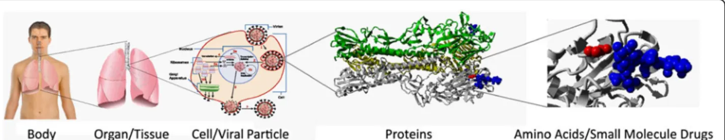
Figure 1The link between an influenza virus mutation and the altered course of infection.Schematic representation showing how a single viral amino acid mutation (right, red balls) can affect host cell receptor (blue balls) interaction, which can alter viral localization and where the infection takes place, which in turn can affect severity and symptoms for the patient (left). A thorough understanding of the effects of mutations on biological mechanisms is also important for other human diseases such as cancer as well as patient-specific response to different treatments. Attribution of images: The 3 left-most images of the composed figure are public domain or under free-to-use licenses at Wikimedia commons from the following sources: patient body and organ [118] and infected cell [119].
rising in occurrence [47], changes in hemagglutinin surface epitopes [48] and glycosylation sites as well as detect known [49] and novel [50-52] mutations in the neuramin- idase drug binding pocket that alter antiviral drug efficacy.
While the wealth of prior work on influenza is crucial for the ability to make relevant computational predictions, it shows that, with a concerted effort, similar successes may be achieved in other areas of high interest.
Conclusions from the sequence of the enterohemorrhagic O104:H4E. colistrain
Next generation sequencing has dramatically brought down the cost of genome sequencing but the current reality is that there usually is a long way from the initial genomic data to information relevant for clinicians.
However, there are exceptions. When an enterohemor- rhagic O104:H4E. colistrain caused a major outbreak in Germany [53] in 2011, the genome sequence was rapidly available through next generation sequencing [54]. At the same time, the Robert Koch Institute provided the microbial characterization including the clinically im- portant antibiotic susceptibility profile [55]. In principle, the information if a specific antibiotic drug is effective against an organism should be encoded in its genome by the presence of the known target gene of the respective drug as well as the absence of associated drug resistance factors. Clearly, the prerequisite for computationally de- riving an antibiotic susceptibility profile depends not only on the availability of the whole genome but also sufficiently complete annotation data for drug targets and resistance mechanisms of closely related strains or organisms. Since E. coli and related bacteria have been widely studied before in this regard, we show here that one can computationally identify antibiotic drugs that, potentially, can effectively target a new pathogen with available genome, such as the enterohemorrhagic O104:
H4E. colistrain. The steps to achieve this are essentially routine bioinformatics work but typically not easily ac- cessible to clinicians.
First, the available genome sequences (http://www.ncbi.
nlm.nih.gov/Traces/wgs/?val=AFOB01) were searched with BLASTX [56] for close to identical sequence matches against a database of known drug targets from DrugBank [57]. Requiring at least 97% sequence identity of theE. coli sequences to the proteins known to be drug targets ensures that also their structure will be highly similar and hence should represent the same drug binding properties. Sec- ond, we repeat the sequence search but this time against a database of known drug resistance factors from ARDB [58]
requiring a lower threshold of at least 60% identity to con- servatively pick up also more remote similarities to possible resistance factors. Third, we use a Perl script to parse the hits from the BLAST outputs as well as the drug target and resistance annotation data from the two databases and
finally identify the list of drugs for which a known target gene was found in the genome but no respective associated resistance factor.
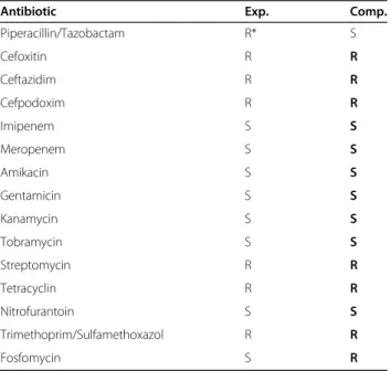
In order to validate the results, we compared our com- putational antibiotic susceptibility profile with the ex- perimental results. To our positive surprise, 15 out of 25 experimentally tested antibiotics were also covered by the existing databases and could, hence, be assessed through our computational workflow. The identity thresholds for the two sequence searches described above have been selected to produce the best possible match with the experimental data. Table 1 shows that thein silicoapproach correctly assigns resistance or sen- sitivity for 13 of the 15 antibiotics. In detail, the new bacterial strain was correctly predicted to be sensitive to 7 antibiotics and resistant to 6 drugs from the list. The only two cases of a mismatch from the prediction with the clinical experimental result are interesting and dis- cussed below.
The first case is the combination drug Piperacillin/
Tazobactam which we flag as sensitive but the Robert Koch Institute as resistant. Sequence searches identified a TEM-1 metallo beta-lactamase in O104:H4 E. coli which causes resistance to penicillins (including Pipera- cillin) by degrading them but we also find that there exists a specific inhibitor against TEM-1 metallo beta- lactamases, Tazobactam, which is given in combination with Piperacillin to inhibit the beta-lactamase and,
Table 1 Predicted potentially effective drugs against enterohemorrhagicE. coli
Antibiotic Exp. Comp.
Piperacillin/Tazobactam R* S
Cefoxitin R R
Ceftazidim R R
Cefpodoxim R R
Imipenem S S
Meropenem S S
Amikacin S S
Gentamicin S S
Kanamycin S S
Tobramycin S S
Streptomycin R R
Tetracyclin R R
Nitrofurantoin S S
Trimethoprim/Sulfamethoxazol R R
Fosfomycin S R
Experimentally measured (Exp.) versus computationally predicted (Comp.) antibiotics susceptibility profile. R. . .resistant; S. . .sensitive; *. . .defined as resistant (AES VITEK). Prediction and experimentally determined results coincide except for two cases (Piperacillin/Tazobactam and Fosfomycin) which are discussed in the text in detail.
therefore, increase efficacy of penicillins to which this strain should otherwise be resistant. In theory, this means that the computational prediction that Piperacil- lin/Tazobactam is effective should be correct. However, it turns out that, in clinical practice, this drug is recom- mended to be avoided due to possible inoculum effects.
Hence, the resistant flag from the clinical judgement according to the used VITEK AES experimental classifi- cation system.
The second case is Fosfomycin, to which the new strain was experimentally found to be sensitive while the computational approach assumed resistance due to the identification of a multidrugefflux pump protein anno- tated to also export Fosfomycin. This means that either the annotation is inaccurate or it would be interesting to further look into the detail of the few sequence differ- ences between the new and the previously known trans- porter (99% identity) to find determinants of activity and substrate specificity which could be considered in a fu- ture more comprehensive approach.
Overall, this crude workflow utilizing available data- bases shows that a computational antibiotics susceptibil- ity profile can be derived with some accuracy by combining next generation genome sequencing with fur- ther computational analysis, but it definitely still needs a critical experienced doctor who further scrutinizes and selects the most suitable treatment according to the cir- cumstances of the infected patient as well as includes any new clinical findings on drug responses of the re- spective strain.
Bacterial communication and cooperation in health and disease
The analysis of human microbiomes and small bacterial communities causing multi-bacterial diseases are among the most challenging and intriguing tasks of medical genome research today [59-61] also including the field of plant diseases [62]. The discovery of chemical com- munication among bacteria in the 1990s has fundamen- tally changed the traditional view that pictures bacteria as single-celled organisms living in isolation [63-66]. In the last fifteen years, it has become increasingly evident that bacteria have the potential to establish highly com- plex communities. Many microbes live in large, multi- species communities in which the participants jointly exploit the resources. Multispecies microbial consortia constitute a major form of life that is found in environ- ments ranging from high-altitude mountains (more than 8 km above sea level) to more than 10 km below the sur- face of the oceans, and have always been among the most important members and maintainers of the planet's ecosystem. The medical importance of this phenomenon is sweeping. Opportunistic pathogenes, such as Pseudo- nomas and Burkholderia species abound in hospital
environments, ready to attack patients weakened by dis- ease or injury. For instance, Pseudomonas aeruginosa usually does not harm a healthy human organism, but can be lethal in the lung of cystic fibrosis (CF) patients, or in burn wounds [67].
Many prokaryotes possess inter-cellular signaling sys- tems which allow species to colonise new habitats, to in- vade hosts and to spread over surfaces [63-66]. A typical example is quorum sensing (QS) which enables bacteria to switch from low activity to high activity regimes using signaling molecules as well as“public goods”(e.g. surfac- tants, enzymes, siderophores) that facilitate movement, nutrient uptake amongst other things [65,66]. We share the widespread opinion that the“change of bacterial life- style”is crucial for colonizing habitats and infecting sus- ceptible hosts–unfortunately the signalling systems that orchestrate the underlying communication and collabor- ation mechanisms are not accurately annotated in bacter- ial genomes. Therefore, a systematic characterization of QS systems in Gram negative bacteria was carried out [68,69] and a modelling effort to map out the theoretic- ally possible consequences of communication and collab- oration in bacterial populations was initiated [70-72].
Virulence and adaptability of many Gram-negative bac- terial species are associated with an N-acylhomoserine lactone (AHL) gene regulation mechanism called quorum sensing (QS). The arrangement of quorum sens- ing genes is variable throughout bacterial genomes, al- though there are unifying themes that are common among the various topological arrangements. A bioinfor- matics survey of 1403 complete bacterial genomes revealed characteristic gene topologies in 152 genomes that could be classified into 16 topological groups [68,69]. A concise notation for the patterns was devel- oped and it was shown that the sequences ofLuxRregu- lators and LuxI autoinducer synthase proteins cluster according to the topological patterns.
The macroscopic behavior of bacterial communities is notoriously difficult to study, colony patterns, invasion/
colonization events depend on a multitude of parameters many of which cannot be reproduced in lab cultures.
Therefore, computational modeling, and particularly the use of simplified minimal models is a very important tool for studying the behavior of populations in rational terms. Agent-based models of communicating and col- laborating bacteria have developed [70]. The bacterial cells are represented by agents randomly moving on a plain (such as an agar surface), while consuming nutri- ents, secreting signal molecules and “public goods”.
Nutrients, signals and public goods are diffusing on the surface, and their local concentration exceeds a thresh- old, the metabolism and movement of bacterial agent switches to a more intensive state. In this model signals are the means of communications, and public goods are
the means of cooperation as can be observed in QS bac- teria. Even though highly simplified, the model reflects the crucial behavior patterns of communicating/cooper- ating bacteria in an open, nutrient/limited environment.
Namely, 1) isolated bacteria cannot survive; only bacteria reaching a critical population size (“quorum”) have a chance for survival. 2) Bacteria self-organize into com- pact communities or “active zones”in which signals and public goods are present in sufficient amounts [70]. 3) Col- laborating communities can collapse if non-cooperating mutants are present [71,72].
Modeling the mutants of QS mechanisms is highly relevant for disease prevention. There is a very vivid interest from the pharmaceutical and pesticide indus- tries, analysts agree that interventions targeting quorum sensing are among the major trends of the future. Since many bacteria use quorum sensing for infection, it is plausible to think about jamming strategies. According to one such scenario, one can saturate the surface of a plant with a signal molecule that will call bacteria to at- tack. If a lonely pathogen lands on the surface, it will im- mediately start to attack, but at the wrong time and place. Since it is alone, it will perish. Or, we can put a gene into the plant that produces an enzyme capable of destroying the signal molecule of the pathogenic bac- teria, so that those will never wage an attack. But both strategies can strike back since they can also destroy the signaling of the beneficial bacteria that are essential to the host. According to a third scenario one may pre- vent the growth of an infecting pathogen by a greedy but antibiotic sensitive mutant of the same species, and then we eliminate the mutant by an antibiotic that specifically acts on that mutant. This is very appealing, but what do we do if the mutant created to heal gets some harmful genes or looses its antibiotic susceptabil- ity? Many similar questions can be studied using com- putational models [73].
Impact of bioimage informatics on healthcare Most likely, the penetration of automated evaluation tools for the analysis of clinically relevant histological images in diagnostic contexts is one of the areas that will experience great changes in the near future. The process of biomedical imaging involves little or no discomfort to the patients, while providing an effective tool for diagno- sis. However, successful usage of images requires a high level of human intelligence, making automated image analysis by machines a challenging task. Currently, the gold standard for diagnosis through imaging is by experi- enced clinicians, typically radiologists or pathologists. It takes many years to train proficient clinicians to analyze images manually and, despite that, this gold standard is not perfect and suffers from subjective variations be- tween different clinicians.
Advances in image processing, pattern recognition and computer vision in the past decades have boosted the possibilities for the application of computing technology.
Currently, the focus is on computer aided diagnosis ra- ther than to achieve a fully automated approach. Soft- ware that can support decision making and reduce the workload of clinicians, especially in routine operations, is extremely useful and valuable. Besides the direct deriv- ation of clinically relevant conclusions from the images, such systems call also for the integration with databases of medical ontologies, the patients’medical records, etc.
Computational image analysis methods can be broadly categorized into those used for assessment, diagnosis and surgery. This section attempts to cover several ex- emplary areas of imaging and image analysis in health- care. Because of the large extent of research work ongoing in academic bioimage informatics and medical image analysis and the growing engagement of the in- dustry, this section cannot be comprehensive but rather we seek to cover a broad spectrum.
Digital pathology
Advances in computer vision and microscopy instru- mentation have made digital pathology an important emerging field. The objective is to aid the pathologist in the analysis of high resolution cellular images obtained through biopsy. For example, highlighting regions of interest or reducing diagnostic variation can generate a big impact. Histological images from various organs such as prostate [74], breast [75] and liver have been the object of algorithm development.
Here, we shall focus our discussion on prostate digital pathology. Prostate cancer has a high prevalence rate worldwide. For example, it is the most common non- cutaneous male cancer in the United States [76] and it is the 3rd most common male cancer in Singapore [16].
The American Cancer Society report in 2009 estimates 192,280 new prostate cancer cases with 27,360 prostate cancer specific death [76]. The severity of prostate can- cer diagnostics is compounded by disagreements be- tween individual pathologists with regard to grading using the Gleason classification [77]. This agreement be- tween different pathologist can be as low as 70% [78]
and up to 29% of Gleason gradings were different be- tween pre- and post-operative prostate cancer specimen [79]. Hence, having objective computer algorithms to aid in prostate pathology assessment is essential to improve diagnosis.
Most computational methods are developed to analyze microscopy images on the standard hematoxylin/eosin stain. The goals are gland segmentation since the archi- tecture of glands is critical for Gleason grading and the identification and segmentation of nuclei since this is useful for detecting nuclei signatures specific to
cancerous cells. Common computer vision techniques used are level sets [80], fractal analysis [81] and machine learning [80,82-86]. These techniques are used to seg- ment glands [80,85] and nuclei [82,84] or to identify regions of malignancy directly [83].
Computer vision in dermatology
Assessment of skin condition and health is both import- ant for clinical medicine as well as for the cosmetics in- dustry. At present, assessment of the skin typically involves a trained dermatologist who will examine fea- tures such as textures and landmarks. While training of dermatologists takes many years, the subsequent diagno- sis suffers from subjective interpretation differing among dermatologists. Hence, a more objective approach is in demand.
Considerable effort is ongoing to analyze skin surfaces through the use of objective computational methods.
Protocols to ensure objective and consistent imaging of human skin (for example, in a well-controlled lighting environment) are vital for reliable diagnosis by computer algorithms [87-89]. Image acquisition is followed by the application of task-dependent image processing and computer vision methods. Liu et al. [90] use texture analysis to create an objective way of evaluating the ef- fectiveness of treatment. A neural network framework has been developed to analyze the human skin condi- tions such as color, roughness, glossiness or tension [91,92]. Skin images have also been studied with data mining methods [88,93] and via modeling/reconstruct- ing the skin surface [89,94].
Computer vision in eye diseases
Imaging methods for eye diseases are unique among bioi- maging techniques because images of the eyes are easily accessible using conventional light cameras. There is no need for expensive and sophisticated machines such as a computer tomograph or magnet resonance imager. A common imaging modality is the optical coherence tom- ography; other imaging methods such as fundus photog- raphy, ultrasound and infra-red imaging are also used.
Although image analysis has been used in the assessment of many eye diseases, we will focus our discussion on glaucoma and dry eye disease in this paper.
Angle closure glaucoma
According to a world health organization report [95], glaucoma is a major global cause of blindness (approxi- mately 5.2 million cases and about 15% of all cases of blindness). The impact of glaucoma on public health will increase with an aging population. However, the lack of a comprehensive measure of glaucoma compounded with its ability to cause sudden blindness makes it hard for treatment planning. Surprisingly, about 50-90% of
potential patients in the world are unaware that they have glaucoma [96,97].
Glaucoma is classified into angle closure and open angle glaucoma according to the drainage angle, the angle between the cornea and iris. Primary angle closure glaucoma is the major form of glaucoma in Asia, in par- ticular, among the Chinese population. It was suggested that angle closure glaucoma causes more blindness than open angle glaucoma in relative terms [98].
A common way for assessment of angle closure glau- coma is through gonioscopy in which the doctor uses an optical instrument to look at the anterior chamber to de- cide if the drainage angle is open or close. Ultrasound [99] and optical coherence tomography (OCT) [100]
images are also used for assessment. Computer vision techniques are used for analyzing eye images derived from the different modalities. As it takes much effort to master the technique of gonioscopy, Cheng et al. [101]
developed a computational technique for RetCam images. A machine-learning based method aids glaucoma diagnosis by analyzing the cup-to-disc ratio measured on fundus images [102]. OCT images provide high resolution and a 3D view of the anterior chamber.
Image analysis software has been developed to make precise measurements of important geometric informa- tion such as anterior chamber area, anterior chamber width, iris thickness, etc. on OCT images [103]. These data can then be correlated to generate new clinical knowledge [104,105].
Image analysis in assessing the dry eye condition
The disease of dry eye has no clear definition; generally, it is a condition in which there is an unstable tear film during the open eye state. The dry eye condition has a prevalence rate of 10-20% in Sweden, Japan, Australia and several other countries. The most common treat- ment of dry eye is application of eye drops [106].
One cause of dry eye disease is meibomian gland dys- function. The meibomian glad is located at the inside of the tarsel plate that supplies meibum, an oily substance, which forms a protective layer to the tear film. Dysfunc- tion of meibomian glands causes lack of meibum and, often, resulted in degeneration of meibomian glands.
The morphology of meibomian glands can be imaged using an infra-red camera mounted on a conventional slit lamp camera [106]. This imaging technology has enabled the application of advanced computer vision techniques for better diagnosis and patient management.
Images from healthy meibomian glands shows a strip like pattern in gland morphology; with the strips being relatively straight, parallel and equally spaced. Images of highly degenerated glands show no strip like patterns at all, but only small isolated regions of remnant glands.