Journal of the American Heart Association
ORIGINAL RESEARCH
Pregnancy Outcomes in Women After
Arterial Switch Operation for Transposition of the Great Arteries: Results From ROPAC (Registry of Pregnancy and Cardiac
Disease) of the European Society of
Cardiology EURObservational Research Programme
Oktay Tutarel, MD†; Karishma P. Ramlakhan , MD†; Lucia Baris, MD; Maria T. Subirana, MD;
Judith Bouchardy, MD, PhD; Attila Nemes, MD, PhD; Niels G. Vejlstrup, MD, PhD; Olga A. Osipova , MD, PhD;
Mark R. Johnson, MD, PhD; Roger Hall, MD, PhD; Jolien W. Roos-Hesselink, MD, PhD ; the ROPAC (Registry of Pregnancy and Cardiac Disease) Investigators Group*
BACKGROUND: In the past 3 decades, the arterial switch procedure has replaced the atrial switch procedure as treatment of choice for transposition of the great arteries. Although survival is superior after the arterial switch procedure, data on preg- nancy outcomes are scarce and transposition of the great arteries after arterial switch is not yet included in the modified World Health Organization classification of maternal cardiovascular risk.
METHODS AND RESULTS: The ROPAC (Registry of Pregnancy and Cardiac disease) is an international prospective regis- try of pregnant women with cardiac disease, part of the European Society of Cardiology EURObservational Research Programme. Pregnancy outcomes in all women after an arterial switch procedure for transposition of the great arteries are described. The primary end point was a major adverse cardiovascular event, defined as combined end point of ma- ternal death, supraventricular or ventricular arrhythmias requiring treatment, heart failure, aortic dissection, endocarditis, ischemic coronary events, and thromboembolic events. Altogether, 41 pregnant women (mean age, 26.7±3.9 years) were included, and there was no maternal mortality. A major adverse cardiovascular event occurred in 2 women (4.9%): heart failure in one (2.4%) and ventricular tachycardia in another (2.4%). One woman experienced fetal loss, whereas no neonatal mortality was observed.
CONCLUSIONS: Women after an arterial switch procedure for transposition of the great arteries tolerate pregnancy well, with a favorable maternal and fetal outcome. During counseling, most women should be reassured that the risk of pregnancy is low.
Classification as modified World Health Organization risk class II seems appropriate.
Key Words: arterial switch operation ■ pregnancy and cardiac disease ■ pregnancy outcomes ■ transposition of the great arteries
Correspondence to: Jolien W. Roos-Hesselink, MD, PhD, Department of Cardiology, Erasmus University Medical Center, Rg-435, PO Box 2040, 3000 CA Rotterdam, the Netherlands. E-mail: j.roos@erasmusmc.nl
*A complete list of the ROPAC (Registry of Pregnancy and Cardiac disease) Investigators Group can be found in Appendix.
†Dr Tutarel and Dr Ramlakhan are co–first authors.
For Sources of Funding and Disclosures, see page 10.
© 2020 The Authors. Published on behalf of the American Heart Association, Inc., by Wiley. This is an open access article under the terms of the Creative Commons Attribution-NonCommercial-NoDerivs License, which permits use and distribution in any medium, provided the original work is properly cited, the use is non-commercial and no modifications or adaptations are made.
JAHA is available at: www.ahajournals.org/journal/jaha
T
he first successful arterial switch procedure for patients with transposition of the great arter- ies (TGA) was reported in 1975 by Jatene and colleagues.1 It establishes a biventricular circulation, where the morphologic left ventricle supports the systemic circulation. This is in contrast to the previ- ously performed atrial switch procedure, after which the morphologic right ventricle acts as a systemic ventricle. This systemic right ventricle is prone to fail, leading to heart failure and a diminished survival rate.2 Therefore, for the past 30 years, the arterial switch has been the surgical approach of choice for TGA.3 Consequently, the number of women after an arterial switch procedure for TGA reaching childbearing age is increasing, although not without complications.Neoaortic valve regurgitation, dilatation of the aor- tic root, ischemia attributable to coronary reinsertion problems, ventricular dysfunction, and right ventric- ular outflow tract obstruction have all been reported after the arterial switch.4 Because pregnancy poses a
hemodynamic challenge to the maternal circulation, it might increase the risk for these complications. Data on the outcome of pregnancies in women after an arterial switch procedure are scarce. Single cases of successful pregnancies were reported in 2001 and 2006,5,6 as well as retrospective series from single centers7–9 and one study from 2 centers.3 These re- ported the outcome of pregnancies in 9, 10, 11, and 15 women, respectively. The rate of adverse events differed significantly between these reports, whereas larger prospective studies are lacking, meaning that uncertainty remains about the impact of pregnancy in women after an arterial switch procedure.3,7–9 Accordingly, TGA after an arterial switch procedure is not yet classified in the modified World Health Organization classification, the most commonly used disease-specific risk assessment of pregnancy in women with cardiac disease.10 Consequently, the aim of this study is to provide a much more accurate assessment of maternal and fetal outcomes of preg- nancy in this group of women.
METHODS Study Design
The ROPAC (Registry of Pregnancy and Cardiac dis- ease) is an international, prospective, observational registry of pregnant patients with structural heart dis- ease, including valvular and congenital heart disease, ischemic heart disease, aortic pathological features, and pulmonary arterial hypertension. Study design and methods have been described previously.11 The European Society of Cardiology working groups on congenital heart disease and valvular heart dis- ease initiated ROPAC in 2007 and subsequently it was embedded in the EURObservational Research Programme of the European Society of Cardiology.
Ethical approval or Institutional Review Board ap- proval as well as patients’ informed consent were ob- tained if necessary, according to local requirements.
Pregnant women who were included in the ROPAC prospectively between January 2007 and January 2018 and had previously undergone an ar- terial switch procedure for TGA were included in this study. The data that support the findings of this study are available from the corresponding author on rea- sonable request.
Data
The ROPAC study protocol and the first results of this registry were published in 2013.11 Patients with a diagnosis of TGA treated with an arterial switch procedure were identified from the registry and compared with the rest of the total ROPAC cohort.
Baseline characteristics collected before pregnancy
CLINICAL PERSPECTIVE
What Is New?
• Women after arterial switch operation for trans- position of the great arteries tolerate pregnancy well, on the basis of 41 pregnancies in a pro- spective worldwide registry.
• In this cohort, there was no maternal mortality, and major adverse cardiovascular events oc- curred in 4.9%, as heart failure (2.4%) and ven- tricular tachycardia (2.4%).
• Prematurity (17.1%) was the most important fetal complication, followed by low birth weight (14.6%), but fetal loss occurred in only 2.4%.
What Are the Clinical Implications?
• Women after arterial switch for transposition of the great arteries can be counseled that pregnancy is safe, with a low risk of cardiac complications.
• Currently unclassified in the modified World Health Organization classification for maternal cardiovascular risk, this study suggests that pregnancy after arterial switch for transposition of the great arteries can be classified as modi- fied World Health Organization risk class II.
Nonstandard Abbreviations and Acronyms
ROPAC Registry of Pregnancy and Cardiac disease
TGA transposition of the great arteries
included age, New York Heart Association functional class, ECG rhythm, diagnosis, risk factors (smoking habits, hypertension, and diabetes mellitus), medi- cation, previous interventions, parity and obstetric history, and echocardiographic measurements. The provision of echocardiographic data was facultative.
Countries were divided into developed or emerging countries, according to the International Monetary Fund Classification.12
Definitions and End Points
The primary combined end point was the occur- rence of a major adverse cardiovascular event, defined as combined end point of maternal death, supraventricular or ventricular arrhythmias requiring treatment, heart failure, aortic dissection, endocar- ditis, ischemic coronary event, and other thrombo- embolic events. The secondary end points were adverse obstetric outcomes and adverse fetal/neo- natal outcomes. Heart failure was defined accord- ing to the American College of Cardiology/American Heart Association guidelines,13 and heart failure episodes were only included when they required hospital admission, new treatment, or change in the existing treatment regimen. Ventricular function was categorized as normal, mildly impaired, mod- erately impaired, or severely impaired. Postpartum hemorrhage was defined as increased blood loss during delivery up to 24 hours postpartum, requir- ing specific interventions. Hemolysis, elevated liver enzymes, low platelets syndrome, preeclampsia and eclampsia, and pregnancy-induced hypertension were defined according to the International Society for the Study of Hypertension in Pregnancy 2018 statement.14 Fetal mortality was defined as the death of a fetus after 20 weeks of gestation until birth.
Neonatal mortality was defined as the death of a live-born baby in the first 6 months of life. Premature birth was defined as birth before 37 weeks of gesta- tion. Low birth weight was defined as a birth weight of <2500 g. Low Apgar score was defined as an Apgar score at 5 minutes of <7. All outcomes were examined for the duration of the pregnancy and up to 6 months postpartum.
Statistical Analysis
Data are presented as mean values with SD if nor- mally distributed and median with interquartile range if skewed. Categorical data are presented as frequen- cies and percentages. Baseline characteristics and outcomes were compared between women after arterial switch for TGA and the other pregnancies in the ROPAC cohort, using Student t tests and χ2 tests where appropriate. P<0.05 (2-sided test) was con- sidered significant. All statistical tests and analyses
were performed with SPSS version 21.0 (SPSS Inc, Chicago, IL).
RESULTS
There were 41 women after arterial switch for TGA among the 5739 patients included in ROPAC from January 2007 to January 2018.15 Baseline character- istics are presented in Table 1. Their mean age was 26.7±3.9 years, 21 women (51.2%) were primigravida, and 2 women (4.9%) were from an emerging country, as opposed to 40% in the rest of the ROPAC cohort (P<0.001). Most of the women (97.6%) were asymp- tomatic or had only mild symptoms (New York Heart Association class I/II) before pregnancy. Only one woman was in New York Heart Association class III. Cardiac medication was used by 24.4% before pregnancy.
Maternal Outcomes
Maternal cardiac outcomes of pregnancy are presented in Table 2. There was no maternal mortality either in pregnancy or up to 6 months after delivery, whereas it occurred in 0.7% of the other ROPAC pregnancies (0% versus 0.7%; P=0.560). Hospital admission for a cardiac reason was required in one woman (2.4%) because of heart failure. The woman had a history of a Blalock-Taussig shunt before the arterial switch pro- cedure and had experienced heart failure symptoms before pregnancy. Unfortunately, a prepregnancy echocardiogram was not available. She was delivered at 36 weeks gestation by an elective cesarean sec- tion. In the third month postpartum, acute heart failure developed. At that point, left ventricular systolic func- tion was moderately impaired, with an ejection fraction of 45%, without significant aortic or pulmonary valve dysfunction.
There were no cases of supraventricular tachy- cardia, and ventricular tachycardia occurred in one woman (2.4%). In the rest of the ROPAC cohort, ven- tricular tachycardia occurred in 1.6% (2.4% versus 1.6%; P=0.652), whereas supraventricular tachycar- dia occurred in 1.7% (0% versus 1.7%; P=0.404).
Before her arterial switch procedure, this woman underwent banding of the pulmonary artery and a Blalock-Taussig shunt. Before pregnancy, echocar- diography showed good biventricular function with- out significant valve dysfunction. During the second and third trimesters, ventricular tachycardia and frequent ventricular ectopic beats occurred, which were effectively treated with metoprolol without re- quiring hospital admission. After induction of labor at 38 weeks of gestation, she had a vacuum-assisted vaginal delivery. After pregnancy, no further arrhyth- mias were noted.
Table 1. Baseline Characteristics
Characteristic TGA After Arterial Switch (n=41) Other ROPAC (n=5698) P Value
Age, mean (SD), y 26.7 (3.9) 29.6 (5.6) 0.002
Living in an emerging country 2 (4.9) 2279 (40) <0.001
Primigravida 21 (51.2) 2552 (44.9) 0.420
Current smoking 1 (2.4) 227 (4.6) 0.617
Underlying cardiac pathological features Associated cardiac defects
Ventricular septal defect 13 (31.7) 703 (12.3) <0.001
PDA 4 (9.8) 145 (2.5) 0.004
Coarctation of the aorta 2 (4.9) 301 (5.3) 0.129
Prior interventions/surgery
Arterial switch 41 (100) 41 (0.7) NA
Time since arterial switch, mean (SD), y 25.7 (0.7) NA NA
Ventricular septal defect closure 13 (31.7) 908 (15.9) 0.006
Pulmonary banding 12 (29.3) NA NA
Rashkind procedure/surgical atrial septostomy 9 (22.0) NA NA
Blalock-Taussig shunt 5 (12.2) NA NA
Pulmonary valve intervention* 4 (9.8) 230 (4) 0.065
Coarctation of aorta resection 2 (4.9) 272 (4.8) 0.150
Percutaneous coronary intervention 1 (2.4) 63 (1.1) 0.418
Aortic surgery 1 (2.4) 235 (4.1) 0.588
Dilatation of pulmonary artery stenosis 1 (2.4) NA NA
PDA closure 1 (2.4) NA NA
Prepregnancy cardiac status
Diabetes mellitus 0 (0) 90 (2) 0.412
Chronic hypertension 1 (2.4) 379 (6.8) 0.271
Signs of heart failure 1 (2.4) 595 (10.6) 0.090
Left ventricular ejection fraction <40% 1 (2.4) 252 (4.4) 0.538
Pulmonary hypertension 1 (2.4) 44 (0.8) 0.568
NYHA class
I 38 (92.7) 4169 (73.2) 0.005
II 2 (4.9) 1189 (20.9) 0.012
III 1 (2.4) 175 (3.1) 0.815
IV 0 (0) 28 (0.5) 0.653
Aortic stenosis 0 (0) 397 (7) 0.077
Aortic regurgitation 14 (34.1) 876 (15.4) <0.001
Mild 13 (31.7) 585 (10.4)
Moderate 1 (2.4) 236 (4.2)
Severe 0 (0) 55 (1)
Pulmonary stenosis 14 (34.1) 443 (8) <0.001
Aortic dilatation 4 (9.8) 170 (3) 0.012
Prepregnancy cardiac medication 10 (24.4) 2059 (36.1) 0.119
β Blockers 2 (4.9) 561 (9.8) 0.287
Diuretics 1 (2.4) 216 (3.8) 0.651
ACE inhibitor 2 (4.9) 155 (2.7) 0.399
Antiplatelet drugs 2 (4.9) 232 (4.1) 0.795
Drug name not included in database 3 (7.3) NA NA
Values are number (percentage) if not otherwise stated. ACE indicates angiotensin-converting enzyme; NA, not available; NYHA, New York Heart Association;
PDA, persistent ductus arteriosus; ROPAC, Registry of Pregnancy and Cardiac disease; and TGA, transposition of the great arteries.
*All valve interventions were surgical repairs of the pulmonary valve; there were no aortic valve interventions or any valve replacements.
On the basis of these 2 events, the total major ad- verse cardiovascular event rate was 4.9%, and no other events or interventions occurred during preg- nancy or in the 6-month follow-up after delivery. An echocardiographic assessment of ventricular systolic function before pregnancy or during the first trimes- ter was available in 23 women (56.1%). Left ventricular systolic function was normal in 22 (95.7%), whereas it was moderately impaired in 1 (4.3%). Right ventricular systolic function was normal in 18 women (78.3%) and moderately impaired in 5 (21.7%). Serial data (prepreg- nancy and postpartum) were available in 13 women (Figure 1). No deterioration in ventricular function or aortic valve function was observed.
Obstetric and Fetal Outcomes
Obstetric and fetal outcomes are presented in Table 3.
Hypertensive disorders of pregnancy occurred in 2 pregnancies (4.9%), compared with 4.4% of the rest of the ROPAC cohort (P=0.865). Median gestational age at delivery was 39 weeks (interquartile range, 37–40 weeks), and mean birth weight was 2962.2±99.8 g, with low birth weight in 14.6% versus 11.7% (P=0.561) in the other ROPAC pregnancies. There were 7 pre- mature births (17.1%) at a median gestational age of 35 weeks (interquartile range, 34–36 weeks), of which 5 were spontaneous labors and 2 were medically in- duced for obstetric reasons. A cesarean section was performed in 46.3% of women after arterial switch for TGA, compared with 49.8% of the rest of the ROPAC cohort (P=0.662). Fetal death occurred once in our co- hort (2.4%), and in 1.3% of the other women included in ROPAC (P=0.778).
DISCUSSION
The ROPAC, an international prospective registry, studies the outcomes of pregnancy in women with structural heart disease. In this article, we have fo- cused on women with TGA treated with an arterial
switch procedure, and our findings are summarized in Figure 2. Nearly all women tolerated pregnancy well, with a low rate of major adverse cardiovascular events (only 4.9%) and without maternal or neonatal mortal- ity. Fetal mortality occurred in only one pregnancy, whereas in terms of maternal morbidity, heart failure and arrhythmias both also occurred only once.
Maternal Outcomes
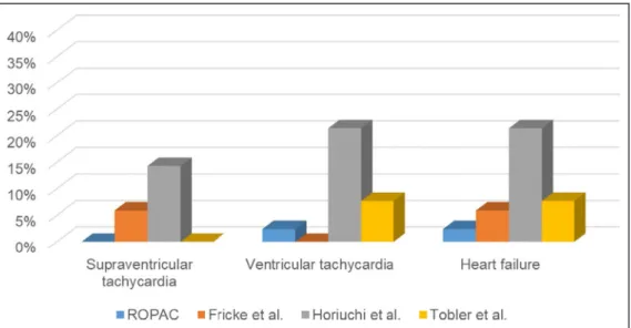
This first large prospective study shows that maternal outcome is favorable, whereas by comparison in pre- vious retrospective studies, maternal cardiac events occurred in 12% to 29% of women,3,8,9 although one recent study (n=15) reported no cardiac events at all.7 Arrhythmias and heart failure were most frequently encountered in these older studies (Figure 3). The wide variation in the number of events is probably because of the small number of women included in these studies. Furthermore, some of these reports included women from an earlier surgical era and with a more severe prior clinical course.3,7 Indeed, both women with cardiac events in our study had had a Blalock-Taussig shunt before the arterial switch pro- cedure, probably illustrating their more complex clini- cal course.
Our contemporary prospective data are reassuring about maternal complications. This information is critically important for women with an arterial switch who are contemplating pregnancy, as they can now be reassured that their chance of complications is low. Because of the low number of events, an analysis to identify risk factors for adverse outcomes was not possible.
Most patients in our study had a normal left (95.7%) and right (78.3%) ventricular systolic function prepreg- nancy. Furthermore, in the women with serial echocar- diographic data, there was no deterioration in left or right ventricular function (Figure 1). In the study of Horiuchi and colleagues, 50% of women with a cardiac event during pregnancy and 20% of those without a cardiac event had
Table 2. Maternal Outcomes of Pregnancy
Outcome TGA After Arterial Switch (n=41) Other ROPAC (n=5698) P Value
Maternal mortality ≤6 mo postpartum 0 (0) 40 (0.7) 0.590
Hospital admission for a cardiac reason 1 (2.4) 757 (13.3) 0.041
Heart failure 1 (2.4) 654 (11.5) 0.070
Supraventricular tachycardia 0 (0) 95 (1.7) 0.404
Ventricular tachycardia 1 (2.4) 89 (1.6) 0.652
Thromboembolic events 0 (0) 87 (1.5) 0.425
Endocarditis 0 (0) 33 (0.6) 0.625
Acute coronary syndrome 0 (0) 24 (0.4) 0.677
Aortic dissection 0 (0) 5 (0.1) 0.849
Values are number (percentage). ROPAC indicates Registry of Pregnancy and Cardiac disease; and TGA, transposition of the great arteries.
a reduced left ventricular function.8 This might explain the higher number of cardiac events in their study. During follow-up, no further deterioration of left ventricular func- tion was observed.8 Similar to our experience, Stoll and colleagues reported no adverse cardiac events, with all
women having normal left ventricular systolic function before their first pregnancy, whereas one woman had a mildly impaired function after her second pregnancy.7 Therefore, an echocardiogram before pregnancy is an important tool for risk assessment.
Figure 1. Echocardiographic prepregnancy and postpartum ventricular systolic function (A and B) and aortic valve function (C) in the women in whom serial echocardiographic data were available.
The postpartum echocardiogram was performed at a mean of 8.2 months postpartum (SD, 1.2 months), with a range of 1 to 15 months.
Table 3. Obstetric and Fetal Outcomes of Pregnancy
Outcome TGA After Arterial Switch (n=41) Other ROPAC (n=5698) P Value
Pregnancy-induced hypertension 0 (0) 77 (1.4) 0.454
(Pre) eclampsia or HELLP syndrome 2 (4.9) 170 (3) 0.478
Gestational diabetes mellitus 0 (0) 160 (2.8) 0.274
Postpartum hemorrhage 2 (4.9) 168 (2.9) 0.468
Cesarean section 19 (46.3) 2662 (49.8) 0.662
Emergency cesarean section 6 (14.6) 760 (13.3) 0.808
Emergency cesarean section for cardiac reason 0 (0) 132 (2.3) 0.324
Fetal death 1 (2.4) 71 (1.2) 0.778
Neonatal death 0 (0) 33 (0.6) 0.625
Fetal congenital heart disease 0 (0) 156 (2.7) 0.283
Premature birth 7 (17.1) 898 (18) 0.947
IUGR 1 (2.4) 253 (4.4) 0.535
Low Apgar scores 2 (4.9) 395 (6.9) 0.606
Low birth weight 6 (14.6) 667 (11.7) 0.561
Birth weight, mean (SD), g 2962.2 (99.8) 2970.9 (639.6) 0.941
Values are number (percentage), except for birth weight. HELLP indicates hemolysis, elevated liver enzymes, low platelet count; IUGR, intrauterine growth retardation; ROPAC, Registry of Pregnancy and Cardiac disease; and TGA, transposition of the great arteries.
In comparison to previous reports of pregnancy after the atrial switch procedure, which reported sev- eral complications, most frequently arrhythmias and heart failure, with a rate between 7% and 22% and between 7% and 21%, respectively, depending on the study design and patient population,16–19 the ma- ternal outcome in our cohort after the arterial switch procedure is much more favorable. These results
emphasize the advantages of the later procedure further.
Obstetric and Fetal Outcomes
In our contemporary cohort, both obstetric and fetal complications were less frequent than reported by previous studies.8,9 Interestingly, the most recent
Figure 2. Pregnancy outcomes in women after arterial switch operation for transposition of the great arteries.
VSD indicates ventricular septal defect.
Figure 3. Rate of adverse cardiovascular events in comparison to previously published reports.
ROPAC indicates Registry of Pregnancy and Cardiac disease.
study reported an unexpected high incidence of ma- ternal obstetric complications (58.8%).9 Most of these complications were attributable to postpartum hemor- rhage, whereas this occurred in only 4.9% of women in our cohort. Maternal obstetric complications are not mentioned in detail in the other 2 series.3,7
The most frequent observed fetal complication in our study was premature birth (17.1%), followed by low birth weight (14.6%). Both are not signifi- cantly different from the rest of the ROPAC cohort.
Iatrogenic premature birth was because of obstet- ric reasons and never because of cardiac causes.
Although one fetal loss was observed, there was no neonatal mortality. Unfortunately, not all previous studies commented on the fetal outcome in detail.
Stoll and colleagues reported premature birth in 9.1%
of pregnancies,7 whereas in the series of Horiuchi and colleagues, 21% of pregnancies did not reach full term.8 In the latter study, low birth weight was observed in 27%,8 whereas Tobler et al reported a lower rate, with 14.3%.3 Therefore, the results of our study provide reassurance about the fetal outcome compared with previous literature.
A cesarean section was performed in 46.3% of women in our study, which is comparable to the high number of women with a cesarean section in the whole ROPAC cohort (49.8%; P=0.662). In the previous stud- ies, the frequency of cesarean sections varied between 36% and 47%.7–9 These high rates could be attributable to the congenital heart defect of the mother, because it is conceivable that women with heart disease in gen- eral are handled with more care and apprehension by treating physicians and are thus given a cesarean sec- tion (as many physicians prefer this apparently more controlled environment). This could be especially true for congenital heart defects, for which data on preg- nancy outcomes are scarce as they are for patients with an arterial switch procedure for TGA. However, the available data do not support this approach,20 and guidelines recommend that a cesarean section should only be considered for obstetric indications or a lim- ited number of cardiac indications, including labor on oral anticoagulants, aggressive aortic pathological fea- tures, acute intractable heart failure, and severe pul- monary hypertension.10 This is also reflected by the reassuringly low number (15%) of emergency cesarean sections in our study, none of which was attributable to cardiac reasons, which confirms the results of Stoll et al,7 whereas it is much lower than the recently re- ported 63%.9 The last report had an unexpectedly high number of obstetric complications. In addition, the use of Cesarean section as a primary mode of delivery is country and center dependent and so could influence the numbers in our multicenter study.
Interestingly, only 4.9% of women from our cohort are living in an emerging country, as opposed to 40%
of the rest of the ROPAC cohort (P<0.001). The main reason for this finding could be that the arterial switch procedure for TGA was often not available to children born in the developing world for a long period.21 In re- cent years, this has changed,22 and therefore, we can expect an increase in the numbers of women after an arterial switch procedure for TGA reaching childbear- ing age in these emerging countries.
A limitation of our study is that serial echocardio- graphic data were available in only a limited number of women. Therefore, analysis on the course and outcome of ventricular function as well as aortic and pulmonary valve function, especially considering the neoaortic valve, was not possible in all women. Despite these limitations, this prospective registry included the largest number of women after an arterial switch procedure for TGA re- ported to date, providing important information related to the maternal and fetal outcome in these women.
CONCLUSIONS
In conclusion, women after an arterial switch proce- dure for TGA tolerate pregnancy well, with a favorable maternal and fetal outcome. Therefore, women after an arterial switch procedure for TGA should be coun- seled that pregnancy is low risk. With a major adverse cardiac event rate of 4.9%, this corresponds to risk class modified World Health Organization II.
APPENDIX
EORP Oversight Committee
Christopher Peter Gale, Chair, GB, Branko Beleslin, RS, Andrzej Budaj, PL, Ovidiu Chioncel, RO, Nikolaos Dagres, DE, Nicolas Danchin, FR, David Erlinge, SE, Jonathan Emberson, GB, Michael Glikson, IL, Alastair Gray, GB, Meral Kayikcioglu, TR, Aldo Maggioni, IT, Klaudia Vivien Nagy, HU, Aleksandr Nedoshivin, RU, Anna-Sonia Petronio, IT, Jolien Roos-Hesselink, NL, Lars Wallentin, SE, Uwe Zeymer, DE.
Executive Committee
Roger Hall GB (Co-Chair), Jolien Roos-Hesselink NL (Co-Chair), Joerg Stein, AT, William Anthony Parsonage, AU, Werner Budts, BE, Julie De Backer, BE, Jasmin Grewal, CA, Ariane Marelli, CA, Harald Kaemmerer, DE, Guillaume Jondeau, FR, Mark Johnson, GB, Aldo P. Maggioni, IT, Luigi Tavazzi, IT, Ulf Thilen, SE, Uri Elkayam, US, Catherine Otto, US, Karen Sliwa, ZA.
ROPAC Investigators
Argentina—Buenos Aires: A. Aquieri, A. Saad, H. Ruda Vega, J. Hojman, J. M. Caparros, M. Vazquez Blanco.
Australia—Elizabeth Vale: M. Arstall, C. M. Chung, G.
Mahadavan, E. Aldridge, M. Wittwer, Y. Y. Chow, Herston:
W. A. Parsonage, K. Lust, New Lambton Heights: N.
Collins, G. Warner, R. Hatton, A. Gordon, E. Nyman.
Austria—Innsbruck: J. Stein, E. Donhauser, Vienna: H.
Gabriel Azerbaijan—Baku: A. Bahshaliyev, F. Guliyev, I. Hasanova, T. Jahangirov, Z. Gasimov Bangladesh—
Dhaka: A. Salim, C. M. Ahmed, F. Begum, M. H. Hoque, M. Mahmood, M. N. Islam, P. P. Haque, S. K. Banerjee, T. Parveen. Belgium—Brussels: M. Morissens, Gent:
J. De Backer, L. Demulier, M. de Hosson, Leuven: W.
Budts, M. Beckx. Bosnia and Herzegovina—Banja Luka:
M. Kozic, M. Lovric, T. Kovacevic-Preradovic. Bulgaria—
Sofia: N. Chilingirova, P. Kratunkov. Canada—Edmonton:
N. Wahab, S. McLean, Hamilton, Ontario: E. Gordon, L. Walter, Montreal: A. Marelli, A. R. Montesclaros.
Colombia—Medellin: G. Monsalve, C. Rodriguez, F.
Balthazar, V. Quintero, W. Palacio, L. A. Mejía Cadavid, E. Munoz Ortiz, F. Fortich Hoyos, E. Arevalo Guerrero, J. Gandara Ricardo, J. Velasquez Penagos. Czech Republic—Hradec Kralove: Z. Vavera, Prague: J.
Popelova. Denmark—Copenhagen: N. Vejlstrup, L.
Grønbeck, M. Johansen, A. Ersboll. Egypt—Alexandria:
Y. Elrakshy, Assiut: K. Eltamawy, M. Gamal Abd-El Aziz, Benha : A. El Nagar, H. Ebaid, H. Abo Elenin, M. Saed, S. Farag, W. Makled, Cairo: K. Sorour, Z.
Ashour, G. El-Sayed, M. Abdel Meguid Mahdy, Minia:
N. Taha, A. Dardeer, M. Shabaan, Zagazig: A. Saad, M. Ali. France—Nice: P. Moceri, Paris: G. Duthoit, M.
Gouton, J. Nizard, L. Baris, S. Cohen, M. Ladouceur, D. Khimoud, B. Iung. Germany—Berlin: F. Berger, A.
Olsson, Bonn: U. Gembruch, W. M. Merz, E. Reinert, S.
Clade, Y. Kliesch, Essen: C. Wald, Hamburg: C. Sinning, R. Kozlik-Feldmann, S. Blankenberg, E. Zengin-Sahm, G. Mueller, M. Hillebrand, P. Hauck, Y. von Kodolitsch, N. Zarniko, Muenster: H. Baumgartner, R. Schmidt, A.
Hellige, Munich: O. Tutarel, H. Kaemmerer, B. Kuschel, N. Nagdyman, Oldenburg: R. Motz. Georgia—Tbilisi:
D. Maisuradze. Greece—Athens: A. Frogoudaki, E.
Iliodromitis, M. Anastasiou-Nana, Marousi, D. Triantafyllis, G. Bekiaris, Thessaloniki: H. Karvounis, G. Giannakoulas, D. Ntiloudi, S. A. Mouratoglou. Hungary—Budapest: A.
Temesvari, H. Balint, D. Kohalmi, B. Merkely, C. Liptai, Szeged: A. Nemes, T. Forster, A. Kalapos, K. Berek, K.
Havasi, N. Ambrus. India—Karad: A. Shelke, R. Kawade, S. Patil. Indonesia—Bandung: E. Martanto, T. M. Aprami, A. Purnomowati, C. J. Cool, M. Hasan, R. Akbar, S.
Hidayat, T. I. Dewi, W. Permadi, D. A. Soedarsono. Iran—
Tehran: M. M. Ansari-Ramandi, N. Samiei, A. Tabib, F.
Kashfi, S. Ansari-Ramandi, S. Rezaei. Iraq—Baghdad:
H. Ali Farhan, A. Al-Hussein, G. Al-Saedi, G. Mahmood, I. F. Yaseen, L. Al-Yousuf, M. AlBayati, S. Mahmood, S. Raheem, T. AlHaidari, Z. Dakhil. Ireland—Dublin:
P. Thornton, J. Donnelly, M. Bowen. Israel—Beer Yakov: A. Blatt, G. Elbaz-Greener, Hadera: A. Shotan, Haifa: S. Yalonetsky, Rehovot: S. Goland, M. Biener.
Italy—Bologna: G. Egidy Assenza, M. Bonvicini, A. Donti, A. Bulgarelli, D. Prandstraller, Bolzano: C. Romeo, R.
Crepaz, Brescia: E. Sciatti, M. Metra, R. Orabona, Massa:
L. Ait Ali, P. Festa, Milan: V. Fesslova, C. Bonanomi, M.
Calcagnino, F. Lombardi, A. M. Colli, M. W. Ossola, C.
Gobbi, E. Gherbesi, L. Tondi, M. Schiavone, M. Squillace, Palermo: M. G. Carmina, Torino: A. Maina, C. Macchi, E.
Gollo, F. M. Comoglio, N. Montali, P. Re, R. Bordese, T.
Todros, V. Donvito, W. Grosso Marra, Trieste: G. Sinagra, B. D’Agata Mottolese, M. Bobbo, V. Gesuete, S. Rakar, F.
Ramani. Japan—Chiba: K. Niwa. Kazakhstan—Almaty:
D. Mekebekova, A. Mussagaliyeva, T. Lee. Kyrgyzstan—
Bishkek: E. Mirrakhimov, S. Abilova, E. Bektasheva, K.
Neronova, O. Lunegova. Lithuania—Kaunas: R. Žaliūnas, R. Jonkaitienė, J. Petrauskaitė, Vilnius: A. Laucevicius, D. Jancauskaite, L. Lauciuviene, L. Gumbiene, L.
Lankutiene, S. Glaveckaite, M. Laukyte, S. Solovjova, V. Rudiene. Malaysia—Kuala Lumpur: K. H. Chee, C. C.-W. Yim, H. L. Ang, R. Kuppusamy, T. Watson.
Malta—Birkirkara: M. Caruana. Norway—Oslo: M.-E.
Estensen. Pakistan—Rawalpindi: M. G. A. Mahmood Kayani, R. Munir. Poland—Bialystok: A. Tomaszuk- Kazberuk, B. Sobkowicz, J. Przepiesc, Krakow: A.
Lesniak-Sobelga, L. Tomkiewicz-Pajak, M. Komar, M.
Olszowska, P. Podolec, S. Wisniowska-Smialek, Lodz:
M. Lelonek, U. Faflik, A. Cichocka-Radwan, Poznan: K.
Plaskota, O. Trojnarska. Portugal—Coimbra: N. Guerra, Lisboa: L. de Sousa, Porto: C. Cruz, V. Ribeiro. Republic of Macedonia—Skopje: S. Jovanova. Romania—
Bucharest: V. Petrescu, R. Jurcut, C. Ginghina, I. Mircea Coman, M. Musteata. Russia—Belgorod: O. Osipova, T. Golivets, I. Khamnagadaev, O. Golovchenko, A.
Nagibina, I. Ropatko, Izhevsk: I. R. Gaisin, L. Valeryevna Shilina, Moscow: N.. Sharashkina, Saint-Petersburg:
E. Shlyakhto, O. Irtyuga, O. Moiseeva, E. Karelkina, I.
Zazerskaya, A. Kozlenok, I. Sukhova. Serbia—Belgrade:
L. Jovovic. Slovenia—Ljubljana: K. Prokšelj, M. Koželj.
Somaliland—Hargeisa: A. O. Askar, A. A. Abdilaahi, M.
H. Mohamed, A. M. Dirir. South Africa—Cape Town:
K. Sliwa, Houghton: P. Manga. Spain—Barcelona:
A. Pijuan-Domenech, L. Galian-Gay, P. Tornos, M. T.
Subirana, M. T. Subirana, Bilbao: N. Murga, Madrid: J.
M. Oliver, B. Garcia-Aranda Dominguez, I. Hernandez Gonzalez, J. F. Delgado Jimenez, P. Escribano Subias.
Sudan—Khartoum: A. Elbushi, A. Suliman, K. Jazzar, M.
Murtada, N. Ahamed. Sweden—Göteborg: M. Dellborg, E. Furenas, M. Jinesjo, K. Skoglund, P. Eriksson, T. Gilljam, Lund: U. Thilen. Switzerland—Basel: D. Tobler, Bern: K.
Wustmann, F. Schwitz, M. Schwerzmann, Lausanne:
T. Rutz, J. Bouchardy, Zurich: M. Greutmann, B. M.
Santos Lopes, L. Meier, M. Arrigo. The Netherlands—
Amsterdam: K. de Boer, T. Konings, Enschede: E. Wajon, L. J. Wagenaar, Geldrop: P. Polak, Groningen: E. P. G.
Pieper, Rotterdam: J. Roos-Hesselink, L. Baris, I. van Hagen, H. Duvekot, J. M. J. Cornette, The Hague: C. De Groot, Utrecht: C. van Oppen. Turkey—Istanbul: L. Sarac,
O. Batukan Esen, S. Catirli Enar. Uganda—Kampala: C.
Mondo, P. Ingabire, B. Nalwanga, T. Semu. United Arab Emirates—Abu Dhabi: B. T. Salih, W. A. R. Almahmeed, S. Wani, F. S. Mohamed Farook, Al Ain, F. Gerges, A.
M. Komaranchath, F. Al bakshi, Dubai: A. Al Mulla, A. H.
Yusufali, E. I. Al Hatou, N. Bazargani, F. Hussain. United Kingdom—Birmingham: L. Hudsmith, P. Thompson, S.
Thorne, S. Bowater, Buckinghamshire: A. Money-Kyrle, P. Clifford, P. Ramrakha, S. Firoozan, J. Chaplin, N.
Bowers, Coventry: D. Adamson, London: F. Schroeder, R. Wendler, S. Hammond, P. Nihoyannopoulos, Norwich Norfolk: R. Hall, L. Freeman, Southampton: G. Veldtman, J. Kerr, L. Tellett. United States—Boston: N. Scott, A. B.
Bhatt, D. DeFaria Yeh, M. A. Youniss, M. Wood, A. A.
Sarma, S. Tsiaras, A. Stefanescu, J. M. Duran, L. Stone, Cleveland: D. S. Majdalany, J. Chapa, Detroit: K. Chintala, P. Gupta, Hershey, PA: J. Botti, J. Ting, W. R. Davidson, Lexington, Kentucky: G. Wells, D. Sparks, Mineola, NY: V. Paruchuri, K. Marzo, D. Patel, Minneapolis: W.
Wagner, S. N. Ahanya, L. Colicchia, T. Jentink, K. Han, M. Loichinger, M. Parker, W. Wagner, C. Longtin, Omaha:
A. Yetman, K. Erickson, J. Cramer, S. Tsai, B. Fletcher, S.
Warta, Phoenix: C. Cohen, C. Lindblade, R. Puntel, K.
Nagaran, N. Croft, Seattle: M. Gurvitz, C. Otto, Stanford, CA: C. Talluto, D. Murphy, M. G. Perlroth.
ARTICLE INFORMATION
Received August 5, 2020; accepted November 2, 2020.
Affiliations
From the Department of Congenital Heart Disease and Paediatric Cardiology, German Heart Centre Munich, Technical University of Munich School of Medicine, Technical University of Munich, Germany (O.T.); DZHK (German Centre for Cardiovascular Research), partner site Munich Heart Alliance, Munich, Germany (O.T.); Department of Cardiology, Erasmus University Medical Center, Rotterdam, the Netherlands (K.P.R., L.B., J.W.R.-H.); Adult Congenital Heart Disease Unit Vall d’Hebrón-Sant Pau, Barcelona, Spain (M.T.S.); Service of Cardiology, University Hospital Lausanne and University of Lausanne, Switzerland (J.B.); Service of Cardiology, University of Geneva, Switzerland (J.B.); 2nd Department of Medicine and Cardiology Centre, Medical Faculty, Albert Szent-Györgyi Clinical Center, University of Szeged, Hungary (A.N.); Department of Cardiology, Rigshospitalet, Copenhagen University Hospital, Copenhagen, Denmark (N.G.V.); Department of Pregnancy Pathology, Perinatal Centre of Belgorod Regional Clinical Hospital of St Iosaph, Belgorod, Russia (O.A.O.); Belgorod State University, Belgorod, Russia (O.A.O.); Department of Obstetric Medicine, Imperial College London, Chelsea and Westminster Hospital, London, United Kingdom (M.R.J.);
and Department of Cardiology, University of East Anglia, Norwich, United Kingdom (R.H.).
Acknowledgments
We acknowledge the EURObservational Research Programme (EORP) Oversight Committee and the ROPAC (Registry of Pregnancy and Cardiac disease) Executive Committee; data collection was conducted by the EORP department from the European Society of Cardiology, by Elin Folkesson Lefrancq as Project Officer and Viviane Missiamenou, Gérard Gracia, and Sebastien Authier as Data Managers. Overall activities were coordinated and supervised by Dr Aldo P. Maggioni (Scientific Coordinator).
Sources of Funding
Funding from “Zabawas Foundation” and “De Hoop Foundation” in addi- tion to the support from EURObservational Research Programme (EORP) is greatly acknowledged. Since the start of EORP, the following compa- nies have supported the program: Abbott Vascular Int (2011–2021), Amgen
Cardiovascular (2009–2018), AstraZeneca (2014–2021), Bayer AG (2009–
2018), Boehringer Ingelheim (2009–2019), Boston Scientific (2009–2012), The Bristol Myers Squibb and Pfizer Alliance (2011–2019), Daiichi Sankyo Europe GmbH (2011–2020), The Alliance Daiichi Sankyo Europe GmbH and Eli Lilly and Company (2014–2017), Edwards (2016–2019), Gedeon Richter Plc (2014–2016), Menarini Int Op (2009–2012), MSD-Merck & Co (2011–2014), Novartis Pharma AG (2014–2020), ResMed (2014–2016), Sanofi (2009–2011), SERVIER (2009–2021), and Vifor (2019–2022).
Disclosures
None.
REFERENCES
1. Jatene AD, Fontes VF, Paulista PP, de Souza LC, Neger F, Galantier M, Souza JE. Successful anatomic correction of transposition of the great vessels: a preliminary report. Arq Bras Cardiol. 1975;28:461–464.
2. Cuypers J, Eindhoven JA, Slager MA, Opic P, Utens EM, Helbing WA, Witsenburg M, van den Bosch AE, Ouhlous M, van Domburg RT, et al. The natural and unnatural history of the Mustard procedure: long- term outcome up to 40 years. Eur Heart J. 2014;35:1666–1674. 10.1093/
eurhe artj/ehu102
3. Tobler D, Fernandes SM, Wald RM, Landzberg M, Salehian O, Siu SC, Colman JM, Sermer M, Silversides CK. Pregnancy outcomes in women with transposition of the great arteries and arterial switch operation. Am J Cardiol. 2010;106:417–420. 10.1016/j.amjca rd.2010.03.047
4. Kirzner J, Pirmohamed A, Ginns J, Singh HS. Long-term management of the arterial switch patient. Curr Cardiol Rep. 2018;20:68. 10.1007/
s1188 6-018-1012-9
5. Siu SC, Sermer M, Colman JM, Alvarez AN, Mercier L-A, Morton BC, Kells CM, Bergin ML, Kiess MC, Marcotte F, et al. Prospective mul- ticenter study of pregnancy outcomes in women with heart disease.
Circulation. 2001;104:515–521. 10.1161/hc3001.093437
6. Ploeg M, Drenthen W, van Dijk A, Pieper PG. Successful pregnancy after an arterial switch procedure for complete transposition of the great arteries. BJOG. 2006;113:243–244. 10.1111/j.1471-0528.2006.00816.x 7. Stoll VM, Drury NE, Thorne S, Selman T, Clift P, Chong H, Thompson
PJ, Morris RK, Hudsmith LE. Pregnancy outcomes in women with transposition of the great arteries after an arterial switch operation.
JAMA Cardiol. 2018;3:1119–1122. 10.1001/jamac ardio.2018.2747 8. Horiuchi C, Kamiya CA, Ohuchi H, Miyoshi T, Tsuritani M, Iwanaga N,
Neki R, Niwa K, Kurosaki K, Ichikawa H, et al. Pregnancy outcomes and mid-term prognosis in women after arterial switch operation for dextro-transposition of the great arteries—tertiary hospital experi- ences and review of literature. J Cardiol. 2019;73:247–254. 10.1016/j.
jjcc.2018.11.007
9. Fricke TA, Konstantinov IE, Grigg LE, Zentner D. Pregnancy out- comes in women after the arterial switch operation. Heart Lung Circ.
2020;29:1087–1092. 10.1016/j.hlc.2019.07.016
10. Regitz-Zagrosek V, Roos-Hesselink JW, Bauersachs J, Blomström- Lundqvist C, Cífková R, De Bonis M, Iung B, Johnson MR, Kintscher U, Kranke P, et al. 2018 ESC guidelines for the management of cardio- vascular diseases during pregnancy. Eur Heart J. 2018;39:3165–3241.
10.1093/eurhe artj/ehy340
11. Roos-Hesselink JW, Ruys TP, Stein JI, Thilen U, Webb GD, Niwa K, Kaemmerer H, Baumgartner H, Budts W, Maggioni AP, et al. Outcome of pregnancy in patients with structural or ischaemic heart disease: re- sults of a registry of the European Society of Cardiology. Eur Heart J.
2013;34:657–665.
12. International Monetary Fund. World Economic Outlook—Recovery Strengthens, Remains Uneven. Washington, DC: International Monetary Fund; 2014.
13. Hunt SA, Abraham WT, Chin MH, Feldman AM, Francis GS, Ganiats TG, Jessup M, Konstam MA, Mancini DM, Michl K, et al. 2009 Focused update incorporated into the ACC/AHA 2005 guidelines for the diagno- sis and management of heart failure in adults: a report of the American College of Cardiology Foundation/American Heart Association Task Force on Practice Guidelines: developed in collaboration with the International Society for Heart and Lung Transplantation. Circulation.
2009;119:e391–e479.
14. Brown MA, Magee LA, Kenny LC, Karumanchi SA, McCarthy FP, Saito S, Hall DR, Warren CE, Adoyi G, Ishaku S. Hypertensive
disorders of pregnancy: ISSHP classification, diagnosis, and man- agement recommendations for international practice. Hypertension.
2018;72:24–43.
15. Roos-Hesselink J, Baris L, Johnson M, De Backer J, Otto C, Marelli A, Jondeau G, Budts W, Grewal J, Sliwa K, et al. Pregnancy outcomes in women with cardiovascular disease: evolving trends over 10 years in the ESC Registry Of Pregnancy And Cardiac disease (ROPAC). Eur Heart J. 2019;40:3848–3855.
16. Canobbio MM, Morris CD, Graham TP, Landzberg MJ. Pregnancy out- comes after atrial repair for transposition of the great arteries. Am J Cardiol. 2006;98:668–672.
17. Metz TD, Jackson GM, Yetman AT. Pregnancy outcomes in women who have undergone an atrial switch repair for congenital d-trans- position of the great arteries. Am J Obstet Gynecol. 2011;205:273.
e1–273.e5.
18. Gelson E, Curry R, Gatzoulis MA, Swan L, Lupton M, Durbridge J, Deans C, Steer P, Johnson MR. Pregnancy in women with a sys- temic right ventricle after surgically and congenitally corrected
transposition of the great arteries. Eur J Obstet Gynecol Reprod Biol.
2011;155:146–149.
19. Drenthen W, Pieper PG, Ploeg M, Voors AA, Roos-Hesselink JW, Mulder BJ, Vliegen HW, Sollie KM, Ebels T, van Veldhuisen DJ. Risk of complications during pregnancy after Senning or Mustard (atrial) repair of complete transposition of the great arteries. Eur Heart J.
2005;26:2588–2595.
20. Ruys TP, Roos-Hesselink JW, Pijuan-Domenech A, Vasario E, Gaisin IR, Iung B, Freeman LJ, Gordon EP, Pieper PG, Hall R, et al. Is a planned caesarean section in women with cardiac disease beneficial? Heart.
2015;101:530–536.
21. Yacoub M, Hosny H, Afifi A. Surgery for TGA in developing countries:
the end of the beginning. J Am Coll Cardiol. 2017;69:52–55. 10.1016/j.
jacc.2016.10.050
22. Schidlow DN, Jenkins KJ, Gauvreau K, Croti UA, Giang DTC, Konda RK, Novick WM, Sandoval NF, Castaneda A. Transposition of the great arteries in the developing world: surgery and outcomes. J Am Coll Cardiol. 2017;69:43–51. 10.1016/j.jacc.2016.10.051