Arterial Tortuosity Syndrome: An Ascorbate Compartmentalization Disorder?
Annekatrien Boel,1Krisztina Veszelyi,2Csilla E. Ne´meth,3Aude Beyens,1 Andy Willaert,1Paul Coucke,1 Bert Callewaert,1,* and E´va Margittai2,*
Abstract
Significance:
Cardiovascular disorders are the most important cause of morbidity and mortality in the Western world. Monogenic developmental disorders of the heart and vessels are highly valuable to study the physiological and pathological processes in cardiovascular system homeostasis. The arterial tortuosity syndrome (ATS) is a rare, autosomal recessive connective tissue disorder showing lengthening, tortuosity, and stenosis of the large arteries, with a propensity for aneurysm formation. In histopathology, it associates with fragmentation and disorganization of elastic fibers in several tissues, including the arterial wall. ATS is caused by pathogenic variants in
SLC2A10encoding the facilitative glucose transporter (GLUT)10.
Critical Issues:
Although several hypotheses have been forwarded, the molecular mechanisms linking disrupted GLUT10 activity with arterial malformations are largely unknown.
Recent Advances:
The vascular and systemic manifestations and natural history of ATS patients have been largely delineated. GLUT10 was identified as an intracellular transporter of dehydroascorbic acid, which con- tributes to collagen and elastin cross-linking in the endoplasmic reticulum, redox homeostasis in the mito- chondria, and global and gene-specific methylation/hydroxymethylation affecting epigenetic regulation in the nucleus. We revise here the current knowledge on ATS and the role of GLUT10 within the compartmentalization of ascorbate in physiological and diseased states.
Future Directions:
Centralization of clinical, treatment, and outcome data will enable better management for ATS patients. Establishment of representative animal disease models could facilitate the study of pathomechanisms underlying ATS. This might be relevant for other forms of vascular dysplasia, such as isolated aneurysm formation, hypertensive vasculopathy, and neovascularization.
Antioxid. Redox Signal. 00, 000–000.Keywords:
arterial tortuosity syndrome, ascorbate, GLUT10, compartmentalization
Introduction
I
n species having lost the ability to biosynthesize ascorbic acid (AA), such asHomo sapiens, AA sparing is of utmost importance (7, 57). AA-dependent reactions are presumably found in all subcellular compartments of ani- mal cells. Aberrant subcellular compartmentalization of AA has been implicated in both inherited and acquired human diseases, including Werner syndrome (58), cancer (11), and subcellular scurvy (100, 101).AA has a ubiquitous antioxidant function and acts es- pecially in organelles characterized by oxidative metabolic reactions, such as mitochondria, peroxisomes, and the en- doplasmic reticulum (ER). Beyond its general antioxidant properties, AA is required for proper functioning of several enzymes, including AA-dependent mono- and dioxy- genases (54, 56, 74) present in various subcellular com- partments; for example, hypoxia-inducible factor prolyl hydroxylases in the cytoplasm, collagen prolyl/lysyl hydrox- ylases in the ER lumen (65, 88), dopamineb-monooxygenase
1Department of Biomolecular Medicine, Center for Medical Genetics Ghent, Ghent University, Ghent, Belgium.
2Institute of Clinical Experimental Research and 3Department of Medical Chemistry, Molecular Biology, and Pathobiochemistry, Semmelweis University, Budapest, Hungary.
*These authors are joint last authors.
ªMary Ann Liebert, Inc.
DOI: 10.1089/ars.2019.7843
1
Downloaded by University of Twente from www.liebertpub.com at 12/22/19. For personal use only.
and peptidylglycine a-hydroxylating monooxygenase in chromaffin granules, synaptic and secretory vesicles (27), and histone and DNA demethylases in the nucleoplasm.
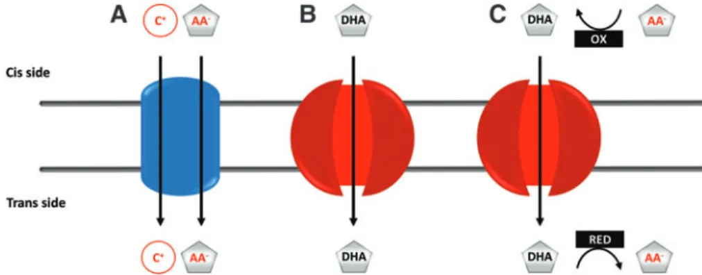
Two families of membrane transporters, namely sodium- dependent vitamin C transporters (SVCTs) and facilitative glucose transporters (GLUTs), accommodate for cellular accumulation and the subcellular distribution of AA. Vita- min C can be transported in both a reduced form (AA) and an oxidized form (dehydroascorbic acid [DHA]). The mechanisms involved are summarized in Figure 1.
SVCTs are highly specific active transporters of anionic AA (13). SVCT1 (SLC23A1) and SVCT2 (SLC23A2) spe- cifically carry AA across membranes otherwise impermeable for the anion. SVCT1 has a high capacity, but low affinity, for AA and is primarily involved in intestinal absorption (13) and renal reabsorption to reduce urinary loss of AA (59, 81).
SVCT2 is a very high-affinity lower-capacity transporter, which is more ubiquitous and responsible for transferring AA into a variety of cells and tissues. This transporter is also present in intracellular membranes, including the inner mi- tochondrial membrane (5, 64). Thus, SVCT1 enables AA accumulation at the level of the whole organism, while SVCT2 ensures the AA supply of each cell and some of their intracellular compartments.
The oxidized form of vitamin C, DHA, can further be transported by several GLUT family members (SLC2, solute carrier protein 2) viafacilitated diffusion [reviewed in (63)].
Under physiological conditions, DHA transport is thought to be less significant since DHA is present at low concentrations in extra- and intracellular compartments due to its unstable nature.
DHA is rapidly reduced to AA by thiol-dependent mechanisms on the trans side of the membrane at the expense of nico- tinamide adenine dinucleotide phosphate and preserves its antiscorbutic properties. However, GLUT-dependent DHA transport may increase substantially under oxidative stress or by oxidase activity in the vicinity of the membrane (68).
GLUT1-4, 8, and 10 are the best-documented, facilitative DHA transporters, which either associate with the plasma membrane or preferentially localize to specific organelles (6).
Overall, the concerted action of both the SVCT and GLUT families results in the intracellular accumulation of tremendous amounts of AA (intracellular: 1–10 mM versusblood plasma:
50–100lM) and in its distribution between organelles (Fig. 1).
Nevertheless, the intracellular distribution of vitamin C and distribution and functioning of AA/DHA transporters remain important, yet incompletely resolved, mechanisms [reviewed in (6)] that are difficult to assess in the absence ofin vivoap- plicable tools for measurement of AA/DHA concentrations in subcellular compartments. A recently proposed approach used high-resolution immunoelectron microscopy and immunogold labeling of AA (99) and demonstrated the presence of AA in organelles of plants at different concentrations that may change under (patho)physiological conditions (45, 84).
Genetic diseases associated with GLUTs are very rare, em- phasizing their vital importance and robustness of the family.
Mutations in SLC2A1, SLC2A2, and SLC2A10 are, respec- tively, associated with neurological disorders with variable intellectual disability and epilepsy [OMIM 606777, (83)], Fanconi–Bickel syndrome [a tubulopathy with intellectual disability, OMIM 227810, (79, 89)], and arterial tortuosity syndrome [ATS, OMIM 208050, (26)]. ATS is a rare con- genital disorder belonging to the group ofcutis laxasyndromes, associating with arterial tortuosity and aneurysm formation (91). The molecular link between mutations in the facilitative glucose transporter GLUT10 (encoded bySLC2A10) and the observed phenotype remains obscure. Hence, it is not surprising that several hypotheses have been forwarded to explain the pathomechanism. We critically review the existing suggestions and draw up a unifying hypothesis.
Clinical Manifestations in ATS
Initial presentation and molecular diagnosis
Most patients present with a cardiac murmur or reduced femoral pulsation. Less frequently, neonatal respiratory fail- ure due to infant respiratory distress syndrome, pulmonary hypertension, or a diaphragmatic hernia may be the initial presentation. A rare initial hint to the diagnosis is a remark- ably stretchable or lax skin. Workup with echocardiography reveals kinking of the aortic arch, for which more extensive vascular imaging with magnetic resonance angiography, com- puted tomography, or percutaneous angiography is initiated.
Upon observation of widespread tortuosity of the aorta and middle-sized arteries, genetic analysis is carried out. The iden- tification of biallelic pathogenic variants inSLC2A10confirms the diagnosis of ATS.
FIG. 1. Possible mechanisms of ascorbate transport through membranes. (A)Secondary active transport coupling the movement of cations (C+) down their gradient with the uphill transport of the ascorbate anion (AA-).(B)GLUTs enable facilitated diffusion of dehydroascorbic acid.(C)The transport of intracellular DHA, present at extremely low concentrations, can be promoted by an ascorbate oxidase on the cis and a DHA reductase on the trans side of the membrane. AA, ascorbic acid;
DHA, dehydroascorbic acid; GLUT, glucose transporter; OX, oxidation; RED, reduction. Color images are available online.
Downloaded by University of Twente from www.liebertpub.com at 12/22/19. For personal use only.
In view of the broad differential diagnosis, most of the pa- tients undergo testing for a panel of genes relevant for arterial aneurysms and tortuosity or undergo targeted exome analysis for a panel of genes related to connective tissue disorders. Of note, a review of evidence for genes that should be included in diagnostic gene panels for heritable thoracic aortic disease does not withholdSLC2A10(75). Although assigned as a potentially diagnostic gene, evidence is currently limited in the context of heritable thoracic aortic diseases based on its association with a syndromic presentation (rather than isolated vascular disease) and the absence of an unequivocal risk for dissection. Hence, some diagnostic panels for heritable thoracic aortic diseases may not includeSLC2A10.
The spectrum ofSLC2A10variants encompasses missense, nonsense, and splice-site mutations, besides large (exonic) and small deletions (4). So far, no genotype–phenotype cor- relation has been observed, impeding prognosis assessment and disease management by early genotyping (9, 14, 16, 77).
Cardiovascular features and natural history
Generalized tortuosity of the aorta and/or middle-sized arteries is invariably present, sporadically being associated with dilated or slightly tortuous large veins (9, 16, 62). Aortic side branches, including the truncus brachiocephalicus and left subclavian artery, may show an aberrant implantation on the aorta (Fig. 2).
A slight majority of patients (57%) have pulmonary artery stenosis, either of the truncus pulmonalis or the main or pe-
ripheral pulmonary branches. Pulmonary hypertension is ob- served in some patients and may or may not be associated with pulmonary artery stenosis. About one-quarter of the patients have a stenosis of the aorta that may be focal (mostly at the isthmus aortae) or present as a long stenotic stretch or hypo- plastic aorta. Other artery stenoses are less common (*15%), but may involve any middle-sized arteries, including the renal arteries. In addition, severe kinking of arteries may cause functional stenosis (9, 16).
Patients are at risk of developing aneurysms of the aortic root (commonly at the sinuses of Valsalva). Some patients have presented with aggressive aortic root dilatation before the age of 4 years, requiring urgent surgery. Others have shown slowly progressive aortic root dilatation in young adulthood. Never- theless, no aortic dissections have been unequivocally recorded to date (9, 16). Mitral valve prolapse and cardiomyopathy have been reported (9). It is unclear if cardiomyopathy is due to secondary remodeling or a primary event (as in the related Marfan syndrome) (18).
Ischemic events (9, 16, 19, 33), including stroke, are a concern. Of note, initial reports have described organ infarc- tions as a cause of death in young children clinically diagnosed with ATS (8, 71, 93), some of whom were confirmed molec- ularly afterward.
While initial mortality rates were reported up to 40%
before the age of 4 (93), larger cohorts of patients with a molecularly confirmed diagnosis revealed a milder disease course (9, 16).
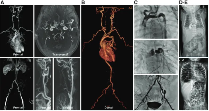
FIG. 2. Imaging studies in five patients with arterial tortuosity syndrome. (A)Magnetic resonance angiography in patient A shows moderate to severe tortuosity of the supra-aortic arteries, including the carotid, the basilar and cerebral arteries, and both iliac arteries. The aortic arch and descending aorta are gracile. There are no stenoses or aneurysms.(B) Computed tomography angiography in patient B shows a tortuous aorta with kinking, tortuosity of the middle-sized arteries, and stenosis of pulmonary arteries. The truncus pulmonalis is dilated and pulmonary arteries have a craniocaudal orga- nization.(C)In patient C, conventional angiography reveals stenosis of the aortic isthmus, tortuosity of the aorta, pulmonary arteries, and supra-aortic, inguinal, and visceral arteries. There is mild narrowing of the pulmonary arteries. (D, E) Conventional radiography studies in patients D and E illustrate the respective presence of a hiatal and diaphragmatic hernia.
Color images are available online.
Downloaded by University of Twente from www.liebertpub.com at 12/22/19. For personal use only.
Pulmonary manifestations
In a recent cohort (9), infant respiratory distress syndrome was a relatively frequent observation that may relate to im- paired lung maturation, diaphragmatic hernia, or pulmonary hypertension. Obstructive sleep apnea may be observed in older children and adults and may relate to hypotonia of the pharyngeal musculature and retrognathia. It remains to be established if patients are at risk to develop pulmonary em- physema, as has been observed in other elastinopathies (24).
Ocular manifestations
Corneal thinning and pellucid corneas are recently de- scribed manifestations in several ATS patients (43, 44). This may result in irregular astigmatism, keratoconus, and kera- toglobus (16, 43). It may also associate with corneal opacities.
Myopia is often present, although it is unclear if it is more fre- quently observed than in the general population. Laser-assisted in situ keratomileusis surgery is a risk factor for developing corneal complications (43).
Connective tissue and craniofacial features
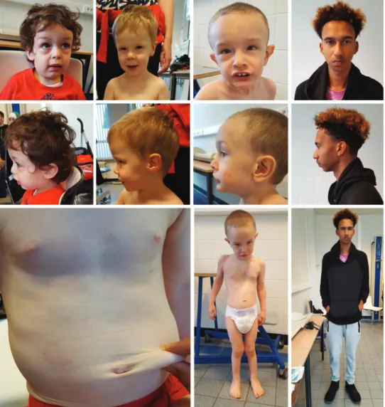
ATS patients may have some distinctive craniofacial fea- tures that become more prominent with age (Fig. 3). Fre-
quently reported features include a long face, hypertelorism, downslanting palpebral fissures, epicanthal folds, periorbital fullness, sagging cheeks, large ears, a highly arched palate, and retrognathia (9, 16). Bifid uvula or a cleft palate seems not to be associated with the disease, which may differentiate it from the Loeys–Dietz syndrome.
Overgrowth results in arachnodactyly and pectus defor- mity, while dolichostenomelia is not routinely observed.
Hypermobility of small and large joints is frequently present and hypotonia may cause isolated motor delay (9, 16). Skin anomalies range from a thin hyperextensible skin with a velvety texture (often described as soft and doughy) to loose skin folds (cutis laxa) (Fig. 3). Wound healing is normal.
Umbilical and inguinal hernias may occur (9) as well as pelvic organ prolapse in women (especially after child bearing) (20).
Other manifestations
Hiatal hernia and diaphragmatic hernia may be present in up to 30% of the patients and their presentation may vary from asymptomatic over postprandial discomfort to neonatal respiratory failure (Fig. 2) (9, 98). About one-third require surgical correction. Pyloric stenosis seems more prevalent (10%) as well as urinary tract anomalies with dilatation of the pyelocaliceal system (9). Finally, some patients may have
FIG. 3. Clinical charac- teristics in four patients with arterial tortuosity syn- drome. Craniofacial features include a long face, hyperte- lorism, downslanting palpe- bral fissures, epicanthal folds, sagging cheeks, large ears, and a hooked nose. Skin in- volvement can range from a thin hyperextensible skin with a velvety texture to marked cutis laxa. Skeletal manifes- tations include joint laxity, pectus deformities, arachno- dactyly, and scoliosis. Images used with permission. Color images are available online.
Downloaded by University of Twente from www.liebertpub.com at 12/22/19. For personal use only.
lengthening of the gastrointestinal tract, resulting in esoph- ageal tortuosity or dolichocolon. Several patients reported symptoms of autonomic dysfunction such as obstipation (that may partly be due to a dolichocolon), Raynaud phenomenon, slow eye accommodation, and orthostatic hypotension.
Many patients have psychological burdens related to both self-esteem and anxiety and distress for an uncertain vascular prognosis (Callewaert, personal observation).
A summary of the clinical characteristics and presentation rates in a series of 50 novel patients and 52 previously re- ported patients with a confirmed molecular diagnosis was recently presented by Beyenset al.(9).
Histopathology
Vascular tissue histology shows fragmentation of the inner elastic membrane and the elastic lamellae in thetunica mediaof the aortic wall. The elastic fibers are short, thick- ened, and disorganized, an observation made as well in other heritable disorders of the connective tissue with cardiovas- cular manifestations (1, 8, 9, 34, 39, 71). Cultured fibroblasts show disorganization of the actin cytoskeleton and multiple extracellular matrix (ECM) components, including fibronec- tin, fibrillin, type 3 collagen, type 5 collagen, and decorin (9, 26, 41, 102).
Transmission electron microscopy on skin biopsies of ATS patients reveals a poorly organized elastin assembly at the periphery of the elastic fiber, accompanied by infiltration of microfibrils in an interrupted elastin core (9).
Patient Management
Given the systemic manifestations associated with ATS, a coordinated and multidisciplinary approach for management and treatment is required. Treatment is largely supportive and symptomatic.
Baseline magnetic resonance arteriography from head to pelvis is required upon diagnosis and should be repeated every 3 years. During the first 5 years of life, echocardiographic follow-up every 3 months is necessary to detect aggressive aneurysm formation or evaluate pulmonary hypertension.
Afterward, yearly follow-up suffices if measurements remain within normal limits (9). In case of aneurysm formation, sur- gical guidelines for Marfan syndrome can be applied as the risk for acute dissections on small aortic diameters seems relatively small (9). Pulmonary artery stenosis can be corrected by per- cutaneous surgical or hybrid approaches (80, 92). Systemic stenosis—if focal—may require excision and end-to-end anastomoses (2, 3, 12, 22, 28, 35, 46, 49, 80, 92). Currently, the experience is limited and is based on expert opinion and on personal experience of the thoracovascular surgeon (14).
In analogy with treatment strategies applied for other heri- table disorders of connective tissue, such as Marfan syndrome and Loeys–Dietz syndrome, agents that reduce hemodynamic stress on the arterial wall, including beta-adrenergic blockers, angiotensin-converting enzyme inhibitors, and angiotensin II receptor 1 antagonists, such as losartan, can be considered. The experience is limited and in case of aortic hypoplasia/stenosis and/or renal stenosis, caution is necessary because of the risk of renal failure. Similarly, caution is needed to use nonsteroidal anti-inflammatory agents (9, 14). Pulmonary hypertension may be severe and related to pulmonary artery stenosis (causing a ventilation–perfusion mismatch), increased vascular wall ten-
sion, or lung hypoplasia due to diaphragmatic hernia. Treatment depends on the cause and may require percutaneous angioplasty and/or surgery or medical treatment (e.g., with sildenafil).
Regular orthopedic (joint laxity and scoliosis), orthodontic (highly arched palate and dental crowding), pulmonary (ob- structive sleep apnea syndrome, also postoperatively), and ophthalmological (keratometry and myopia) assessments are advised. Corneal cross-linking has been done in two ATS pa- tients with corneal thinning, but long-term effectiveness needs to be established (Callewaert, personal communication). Early rehabilitation may improve motor function in case of joint hy- perlaxity and hypotonia. Above all, patients should remain ac- tive in moderation (aerobic joint-gentle activities, such as swimming), as advised for other connective tissue disorders.
Differential diagnosis
ATS clinically shows some overlapping vasculopathy with other heritable disorders of the connective tissue. More specifi- cally, autosomal recessivecutis laxatype 1B (ARCL type 1B) (76) caused by mutations in fibulin-4, encoding for the elastin- binding protein fibulin-4, is also associated with arterial tortu- osity, arterial aneurysms, and stenosis, although ARCL type 1B more commonly leads to focal stenosis at the aortic isthmus and a more aggressive arterial phenotype. Furthermore, Loeys–Dietz syndrome, caused by heterozygous pathogenic variants in genes involved in transforming growth factor beta (TGFb) pathway signaling, such as TGFbcytokines (TGFB2andTGFB3), TGFb receptors (TGFBR1 and TGFBR2) and signal transducers (SMAD2andSMAD3), is characterized by cerebral, thoracic, and abdominal arterial aneurysms and/or dissections (55).
Occipital horn syndrome, an X-linked disorder caused by mutations in ATPase copper-transporting alpha (ATP7A), presents not only with intracranial vascular tortuosity but also with distinctive skeletal and urogenital features (10). Loose skin may be reminiscent of different types ofcutis laxa, in- cluding ARCL type 1A, type 1B, type 1C, and ARCL type 2.
When the skin is rather hyperextensible, it should be differ- entiated from the Ehlers–Danlos syndrome, hypermobility and vascular type (14).
Phenotypic Analysis of Animal Models for ATS
To date, two mouse models have been described for ATS.
These models were generated throughN-ethyl-N-nitrosourea mutagenesis in mice with a C3HeB/FeJ background. Two lines harboring missense mutations in Slc2a10 (c.383G>A—
p.G128E and c.449C>T—p.S150F) were recovered, and these nucleotide alterations were predicted to be deleterious (15).
Callewaert et al. executed comparative studies on wild- type, heterozygous, and homozygous mice at 5 months of age, including ultrasound analysis of the abdominal aorta, whole-animal vascular corrosion casting, and histological analysis of the tail and popliteal arteries. For both lines, none of the examinations revealed ATS-related phenotypic ab- normalities. Suggested explanations for an absent phenotype include (i) several GLUT family members may show func- tional redundancy, thus compensating for the loss of GLUT10;
(ii) phenotypic penetrance is hindered by the used genetic background (C3HeB/FeJ); and (iii) GLUT10 does not con- tribute to vasculogenesis in mice (15).
Utilizing the same mouse models, Chenget al.executed a series of additional experiments (21). Again, echocardiograms
Downloaded by University of Twente from www.liebertpub.com at 12/22/19. For personal use only.
did not reveal any abnormalities, and using brain magnetic resonance angiography, architectural anomalies in the cerebral arteries such as tortuosity, stenosis, or aneurysms could not be detected at 10 months of age. Histopathological analysis, however, disclosed a mild vascular phenotype, which was more severe in mutant mice carrying the p.G128E mutation (in comparison with the p.S150F mutant) and in older mice (10 months of agevs.6 months of age). Older mutant mice had a higher blood pressure and displayed a thickened and irregularly shaped vessel wall, fragmented elastic fibers with a disruption of the internal elastic lamina, and hypertrophy of endothelial cells in thetunica intima(21).
In 2012, Willaertet al.published a report on anslc2a10 zebrafish knockdown model (95). Morpholino-injected em- bryos (morphants) showed several cardiovascular abnor- malities: cardiac edema, a reduced heart rate and blood flow, and an incomplete and irregular patterning of the vasculature, resulting in disrupted blood flow in the sinus venosus. These anomalies possibly represent developmental precursors to vascular anomalies, as observed in ATS patients.
Molecular Background
ATS exhibits autosomal recessive inheritance and ap- proximately a decade ago, its causal gene, SLC2A10, was identified (26). The protein it encodes (GLUT10) is a member of the facilitative GLUTs, which are involved in assisting the transport of monosaccharides, polyols, and other small car- bon compounds across eukaryotic cell membranes (63). The GLUT family consists of 14 proteins, which are categorized into three classes based on sequence similarity: class 1 (GLUTs 1–4 and 14), class 2 (GLUTs 5, 7, 9, and 11) and class 3 (GLUTs 6, 8, 10, 12, and H+/myo-inositol co- transporter). The general structure of each GLUT protein is similar: 12 transmembrane domains connected by linker domains, cytoplasm-pointing amino and carboxy termini, and a unique N-linked oligosaccharide side chain, positioned in the first (classes 1 and 2) or fifth (class 3) exofacial linker domain (63).
In GLUT10, this particular extracellular loop is distinctively larger in size compared with the other GLUT family members.
Some authors have speculated that this loop may attribute al- ternative functions to GLUT10 (60). However, none of the 34 known mutations causing ATS reside in this exofacial linker domain (9), possibly reflecting a less critical role of this domain.
Hypotheses for the Pathophysiology of ATS
GLUT10 is widely expressed, but is most abundant in smooth muscle-rich organs (97), which correlates with its importance in blood vessel development and homeostasis. Other organs showing expression are the heart, placenta, lung, liver, skeletal muscle, pancreas, brain, kidney (32, 60), and adipose tissue (96).
On a subcellular level, GLUT10 has been localized to the nuclear envelope (NE) (26), ER (82), mitochondria, and Golgi apparatus (53). The diverse expression patterns indi- cate that GLUT10 may translocate in different cellular states.
Initially, human GLUT10—expressed in Xenopus laevis oocytes—was shown to facilitate transport of D-glucose, D- galactose, and 2-deoxy-D-glucose (32) with high affinity.
Subsequent studies favored DHA as a substrate. Based on the subcellular localization of GLUT10 and the range of func- tions its presumable substrates can fulfill, a number of disease hypotheses for ATS have been postulated and investigated.
Inhibition of glucose-mediated transcriptional regulation in the nucleus
Couckeet al.localized GLUT10 in the perinuclear region and identified TGFb pathway upregulation in ATS patients (26). According to the hypothesis, deficiency of GLUT10 would result in reduced nuclear glucose levels, downregulating transcription of genes with glucose-responsive elements in their promoter regions. One such gene isDCNencoding the proteoglycan, decorin. Decorin sequesters TGFb(37) and its expression was reduced in cultured vascular smooth muscle cells (SMCs) of ATS patients. Consequently, TGFb-driven expression of the proteoglycan, versican, was increased (26).
Versican has an inhibitory role on elastic fiber assembly (94)
FIG. 4. Schematic repre- sentation of the hypothesis proposed by Coucke et al.
(26), in which GLUT10 serves as a nuclear trans- porter of glucose. Intra- nuclear glucose can trigger transcription of genes har- boring glucose-responsive elements (GLEs) in their promoter regions, such as decorin, which is known to inhibit TGFb pathway activa- tion by sequestering TGFb. A GLUT10 defect (indicated by a green X) would lead to downregulation of decorin and triggering of the TGFb path- way (green arrows). GLU, glucose, Na, sodium; TGFb, transforming growth factor beta. Color images are avail- able online.
Downloaded by University of Twente from www.liebertpub.com at 12/22/19. For personal use only.
and its increased expression could explain failed elastogenesis in ATS (Fig. 4).
Nevertheless, the role of GLUT10 as a nuclear GLUT is questionable based on the following: (i) small polar molecules such as glucose are able to enter the nucleusviadiffusion through the nuclear pore, hence not requiring specialized transporters, and (ii) glucose-dependent transcription is not directly mediated by intranuclear glucose, but rather by indirect mechanisms transducing the glucose signal to the genome (82).
Oxidative stress
Several studies, described in detail below, pointed toward a contribution of oxidative stress in the pathogenesis of ATS.
Disturbed redox status and increased reactive oxygen species (ROS) production, promoting protein oxidative modifica- tions, can modulate signaling molecule activities and dysre- gulate key pathological cellular processes, such as apoptosis, migration, proliferation, inflammation, and ECM component degradation, accumulating in vascular remodeling as ob- served in ATS (38).
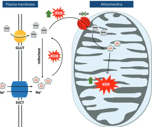
A first study, performed by Leeet al., confirms that GLUT10 operates as a DHA transporter (53). They observed that exog- enous tagged GLUT10 localized primarily to the mitochondria in rat A10 SMCs and insulin-stimulated mouse 3T3-L1 adipo- cytes, while the protein localized to the Golgi apparatus in mouse NIH3T3 fibroblast cells. Mitochondrial import of DHA increased in a dose-dependent manner and following oxidative stress upon H2O2administration.
These findings prompted a second hypothesis (Fig. 5) where GLUT10 has a major role in replenishing mitochon- drial antioxidative capacities through DHA transport. Under normal conditions, both DHA and AA are transported into the
cytoplasm, where most DHA is reduced to AA. Under oxi- dative conditions, DHA accumulates in the cytoplasm and is transported to the mitochondria in a GLUT10-dependent manner, followed by its regeneration to AA. There, AA ex- erts its function as an antioxidant, quenching ROS molecules and free radicals, protecting the cell from damage due to oxidative stress. A GLUT10 defect would result in accumu- lation of ROS molecules, which might trigger development of ATS-related anomalies (53).
A couple of arguments, however, dispute this pathogenesis model. A recentin silicostudy identified GLUT10 as one of the transporters with the lowest predicted mitochondrial lo- calization score (87). In addition, the sodium-coupled ascorbic acid transporter-2 (SVCT2) has already been identified as a mitochondrial AA transporter, questioning the necessity of GLUT10 for mitochondrial DHA transport (5, 64).
This hypothesis was nonetheless reinforced by the same laboratory with additional data obtained on rat, mouse, and human SMCs. This study revealed that stress and aging condi- tions increased intracellular and mitochondrial ROS levels and promoted targeting of GLUT10 to the mitochondria, increasing mitochondrial DHA uptake. In addition, GLUT10 knockdown and overexpression models in mouse MOVAS SMCs, respec- tively decreased or increased the inner mitochondrial membrane potential and oxygen consumption rate (86). Compared with control SMC, SMCs from GLUT10G128Emice showed higher ROS levels, both intracellularly and in the mitochondria, a re- duced oxygen consumption rate, and altered mitochondrial morphology and density. These features were also confirmed in aortic tissue of 15-month-old GLUT10G128Emice. (86).
Comparative transcriptome analysis at 48 h postfertilization between slc2a10 morphants (following morpholino-based slc2a10 knockdown) and control embryos revealed altered
FIG. 5. Graphical repre- sentation of the mitochon- drial dysfunction ATS pathogenesis hypothesis (53).Both DHA and AA are transported into the cytosol by GLUT or SVCT, respec- tively. In the cytosol, reduc- tase converts most DHA to AA. Under oxidative con- ditions (indicated by red icons), however, cytosolic DHA accumulation is fol- lowed by its transportation into the mitochondria in a GLUT10-dependent man- ner. There, regenerated AA acts as an antioxidant. A GLUT10 defect (indicated by a green X) leads to ac- cumulation of ROS molecules (green arrows). Na, sodium;
ROS, reactive oxygen species;
SVCT, sodium-dependent vi- tamin C transporter. Color images are available online.
Downloaded by University of Twente from www.liebertpub.com at 12/22/19. For personal use only.
expression of genes involved in oxidative phosphorylation, reactive oxygen production, the Szent–Gyo¨rgyi–Krebs cycle, the glycolysis/gluconeogenesis pathway, and glycogen me- tabolism. These findings suggested mitochondrial dysfunction as a pathomechanism, which was further validated by impaired oxygen consumption rates in morphants. In contrast to ob- servations made in the ATS mouse model, electron micros- copy analysis ofslc2a10morphants did not reveal any changes in mitochondrial morphology (95).
Disturbed post-translational modification in the ER
In silicotools predict GLUT10 localization in the ER with high probability (40) because of the presence of an ER retention
signal YXXI/V (48) and a Lys-Arg-Arg (KRR) motif (42) in the C-terminus of the protein. Since ATS patients show a remark- able clinical overlap with heritable disorders of the connective tissue with abnormal collagen and/or elastin homeostasis, Se- gade hypothesized that GLUT10 operates as a transporter of AA, a necessary component for post-translational modification of collagen and elastin molecules in the ER (82).
AA—being a cofactor of prolyl and lysyl hydroxylases—
catalyzes the hydroxylation of proline and lysine residues in elastin and collagen, thereby facilitating cross-link forma- tion. Lowered AA levels in the ER would result in defective and immature collagen and elastin molecules, contributing to histopathology observed in collagen- and elastin-rich tissues in ATS patients (Fig. 6). In this scenario, the observed
FIG. 6. Schematic representation of the hypothetical model proposed by Segade (82) and Ne´methet al.(67), where GLUT10 functions as a DHA transporter in the ER-NE continuum.Segade proposes that GLUT10 facilitates DHA transport to the ER, after which conversion to AA by PDI takes place. In the ER, AA functions as a cofactor of prolyl and lysyl hydroxylases, which modify proline and lysine residues in collagen and elastin molecules, leading to their stabili- zation. A GLUT10 defect would lead to deposition of immature collagen and elastin molecules, weakening of the extra- cellular matrix, and subsequent triggering of the TGFbpathway. Ne´methet al.suggest that GLUT10 transports DHA into the nucleoplasm, either directly through the membranes of the nuclear envelope or via the ER, through the ER-NE continuum, where regenerated AA functions as a cofactor for DNA and histone demethylases. ER, endoplasmic reticulum;
Fe, iron; HyPro, hydroxyproline; Na, sodium; NE, nuclear envelope; PDI, protein disulfide isomerase; Pro, proline. Color images are available online.
Downloaded by University of Twente from www.liebertpub.com at 12/22/19. For personal use only.
upregulation of the TGFbpathway in ATS patients would result from weakening of the ECM and increased activation of latent TGFbmolecules (82).
This hypothesis has been experimentally supported by identification of GLUT10 as a DHA transporter in the ER.
Transient transfection of GLUT10-green fluorescent protein constructs in rat SMCs confirmed the presence of GLUT10 in the ER (82). Furthermore, immunocytochemistry-supported localization experiments, either employing ATS patient cells or exogenous tagged GLUT10, showed a reticular distribu- tion and perinuclear abundance of the protein in fibroblast cells (40, 102), and GLUT10 was found to be present in microsomal (ER-derived) fractions obtained from human fi- broblasts and liver tissue (40).
Incorporation ofin vitro-produced GLUT10 into liposomes resulted in efficient transport of DHA in a concentration- dependent manner. Moreover, selective permeabilization ex- periments in fibroblasts showed that the transport of DHA through endomembranes was markedly reduced in ATS pa- tient cells when compared with control fibroblasts (66).
Epigenetic modulation through AA deficiency in the nucleoplasm
Recently, nucleoplasmic JmjC domain-containing demethy- lases (50, 90) and ten-eleven translocation methylcytosine dioxygenases (47, 51), all members of the AA-dependent Fe2+/2-oxoglutarate-dependent dioxygenases, gained atten- tion since they promote demethylation of histones and DNA, respectively. Both mechanisms are involved in epigenetic regulation of transcription (17).
It has recently been shown that by applying high- resolution immunoelectron microscopy and immunogold labeling of AA (99), AA accumulation in the nucleoplasm was diminished in ATS fibroblasts (67). Since the syn- drome is due to mutations in GLUT10, it can be supposed that GLUT10 is required for the proper AA concentration in the nucleus.
Because the membranes of the ER and the NE are con- tinuous, DHA uptake by GLUT10 in the nucleoplasm can theoretically follow two routes. DHA can be transferred from the ER across the inner membrane of the NE to the nucleoplasm. Alternatively, DHA could be transported directly through the membranes of the NE (Fig. 6). As a consequence, altered DNA hydroxymethylation patterns at both the global level and at specific gene regions (including PPARG encoding peroxisome proliferator-activated re- ceptor [PPAR]c) were found in ATS patient fibroblasts (67), which suggests an epigenetic role of AA transport in the ATS pathomechanism.
Discussion
Vitamin C compartmentalization:
unifying the different disease mechanisms?
Scurvy, a generalized AA deficiency due to insufficient AA intake, has been known long before the discovery of AA and causes muscular weakness, pain, generalized bleeding, and impaired wound healing. Scurvy at the cellular level can manifest with normal intake and serum concentrations of AA.
This latent or tissue scurvy may contribute to complications of insulin-dependent diabetes mellitus, such as endothelial dys-
function and atherosclerosis (73), due to competition between glucose and DHA for GLUTs during hyperglycemia (29, 72).
In a series of elegant experiments, it has been demon- strated that subcellular scurvy may also result from either increased consumption of AA in a cellular compartment or defective transport into a specific organelle. Zitoet al.studied the effect of the combined loss-of-function mutations in genes encoding the ER thiol oxidases ERO1a, ERO1b, and peroxiredoxin 4 in a murine model. These enzymes catalyze electron transfers implicated in oxidative protein folding (101). Surprisingly, only minor alterations in disulfide bond formation were found, suggesting the presence of alternative electron transfer routes. However, low tissue AA concen- trations and decreased 4-hydroxyproline content of pro- collagen were observed.
Hence, in the absence of ER thiol oxidases, cysteinyl thiol groups are oxidized to sulfenic acid through an alternative hydrogen peroxide-dependent way, consuming AA as a re- ductant (100). This competes with prolyl hydroxylases for AA in the ER lumen, resulting in impaired procollagen hy- droxylation, and impairs ECM homeostasis, causing scurvy- like disease. Thus, neither AA synthesis (mice are able to produce AA) nor AA transport could keep pace with the increased consumption.
Due to the presence of transporters, competition may arise between AA-requiring reactions of two (or more) compart- ments as well. For instance, in pluripotent stem cells, in- creased prolyl-4-hydroxylase activity in the ER hampers 5-methylcytosine demethylation in the nucleoplasm, affect- ing epigenetic regulation, and vice versa (30). Genetic ablation of prolyl-4-hydroxylase subunit alpha-2 or its pharmacological inhibition reverted epigenetic changes and antagonized cell state transition. Thus, it can be hypothesized that AA consumption in the ER by collagen hydroxylation reduces AA availability for DNA and histone demethylases in the nucleoplasm, preventing mesenchymal transition.
Finally, mislocalization or translocation of AA/DHA trans- porters can also influence vitamin C distribution between two neighboring compartments. Indeed, Pena et al.demonstrated that normal and cancer cells handle AA differently (69). Hu- man breast cancer tissues expressed SVCT2, but were not able to take up AA. This was explained by the fact that this form of SVCT2 was absent from the plasma membrane and expressed in mitochondria of breast cancer cells (78). Augmented ex- pression of the SVCT2 mitochondrial form and mitochondrial sequestration of AA might profoundly alter AA-dependent mechanisms in other subcellular compartments and might represent an adaptation mechanism to the special metabolic features in cancer cells.
These mechanisms balancing transport and consumption within and between cellular compartments may participate in the pathogenesis of ATS, although still several aspects re- main to be addressed.
Pathophysiological mechanisms related to oxidative stress
Different lines of evidence support a role for GLUT10 in DHA transport through the ER-NE continuum or the contri- bution of oxidative stress to the ATS pathomechanisms. The complementarity and hierarchy of these identified disease mechanisms need further investigation, as well as the exact
Downloaded by University of Twente from www.liebertpub.com at 12/22/19. For personal use only.
role of GLUT10 herein. However, molecular alterations linked to ATS pathogenesis were rescued upon reexpression of GLUT10 in ATS patient fibroblasts (under low, non- physiological AA concentrations), suggesting that GLUT10 might also harbor transport-independent functions (102, 103).
Comparable with the zebrafish morpholino model, tran- scriptome analysis comparing ATS patient and control cells identified affected redox homeostasis, energy and lipid me- tabolism, TGFb signaling, and ECM architecture (102). Of note, the most upregulated gene wasALDH1A1, encoding a member of the aldehyde dehydrogenases (ALDHs). ALDHs act in detoxification induced by sustained presence of lipid peroxidation (LPO)-derived molecules that were found to be increased in ATS fibroblasts. Elevated ROS levels in ATS fibroblasts were suggested to initiate the LPO mechanism by interaction with polyunsaturated fatty acids (PUFAs).
Furthermore, PPARc activity is boosted by peroxidized PUFAs and an altered cellular energy metabolism. PPARcis a master regulator in multiple (patho)physiological processes and influences transcription and post-translational modifica- tion events. Oxidative stress in ATS might depend on a PPARc-dependent mechanism through a maladaptive feed- back, upregulatingb-oxidation, and PUFA metabolism, es- tablishing a vicious circle of oxidative stress induction. ROS directly promote ECM protein fragmentation and induce LPO-derived molecules that could hamper the secretion and organization of collagen and elastin molecules in the ECM, causing ER stress (102).
TGFbsignaling: a delicate balance at the crossroads of oxidative stress, defective ECM production, and epigenetic modification
A key pathway in multiple heritable disorders of connective tissue with cardiovascular implications is TGFbsignaling. In- itial immunostaining experiments for connective tissue growth factor and pSMAD2 on arterial tissue of one ATS patient demonstrated an extreme increase in signal intensity in com- parison with control tissue (26). Similar analyses in skin and artery tissue in a subsequent study could not confirm this ob- servation (9). These observations likely point to variability in spatiotemporal TGFbsignaling and add to the uncertainty re- garding the (primary) role of TGFbsignaling in the disease mechanisms underlying elastic fiber diseases.
TGFbis secreted in the ECM in its latent form, called the large latent complex (LLC), comprising TGFb, latency- associated peptide (LAP), and latent TGFb-binding proteins, which are covalently cross-linked to ECM components.
TGFbcan be released from this complex through LAP pro- teolysis, interaction with av-containing integrins such as avb3 integrin, or ROS-induced mechanisms.
Disturbed TGFb signaling has been claimed to be a primary actor in ATS pathogenesis (26), or to result sec- ondarily from increased release of latent TGFb from a damaged ECM (82), due to ineffective hydroxylation of collagen and elastin. Furthermore, epigenetic inhibition of PPARcwill enhance TGFbsignaling, and ROS contribute to the release of active TGFbfrom the LLC.
Finally, Zoppi et al. found a key role foravb3 integrin, cross talking with noncanonical TGFbsignaling. More spe- cifically,avb3 integrin-dependent release of TGFbfrom the LLC inducesavb3 integrin association with TGFbreceptor II
and noncanonical TGFb pathway stimulation through p38 MAPK (102). This is supported by an increased presence of TGFbreceptor II,avb3, integrin, and p38 MAPK, but low- ered pSMAD2, a biomarker for canonical TGFbsignaling, in mock-transfected ATS patient fibroblasts compared with GLUT10 transfected ATS cells. Although p38 MAPK is a negative regulator of cellular stress, it does not seem to correct ECM organization, as demonstrated by staining of several ECM components in ATS patient fibroblasts.
Future Perspectives
The different hypotheses provide paradigms to understand ATS pathophysiology and developmental biology of the ar- teries, but none of them are currently supported by sufficient evidence to be regarded as the sole, or even main, mechanism.
Nevertheless, the formulated hypotheses are not mutually exclusive.
Indeed, GLUT10 may translocate to different organelles depending on the cell state (25, 40, 53, 82, 102). Moreover, intracellular membranes as well as the plasma membrane are no longer regarded as static structures, but instead are at- tributed to plasticity, interchanging phospholipids, and membrane-bound (or enclosed) molecules through physical links and/or transport mechanisms. Of note, a complex, ER- mitochondria encounter structure, brings ER and mitochon- drial membranes in close proximity and is necessary for cell survival (52). In a related disorder, occipital horn syndrome caused by deficiency of the copper ATPase, encoded by ATP7A, it has been shown that the molecule translocates from the cell membrane to trans- and post-Golgi compartments depending on copper concentration (70).
Finally, as discussed above, it is crucial to understand the balance between AA use, import, and transport between or- ganelles both under physiological and GLUT10-deficient states. Accumulating evidence shows that local subcellular concentrations of vitamin C are equally or even more impor- tant than the global cellular or blood levels. Mapping of sub- cellular AA concentrations, determination of the redox state of the AA/DHA couple in the different cellular compartments, and exploration of AA/DHA transport mechanisms between the cytoplasm and various organelles, both under physiolog- ical and diverse pathological situations, are future challenges.
This knowledge will lead to a more detailed understanding of known and yet unknown functions of AA in the different cellular compartments in health and disease. Knowing the importance of AA in vascular development and homeostasis (as evidenced by, respectively, ATS and scurvy), this knowl- edge will contribute to understanding neovascularization both in desirable (wound healing, neovascularization after vascular injury, or thrombosis) or undesirable (tumor formation) states.
Furthermore, we should keep in mind thatin vivo versus in vitroexperiments may reveal completely different biological outcomes, as has been observed for the Loeys–Dietz syndrome, caused by mutations inTGFBR2(31, 61). In addition, observed pathophysiological aberrations in postdevelopmental life may be protective in early development (when the arterial system is being developed) (23). Similarly, the damaged arterial wall may behave differently on blood pressure when still developing or in postnatal life (as hypothesized for intra- cranial tortuosity in autosomal dominantcutis laxadue to ALDH18A1defects) (36, 85). Finally, it may prove difficult
Downloaded by University of Twente from www.liebertpub.com at 12/22/19. For personal use only.
to distinguish primary versus secondary effects. Indeed, while increased TGFb signaling seems a common final pathway in many vascular disorders, it remains to be re- solved whether it precedes ECM remodeling or whether it is a secondary effect of altered ECM homeostasis.
Animal models could be extremely helpful to test the different hypotheses. Unfortunately, the reported mouse models with missense mutations showed extremely mild phenotypes, not reminiscent of the severe cardiovascular anomalies encountered in ATS patients. Therefore, it is questionable if the mild phenotypic differences observed in older GLUT10G128Emice are relevant to study ATS path- ogenesis. Genetic compensation might be at play to coun- teract the mechanisms induced upon Slc2a10 mutations.
Moreover, since mice are able to synthesize AA, it can be anticipated that the abovementioned results should be in- terpreted with caution. It is possible that mice will present with a more relevant ATS-related phenotype if made in- capable of synthesizing AA (82). Alternatively, CRISPR/
Cas9 knockout and knockin approaches in zebrafish might be a good alternative, especially since results on the mor- phants were motivating (95).
Acknowledgments
B.C. is a senior clinical investigator of the Research Foundation–Flanders (FWO). E´ .M. was supported by the Ja´nos Bolyai Research Scholarship from the Hungarian Academy of Sciences and by the New National Excellence Program from the Ministry of Human Capacities.
Author Disclosure Statement
No competing financial interests exist.
Funding Information
This research is supported by FWO funds VS.080.16N, FWOOPR2013025301, and by the Hungarian National Re- search, Development and Innovation Office (NKFIH grant number: FK124442).
References
1. Ades LC, Knight WB, Byard RW, Bateman JF, Esquivel JA, Mee RB, Haan EA, and Milewicz DM. Clinicopathologic findings in congenital aneurysms of the great vessels.Am J Med Genet66: 289–299, 1996.
2. Al-Khaldi A, Alharbi A, Tamimi O, and Mohammed Y.
Successful surgical pulmonary artery reconstruction in arterial tortuosity syndrome.Ann Thorac Surg88: 1343–
1345, 2009.
3. Al-Khaldi A, Mohammed Y, Tamimi O, and Alharbi A.
Early outcomes of total pulmonary arterial reconstruction in patients with arterial tortuosity syndrome.Ann Thorac Surg92: 698–704; discussion 704, 2011.
4. Albuisson J, Moceri P, Flori E, Belli E, Gronier C, and Jeunemaitre X. Clinical utility gene card for: arterial tor- tuosity syndrome.Eur J Hum Genet23:1432, 2015.
5. Azzolini C, Fiorani M, Cerioni L, Guidarelli A, and Can- toni O. Sodium-dependent transport of ascorbic acid in U937 cell mitochondria.IUBMB Life65: 149–153, 2013.
6. Banhegyi G, Benedetti A, Margittai E, Marcolongo P, Fulceri R, Nemeth CE, and Szarka A. Subcellular com-
partmentation of ascorbate and its variation in disease states.Biochim Biophys Acta1843: 1909–1916, 2014.
7. Banhegyi G, Braun L, Csala M, Puskas F, and Mandl J.
Ascorbate metabolism and its regulation in animals.Free Radic Biol Med23: 793–803, 1997.
8. Beuren AJ, Hort W, Kalbfleisch H, Muller H, and Stoer- mer J. Dysplasia of the systemic and pulmonary arterial system with tortuosity and lengthening of the arteries. A new entity, diagnosed during life, and leading to coronary death in early childhood.Circulation39: 109–115, 1969.
9. Beyens A, Albuisson J, Boel A, Al-Essa M, Al-Manea W, Bonnet D, Bostan O, Boute O, Busa T, Canham N, Cil E, Coucke PJ, Cousin MA, Dasouki M, De Backer J, De Paepe A, De Schepper S, De Silva D, Devriendt K, De Wandele I, Deyle DR, Dietz H, Dupuis-Girod S, Fontenot E, Fischer-Zirnsak B, Gezdirici A, Ghoumid J, Giuliano F, Diez NB, Haider MZ, Hardin JS, Jeunemaitre X, Klee EW, Kornak U, Landecho MF, Legrand A, Loeys B, Lyonnet S, Michael H, Moceri P, Mohammed S, Muino- Mosquera L, Nampoothiri S, Pichler K, Prescott K, Rajeb A, Ramos-Arroyo M, Rossi M, Salih M, Seidahmed MZ, Schaefer E, Steichen-Gersdorf E, Temel S, Uysal F, Vanhomwegen M, Van Laer L, Van Maldergem L, War- ner D, Willaert A, Collins TR, Taylor A, Davis EC, Zarate Y, and Callewaert B. Arterial tortuosity syndrome: 40 new families and literature review.Genet Med10:1236–1245, 2018.
10. Beyens A, Van Meensel K, Pottie L, De Rycke R, De Bruyne M, Baeke F, Hoebeke P, Plasschaert F, Loeys B, De Schepper S, Symoens S, and Callewaert B. Defining the clinical, molecular and ultrastructural characteristics in occipital horn syndrome: two new cases and review of the literature.Genes10: pii: E528, 2019.
11. Blaszczak W, Barczak W, Masternak J, Kopczynski P, Zhitkovich A, and Rubis B. Vitamin C as a modulator of the response to cancer therapy. Molecules (Basel, Swit- zerland)24: pii: E453, 2019.
12. Bottio T, Bisleri G, Piccoli P, and Muneretto C. Valve- sparing aortic root replacement in a patient with a rare connective tissue disorder: arterial tortuosity syndrome.J Thorac Cardiovasc Surg133: 252–253, 2007.
13. Burzle M, Suzuki Y, Ackermann D, Miyazaki H, Maeda N, Clemencon B, Burrier R, and Hediger MA. The sodium- dependent ascorbic acid transporter family SLC23. Mol Aspects Med34: 436–454, 2013.
14. Callewaert B, De Paepe A, and Coucke P. Arterial Tor- tuosity Syndrome. In:GeneReviews((R)),edited by Adam MP, Ardinger HH, Pagon RA, Wallace SE, Bean LJH, Stephens K, Amemiya A. Seattle, WA: University of Washington, 1993.
15. Callewaert BL, Loeys BL, Casteleyn C, Willaert A, De- wint P, De Backer J, Sedlmeier R, Simoens P, De Paepe AM, and Coucke PJ. Absence of arterial phenotype in mice with homozygous slc2A10 missense substitutions.
Genesis (New York, N.Y.: 2000)46: 385–389, 2008.
16. Callewaert BL, Willaert A, Kerstjens-Frederikse WS, De Backer J, Devriendt K, Albrecht B, Ramos-Arroyo MA, Doco-Fenzy M, Hennekam RC, Pyeritz RE, Krogmann ON, Gillessen-kaesbach G, Wakeling EL, Nik-zainal S, Francannet C, Mauran P, Booth C, Barrow M, Dekens R, Loeys BL, Coucke PJ, and De Paepe AM.
Arterial tortuosity syndrome: clinical and molecular findings in 12 newly identified families.Hum Mutat29:
150–158, 2008.
Downloaded by University of Twente from www.liebertpub.com at 12/22/19. For personal use only.
17. Camarena V and Wang G. The epigenetic role of vitamin C in health and disease.Cell Mol Life Sci73: 1645–1658, 2016.
18. Campens L, Renard M, Trachet B, Segers P, Muino Mos- quera L, De Sutter J, Sakai L, De Paepe A, and De Backer J.
Intrinsic cardiomyopathy in Marfan syndrome: results from in-vivo and ex-vivo studies of the Fbn1C1039G/+ model and longitudinal findings in humans.Pediatr Res78: 256–
263, 2015.
19. Cartwright MS, Hickling WH, and Roach ES. Ischemic stroke in an adolescent with arterial tortuosity syndrome.
Neurology67: 360–361, 2006.
20. Castori M, Ritelli M, Zoppi N, Molisso L, Chiarelli N, Zaccagna F, Grammatico P, and Colombi M. Adult pre- sentation of arterial tortuosity syndrome in a 51-year-old woman with a novel homozygous c.1411+1G>A muta- tion in the SLC2A10 gene. Am J Med Genet A 158A:
1164–1169, 2012.
21. Cheng CH, Kikuchi T, Chen YH, Sabbagha NG, Lee YC, Pan HJ, Chang C, and Chen YT. Mutations in the SLC2A10 gene cause arterial abnormalities in mice.Cardiovasc Res 81: 381–388, 2009.
22. Cine N, Basaran M, Guzelmeric F, and Sunar H. Repair of ascending aortic aneurysm in a patient with arterial tor- tuosity syndrome. Interact Cardiovasc Thorac Surg 12:
1051–1053, 2011.
23. Cook JR, Clayton NP, Carta L, Galatioto J, Chiu E, Smaldone S, Nelson CA, Cheng SH, Wentworth BM, and Ramirez F. Dimorphic effects of transforming growth factor-beta signaling during aortic aneurysm progression in mice suggest a combinatorial therapy for Marfan syndrome.
Arterioscler Thromb Vasc Biol35: 911–917, 2015.
24. Corsico AG, Grosso A, Tripon B, Albicini F, Gini E, Mazzetta A, Di Vincenzo EM, Agnesi ME, Tsana Tegomo E, Ronzoni V, Arbustini E, and Cerveri I. Pulmonary in- volvement in patients with Marfan Syndrome. Panmi- nerva Med56: 177–182, 2014.
25. Coucke PJ, Wessels MW, Van Acker P, Gardella R, Barlati S, Willems PJ, Colombi M, and De Paepe A.
Homozygosity mapping of a gene for arterial tortuosity syndrome to chromosome 20q13.J Med Genet40: 747–
751, 2003.
26. Coucke PJ, Willaert A, Wessels MW, Callewaert B, Zoppi N, De Backer J, Fox JE, Mancini GM, Kambouris M, Gardella R, Facchetti F, Willems PJ, Forsyth R, Dietz HC, Barlati S, Colombi M, Loeys B, and De Paepe A. Muta- tions in the facilitative glucose transporter GLUT10 alter angiogenesis and cause arterial tortuosity syndrome.Nat Genet38: 452–457, 2006.
27. Crivellato E, Nico B, and Ribatti D. The chromaffin vesicle: advances in understanding the composition of a versatile, multifunctional secretory organelle. Anat Re- cord (Hoboken, N.J.: 2007)291: 1587–1602, 2008.
28. Cubero A, Ayala J, Hamzeh G, Cortes A, Udaondo J, and Aramendi JI. Severe arterial tortuosity. World J Pediatr Congenital Heart Surg8: 231–234, 2017.
29. Cunningham JJ. The glucose/insulin system and vitamin C: implications in insulin-dependent diabetes mellitus.J Am Coll Nutr17: 105–108, 1998.
30. D’Aniello C, Cermola F, Palamidessi A, Wanderlingh LG, Gagliardi M, Migliaccio A, Varrone F, Casalino L, Ma- tarazzo MR, De Cesare D, Scita G, Patriarca EJ, and Minchiotti G. Collagen prolyl hydroxylation-dependent metabolic perturbation governs epigenetic remodeling and
mesenchymal transition in pluripotent and cancer cells.
Cancer Res79: 3235–3250, 2019.
31. Davis JM, Kugler G, and Nixon BP. Nonclostridial cel- lulitis with subcutaneous emphysema.J Foot Surg31: 85–
87, 1992.
32. Dawson PA, Mychaleckyj JC, Fossey SC, Mihic SJ, Craddock AL, and Bowden DW. Sequence and functional analysis of GLUT10: a glucose transporter in the Type 2 diabetes-linked region of chromosome 20q12–13.1. Mol Genet Metab74: 186–199, 2001.
33. Drera B, Barlati S, and Colombi M. Ischemic stroke in an adolescent with arterial tortuosity syndrome. Neurology 68: 1637; author reply 1637, 2007.
34. Ertugrul A. Diffuse tortuosity and lengthening of the ar- teries.Circulation 36: 400–407, 1967.
35. Faiyaz-Ul-Haque M, Zaidi SH, Al-Sanna N, Alswaid A, Momenah T, Kaya N, Al-Dayel F, Bouhoaigah I, Saliem M, Tsui LC, and Teebi AS. A novel missense and a recurrent mutation in SLC2A10 gene of patients affected with arterial tortuosity syndrome.Atherosclerosis203: 466–471, 2009.
36. Fischer-Zirnsak B, Escande-Beillard N, Ganesh J, Tan YX, Al Bughaili M, Lin AE, Sahai I, Bahena P, Reichert SL, Loh A, Wright GD, Liu J, Rahikkala E, Pivnick EK, Choudhri AF, Kruger U, Zemojtel T, van Ravenswaaij- Arts C, Mostafavi R, Stolte-Dijkstra I, Symoens S, Paju- nen L, Al-Gazali L, Meierhofer D, Robinson PN, Mundlos S, Villarroel CE, Byers P, Masri A, Robertson SP, Schwarze U, Callewaert B, Reversade B, and Kornak U.
Recurrent De Novo Mutations Affecting Residue Arg138 of Pyrroline-5-Carboxylate Synthase Cause a Progeroid Form of Autosomal-Dominant Cutis Laxa. Am J Hum Genet97: 483–492, 2015.
37. Fischer JW, Kinsella MG, Levkau B, Clowes AW, and Wight TN. Retroviral overexpression of decorin differ- entially affects the response of arterial smooth muscle cells to growth factors.Arterioscler Thromb Vasc Biol21:
777–784, 2001.
38. Fortuno A, San Jose G, Moreno MU, Diez J, and Zalba G.
Oxidative stress and vascular remodelling. Exp Physiol 90: 457–462, 2005.
39. Franceschini P, Guala A, Licata D, Di Cara G, and Franceschini D. Arterial tortuosity syndrome.Am J Med Genet91: 141–143, 2000.
40. Gamberucci A, Marcolongo P, Nemeth CE, Zoppi N, Szarka A, Chiarelli N, Hegedus T, Ritelli M, Carini G, Willaert A, Callewaert BL, Coucke PJ, Benedetti A, Mar- gittai E, Fulceri R, Banhegyi G, and Colombi M. GLUT10- Lacking in Arterial Tortuosity Syndrome-Is Localized to the Endoplasmic Reticulum of Human Fibroblasts.Int J Mol Sci 18, 2017.
41. Gardella R, Zoppi N, Assanelli D, Muiesan ML, Barlati S, and Colombi M. Exclusion of candidate genes in a family with arterial tortuosity syndrome. Am J Med Genet A 126A: 221–228, 2004.
42. Girard C, Tinel N, Terrenoire C, Romey G, Lazdunski M, and Borsotto M. p11, an annexin II subunit, an auxiliary protein associated with the background K+channel, TASK- 1.EMBO J21: 4439–4448, 2002.
43. Hardin JS, Zarate YA, Callewaert B, Phillips PH, and Warner DB. Ophthalmic findings in patients with arterial tortuosity syndrome and carriers: a case series.Ophthal- mic Genet39: 29–34, 2018.
44. Hasler S, Sturmer J, and Kaufmann C. Keratoglobus and deep stromal corneal opacification in a case of arterial
Downloaded by University of Twente from www.liebertpub.com at 12/22/19. For personal use only.
tortuosity syndrome. Klin Monbl Augenheilkd228: 345–
346, 2011.
45. Heyneke E, Luschin-Ebengreuth N, Krajcer I, Wolkinger V, Muller M, and Zechmann B. Dynamic compartment specific changes in glutathione and ascorbate levels in Arabidopsis plants exposed to different light intensities.
BMC Plant Biol13: 104, 2013.
46. Hoop R, Steinmann B, and Valsangiacomo Buechel ER.
Cardiovascular findings in arterial tortuosity syndrome.
Eur Heart J27: 2045, 2006.
47. Ito S, Shen L, Dai Q, Wu SC, Collins LB, Swenberg JA, He C, and Zhang Y. Tet proteins can convert 5-methylcytosine to 5-formylcytosine and 5-carboxylcytosine. Science (New York, N.Y.)333: 1300–1303, 2011.
48. Joost HG and Thorens B. The extended GLUT-family of sugar/polyol transport facilitators: nomenclature, sequence characteristics, and potential function of its novel members (review).Mol Membr Biol18: 247–256, 2001.
49. Kalfa D, Gronier C, Ly M, Le Bret E, Roussin R, and Belli E. Giant aortic aneurysm in an infant with arterial tortuosity syndrome.Ann Thorac Surg94: e51, 2012.
50. Klose RJ, Kallin EM, and Zhang Y. JmjC-domain-containing proteins and histone demethylation.Nat Rev Genet7: 715–
727, 2006.
51. Kohli RM and Zhang Y. TET enzymes, TDG and the dy- namics of DNA demethylation.Nature502: 472–479, 2013.
52. Kornmann B, Currie E, Collins SR, Schuldiner M, Nun- nari J, Weissman JS, and Walter P. An ER-mitochondria tethering complex revealed by a synthetic biology screen.
Science (New York, N.Y.)325: 477–481, 2009.
53. Lee YC, Huang HY, Chang CJ, Cheng CH, and Chen YT.
Mitochondrial GLUT10 facilitates dehydroascorbic acid import and protects cells against oxidative stress: mech- anistic insight into arterial tortuosity syndrome.Hum Mol Genet19: 3721–3733, 2010.
54. Loenarz C and Schofield CJ. Expanding chemical biology of 2-oxoglutarate oxygenases.Nat Chem Biol4: 152–156, 2008.
55. Loeys BL and Dietz HC. Loeys-Dietz Syndrome. In:
GeneReviews((R)), edited by Adam MP, Ardinger HH, Pagon RA, Wallace SE, Bean LJH, Stephens K, Amemiya A. Seattle, WA: University of Washington, 1993.
56. MacPherson IS and Murphy ME. Type-2 copper-containing enzymes.Cell Mol Life Sci64: 2887–2899, 2007.
57. Mandl J, Szarka A, and Banhegyi G. Vitamin C: update on physiology and pharmacology.Br J Pharmacol157: 1097–
1110, 2009.
58. Massip L, Garand C, Paquet ER, Cogger VC, O’Reilly JN, Tworek L, Hatherell A, Taylor CG, Thorin E, Zahradka P, Le Couteur DG, and Lebel M. Vitamin C restores healthy aging in a mouse model for Werner syndrome.FASEB J 24: 158–172, 2010.
59. May JM. The SLC23 family of ascorbate transporters:
ensuring that you get and keep your daily dose of vitamin C.Br J Pharmacol 164: 1793–1801, 2011.
60. McVie-Wylie AJ, Lamson DR, and Chen YT. Molecular cloning of a novel member of the GLUT family of transporters, SLC2a10 (GLUT10), localized on chromo- some 20q13.1: a candidate gene for NIDDM susceptibil- ity.Genomics72: 113–117, 2001.
61. Mizuguchi T, Collod-Beroud G, Akiyama T, Abifadel M, Harada N, Morisaki T, Allard D, Varret M, Claustres M, Morisaki H, Ihara M, Kinoshita A, Yoshiura K, Junien C, Kajii T, Jondeau G, Ohta T, Kishino T, Furukawa Y,
Nakamura Y, Niikawa N, Boileau C, and Matsumoto N.
Heterozygous TGFBR2 mutations in Marfan syndrome.
Nat Genet36: 855–860, 2004.
62. Moceri P, Albuisson J, Saint-Faust M, Casagrande F, Giuliano F, Devos C, Benoit P, Hugues N, Ducreux D, Cerboni P, Dageville C, and Jeunemaitre X. Arterial tor- tuosity syndrome: early diagnosis and association with venous tortuosity.J Am Coll Cardiol61: 783, 2013.
63. Mueckler M and Thorens B. The SLC2 (GLUT) family of membrane transporters. Mol Aspects Med 34: 121–138, 2013.
64. Munoz-Montesino C, Roa FJ, Pena E, Gonzalez M, So- tomayor K, Inostroza E, Munoz CA, Gonzalez I, Mal- donado M, Soliz C, Reyes AM, Vera JC, and Rivas CI.
Mitochondrial ascorbic acid transport is mediated by a low-affinity form of the sodium-coupled ascorbic acid transporter-2.Free Radic Biol Med70: 241–254, 2014.
65. Myllyharju J. Prolyl 4-hydroxylases, key enzymes in the synthesis of collagens and regulation of the response to hypoxia, and their roles as treatment targets.Ann Med40:
402–417, 2008.
66. Nemeth CE, Marcolongo P, Gamberucci A, Fulceri R, Benedetti A, Zoppi N, Ritelli M, Chiarelli N, Colombi M, Willaert A, Callewaert BL, Coucke PJ, Grof P, Nagy SK, Meszaros T, Banhegyi G, and Margittai E. Glucose trans- porter type 10-lacking in arterial tortuosity syndrome- facilitates dehydroascorbic acid transport.FEBS Lett590:
1630–1640, 2016.
67. Nemeth CE, Nemoda Z, Low P, Szabo P, Horvath EZ, Willaert A, Boel A, Callewaert BL, Coucke PJ, Colombi M, Banhegyi G, and Margittai E. Decreased nuclear ascorbate accumulation accompanied with altered genomic methylation pattern in fibroblasts from arterial tortuosity syndrome patients.Oxid Med Cell Longev2019: 8156592, 2019.
68. Nualart FJ, Rivas CI, Montecinos VP, Godoy AS, Guai- quil VH, Golde DW, and Vera JC. Recycling of vitamin C by a bystander effect. J Biol Chem 278: 10128–10133, 2003.
69. Pena E, Roa FJ, Inostroza E, Sotomayor K, Gonzalez M, Gutierrez-Castro FA, Maurin M, Sweet K, Labrousse C, Gatica M, Aylwin CF, Mendoza P, Maldonado M, Del- gado C, Madariaga J, Panes J, Silva-Grecchi T, Concha, II, Moraga-Cid G, Reyes AM, Munoz-Montesino C, Vera JC, and Rivas CI. Increased expression of mitochondrial sodium-coupled ascorbic acid transporter-2 (mitSVCT2) as a central feature in breast cancer.Free Radic Biol Med 135: 283–292, 2019.
70. Petris MJ, Mercer JF, Culvenor JG, Lockhart P, Gleeson PA, and Camakaris J. Ligand-regulated transport of the Menkes copper P-type ATPase efflux pump from the Golgi apparatus to the plasma membrane: a novel mech- anism of regulated trafficking.EMBO J15: 6084–6095, 1996.
71. Pletcher BA, Fox JE, Boxer RA, Singh S, Blumenthal D, Cohen T, Brunson S, Tafreshi P, and Kahn E. Four sibs with arterial tortuosity: description and review of the lit- erature.Am J Med Genet66: 121–128, 1996.
72. Price KD, Price CS, and Reynolds RD. Hyperglycemia- induced latent scurvy and atherosclerosis: the scorbutic- metaplasia hypothesis. Med Hypotheses 46: 119–129, 1996.
73. Price KD, Price CS, and Reynolds RD. Hyperglycemia- induced ascorbic acid deficiency promotes endothelial dys-
Downloaded by University of Twente from www.liebertpub.com at 12/22/19. For personal use only.