Arterial tortuosity syndrome and GLUT10
PhD. Thesis
Csilla Emese Németh
Doctoral School of Molecular Medicine Semmelweis University
Supervisors: Gábor Bánhegyi, D.Sc.
Éva Margittai, Ph.D.
Official reviewers: Szilvia Zita Tóth, Ph.D.
Zoltán Zádori, Ph.D.
Head of the Final Examination Committee:
Gábor Varga, D.Sc.
Members of the Final Examination Committee:
Katalin Monostory, Ph.D.
István Voszka, Ph.D.
Budapest 2018
1 Introduction
Arterial tortuosity syndrome (ATS, OMIM #208050) is an autosomal recessive heritable disorder caused by the loss-of- function mutation of GLUT10 protein encoded by SLC2A10 gene. The clinical phenotype is mainly characterised by connecting tissue abnormalities, such as elongated and tortuous arteries and arterial stenosis – symptoms, which may severely worsen blood supply of vital organs and decrease cardiac output. As a consequence of the weaker arterial wall, aorta aneurysm may also occur, which eventual rupture is a life- threatening condition. Even though the role of GLUT10 in the development of ATS has been described long ago, the precise cellular location and the exact function of the transporter have not been clarified yet – so the pathomechanism of ATS is still to be resolved. Earlier hypothesis for the pathomechanism of the disease assumes that i) the lack of GLUT10 in the perinuclear membrane leads to glucose-dependent upregulation of TGFβ pathway, leading to the proliferation of cells residing in the arterial wall; ii) the lack of mitochondrial ascorbate accumulation leads to oxidative stress within the smooth muscle cells of the arterial wall.
As humans lack the gulonolacton-oxidase enzyme, catalysing the last step of vitamin C synthesis, the daily requirement of the vitamin must be maintained through diet. The two forms of vitamin C are ascorbate and dehydroascorbate (DHA), both absorbed throughout the whole intestinal tract. The uptake of the two compounds is saturable, suggesting the role of transport proteins in the process. The reduced form of vitamin
2
C, ascorbate is taken up by an active transport mechanism through sodium-dependent vitamin C carriers, functionally linked to Na+/K+ ATPases. Uptake of DHA is executed by facilitated diffusion through glucose transporters. Even though these carriers for the cellular uptake of the vitamin are well- known, the intracellular transport of the two molecules are mainly unresolved.
Previously our laboratory detected a remarkable difference between ascorbate and DHA transport in endoplasmic reticulum (ER) fractions isolated from rat liver. While the ascorbate uptake was negligible, the significant DHA transport to the ER suggested the presence of a transport protein.
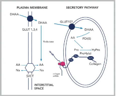
According to our hypothesis, GLUT10 may act as a DHA transporter in the ER membrane. Within the lumen, DHA is reduced to ascorbate and acts as a cofactor for the hydroxylation of prolyl and lysyl residues, a biochemical reaction crucial for collagen and elastin maturation and folding (Figure 1).
Fibroblasts cells isolated from ATS patients were used as a model system in our experiments, in which GLUT10 carried one of the 35 already described and published mutations.
3
Figure 1. Potential localization and role of GLUT10 in the endoplasmic reticulum
Aims
Clarify the intracellular positioning of GLUT10 protein in human fibroblasts;
examine the transport of ascorbate, DHA and other possible ligand(s) of in vitro translated GLUT10 protein in proteoliposomes;
prove the role of GLUT10 in the transport of ascorbate and DHA in fibroblasts isolated from ATS patients and controls.
4 Methods
Subcellular fractionation of human fibroblasts
Subcellular fractions were prepared from human control fibroblasts. Cells were sonicated, and then cellular homogenates were centrifuged for 10 min at 1000 g. The pellet – containing cellular debris and nuclear fraction - was washed with a further centrifugation step. Postnuclear supernatant was further centrifuged in clean tubes for 20 min at 18 000 g, which resulted the mitochondrial fraction. Microsomes were recovered by ultracentrifugation of the postmitochondrial supernatant for 60 min at 195 000 g.
hTERT fibroblast cultures and silencing
BJ-5ta hTERT immortalized fibroblasts from LGC Standards were silenced for GLUT10. shRNA-mediated knockdown of the SLC2A10 gene was achieved with human GIPZ lentiviral shRNAmir constructs containing viral particles. For stable expression experiments, selection of transduced cells was performed with puromycin.
RNA isolation and PCR analysis
Silencing of SLC2A10 gene in hTERT cells was checked by PCR technique. RNA was isolated by using RNeasy Plus Mini Kit according to the manufacturer’s instructions. Two micrograms of RNA were reverse transcribed with SuperScript III First-Strand Synthesis System for RT-PCR analysis.
5 Western blot analysis of gene silencing
Equal protein amounts (15–20 µg) prepared from hTERT cells were loaded to 10–12% SDS polyacrylamide gel. Proteins were consequently transferred to PVDF membranes with electro-blot apparatus. Protein content of the samples was checked by Ponceau staining and consequent densitometry. Membranes were probed using specific rabbit polyclonal anti-glucose transporter GLUT10 antibody (1:1000) and GAPDH antibody (1:7500).
Western blot analysis of cellular fractions
Equal protein amounts (20 µg) prepared from subcellular fractions were subjected to 12% SDS polyacrylamide gelelectrophoresis. Antibodies used were GLUT10 (1:1000), VDAC1 (1:1000), Cyclophylin D (1:2000) and Grp94 (1:5000).
Preparation of cells for transport measurements
After centrifugation, fibroblasts were resuspended in MOPS buffer (pH 7,2). Mitochondrial functions were inhibited with 1 mM NaN3, and cells were permeabilized with 40 μM digitonin.
Liposome preparation
Large unilamellar liposomes (LUV) were prepared from phosphatidylcholine from egg yolk. LUV were obtained by extrusion of the multilamellar vesicle suspension through a polycarbonate filter (pore size 0,4 nm). At least 40 extrusion cycles were performed at 40 °C.
6
In vitro translation of GLUT10 transporter
GLUT10 protein was synthesized from in vitro transcribed mRNA by using wheat germ extract, containing all of the necessary components for the translation (except amino acids).
Transport assays by rapid filtration technique
Intact and plasmamembrane-permeabilized cells were incubated in the presence of 14C-labeled ascorbate or DHA and were consequently filtered through glass fiber paper (pore size 0.45 nm) by using vacuum pump. The membrane was then washed with 2 ml MOPS buffer. The radioactivity retained on the filter was measured by liquid scintillation counting.
Measurement of ascorbate by HPLC
Ascorbate content of control and ATS cells was determined by high-pressure liquid chromatography (HPLC). Cells were incubated in the presence of 50 μM ascorbate for 0, 6, 12, 24 hrs. After incubation, the cells were collected in 25 w/v%
metaphosphoric acid solution. Ascorbate was measured from the clear supernatant after centrifugation.
Transfection of ATS fibroblasts
SLC2A10 cDNA was synthesized from the RNA isolated from control fibroblasts, by using SuperScriptIII One-Step Synthesis System, and was amplified by PCR technique. The PCR product was then inserted into an eukaryotic expression vector
7
pEF6/V5-His-TOPO™. Turbofect transfection reagent was used to transfect ATS cells with the expression vector. As a control, empty vector was transfected into ATS cells as well (mock).
Immunofluorescence Microscopical Analysis
To immunoreveal the GLUT10 protein, control fibroblasts were immunoreacted with rabbit polyclonal anti-human GLUT10 antibody and anti-rabbit secondary antibody conjugated to Alexa Fluor 488. Anti-cytochrome c monoclonal antibody and anti-mouse secondary antibody conjugated to Alexa Fluor 594 were used to reveal mitochondria. The co- localization of GLUT10 with the protein disulfide isomerase (PDI) was analysed in ATS fibroblasts transiently transfected with tagged GLUT10. Fibroblasts were immunoreacted with rabbit polyclonal anti-PDI antibody, which labels ER, and anti- V5 monoclonal antibody to label tagged GLUT10. Cells were incubated with anti-rabbit and anti-mouse secondary antibodies conjugated to Alexa Fluor® 488 and 594. To control the re- expression of GLUT10 in ATS cells, we used polyclonal anti- human GLUT10 antibody and rhodamine-linked secondary anti-rabbit antibody. Immunofluorescence signals were acquired by a CCD camera mounted on Zeiss fluorescence Axiovert microscope.
Statistical analyses
In each experiment we performed three parallel measurements.
Results are presented as mean values ± S.D. Statistical
8
comparisons were made by ANOVA and Student’s t-test. 5%
probability was accepted as significance level.
Results
At first, subcellular localization of GLUT10 on guinea pig liver fractions was examined. To test the purity of subcellular fractions, we have used antibodies reacting with proteins specific for a certain fraction (VDAC - mitochondria, Grp78 - ER). We found that GLUT10 was mainly localized in the ER.
Localization of GLUT10 was further examined on our human fibroblast model system, from which we have isolated nuclear, cytosolic, mitochondrial and ER fractions. The purity of each fraction was controlled as described above. Western blot analysis of the fractions showed that GLUT10 was present mainly in the ER of human fibroblasts, too.
Subcellular localization of GLUT10 was further studied with immunohistochemistry in human fibroblasts. In fibroblasts, a remarkable co-localization was detected between GLUT10 and PDI, while the transporter did not show co-localization with the mitochondrial cytochrome c.
After clarifying the localization of GLUT10, transport measurements were first executed on hTERT immortalized human skin fibroblasts silenced for SLC2A10. Stable clones expressing the silencing vector were selected and were checked for the efficiency of silencing. Semi-quantitative RT-PCR analysis of the stable clones showed that two silencing constructs out of three were effective.
Plasmamembrane of these GLUT10 silenced cells were selectively permeabilised by digitonin, while the
9
endomembranes were left intact (semipermeabilization).
Mitochondrial functions were inhibited by sodium-azid to exclude the possible mitochondrial transport mechanisms.
Transport measurements on these semipermeabilized hTERT fibroblasts showed that DHA uptake severely decreased in the effectively silenced cells respect to the control ones. The level of DHA transport showed a strong correlation with the amount of GLUT10 protein.
Transport measurements were then done on intact human fibroblasts isolated from ATS patients and healthy individuals.
While ascorbate transport on ATS cells remained unaltered, the transport of DHA decreased in the cells carrying a mutated GLUT10. The phenomena might be explained by the fact that a small amount of GLUT10 is present on the plasmamembrane.
Another possibility is that DHA uptake through endomembranes may affect the plasmamembrane transport of the molecule; in control cells, where the DHA is carried easily towards the luminal side of the endomembranes, these organelles may “suck” greater amount of externally added DHA. This effect is missing in ATS cells, therefore resulting a lower cellular DHA uptake.
Long-term ascorbate uptake was measured by HPLC in intact fibroblasts. Using physiological ascorbate concentration, ascorbate accumulation of ATS cells was lower respect to control cells. After 24 hrs, we found significantly less ascorbate in patient cells. Ascorbate concentration severely decreased during the 24 hrs of incubation in the cellular medium, which is probably due to the extensive oxidation of the vitamin. However, we didn’t find significant difference
10
between the ascorbate level of the medium of control and ATS cells.
To elucidate the transport through endomembranes, digitonin semipermeabilization – as described above - was applied in fibroblast cells. While ascorbate uptake did not change significantly between the control and patient semipermeabilized fibroblasts, we found that DHA transport severely decreased in ATS cells respect to controls.
In subsequent experiments, ER fractions isolated from fibroblast cells were fused with unilamellar liposomes to measure transport activity of the endomembranes. Microsomal fractions isolated from ATS cells showed diminished DHA transport when compared with the control group – even though the result was statistically not significant.
To directly measure DHA transport activity and the transport of potential other ligands of GLUT10, in vitro translated GLUT10 was incorporated into liposomes. The protein did not transport UDP-glucuronic acid and saccharose, while ascorbate showed a modest uptake. Both glucose and DHA (1 mM) were effectively transported by 3-5 nmol/mg liposome concentrations. In case of DHA, the uptake was dependent on its concentration.
In order to examine, whether GLUT10 is functional in transfected ATS cells, transport measurements were executed after the re-expression of the protein. Plasmamembrane of these cells were selectively permeabilized by digitonin – as described above – and DHA uptake was measured by rapid filtration technique. We found that DHA transport activity of these cells were restored upon the re-expression of GLUT10,
11
and became similar to the semipermeabilized control fibroblasts.
Conclusions
Present study verifies that GLUT10 is localized to the ER of human fibroblasts.
We demonstrated - by incorporating in vitro-translated GLUT10 protein into liposomes - that it acts as a specific DHA transporter.
The results obtained on GLUT10 mutant or silenced fibroblasts demonstrate that DHA transport decreases both on the plasmamembranes, but primarily on the endomembranes when compared with wild-type fibroblasts.
12
Bibliography of the candidate’s publications
Publications related to the thesis
Bánhegyi G., Benedetti A., Margittai E., Marcolongo P., Fulceri R., Németh C.E., Szarka A.: Subcellular compartmentation of ascorbate and its variation in disease states. Biochim Biophys Acta. 2014. (IF= 5.019)
Németh C.E., Marcolongo P., Gamberucci A., Fulceri R., Benedetti A., Zoppi N., Ritelli M., Chiarelli N., Colombi M., Willaert A., Callewaert B.L., Coucke P.J., Gróf P., Nagy S.K., Mészáros T., Bánhegyi G., Margittai É.: Glucose transporter type 10 - lacking in arterial tortuosity syndrome - facilitates dehydroascorbic acid transport. FEBS Lett. 2016. (IF= 3.623)
Gamberucci A.1, Marcolongo P.1, Németh C.E.1, Zoppi N., Szarka A., Chiarelli N., Hegedűs T., Ritelli M., Carini G., Willaert A., Callewaert B.L., Coucke P.J., Benedetti A., Margittai É., Fulceri R., Bánhegyi G., Colombi M.: GLUT10- Lacking in Arterial Tortuosity Syndrome-Is Localized to the Endoplasmic Reticulum of Human Fibroblasts. Int J Mol Sci.
2017. (IF= 3.226)
Publications not related to the thesis
Németh C.E., Bernert Z., Gallina Z., Varga M., Pap I., Hajdu T.: Kaposvár 61-es út 2. Lelőhely Árpád-kori embertani anyagának paleopatológiai vizsgálata. AnthropKozl. 2015.