Calcium Transport by Selected Animal Cells and Tissues*
R. H. Wasserman
I. Introduction 351 II. Isolated Kidney and HeLa Cells 352
III. Calcium Transport by the Erythrocyte 355 IV. Calcium Transport in Bone Cells 357
V. Chorioallantoic Membrane of the Avian Egg 362
VI. The Avian Shell Gland 366 VII. Intestinal Calcium Transport 372
VIII. Summation 378 References 381
I. INTRODUCTION
Calcium represents one of the key elements in bodily function and is required for diverse processes, including bone formation, nerve impulse transmission, the action potential, blood clot formation, membrane in- tegrity, and membrane function. This cation also has a central role in muscle contraction, and the calcium translocating system in endoplas- mic reticulum is discussed in this treatise by Martonosi (Chapter 8).
Because of the critical nature of calcium in these different systems, spe- cific processes have evolved to assure that adequate calcium is made available to the animal, and these involve absorption by the intestine, reabsorption in the kidney, and net resorption from the skeleton. These processes, together with the functioning of the calcium-regulating hor- mones, provide a tight homeostatic control of calcium levels in the extra- cellular fluids.
* The investigations from the author's laboratory were supported by NIH Grant AM-04652, NIDR Training Grant 5 T01DE00090-09, and U.S. A.E.C. Contract AT(30-l)-4039.
351
The " typical" mammalian cell is, of course, a complex system, con
taining a variety of subunits, substructure, organelles, etc. Despite the need for calcium for many reactions, each cell seems to be provided with a system for extruding calcium in order to maintain intracellular ionic calcium at levels of the order of 10"5 to 10"7 Μ [1 ]. No doubt one reason for this is that certain enzymes and biological reactions are calcium- inhibitable and do not operate optimally in a high calcium environment
[2,3]. In addition to membrane-bound extruding pumps, the mitochond
ria can accumulate calcium and, thereby, operate in essence as a calcium buffer to maintain cytoplasmic Ca within the required range [4]. Fixed anionic sites and other intracellular entities, such as the granules de
scribed by Martin and Matthews [5], provide additional mechanisms for maintaining low intracellular calcium ion concentrations.
The purpose of this chapter is to review some phases of the problem of calcium translocation. First, attention will be given to single cells studied in tissue culture and to the erythrocyte. Next, contemporary thoughts on how a specialized "single" cell, the bone cell, handles calcium will be presented, and, finally, consideration will be given to three types of cellular membranes across which substantial amounts of calcium are moved, the chorioallantoic membrane of the developing avian egg, the shell gland of the laying hen, and the intestine of several animal species.
Monographs, compilations, and reviews on some of these aspects have appeared recently [1-4, 6-9], and they should be consulted for details, additional information, and model formulations.
II. ISOLATED KIDNEY A ND HeLa CELLS
A systematic study of calcium transport by cells in tissue culture has occupied the attention of Borle [10-15] in recent years, and from these investigations have come valuable concepts and information, particularly with regard to the cellular handling of calcium and certain hormonal effects. The general plan was to subject HeLa or kidney cells, grown in tissue culture, to a medium containing radiocalcium and, with time, determine the pattern of radiocalcium accumulation (influx). The efflux pattern was assessed by preloading cells with the radionuclide and fol
lowing its release into nonlabeled medium.
The influx curve revealed two distinct components, one with a half- time of about 1.3 minutes and a slower one with a half-time of about 31 minutes. Assigning cellular localities to the two components is some
what hazardous but, in this case, it was reasonable to suppose that the fast component represented interaction with a surface-oriented structure and the slower phase, with a deeper intracellular component(s).
By treating the cells with trypsin-EDTA, the extracellular structures could be removed without presumably damaging the plasma membrane.
It was then possible to examine the compartment sizes of intact and
" naked " cells. From the data in Table I, it was apparent that a sizable
TABLE ι
COMPARISON BETWEEN CALCIUM COMPARTMENT SIZE OF CELLS AND CELL COAT OBTAINED BY CHEMICAL ANALYSIS
AND BY TRACER TECHNIQUES IN HeLa CELLS0
Chemical Kinetic analysis analysis Extracellular Caf t 37.78 1.06
Cell Ca* 5.74 2.69
Cell Cac 0.47 0.224
a From Borle [10].
b m/z moles/mg protein.
° mmoles/kg cell water; calculated on basis of water con
tent of HeLa cells which is about 12.3 mg water/mg cell protein.
discrepancy existed between extracellular Ca (cell coat) as determined by direct analysis of cells and as calculated from the kinetic data. This would indicate that, even though external to the cell, a large part of this calcium is tightly bound to the functional groups of the glycocalyx and is not readily exchangeable. The cell calcium (intracellular) values obtained by the two methods were reasonably close, and the data suggest that about half of the cell calcium is in a deeper intracellular compart
ment, perhaps mitochondria (see below).
The distribution of calcium in these various fractions can be denoted by the accompanying scheme, which pertains when the calcium content of the buffer medium is 1.3 mM [11]. This scheme gives an indication of the magnitude of the extracellular and intracellular compartments as derived from the Borle analysis.
[Ca] medium
[Ca] 84.4%
unexchangeable
[Ca] 2.4%
exchangeable
[Ca] 7.0%
' unexchangeable (subcellular
\ structures) [Ca] 6.2%
exchangeable Glycocalyx-membrane complex Intracellular
Several additional properties and characteristics of the system were revealed by these studies. First, when the medium calcium concentration was increased over the range 0—2.5 mM, the calcium content of the total cell was increased proportionately. However, if the same study was done on " naked " cells, cellular Ca showed saturation. Thus, the pro
portion of calcium deposited external to the plasma membrane becomes greater as the medium Ca concentration increases, indicating that the glycocalyx-membrane complex comprises a sizable depot for calcium.
The second significant finding was that the exchange rate of the slow component is very similar (~0.055 μμιηοίε cm"2 sec"1) to calcium influx into squid axon [16], sartorius muscle [17], and heart muscle [18], and that this flux is not altered by the inhibitors dinitrophenol and iodo
acetate, and anaerobiosis. The passive nature of the process is consistent with the direction of the electrochemical gradient. The third finding was that parathyroid hormone significantly enhanced the influx of calcium into "naked" cells. This led to the suggestion that the parathyroid hor
mone affected the passive diffusion of calcium into the cell interior, although an interaction with some membrane component facilitating the downhill transfer of calcium cannot be excluded. In fact, subsequent studies with isolated monkey kidney cells [13] indicated that the Km of the slow influx component increased from 0.3 to 1.15 mM and that the
Km a x increased by a factor of two. These findings were taken to suggest
that PTH stimulates the translocation of a calcium-carrier complex across the membrane.
The efflux of 4 5C a from cells preloaded with the radioisotope also revealed two kinetic components, a fast and a slow phase [12]. The half- times of exchange of each of these were almost identical with the half- times estimated from the influx studies. But one significant difference was noted. The slow efflux component was inhibited by DNP and IAA, extending the half-time of exchange by two- to sixfold, and was also highly temperature dependent, indicating that Ca efflux was, in part, an active process. Calcitonin appears to inhibit this active extrusion phase in kidney cells, since a significant effect of the hormone on the slow efflux component was readily demonstrable and no effect on Ca exchange during influx was noted.
These studies with the calcium regulating hormones indicated that PTH affects the passive, though possibly carrier-mediated, entrance into the cell, and calcitonin depresses the Ca efflux pump. Both would have similar effects on cell calcium levels, but the mechanisms proposed are certainly different.
In more recent experiments, Borle [15] was able to demonstrate an
other intracellular compartment. The evidence was that the amount of
4 5C a removable from cells by "wash-out" experiments was an inverse function of the length of the labeling period. For example, those cells labeled for 10 minutes lost 98% of their 4 5C a and those labeled for 2 hours lost less, about 86 %, and this effect was dependent on the pre
sence of phosphate in the medium. The involvement of mitochondria in sequestering the intracellular calcium (presumably as the phosphate) was proposed and tentatively verified by the additional finding that anti- mycin A and warfarin obliterated the second intracellular compartment.
The transfer of Ca between the two intracellular compartments (cyto
plasm and mitochondria) is rapid and reversible. Considering compara
tive transfer rates, Borle [15] further suggested that the intracellular calcium ion activity is controlled mainly by the exchange of cation be
tween cytoplasm and mitochondria, and less by the influx and efflux of calcium across the plasma membrane.
III. CALCIUM TRANSPORT BY THE ERYTHROCYTE
The mammalian erythrocyte has proved to be an important tool for the study of the membrane transport of C a2 + and other ions and non- electrolytes. Advantage can be taken of the ability to alter the internal milieu of the cell by reverse hemolysis. Erythrocytes, suspended in isotonic buffer, are diluted fivefold with water, producing "holes" in the membrane and releasing hemoglobin and other soluble constituents.
The ghosts, after being loaded with ATP, Mg, labeled Ca, etc., are re
sealed by adding sufficient amount of concentrated salts to reinstate the required osmotic relationships. With this technique the effect of various intracellular substances on the efflux and influx of calcium across the membrane, and on the calcium-activated adenosine triphosphatase, can be studied.
Direct analysis of the erythrocyte showed that intracellular Ca was of the order o f 4 x l O "5M , a value considerably below that of ionic serum Ca (~ 1.25 χ 10"3 M) [19]. Since Passow [20] previously demonstrated that the erythrocyte membrane was finitely permeable to calcium, the maintainance of the concentration gradient would require the operation of a Ca pump. Evidence for the existence of the membrane Ca pump was obtained by Schatzmann [21] and Schatzmann and Vincenzi [19]. It was demonstrated, for example, that, when washed cells were reversibly hemolyzed in medium containing ATP and M g2 +, calcium was extruded against a chemical gradient, a situation not occurring in cells resealed in the absence of ATP. In the ATP-loaded cells, calcium extrusion was
accompanied by the release of phosphate, arising in large part from the hydrolysis of the added ATP.
In a parallel study the relationship between ATP hydrolysis and Ca extrusion was quantitated, and the molar ratio was about 1.3 moles ATP hydrolyzed/mole calcium transported. Another interesting and important feature of the system was that ATPase activity was stimu
lated only by intracellular, and not extracellular, calcium. This is remin
iscent of the Na, K-ATPase enzyme, which is stimulated by internal N a+ and external K+. The asymmetry of ATPase activation provides addi
tional support for the involvement of the enzyme in calcium transport, and for the contention that the asymmetry of the binding site represents part of the molecular basis of the translocation process. Magnesium ion was an obligatory ingredient for the optimal operation of both the pump and the ATPase enzyme. The result was further obtained that Sr2 + could be actively extruded from the cell, as was also shown by Olsen and Cazort [22].
The erthrocyte calcium transport system is not inhibited by ouabain or oligomycin, specific inhibitors of the sodium-potassium pump and the Na, K-stimulated, Mg-activated ATPase. This indicates that the two transport systems are distinct. This is also suggested by the observation that N a+ and K+ have no significant effect on the Ca, Mg-ATPase enzyme. In contrast to ouabain and oligomycin, sulfhydryl reagents, such as salygran and ethacrynic acid [23], inhibit calcium transport when present in sufficient concentrations, but this effect only indicates that a sulfhydryl group is somewhere involved in the transport process.
Others have noted a Ca-ATPase in erythrocyte membrane in associ
ation with an actomyosin-like protein [24-26]. The recent report of Horton et al [27] gives evidence that the membrane contains two dif
ferent Ca-ATPases, one maximally activated at about 10"6 Μ Ca and the other at 3 χ 10" 4 Μ Ca. One may represent the transport ATPase and the other, the contractile ATPase. Wolf [28] purified erythrocyte mem
branes and obtained a fraction which, electron microscopically, appeared as ghosts with a large number of holes and had an ATPase activity that was strongly stimulated by Ca. This preparation was devoid of alkaline phosphatases and had only slight Na, K-ATPase activity. Evidence was also obtained for two Ca-ATPases, one with optimal activity at 25 X 10" 6 M C a and the second, at 0.04-0.10 x 10"3 Μ Ca.
The maintenance of a low intracellular Ca seems to be a universal requirement of all animal cells, and this was commented upon before.
Vincenzi [29,29a] produced a specific reason for this situation. Ca ions depress the Na, K-ATPase of membranes, and this inhibitory effect
occurs only when calcium is intracellular, not extracellular. The extrusion of intracellular Ca by the pump, coupled with the low cell permeability, assures a low concentration of cell calcium. With these relationships one would predict that the activation of the Ca pump would occur at a lower Ca concentration than that which would inhibit the ATPase of the sodi
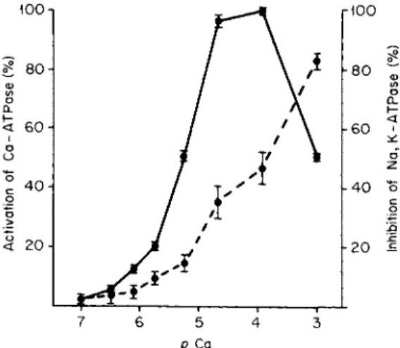
um pump. In Fig. 1 the predicted relationship is evident. Activation of
ρ Ca
FIG. 1. Effect of calcium concentration on the activation of Ca-ATPase ( # — φ ) and inhibition of Na, K-ATPase ( # — # ) in isolated erythrocyte membranes. ATPase activity was assayed by determining phosphate release in 60 minutes at 37°C in the presence of ATP (3 mM), MgCl2 (3 mM), NaCl (80 mM), KC1 (15 mM), tris-HCl (30 mM), EGTA ( 4 m M ) , p H 7 . 1 . Ca-ATPase activity represented the difference between zero Ca and a given concentration of Ca in the presence of 1 0 "4 Μ ouabain. Na, K-ATPase activity was taken as the ouabain-inhibitable activity at each Ca level. From Vincenzi [29] and Davis and Vincenzi [29a].
Ca-ATPase begins at about 10" 6 Μ Ca and becomes about maximal at 10"5 Μ Ca. The Na, K-ATPase is 35% inhibited at 10"5 Μ Ca, a con
centration at which the Ca-ATPase is maximally stimulated. Certainly this represents a clear-cut role of the Ca pump in the control of cell activity, mediated through the intracellular concentration of Ca.
IV. CALCIUM TRANSPORT IN BONE CELLS
Investigative progress on transport processes in bone cells has, of course, been substantially hindered by the inaccessibility of these cells. The osteoblasts, osteoclasts, and osteocytes are either intimately associated
with the bone or cartilage surface or buried within the bone proper.
However, techniques have been developed to release bone cells, such as exposure of calvaria to collagenase [30] and controlled hand-grinding of a bone specimen in a mortar and pestle [31]. Some cell damage might be expected to occur, and certainly the orientation of the bone cell is dis
rupted since osteoblasts and osteocytes are polarized with respect to the bone surface [32]. Studies of calcium transport by such cells will yield only part of the total picture. Furthermore, recent data [33-35] support the concept, first suggested by Thomas and Howard [36], that the bone surface is enveloped by bone cells and bone cell processes, forming a membrane-like barrier. This differs from the previous idea in which extracellular fluid was considered to have immediate access to the calci
fying surface.
Considerable progress in understanding calcium metabolism in bone cells per se has been achieved by Nichols and co-workers [31,37,38].
Using the grinding technique and repeated differential centrifugation over a dense layer of dextran (d= 1.11), bone cells were isolated and were found to contain a high concentration of total calcium ( ~ 100-
170 mM). The problem of contamination of the bone cells by bone fragments or calcified cartilage was systematically examined, and the evidence indicated that the calcium was within the cell. Treatment of the cells with trypsin-EDTA, collagenase, neuramidase, etc., did not release cell-bound calcium, and, in addition, bone cells isolated by three different methods, including the use of collagenase, yielded cell popula
tions containing high concentrations of calcium of the order of 150 mM.
Along with the dense, calcium-containing cells, bone cells of lighter density were also released. The latter cells have a higher capacity to hydroxylate proline than the heavier cells, and the latter have higher rates of 02 uptake and lactate production. This is suggestive of special
ization of function, some cells being concerned with calcium transloca- ation and others with organic matrix synthesis.
Centrifugation of bone cell homogenates revealed that most of the intracellular calcium was associated with the most dense fraction. Fur
ther purification of this fraction yielded calcium-containing particles composed of about 6.8% calcium, 3.4% phosphate, 52% protein, and 34 % phospholipid of the total dry weight. Only traces of hydroxyproline were present, indicating that little, if any, of the cell calcium was associ
ated with engulfed bony fragments.
These biochemical studies are consistent with the prior histochemical demonstration of calcium phosphate spherules in bone cells by Kashiwa [38a]. Evidence suggested that these calcium phosphate spherules in the cytoplasm of epiphyseal chondrocytes also contain bound lipid, and that
these mineralized entities are secreted into the perilacunar matrix, co
alescing to form larger spherules [38b].
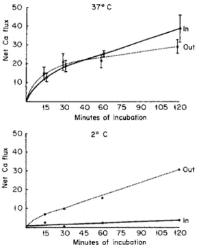
Kinetic studies suggested that calcium (and phosphorus) in the intra
cellular mineral deposit can exchange with external calcium to a sizable degree. As shown in Fig. 2, the fluxes of calcium into and out of the isolated cells were about equal at 37°C and each represented a sizable fraction of the total cell calcium. Therefore, despite its apparent insolu
bility, the calcium in this pool has a high rate of exchange and appears to be reasonably labile. When incubated at 2°C (Fig. 2), the efflux rate exceeded the influx rate, and a net loss of calcium occurred. Further, the depression of the influx rate to a low value at the lower temperature suggests that influx is a metabolically dependent process, whereas efflux is not, a situation opposite that in erythrocytes, and isolated HeLa and kidney cells. On the other hand, Stern and Austin [38c], using bone cells from fetal rat calvaria by collagenase treatment, stated that 4 5C a influx was unaffected by anoxia, whereas an efflux component was markedly inhibited. Their results suggested active extrusion of 4 5C a from the bone cells, and entrance into the cell by diffusion only.
50
Out In
15 30 40 60 75 90 105 120 Minutes of incubation
50 3 40
a Out
υ 30
Φ
20
In 15 30 45 60 75 90 105 120
Minutes of incubation
FIG. 2. The effect of temperature on the influx and efflux of calcium from isolated bone cells (in mM/kg cells). The cells were incubated in Krebs-Ringer bicarbonate buffer containing 1.25 mM Ca and glucose as substrate. From Nichols et al. [31].
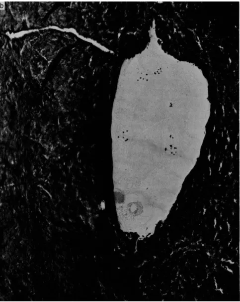
FIG. 3. Electron micrograph of an osteocyte in a lacunae (a) before and (b) after incinerating at 500°C for 15 minutes. The unashed preparation shows several dense granules that persist after ashing. Magnification χ 14,000. From Martin and Matthews [39].
Various factors related to calcium metabolism have also been exam
ined, and such factors as parathyroid hormone, calcitonin, and phos
phate ions appear to effect either efflux, influx, total cell calcium content, or combinations of these. The indication was that parathyroid increased the active uptake of C a2 + by the bone cell, an effect different from that proposed by Borle (see above). Both calcitonin and phosphate appeared to block the exit of calcium from the cell, which does coincide with the observation made by Borle on isolated kidney cells.
The model put forth by Nichols [31] considers that packets of calcium phosphate accumulated within the bone cell are translocated as such to the bone surface, where the mineral crystals are released and deposited at the calcification front. The other function of bone cells, i.e., mainten
ance of circulating levels of calcium and phosphorus, is accomplished by the same packets releasing their contents into a solubilizing environ
ment near the capillaries. The calcium-phosphate packet idea has con
siderable support from the studies of Matthews and colleagues [5,39], who showed that mitochondria of bone cells contain granules that persist after ashing (Fig. 3). The intracellular concentration of these granules varies with cell type, in bone tissue the granules being less numerous in chondrocytes in the zone of provisional calcification and more numerous in cells in the hypertrophic zone. This and other evidence indicate a direct association between mitochondrial granules and calci
fication. Lehninger [4] and Talmage [40], in recent reviews, also consider the calcium-containing packets as central to the operation of bone cells in calcification and calcium homeostasis. X-ray diffraction data of Posner (cited by Lehninger [4]) showed that the calcium accumulated by mitochondria to be amorphous in nature, and not hydroxyapatite.
Other reports by Termine and Posner [41] suggest that, indeed, the calcium deposited in newly mineralizing bone is amorphous and later undergoes a transition to the crystalline type. Lehninger [4] further con
siders that amorphous calcium phosphate exists as "micropackets" of colloidal dimensions in bone cells and can dissociate from large aggre
gates and traverse across membranes intact, either by diffusion or by reverse pinocytosis.
V. CHORIOALLANTOIC MEMBRANE OF THE AVIAN EGG
The developing avian skeleton obtains most of its calcium from the egg-shell, a conclusion that is obvious but, at one time, had been a source of controversy [42,43]. Chemical balances clearly showed that about 80-82 % of calcium in the embryo skeleton was derived from the shell
and the rest from the yolk [44]. How is the egg shell calcium resorbed and made available to the embryo ? What physiological and biochemical processes are involved ?
In the early stages of development of the avian embryo, the allantois grows from the hind-gut into the extraembryonic region and provides a diverticulum for the storage of the nitrogenous waste products until hatching. Before this, other membranes are formed, among them the outer chorion, which is of ectodermal origin. The chorion and allantois begin to fuse on the fifth day and, by the ninth or tenth day, proliferate such that most of the inner aspect of the eggshell is lined with this double membrane [45]. The membrane is comprised of three layers, the outer ectodermal layer, an inner endodermal layer, and a mesodermal layer between the two. The membrane becomes highly vascularized and the ectoderm (adjacent to, and underlying, the shell) undergoes an intensive differentiation and proliferation at days 10-11. These histological stud
ies, as well as those of Leeson and Leeson [46], indicate that the chorio
allantoic membranes are concerned with other functions besides gas ex
change during respiration. The closeness of attachment of the ectodermal layers to the inner surface of the eggshell suggested that one such func
tion might be calcium translocation.
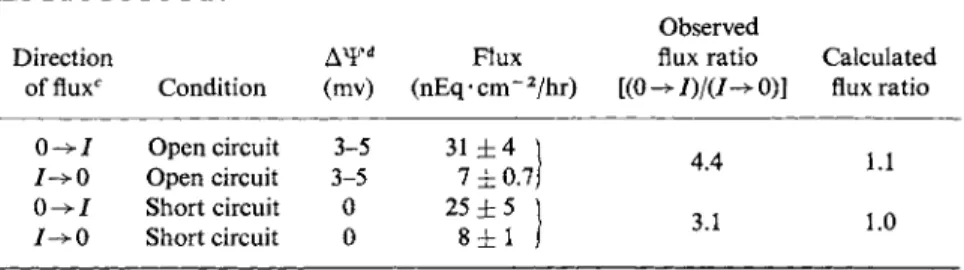
Terepka and associates [47,48] undertook a series of studies with the chorioallantoic membrane, which could be separated from the eggshell as a flat sheet. By placing the membrane between two chambers of an Ussing-Zerhan device [49], measurements of the fluxes of calcium into and across the membrane could be made. The data summarized in Table II showed that, either under open- or short-circuited conditions, the uni
directional flux of Ca from the outer bathing fluid into the inner fluid (0-^7) exceeded that in the opposite direction ( 7 ^ 0 ) by a factor of three to four. Since the expected flux ratio in either condition, as cal
culated from Ussing's equation (see footnote to Table II) was about unity, these observations suggested that the calcium ion was being active
ly transported against a thermodynamic gradient. Net transport oc
curred in the direction of shell to the inner aspect of egg. The 0 -> I flux was inhibited by replacing 02 with N2 and by the addition of dicoumarol (10"4 M), oligomycin (5 χ 10"5 Μ), and ouabain (10"5 M) to the bath
ing media. Inhibitors of protein synthesis (actinomycin D, cyclohexa- mide) were without effect over the period of incubation (6 hours). Clear
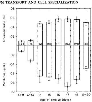
ly, then, the membranes associated with the eggshell were shown to have the capacity to participate in the transfer of calcium from the main reservoir (the shell) to the developing embryo. This function becomes manifest at the time in development when relatively large amounts of calcium are required for bone formation, i.e., after day 13 (Fig. 4).
More detailed investigations of the isolated C-A membrane suggested
TABLE II
UNIDIRECTIONAL FLUXES OF Ca ACROSS THE C-A MEMBRANE in vitro0'·b
Direction
offluxc Condition (mv)
Flux (nEq-cm~2/hr)
Observed flux ratio [(0 ->/)/(/-> 0)]
Calculated flux ratio 0 -» I Open circuit 3-5
I->0 Open circuit 3-5 0 - > 7 Short circuit 0 7->0 Short circuit 0
31 ± 4 \ 7 ± 0 . 7 / 25 ± 5
8 ± 1
4.4 3.1
1.1 1.0
a From Moriorty and Terepka [48].
&Inner and outer bathing fluids were identical and had the following composition:
NaCl (140 mM), KC1 (4 mM), CaCl2 · 2 H20 (1 mM), glucose (4.4 m M ) , Tris (25 m M ) ; pH 7.4. Gassed with 100% 02.
c 0->7 represents the flux from ectodermal (shell side) to the endodermal side. I - > 0 is the flux in the opposite direction.
d Outer or ectodermal side is positive.
e Calculated from Ussing's flux ratio equation:
/ - > 0 {Ca},
where {Ca}0 = concentration of Ca in outer bathing fluid, {Ca}/ = concentration of Ca in inner bathing fluid, Δ Ψ = electropotential difference across the membrane, and Z, F, R, and Tare, respectively, the valence of the ion, the Faraday constant, the gas constant, and the absolute temperature.
that the calcium "in transit" in the membrane is compartmentalized and may move as discrete " packages " of calcium. The evidence for this concept is primarily indirect. First, the specific activity of the calcium in the C-A is always less than that of precursor solution (outer fluid), indi
cating that transported cation does not mix with the total calcium pool in the membrane. Second, the flux of calcium was unaffected by either the spontaneous or an imposed membrane potential difference, indicating that Ca transfer was not electrogenic. Third, the study of the uptake of calcium by the membrane showed that the magnitude of accumulation exceeded that of the transcellular movement. If, after radiocalcium was taken up by the membrane from labeled outer fluid, the outer fluid was replaced by unlabeled medium, membrane radiocalcium moved pri
marily in the " proper " direction, i.e., 0 -> /. Also the specific activity of the transported calcium was higher than tissue 4 5C a and similar to that in the precursor compartment, suggesting that calcium is transferred through the cell in distinct " compartments." Electron probe analysis of the calcium-transporting chorioallantoic membrane showed that the cal
cium was compartmentalized in only certain cells of the ectodermal layer, those cells found immediately adjacent to respiratory capillaries, and
^ .06 4 .04 .02
Ε α>
.02 -{
.04 .06 .08 1.0
(4)| 1(8) (6) (11) 1(12) (16)| (19)
Ψ
(11)
10-11 12-13 14 15 16 17 18 19-20 Age of embryo (days)
FIG. 4 . Change in transport capacity of the membranes of eggshell during embyronic development. The flux of calcium from outer to inner fluid is denoted by the height of the bar above the zero line, the uptake of calcium by the length of the lower bar, and the total transfer from the outer medium by the total length of the bar. Between 1 2 - 1 3 and 1 4 days a considerable increase in transmembrane flux, uptake by the membrane, and total move
ment from the outer solution occurred. The numbers in parentheses are the number of experiments, and the S.E. of the mean value is shown. Flux and uptake both in /xEq/cm2/ hour. From Terepka et al. [47].
which have long cytoplasmic processes that surround these capillaries [50], A direct relationship between these calcium-containing bodies and calcium translocation is suggested, particularly since these "com
partments" are not evident in young, nontransporting membranes.
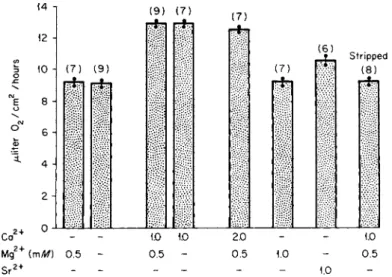
The stimulation of 02 uptake by the membranes by calcium was also detected (Fig. 5) and maximal stimulation occurred at 1 mM Ca, a value comparable to that which saturates the transport process [51]. The cal
cium effect is only evident when the ectodermal surface is exposed to the cation; increasing the calcium content of the fluid bathing the endo- dermal surface yielded no increment in 02 uptake. Using such data, it was
estimated that, for every molecule of 02 consumed above the basal level, about one atom of Ca was transported. For the N a+ translocation sys
tem in several diverse tissues [52], the N a / 02 ratio is about 20 atoms of Na transported/molecule of 02 consumed. This comparison indicates that calcium transport places a high demand on available cellular energy, or that a different mechanism is available for Ca2 + transport in contrast to that for N a+ [51].
.08 •
14 -ι (9) (7) (7)
ε
ο
ο"
12 Η
ίο Η (7) (9) (7)
(6) C | ' Μ
* Stripped
^ (8)
2 i
Ca2+ - - 1.0 1.0 2.0 - - 1.0
Mg2+ (mM) 0.5 - 0.5 - 0.5 1.0 - 0.5
S r2 + _ _ 1.0
FIG. 5. Effect of alkaline earth cations on the 02 uptake by the chorioallantoic mem
brane. Oxygen consumption was determined manometrically. Note that C a2 + significantly increased 02 uptakes with or without M g2 + . S r2 + also elicited an effect but less than C a2 + . From Terepka et al. [51].
Sulfhydryl reagents, such as parachloromercuribenzoate (PCMB) ap
peared to have a specific effect on the calcium entrance step [51]. This is evidenced by the observation that, in the presence of PCMB, calcium translocation was suppressed and, significantly, that only Ca-stimulated 02 uptake was inhibited. Furthermore, PCMB had no effect on 02 up
take when only the endodermal surface was subjected to the sulfhydryl reagent, whereas the 02 uptake of the ectodermal aspect of the membrane was equally suppressed by PCMB in the presence of calcium or when calcium and PCMB were both absent. These data were interpreted as indicating that an — SH-containing compound close to the ectodermal surface is directly involved in calcium translocation and might involve a calcium-dependent adenosine triphosphatase, an enzyme inhibitable by — SH reagents in other vertebrate tissues.
VI. THE AVIAN SHELL G L A ND
The shell-forming gland (also called the uterus) is that part of the avian reproductive tract involved in eggshell calcification. It comprises about 17% of the total oviduct and "secretes" about 2.2 gm calcium
over a 15-hour period or 100-150 mg Ca/hour. If blood calcium were not rapidly replenished by calcium from bone or from the intestine, it would be depleted in 10—15 minutes [43]. Along with calcium, the car- bonate ion is also transferred and together deposit in the shell as in- soluble CaC03 in the form of calcite. The egg, when it first enters the shell gland region, is surrounded by a loose protein-mucopolysaccharide membrane, which provides part of the matrix for calcium deposition.
In this same region, a "watery" secretion is also produced which pene- trates into the egg and distends the membrane so that it comes into close contact with the uterine epithelium.
The epithelial lining cells (those facing the lumen of the shell gland) are of two types, the basal cells and the apical cells, and these appear to alternate with one another. The basal and apical terms designate the location of the nucleus within each cell type. The basal cells have micro- villi, and the apical cells have both microvilli and cilia, on their luminal surface. Another prominent feature of the uterine tissue are the tubular glands, the cells of which surround a small duct in acinar configuration.
The cells of the tubular glands are devoid of histological components that are indicative of secretory activity. On the other hand, both the basal and apical cells contain numerous secretory granules which discharge their contents into the uterine cavity during eggshell formation.
During calcification the microvilli on the tubular cells change and be- come enlarged and distend into bloblike structures [53]; this, also noted by Johnston et al. [54], was described as vacuolization. Comparing these changes in structure to other cell types involved in water transport, Breen and DeBruyn [53] suggested that the tubular cells secrete a mineral solution which might constitute the primary source of shell calcium. This assumption was reinforced by the absence of complex organic mol- ecules, such as mucopolysaccharides and proteins, in the secretion.
Johnston et al. [54] also suggested, on the basis of ultrastructural analy- sis, that the uterine gland cells are concerned with the transfer of calcium to the calcifying eggshell.
Calcium deficiency in the hen leads eventually to a cessation of egg laying; the microvilli lining the tubular gland cell disappear, and other degenerative changes are noted in the tubular cell proper [55]. The con- figuration of the lining epithelia cells also is altered by low calcium in- take, but the cells preserve their capacity to maintain secretory granules.
It would be tempting to correlate directly the cell type most altered by calcium deficiency with the cell type that is involved in calcium transloca- tion. However, this becomes difficult when it is recognized that the loss of egg production might be an indirect effect of an insufficient secretion of gonadotrophin from the pituitary.
Evidence implicating the lining epithelium cells, rather than the tubu
lar cells, in calcium translocation is derived from autoradiographic local
ization of 4 5C a in shell gland tissue obtained either during active shell formation or in the absence of calcification [56]. The tissue was taken 5 minutes after 4 5C a was injected into hens, and processed by freeze- substitution in order to decrease translocation of the isotope. Grain counts of the autoradiograph showed that, during active shell calcifica
tion, the columnar lining cells increased in 4 5C a content by about 68%
whereas the tubular lining cell 4 5C a decreased by about 32%, with re
spect to the noncalcifying shell gland. Further, the in vitro accumulation of radiocalcium by lining epithelium was considerably greater than that of the tubular gland cells (also measured autoradiographically), and the former was more susceptible to inhibition by 2,4-dinitrophenol. Be
cause of these patterns of uptake, Schraer and Schraer [57] stated that the lining cells are the major site of calcium transport in the shell gland.
In addition, low temperature microincineration of thin sections of uterine tissue yielded patterns indicating that the lining epithelium had heavier mineral deposits than the tubular gland cells [58].
Thus, the morphological and ultrastructural observations by Breen and DeBruyn [53] and Johnston et al. [54] suggest that the tubular gland cells are most likely involved in the calcium secretion process, whereas Schraer and Schraer [57], from autoradiographic and microincineration studies, strongly implicate the epithelium lining cells. The Breen-De- Bruyn-Johnston evidence, highly indirect, is primarily that the number of secretory granules containing polymers in the tubular gland cells were minimal and that the character of the microvilli changed during active calcification. The evidence of Schraer and Schraer is based on the ap
parent high concentration of mineral in the lining cells during calcifi
cation when shell is being formed; the opposite pattern emerged in the tubular gland cells. This latter evidence is also indirect and could be interpreted somewhat differently. The decreased calcium content of the tubular gland cells could reflect an increased release, with a concomitant increased turnover, of calcium as a consequence of transport. The in
creased level of calcium in the lining cells might only reflect the higher concentration of calcium in the uterine fluid, which enhances the level of absolute accumulation of cation. Other procedures and additional evi
dence are required to resolve this problem. As pointed out by Nevalainen [55], the demonstration of the vitamin D-induced, calcium-binding pro
tein (CaBP) in the shell gland by Corradino et al. [59] suggests such an approach. Since CaBP has been implicated in calcium translocation across the intestine, the cellular localization of CaBP in shell gland tissue, as done by Taylor and Wasserman [60] with intestine, might aid in defin
ing the cell type involved in calcium transport.
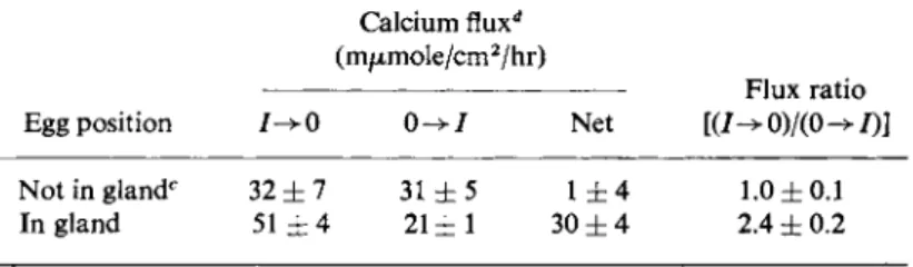
The processes by which calcium is transported across the shell gland were investigated in considerable detail by Schraer and Schraer and colleagues [57,61 ]. By placing the shell gland across the orifice of a Ussing- Zerhan chamber, the unidirectional fluxes of radiocalcium, in addition to the electropotential gradient and the short circuit current, were meas- ured. In one study the shell gland was taken from hens in one of two states, either with or without an egg in the uterus. The data, shown in Table III, show several interesting features of the system. First, the uni- fluxes of calcium across the "egg-less" tissue were equal, yielding a flux
TABLE III
UNIDIRECTIONAL FLUXES AND N E T FLUX OF CALCIUM ACROSS THE ISOLATED SHELL GLAND OF THE LAYING H E N ; EFFECT OF LOCATION OF E G G AT TIME
OF SACRIFICE."* *
Calcium fluxd
(m/xmole/cm2/hr)
Flux ratio Egg position 7 ^ 0 0 - > 7 Net [(7->0)/(0->7)]
Not in glandc 32 ± 7 31 ± 5 1 ± 4 1.0 ± 0 . 1 In gland 51 ± 4 2 1 ± 1 30 ± 4 2.4 ± 0.2
a From Ehrenspeck et al. [62]. Values = mean ± SEM.
* Inner and outer bathing solutions were identical and had the following composition: NaCl (136 mM), KC1 (22.8 mM), CaCl2 (1.80 mM), M g S 04
(1.66 mM), tris (23.8 mM), and glucose (16.7 mM). Gassed with pure 02.
c Three of the five tissues came from hens with no egg in the oviduct, one with an egg in the magnum, and one with the egg in the isthmus.
d 7-^0 represents the flux in the direction from blood to lumen and 0->7, the flux in the opposite direction.
ratio of unity. Second, the presence of an egg in the uterus stimulated, in some fashion, the flux of calcium from serosal side to luminal side 60%
and decreased the flux in the opposing direction 32%. The flux ratio of the calcium fluxes was now 2.4. Third, only the "stimulated" uterus yielded a net flux of calcium, this being in the luminal direction. Under similar conditions, but in a different experiment, the potential difference across the shell gland in vitro was observed to be only 1 mV with luminal side negative (Erhenspeck, 1969; cited in Schraer and Schraer [57]); the deviation of the flux ratio of the "stimulated" tissue from unity could not, therefore, be due to the existing electropotential gradient. Thus, the presence of an active transport of calcium in the shell gland can be in- ferred from this analysis, but this proposal only pertains to those shell glands obtained when an egg was present therein. Erhenspeck et al. [62]
also demonstrated that the flux of calcium from blood to lumen was depressed by 2,4-dinitrophenol (5 χ 10~5 M), KCN (5 χ 10"2 M) or an N2 atmosphere, and was stimulated by sodium succinate as substrate.
Thus the shell gland apparently contains a special mechanism for translocating calcium during eggshell formation, a process subjected to exogenous control. The latter is also evidenced by the differing calcium content of the tissue during different phases of the egg-laying cycle and the different behavior of gland mitochondria during these phases [62].
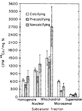
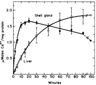
When no eggshell is being laid down in the gland, the calcium content of the tissue and the degree of accumulation of 4 5C a by the mitochondria of that tissue are about twice those when calcium is being secreted (Fig. 6) [57,62a].
Proceeding under the hypothesis that mitochondria in calcium-trans
locating tissues might have special properties related to calcium transfer, Schraer and Schraer [61] compared certain aspects of calcium metab-
3600 y 3300 - 3000 - 2700- 2400-- 2100 -•
1800--
«500-- 1200-- 900-- 600-- 300-- 0
• Calcifying
• Precalcifying ϋ Noncalcif ying
ft)
Homogenate Mitochondrial Supernatant Nuclear Microsomal Subcellular fraction
FIG. 6. Influence of the phase of the egg-laying cycle of the donor animal on the 4 5C a in cell fractions prepared from shell gland mucosa. T h e4 5C a was administered to the animal before the tissue was excised and fractionated. The three phases represented are calcifying (active calcification of the eggshell), precalcifying (phase just before the entrance of the egg into the shell gland), and noncalcifying (no egg in oviduct). From Hohman and Schraer [62a] and Schraer and Schraer [57].
olism of shell and liver mitochondria. In the presence of ATP in vitro, gland mitochondria accumulated 4 5C a (4 mM 4 0Ca) more rapidly than liver mitochondria and tended to discharge the accumulated 4 5C a after about 20 minutes (Fig. 7) [61,62b]. Liver mitochondria, on the other hand, took up 4 5C a more slowly and, over the period reported (150 minutes), showed no release of the radionuclide. Another difference was that gland mitochondria accumulated more 4 5C a at pH 6.2 than at pH 7.4 (Fig. 8), whereas the process in liver mitochondria did not change over this pH range.
τ 1 1 1 1 1 1 Γ
Minutes
FIG. 7. Uptake of calcium by isolated mitochondria from shell gland and liver of actively laying hens. The incubation medium contained tris (10 mM), ATP (3 mM), succin
ate (4 mM), Mg (10 mM), and Ca (4 mM), pH 6.2. Temperature 25°C. The bars indicate the range. From Schraer and Schraer [61] and Elder [62b].
It is tempting to speculate that shell gland mitochondria have a signifi
cant and unique role in the formation of the eggshell and the attendant translocation of calcium. The fact that the calcium content varies with the state of the gland and that the in vitro turnover of calcium is greater in gland mitochondria than in liver mitochondria suggests such a role.
It is, of course, reasonable to suppose also that the mitochondrion has as its major function the maintenance of a low intracellular calcium concentration during translocation, as proposed for other tissues [4].
However, one would suspect that, if this were the case, the greatest con
centration of Ca in the gland mitochondria would occur during, and not
τ 1 1 Γ
Live
I ι ι ι ι ι ι ΐ—ΐ 6.2 6.4 6.6 6.8 7.0 7.2 7.4
ΡΗ
FIG. 8. Effect of pH on the uptake of calcium by isolated mitochondria from the shell gland and liver of the laying hen. The incubation medium contained Hepes buffer (20 m M ) , ATP (3 mM), Mg (4 mM), Ca (3 mM). Incubation time 15 minutes. From Schraer and Schraer [61] and Elder [62b].
before, marked calcium movement. Mitochondrial Ca might also repre
sent an accessible store of the element, but, from Figs. 6 and 7, it is apparent that the high 4 0C a content of the gland is discharged before the uncalcified egg reaches the shell gland.
VII. INTESTINAL CALCIUM TRANSPORT
Considerable effort has been given to elucidating both the mechanism by which calcium moves across the epithelial membrane of the gut and the action of vitamin D thereon, and several reviews on these aspects have appeared recently, viz., DeLuca [63], Norman [64], Wasserman [65], Wasserman and Taylor [66], Wasserman and Kallfelz [67], and Kenny [68].
The intestinal epithelium has several important features that might bear mentioning, as summarized and documented previously [66,69].
The luminal-facing surface is covered by a glycocalyx ("fuzz") com
prised mainly of glyco-proteins and mucopolysaccharides. Some of these elements are closely attached to the microvillar membrane and cannot be easily removed. The microvillus membrane itself shows the typical tri-
partite structure on electron microscopic examination although, in cer- tain preparations, a globular substructure^ suggested. Within the core of the microvillus are rod-shaped elements which are considered by some to be microtubules; and these elements extend into the terminal web, the latter residing in the region immediately beneath the microvillus membrane. At about this level there is a cooperative structure between adjacent cells; it is termed the tight junction or "zona occludens" and presumably prevents the transfer of materials directly from the lumen to the intercellular space. This junction might be analogous to the intercellular bridge studied by Lowenstein [70], which is involved in intercellular communication. The intercellular region contains'muco- polysaccharide material [71] and specialized structures, such as desmosomes. The terminal web region does not appear to contain sub- cellular elements, although vesicles are occasionally observed. Beneath this region are the usual intracellular particles, such as mitochondria, endoplasmic reticulae, and nucleus. The basal border rests on a loose basal membrane just apical to the lamina propria wherein the capillaries and lymphatics are located. Beneath the lamina propria are the longi- tudinal and circular muscle layers and, in the everted gut sac preparation, the calcium ion must traverse across these layers in addition to the mucosal cells and lamina propria.
The transfer of calcium across the intestine occurs by at least two processes, active transport and diffusion (simple or facilitated). The active transport of calcium was first shown by Schachter and Rosen [72]
and adequately confirmed by others, using both in vitro and in vivo tech- niques (cf. [66,68]). However, the dual mode of transfer is suggested by the data illustrated in Fig. 9, which shows the unidirectional fluxes of calcium across chick duodenum as a function of intraluminal calcium concentration. As may be noted, the slope of the efflux curve (from lumen to blood) markedly changes at about 5 mM [Ca], indicating a transition from a rapid efflux process to a slower one. The essential features of the efflux curve (Fig. 9) [72a] were recently verified by Sammon et al [73], using a balance technique, and by Papworth and Patrick [74], using an in vitro procedure. Each of these dual component curves can be described by equation [66]:
7
"=isrr!;
+0<Ca)<"
where JLB = total flux from lumen to blood, /m a x = maximum flux through the saturable component, [Ca] = calcium ion concentration in the lumen, Kt = transport constant (analogous to the Michaelis-Menten constant), and D = diffusional coefficient of the second component. The
Ε
16 ι ι — —ι— ? " I I I
14 Efflux
12
Χ
Vitamin D3
10 - /
8 6 -
4 - Rachitic
2 -
0
2 —..^ j-Rachitic
4 Influx
ι I
Vitamin D3 —s * , ^ ^
6ο 20 4 0 6 0 8 0 100 120 140 Intraluminal Ca
FIG. 9. Effect of vitamin D3 on the undirectional fluxes (ftmoles/cm hr) of calcium across chick duodenum in situ. Chicks were raised on a rachitogenic diet for 3-4 weeks, and half were given 500IU vitamin D3 orally 24 hours before experiment. The other half (rachitic control) were given vehicle alone. The fluxes were assessed at different levels of intra
luminal Ca(mM). Efflux refers to movement out of the lumen and influx to movement in the opposite direction. From Wasserman and Kallfelz [72a].
validity of the above two-component flux equation [Eq. (1)], has support from the data of Papworth and Patrick [74], who showed that dinitrophenol primarily inhibited the first (saturable) component and left nearly unchanged the second or diffusional component.
One of the more important factors affecting calcium absorption is vitamin D, and understanding its role in the process should certainly aid in understanding the total mechanism. It has been known for many years, of course, that the vitamin D-deficient animal absorbs less calcium than a normal animal, and this results in rickets in the growing animal and osteomalacia in the adult. In addition to altering calcium absorp
tion, vitamin D elicits effects on the kidney and bone, and perhaps else
where.
Effects of vitamin D on the calcium fluxes are apparent from the data depicted in Fig. 9. The total efflux was increased by the vitamin, as was the unidirectional flux in the opposite direction. The calculation of the flux ratios at the different calcium concentrations indicated that, with respect to the theoretical flux ratio calculated from Ussing's equation [75], active transport was occurring below 5 mM Ca and diffusion above that concentration. Also above 5 mM Ca, the flux ratios of the rachitic and vitamin D intestine were identical, indicating that the steriod was affecting primarily diffusion rather than active transport. The altered permeability of the intestinal membrane by vitamin D is further sug- gested by the greater rate of transfer of Ca from blood to lumen in the vitamin D-treated chicks. The influence of vitamin D on calcium diffu- sion across everted gut sacs in vitro was proposed previously by Harrison and Harrison [76]. Schachter et ah [77], on the other hand, showed by the everted sac procedure that the active transport of calcium is enhan- ced by vitamin D.
The active transport component in the rat is, as shown by Schachter and Rosen [72], inhibited by anaerobiosis, DNP, iodoacetate, malonate, etc., whereas in the chick ileum the process is not inhibited by anaeroboi- sis, DNP, or NaCN but is significantly depressed by 5°C, iodoacetate, JV-ethylmaleimide [78]. These data suggest a species difference; in the rat, calcium transport is primarily supported by oxidative phophoryl- ation and, in the chick, by nonoxidative processes. It was also interesting to note that N2, NaCN, and DNP, while not affecting translocation by intestine from vitamin D-treated chicks, did accelerate the transfer of calcium across the intestine of rachitic chicks [78]. The phenomenon whereby an inhibitor depresses calcium absorption in the vitamin D- treated chick and accelerates calcium transfer in the rachitic animal has been our usual experience. Such data denote that two energy-dependent reactions are involved in determining Ca absorption, one related to active Ca transport and the other to the maintenance of Ca permeability of the gut. Therefore, alterations in either the resistance of the calcium path or changes in Ca pump units become feasible ways in which a given treatment might affect calcium absorption.
Vitamin D undergoes various transformations in the animal, and metabolites more active than vitamin D have been identified [79]. One of these is 25-hydroxycholecalciferol, which elicits a physiological response more rapid than cholecalciferol (D3) and, in the rat, appears more bio- logically potent by a factor of about 1.4 [63]. In the chick, 25-HCC does act more rapidly, but a difference in total biological potency is as yet uncertain. The effects of both vitamin D3 and 25-HCC can be blocked by actinomycin D and other inhibitors of protein synthesis, and the
incorporation of labeled RNA precursors into RNA are stimulated by these steroids [63,64]. These observations, as well as the fact that a considerable period of time is required for their action, attests to a pro
tein synthetic event being involved in the action of vitamin D.
More recent investigations suggest that another metabolite [81a] is formed from 25-HCC by the kidney [81b] which has been identified as 1,25-dihydroxycholecalciferol [81c,d]. This metabolite is more active and acts more than rapidly 25-HCC or vitamin D3 in enhancing cal
cium absorption [81e-g] and is considered by some to be the active form.
Before the studies with protein inhibitors, we had observed that a factor is produced in mucosal tissue of intestine in response to vitamin D3 which alters the distribution of radiocalcium in a homogenate of that tissue [80,81]. Later, more definitive studies disclosed that this factor was a calcium-binding protein [82] with a molecular weight of about 28,000 in the chick [83] and about 12,000 in the bovine [84] and rat [85].
The apparent affinity constant and number of high affinity calcium- binding sites of chick CaBP were estimated, initially, to be about 2.6 χ
105 M'1 and 1, respectively, by an ion exchange procedure [83]. The availability of greater amounts of purified protein allowed a reinvesti
gation of some of these parameters and, from equilibrium dialysis studies, the affinity constant appears to be higher, about 106 Μ "1, and the number of high affinity binding sites, about 4. A number of low affinity sites are also present, as expected [86]. The existence of a unique calcium-binding protein in intestine has been verified in other laboratories, including those of Bronner [87], Schachter [77], Avioli [88], Hurwitz [89], DeLuca [90] and Cohn [91]. CaBP has been identified in diverse species, such as the chick, dog [92], bovine [84], monkey [93], pig [94], and human [95]
and, in terms of its tissue localization, occurs in all segments of the small intestine, colon, kidney [60,96], and shell gland of the laying hen [59].
Thus, it does appear to be associated with epithelial tissues across which considerable quantities of calcium move.
The amount of CaBP in intestinal mucosa varies with the nutritional and physiological status of the individual, being greater in younger than older animals [96], in laying than nonlaying hens [97], in animals adapted to a low calcium diet [98], and depressed by high levels of dietary stron
tium [99]. In each one of these states, the absorption of calcium is direct
ly related to the amount of CaBP in the intestine, thereby supporting the concept that CaBP is intimately involved in the calcium transport reaction. By immunofluorescent techniques, CaBP is found in associ
ation with goblet cells and the brush border region of the intestine [60].
In addition to CaBP, a brush border Ca-ATPase, which increases after vitamin D administration, has been identified by Melancon and DeLuca
[100] and verified by Norman et al [101] and our group [102]. This enzyme requires both M g2 + and C a2 +, and its Km is about 2 mM C a2 + [100]. Another enzyme that varies with the vitamin D status of the animal is alkaline phosphatase [101]. However, evidence produced by Nagode et al [103] suggests that Ca-ATPase and alkaline phosphatase are one and the same enzyme although, in our hands, the activities of these enzymes do not vary together after vitamin D is given to a rachitic animal.
There are many other factors and conditions that affect calcium ab
sorption, as reviewed recently [66,68,79]. How all of these fit into a unified picture of the translocation process in intestine is not yet known, but these must be considered when establishing a suitable model of the intestinal calcium transport complex. It has been often proposed by ourselves and others that the general scheme of calcium absorption is as follows: (1) Ca diffuses across the mucosal border into the cell, (2) Ca then moves across the cell by diffusion or in association with an organic binding molecule or as an inorganic-organic granule, and (3) Ca is next actively transported out of the cell by a pump attached to the basal or lateral borders or both. The evidence for this model comes largely from the in vitro experiments of Schachter et al [104] and data from in vivo- based studies [66,69]. However, much remains unanswered by this simple model, as would be expected. For example, the Ca-ATPase of the brush border would presumably represent a component of a pump, transport
ing C a2 + into the cell. But why is this necessary if the energy gradient is in favor of simple diffusion, assuming that the intracellular C a2 + ion concentration is 10"5 to 10"7 Μ and the intracellular electropotential is at least 30 mV less than the outside potential? Also, why does the Ca pump cut off just at the concentration of luminal Ca at which the lumen to blood gradient becomes favorable for diffusion? Perhaps the function of the Ca-ATPase-related pump is only to produce a local Ca gradient in or near the luminal surface (on either side of the luminal membrane), such that transcellular diffusion becomes feasible. It then becomes essential to evaluate whether sufficient energy is available in order to transport Ca2 + out of the chemical potential sink within the cell and into the extracellular fluid. As it turns out, insufficient energy is avail
able under certain circumstances, if the intracellular-extracellular Ca gradient and the electropotential gradient are as noted above. Because of these considerations, alternative models with alternative routes of transfer should be considered, and presently considerable thought is being given to one which would have the following steps: (1) diffusion of Ca across the microvillar membrane into the intramicrovillar space, if luminal Ca > 5 mM or active transport by the microvillar pump into
this microvillar region if luminal Ca2 + < 5 mM; (2) transfer into, by, and/or through the microtubule complex in the terminal web region, (3) transfer through the microtubule system or vesicles to the lateral borders; (4) movement within the intracellular space to the basal portion of the cell; and (5) transfer finally into the capillaries. The data support
ing this idea, in addition to energy considerations will be documented elsewhere [105].
VIII. SUMMATION
An attempt was made to summarize some of the features associated with the specific calcium translocating systems discussed in this chapter, and this will be done in reference to the "models" depicted in Fig. 10.
Recognition should be given to the speculative nature of this endeavor, the paucity of information on some aspects, and the disagreement sur
rounding specific concepts. Perhaps the erythrocyte represents the simp
lest situation in which calcium enters the cell by diffusion across the relatively Ca-impermeable membrane; the low intracellular ionic cal
cium level is maintained by a Ca-ATPase-related pump. In addition, the external surface of the erythrocyte membrane contains anionic groups which bind calcium, and some of these are neuramidase-sensitive [106].
Since the existence of glycoprotein and/or mucopolysaccharide coats on cell membranes seems to be a universal observation [107], it is presumed that each of the following more complex systems contains a surface coat capable of exchanging and sequestering ambient Ca.
The kidney and HeLa cells, as studied by Borle, show additional characteristics, such as the intracellular trapping of Ca by mitochondria or other structures. In such cells, the mitochondria might act as calcium buffers to maintain intracellular Ca at the 10"5 to 10"7 Μ level.
The bone cells have special problems. The osteoblast, for example, is exposed, on one aspect, to a calcium phosphate solution supersaturated with respect to hydroxyapatite [108] and, on the other, to the bone sur
face composed of amorphous calcium phosphate or hydroxyapatite or both. The osteocyte is buried within the bone proper, with cellular ex
tensions into the canaliculae. Both cell types are concerned, on the one hand, with managing the accumulation and laying down of new bone Ca and, on the other, with removing bone Ca when required to regulate blood Ca levels. Nichols et al. [31], Talmage [40], Martin and Matthews [39], and Lehninger [4] consider the formation and transit of mineral granules or CaP, micropackets central to the operation of bone cells,
FIG. 10. Models of various calcium transport systems depicting specializations. These are described in the text and are documented therein. The speculative character of some of these models should be evident. ECF = extracellular fluid; ~P = energy input into a Ca pump. Components: (A) Influx leak; (B) efflux pump; (C) surface exchange and deposition;
(D) mitochondrial influx pump; (E) mitochondrial efflux leak; (F) influx pump (into granule-forming structure); (G) CaP, micropackets; (H) micropackets depositing in bone;
(I) Micropackets supplying C a2 + to E C F ; (J) influx pump; (K) compartmentalization;
(L) vesicle transfer; (M) release by dissolution or reverse pinocytosis; (N) lumen to
"terminal w e b " pump; (O) leak to "terminal w e b " region; (P) leak of Ca to cytosol;
(Q) path to intercellular space; (R) intercellular path; (S) intracellular path.