CHAPTER 3
Carbohydrate Transport in Bacterial Cells*
Saul Roseman
I. Introduction 42 II. Modes of Transport 43
A. General Comments 43 B. Passive Diffusion 44 C. Transport across Cell Membranes 44
D. Transport of Sugars by Bacterial Cells 46 E. Approaches to the Mechanism of Carbohydrate Transport 47
III. The Phosphotransferase System 48
A. General Comments 48 B. Proteins of the Phosphotransferase System 48
C. Role of the Phosphotransferase System in Sugar Transport 60
D. Occurrence of the Phosphotransferase System 64
E. Cation-Dependent Sugar Transport 67 F. Regulation of Induced Enzyme Synthesis by the Phosphotransferase
System 70 IV. Transport Studies with Membrane Vesicles 76
A. Properties of Bacterial Membrane Vesicles 76
B. Advantages and Limitations 77 C. Group Translocation of Sugars by Membrane Vesicles 79
D. Active Transport by Membrane Vesicles 80 E. Relationship between Electron and Solute Transport 83
V. Concluding Remarks 86
References 87
* Contribution No. 668 of the McCollum-Pratt Institute. These studies were supported by Grant AM09851 of the National Institutes of Arthritis and Metabolic Diseases of the National Institutes of Health, and by Grant P-544 of the American Cancer Society. This review was completed in August, 1971.
41
42 SAUL ROSEMAN
I. INTRODUCTION
While neither the structures of natural membranes nor the molecular bases for their varied functions have been established, these subjects have been extensively reviewed [1-10]. The reviews cover different aspects of transport in both bacterial and animal cells. This chapter is therefore restricted to attempts to define the molecular basis under- lying the transport of sugars across bacterial membranes; recent studies with a bacterial phosphotransferase system and with bacterial membrane vesicles are emphasized. Although the precise molecular mechanism of sugar transport is not yet known, substantial insight into the process has been gained.
Early studies on transport in animal cells provided evidence for the existence of "carriers," but the nature of these membrane components was a mystery. The work of Gale, Mitchell, and their co-workers and of other investigators showed that transport processes in bacterial cells resembled or were perhaps identical with those in animal cells, which had been studied much more extensively. Mitchell and Moyle also suggested [11] that the "carriers" might be solute-specific enzymes (or at least proteins) located in the plasma membrane. The most important single event in the history of research on transport was, in our opinion, the application of bacterial genetics to this complex problem. This approach was initiated by the discovery and isolation of various mutants of yeast and Escherichia coli which were cryptic, i.e., were unable to ferment or utilize various sugars although the mutants con- tained the normal array of enzymes that catabolized these substances (see review [12]). One of the best characterized mutants of this type was isolated by Doudoroff et al. [13]. The mutant could utilize maltose but not glucose, despite the fact that fermentation of maltose involved its cleavage to glucose, which was metabolized when liberated inside the cell. We now know that the defect in the mutant was in the imper- meability of its membrane to glucose. The genetic approach to the mechanism of transport was considerably extended by the group at the Pasteur Institute and has been described in what must be considered a seminal review of the problem [12]. The review focuses on transport, the nature of the carriers, and the use of bacterial genetics to solve the key questions at that time. Workers at the Pasteur Institute showed that the carriers were indeed solute-specific membrane proteins, many of which were induced by their respective substrates and by nonmeta- bolizable analogs of these substrates. In addition, the problem of cryp- ticity was explained by the finding that a cryptic mutant was unable to
3. CARBOHYDRATE TRANSPORT IN BACTERIAL CELLS 43 synthesize a particular solute-specific membrane protein, although it was perfectly capable of synthesizing membrane proteins that served as carriers for other solutes. This, then, laid the foundation for modern studies on the mechanism of the transport process; by the usual methods of bacterial genetics it became possible to isolate mutant cells defective in one or more proteins involved in the transport of specific solutes.
Thus, the isolation of cells defective in single proteins permits the direct test of any hypothesis that requires that protein to be involved in the transport process. Unfortunately, this technique is not yet applicable to animal cells, where a more indirect approach is generally required.
There seems little doubt that genetic techniques will ultimately provide the tools for solving the question of selective membrane permeability.
Bacterial genetics is insufficient by itself. The defects in the isolated mutants must be characterized biochemically, and the physiological consequence(s) of the mutation must also be defined. It is the latter area that has proved to be the most difficult to evaluate. For example, if a cell membrane is freely permeable to glucose, but not at all to glucose 6-phosphate,* and, if the cell contains an active glucokinase and a sufficient supply of ATP, then transport studies would lead to the conclusion that glucose was taken up at a rapid rate by the cell and accumulated at a concentration well above the outside level (if the intracellular sugar was not characterized). A mutant defective in glucokinase, and thus not able to phosphorylate and metabolize it, would take up only a limited amount of glucose (to the same con- centration as that outside the cell), and the mutant would therefore appear to be defective in transport, although the ability of the mutant to transport the sugar is in fact identical with that of the parent strain.
This simple example indicates the importance of a detailed kinetic analysis of mutants (or membrane vesicles) when the attempt is made to determine the underlying basis of their physiological defects. Unfor- tunately, the minimal kinetic analyses have not always been conducted.
For this reason, the next section briefly reviews the characteristics of the different transport processes.
II. MODES OF T R A N S P O RT A. General Comments
A detailed presentation of transport kinetics is presented in the first chapter of this book by Schachter, a treatment that does not depend on mechanism. The general equations apply whether the mechanism of
* All sugars are of the D configuration and glycosides are pyranosides unless otherwise indicated.
44 SAUL ROSEMAN
solute translocation involves diffusion of a solute-carrier complex across the lipid membrane, conformational change in a solute-carrier (i.e., solute-protein complex), or any of a variety of other mechanisms that have been proposed [1-10]. It is apparent that even the simple process of facilitated diffusion (see below) involves extremely complex kinetic equations, comprising eight rate constants. The treatment given below considers only the most fundamental and simplest charac- teristics of the various transport processes. More extensive analyses have been presented elsewhere [1,4,5].
B. Passive Diffusion
If a cell membrane behaved like a dialysis bag, it would have certain predictable characteristics, and these represent a good reference point for considering biological membranes. The relevant properties of the dialysis bag may be summarized as follows, (a) The pore size of the bag would determine the maximum size of a solute that could diffuse through it. (b) If the solute was neutral, diffusion would occur down its concen- tration gradient, i.e., net diffusion would occur toward the surface of lower concentration, (c) At equilibrium, the concentration of solute on each side of the membrane would be equal, (d) The membrane would not be stereospecific, i.e., a compound such as D-glucose would penetrate the membrane at the same rate as L-glucose. (e) The rate of diffusion would behave according to Fick's first law. That is, the steady-state rate of diffusion across the membrane would be strictly proportional to the concentration difference within reasonable limits of concentration, (f) Structural analogs would not compete, but their diffusion rates would be independent of each other. Other parameters of diffusion across such an inert membrane may be given, but those mentioned are the most important and are sufficient for our purposes.
C. Transport across Cell Membranes
Transport processes across living cell membranes can be simply considered by referring to Fig. 1. Facilitated diffusion exhibits the first three properties described above for passive diffusion. However, it differs from passive diffusion with respect to the latter three properties.
Facilitated diffusion is stereospecific. For example, L-glucose enters cells at low to insignificant rates compared to D-glucose. The rate of penetration of permeants by facilitated diffusion is usually much more rapid than passive diffusion across lipid bilayers, sometimes being 104 times as fast. The rate of penetration is not strictly proportional to
3. CARBOHYDRATE TRANSPORT IN BACTERIAL CELLS 45
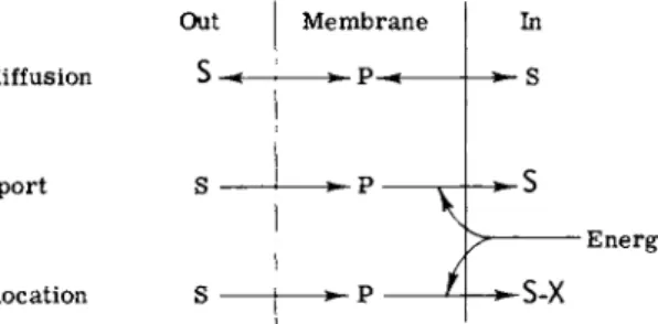
Out Membrane In
Facilitated diffusion s ρ s
Group translocation
Active transport S
S S-X
Energy
FIG. 1. Transport systems. The relative concentrations of solute, high and low, are indicated by S and s, respectively.
the concentration difference, but, when the initial rate is plotted as a function of the difference, the resultant curve is a hyperbola, i.e., the rate tends to reach a limit or, to use the common terminology, the system is "saturable." This property resembles that of a simple enzyme reaction and is frequently treated by Michaelis-Menten kinetics.
Facilitated diffusion, like passive diffusion, is driven by an existing concentration difference, and metabolic energy is only required to keep the membrane intact. In theory, the equilibrium state of a facilitated diffusion process should be achieved when the concentrations of solute are identical on both sides of the membrane. This assumes that the process of facilitated diffusion is symmetrical, and that the various rate constants involved in the process on one side of the membrane are equal to those on the other. Or, to put it more exactly, that the constants in Fig. 1 of Chapter 1, k3 = k-l9 klS1 = k-3S2, etc. In fact, it has rarely been shown that a process which resembles facilitated diffusion in all respects achieves an equilibrium state where internal and external concentrations are precisely the same. Perhaps this is not too surprising.
The internal milieu certainly differs markedly from the external solu
tion, and conceivably these differences could affect some of the kinetic constants.
The characteristic features of the active transport process are identical with those of facilitated diffusion with only one key difference. In the case of active transport, solute can be pumped against a concentration difference (Fig. 1). Active transport requires energy, and metabolic energy must therefore be coupled to this process. One of the major areas of research in the field of transport attempts to define exactly how metabolic energy is coupled to solute pumps. As will be seen, energy coupling may be different in different active transport systems.
The third type of transport shown in Fig. 1 is designated group translocation. The characteristic feature of this process is that the
46 SAUL ROSEMAN
solute is changed or converted into a derivative during the transport process. If the cell membrane is impermeable to the derivative (S-X in Fig. 1), then high concentrations of the derivative can be accumulated on the inside of the membrane. This process resembles active transport in the sense that the cell appears to concentrate or accumulate solute, but it is a very different phenomenon. The membrane component in the case of group translocation is acting as an enzyme(s) with vectorial properties.
Group translocation may not necessarily require metabolic energy as depicted in Fig. 1. If the chemical reaction is endergonic then meta- bolic energy is indeed required, but, if the reaction is exergonic, then the transport process may provide energy to the cell. One may speculate that organisms that oxidize a variety of compounds as the first step in their catabolic pathways might be excellent candidates for this type of transport process.
The processes shown in Fig. 1 are the major ones thus far described in bacterial systems. In this sense, bacteria appear to differ from animal cells, where other mechanisms are also operative. Active transport of most solutes in animal tissues is coupled to cation transport [4,5,14], a subject considered later in this chapter.
While the simple mechanisms shown in Fig. 1 would seem to be readily distinguishable from each other, this is not always the case.
The tight coupling of a facilitated diffusion process to an active sugar kinase would yield sugar phosphate as the apparent transport product, and it would be concluded that the process was really group transloca- tion. In an analogous manner, if a sugar phosphate were a transient intermediate during transport via group translocation, and this system were coupled to a phosphatase, then the net process would resemble active transport. Proper identification of the mode of transport is, of course, a necessary prerequisite for the study of mechanism, although the characterization presents formidable problems in a tightly coupled enzyme system.
D. Transport of Sugars by Bacterial Cells
Bacterial cells exhibit so many variations that no generalization concerning transport is possible. Any discussion of the process must define both the organism and the sugar. Different genera exhibit different modes of transport and may or may not transport a particular sugar by the same route. Further, a specific cell type may use different mechanisms for transporting different sugars or even the same sugar.
The following examples illustrate some of the kinds of variability:
3. CARBOHYDRATE TRANSPORT IN BACTERIAL CELLS 47 (a) Staphylococcus aureus apparently transports most, if not all, sugars by the process of group translocation [15,16]. On the other hand, Escherichia coli and Salmonella typhimurium transport sugars by all three processes described above: facilitated diffusion, active transport, and group translocation. Finally, Pseudomonas aeruginosa, which transports only a few carbohydrates [17], may transport these com
pounds by the process of facilitated diffusion, (b) A particular sugar, such as lactose, is transported by different mechanisms in different genera, (c) Active transport and facilitated diffusion may be linked in the sense that wild type cells may actively transport a particular carbo
hydrate, whereas the same cells carry out facilitated diffusion on this substrate when they are poisoned by energy inhibitors [18]. Similarly, mutants of the parent strain may be unable to actively transport the carbohydrate, while they are capable of conducting facilitated diffusion, (d) A single sugar in a single cell may be transported by more than one permease system. In E. coli, galactose is a particularly good example of this phenomenon; four separate galactose permease systems have been detected [19],
In contrast to the various modes of influx, which are briefly consid
ered above, relatively little is known about the process of efflux. Differ
ent investigators have suggested that it occurs by passive diffusion, facilitated diffusion, and active transport. Some believe that efflux occurs through the same permeases utilized for influx, whereas others present evidence that efflux occurs through different systems. Possibly many of the apparent discrepancies are related to both the cell types and the sugars that have been studied. Further, sugar permeases are not always specific for their major substrates, and more than one permease for a given carbohydrate can exist in a single cell.
E. Approaches to the Mechanism of Carbohydrate Transport
The early kinetic and genetic data on sugar transport in bacteria provided the groundwork for the more recent work in this field, which now emphasizes studies on subcellular systems. Experiments have been conducted along the following lines, (a) The isolation of solute-binding proteins. In the first of this class of experiments, Fox and Kennedy [20]
devised an elegant technique for differentially labeling a membrane protein of E. coli, called Μ protein, the synthesis of which is controlled by the y gene of the lactose operon (the y "permease"). The labeled protein was then solubilized with detergent and isolated. Work on the Μ protein has recently been reviewed [21] and is not further considered in the present discussion, (b) Isolation of solute-binding proteins by
48 SAUL ROSEMAN
employing an osmotic shock procedure [22], which liberates proteins into the extracellular fluid. A number of these proteins bind amino acids and sugars. The osmotic shock technique and the properties of the binding proteins are reviewed elsewhere in this volume. To the best of our knowledge, the location of the binding proteins appears to be primarily in the periplasmic space, between the cell wall and plasma membrane, and these proteins, unlike the Μ protein, have not yet been shown to be present in the plasma membrane, (c) The characterization of a phosphoenolpyruvate-dependent phosphotransferase system and attempts to define its physiological functions, (d) Attempts to define transport mechanisms by use of membrane vesicles. This subject has been reviewed recently [10].
The remainder of this discussion describes recent studies with the phosphotransferase system and with bacterial membrane vesicles.
III. THE PHOSPHOTRANSFERASE SYSTEM A. General Comments
In 1964 [23], a phosphotransferase system (PTS) was detected in extracts of bacterial cells. The enzyme system phosphorylated several sugars and was unique in the sense that several protein fractions were required for the reaction, and the phosphoryl donor was phosphoenol- pyruvate (PEP) rather than nucleotide triphosphate. A general descrip
tion of the PTS, the experimental observations that demonstrate that it is involved in sugar transport, and its potential role in cell metabolism have been presented [9]. This chapter therefore describes more recent studies with emphasis on the enzymological aspects and some additional data on the physiological behavior of mutants lacking one or more proteins of the PTS. The latter includes a brief description of the transport properties of such mutants and also of the relationship between the PTS and the phenomena of repression of enzyme syn
thesis.
B. Proteins of the Phosphotransferase System
The system catalyzes the reaction shown in Fig. 2, the transfer of phosphate from PEP to sugar. Hexose, hexitols, and other sugars utilized by the PTS are phosphorylated [23] at the terminal carbon atom (e.g., C-6 in the hexoses), with the exception of fructose, which is phosphorylated at C-l [24,25]. The phosphorylated disaccharides produced by the PTS have not been characterized, with the exception
3. CARBOHYDRATE TRANSPORT IN BACTERIAL CELLS 49
Sugar
Mg,2+ 5 Sugar - P 03 2 _
+ PTS +
C H2= C — C O "
o - p oI 3 2"
CH3COC02"
Phosphoenolpyruvate Pyruvate (PEP)
FIG. 2. Reaction catalyzed by the phosphotransferase system. PTS = phosphoenol- pyruvate-dependent phosphotransferase system.
of lactose-P, a product of the S. aureus system, in which the phosphoryl group is linked to C-6 of the galactose moiety [26].
Most of the biochemical work on the phosphotransferase system has been conducted with cell-free preparations of three genera, the gram- negative organisms E. coli and S. typhimurium, and the gram-positive organism S. aureus. The emphasis in this laboratory has been to attempt to isolate each of the proteins in highly purified or homogen
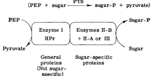
eous form and then to define the function of each protein. Figure 3 shows the generalization that can now be made for the systems thus far studied. The phosphorylation of any sugar requires four proteins.
Two of them are designated general proteins of the PTS because they are required far all sugars phosphorylated by the system and therefore lack sugar specificity. Two of the proteins are sugar-specific.
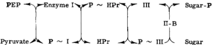
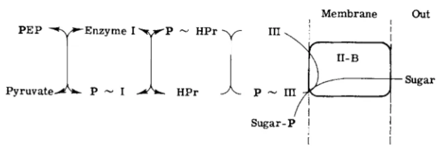
Experiments with purified components of the phosphotransferase system from S. aureus gave the results shown in Fig. 4. The phosphoryl group is sequentially transferred from PEP to the first general protein, Enzyme I, to the next general protein, HPr, to one of the two sugar- specific proteins (which may or may not be a membrane component), and finally to the sugar. The last step requires another sugar-specific protein, which is a membrane component. One or both of the two sugar-specific proteins may therefore be constituents of the membrane, but, in all systems which have been defined, the terminal step, the trans
fer of phosphate from phosphoprotein to sugar always requires a sugar-specific membrane protein.
(PEP + sugar PTS
s u g a r - Ρ + pyruvate)
General Sugar - spec if ic proteins proteins (Not sugar-
specific)
FIG. 3. Phosphate transfer via the phosphotransferase system.
50 SAUL ROSEMAN
P E P ^ > - E n z y m e I > * ^ P
Pyruvate Ρ HPr
Sugar-Ρ
Sugar
FIG. 4. Phosphate transfer via the phosphotransferase system. The following abbrevia
tions are used: PEP, phosphoenolpyruvate; HPr, the low molecular weight, phosphate- carrier protein of the phosphotransferase system; III, Factor III (soluble or cytoplasmic sugar-specific protein); II-B, membrane-bound sugar-specific protein.
1. NOMENCLATURE
As the different proteins of the phosphotransferase system were detected, a purely operational, cumbersome nomenclature was devel
oped. These designations have now been incorporated into the litera
ture, and will probably be used until the function of each protein and lipid in the phosphotransferase system has been accurately defined. A temporary, systematic nomenclature has been suggested [27,28].
Enzyme I, the first general protein of the phosphotransferase system, is a phosphoenolpyruvate:HPr phosphotransferase. HPr is a low molecular weight phosphate-carrier protein where the phosphoryl residue is linked to histidine. Enzyme II is the sugar-specific protein complex (including lipid and divalent cation) located in the membrane;
in some cases (see below), the two proteins specific for a given sugar are located in the membrane and are designated II-A and II-B, while in other cases the membrane contains only one of the proteins of a pair required for phosphorylation of specific sugar and is called II-B.
The soluble or 4 4 cytoplasmic " sugar-specific proteins are called Factors (or sometimes Enzymes) III. Factors III (soluble) and Enzymes II-A (membrane-bound) may be functionally the same, but, until functions are specified, the II-A, Factor III designations will continue to be employed.
As discussed below, most sugar-specific proteins are inducible;
strain, growth conditions, presence or absence of inducer, and sugar specificity of a given protein fraction must all be specified. The follow
ing abbreviations are suggested. Subscripts are to be used to identify strain of organism, growth conditions employed, and the presence of inducer, the latter by parentheses. Superscripts are to be used to designate sugar specificity and/or the proteins with which the designated fraction interacts. Thus, H P r£ c K 2 3 5 > b r o t h refers to HPr isolated from E. coli strain K235 grown in broth culture. Similarly, IIIsaac 5 6 o i synthetic (gai-6-p) refers to Factor III specific for lactose from S.
3. CARBOHYDRATE TRANSPORT IN BACTERIAL CELLS 51 aureus strain 5601 grown in synthetic medium in the presence of the in- ducer galactose-6-P. II-A|lcc, c o n s t i t u t i v e refers to the II-A protein that phosphorylates glucose, isolated from E. coli membranes grown in any medium.
2. ENZYME I
This enzyme has been partially purified from extracts of S. aureus and E. coli and purified to apparent homogeneity from S. typhimurium [28a]. Only preliminary physical studies have been performed with the purified Salmonella Enzyme I. The enzyme is easily inactivated by sulfhydryl reagents. Disc gel electrophoresis gives a number of bands from the purified protein, but a single band is obtained when the protein is first treated with N-ethylmaleimide. The properties of the purified protein lead to the tentative conclusion that it is readily dis- sociable either into its monomeric form, or into subunits of unknown size, which may or may not be identical.
Using the purified protein, the following reaction has been demon- strated
M g2 +
PEP + I ^ P-I + pyruvate
The phosphoryl-Enzyme I has been isolated, and on the basis of its hydrolytic behavior it appears that the phosphoryl residue is attached to an imidazole ring of a histidinyl residue in the protein (see below).
The equilibrium constant for the reaction written above has not yet been established.
The apparently complex nature of Enzyme I requires further study, particularly to determine whether the enzymatic activity is regulated by intermediates or products of the phosphotransferase system, or conceivably by other metabolites. As indicated in an earlier review [9], the transport process through the phosphotransferase system is linked to general metabolism in the cell, and an important potential point of control or regulation would be at the first protein in the process, Enzyme I.
3. HPr
This protein, which should more properly be called phospho-carrier protein, was originally designated HPr [23] because it appeared to be heat-stable. We now know that heating the purified protein at 100°C results in the loss of amide groups with concomitant loss of activity.
Nevertheless, the HPr designation is properly descriptive since, as
52 SAUL ROSEMAN
discussed below, the functional amino acid of this protein is a histidine residue.
The protein has been isolated in homogeneous form from all three organisms, and S. typhimurium HPr has been crystallized [28a]. The E. coli and S. typhimurium HPr proteins appear to be identical in all respects. However, they differ in amino acid composition from S. aureus HPr (Table I). The S. aureus HPr has but one histidine residue compared with two such residues in E. coli and S. typhimurium HPr, and S. aureus HPr contains tyrosine, which is not present in the HPr from the gram- negative organisms. The methods used for establishing homogeneity of HPr and some of its properties are illustrated in Table II.
When S. typhimurium or E. coli HPr is heated at 100°, depending on the time of heating, one to three (or more) amide residues are lost.
TABLE ι
AMINO ACID COMPOSITION OF H P R PROTEINS FROM
S. aureus AND E. coli
Amino acid S. aureus0 E. colib
Histidine 1.02 2.0
Tyrosine 2.2 0.0
Lysine 9.4 8.1
Arginine 1.04 0.96
Aspartic acid 9.4 3.6
Threonine 4.6 10.0
Serine 6.2 6.9
Glutamic acid 8.3 13.5
Proline 1.2 2.2
Glycine 6.0 6.7
Alanine 5.7 9.4
Valine 4.2 7.1
Methionine 3.2 2.1
Isoleucine 6.8 3.4
Leucine 5.1 8.5
Phenylalanine 0.94 4.0
Cysteine 0.0 0.0
Tryptophan 0.0 0.0
Molecular weight (sum) 8630 9537
a Residues per 6 moles of glycine.
b Residues per 2 moles of histidine; S. typhimurium gave the same results within experimental error; E. coli HPr contains 9.2 amide residues.
3. C A R B O H Y D R A T E T R A N S P O R T I N B A C T E R I A L C E L L S 53
1. Homogeneous
a. Disc gel electrophoresis; pH 2.3, 4.3, 6.6, 9.5 b. Ultracentrifuge
2. Molecular Weight
9340 ± 230 by ultracentrifugation (Yphantis) 9639 by £ amino acid residues 3. Analysis
a. No carbohydrate or Ρ
b. Usual amino acids except Cys, Tyr, Trp c. 2 His/mole
Two of the resultant derivatives of HPr, designated HPr-1 and HPr-2, have been isolated in homogeneous form [29], and their properties have been studied. HPr-1 is about 50% as active as native HPr in the sugar phosphorylating assay system, while HPr-2 is about 25% as active. The location of these amide residues in native HPr may be determinable by amino acid sequence studies (now in progress): the first 25 amino acid residues have been identified with an automatic sequenator.
As noted above and in Table I, the HPr proteins from S. aureus and E. coli (or S. typhimurium) are not identical, This is also true with respect to their utilization by the other proteins of the phosphotrans
ferase system. Enzyme I from one organism can phosphorylate HPr from the other at a very low rate compared to the rate of phosphoryl
ation of homologous HPr, while P-HPr is only utilized at a negligible rate by the heterologous sugar-specific proteins.
The phospho-HPr proteins have been isolated. In all three cases only one phosphoryl group can be introduced into the HPr protein, and in each case it is linked to a histidine residue. Hydrolysis studies of E. coli phospho-HPr as a function of pH [23,27,29] indicated that the phosphoryl group was attached to an imidazole ring of a histidine residue and the phospho-histidine was isolated after alkaline hydrolysis [23,27]. Recent results [29] have shown the phosphoryl group to be attached at the N-l position of the imidazole ring (Table III), and, in this respect, the phospho-HPr differs from several other characterized phospho-histidinyl proteins, in which the phosphoryl group is attached at the N-3 position [30-34]. Phospho-HPr is hydrolyzed at significant rates between pH 7 and 8, and very rapidly at pH values below 7.
An important point concerning phospho-HPr is the free energy of
T A B L E I I
SOME PROPERTIES OF E. coli H P R
54 S A U L R O S E M A N T A B L E III
LINKAGE OF THE PHOSPHORYL G R O U P IN PHOSPHO-HPR (E. coli)
PEP + HPr
M g2
phospho-HPr + pyruvate
- N H — C H — C O C Hi 2
1 mole Ρ incorporated/mole HPr Ρ linked to Ν-1 of His imidazole ring
N H — C H — C O v CHI 2
N ^ N - P Os2-
1 - P - H i s t i d i n e
"',ΟΡ-Ν Ν
3 - P - H i s t i d i n e
formation of the phosphoryl protein. As indicated above, direct trans
fer of phosphate from PEP to Enzyme I has been demonstrated, and, likewise, the direct transfer of phosphate from phospho-I to HPr has also been demonstrated [28a]. Interestingly, the latter reaction does not appear to require Mg2 + , in contrast to the first step. Preliminary studies have been conducted to determine the equilibrium constant of the overall reaction
PEP + HPr I, M g
2
P-HPr + pyruvate
The marked lability of P-HPr has thus far prohibited exact determina
tion of the equilibrium constant, but the apparent Ke q is approximately 10. This result shows that the linkage between the phosphoryl group and HPr is truly of the " high energy " type, somewhat less than that of PEP, and almost twice that in the pyrophosphate bonds of ATP.
4. SUGAR SPECIFIC PROTEINS
The sugar-specific proteins of the phosphotransferase system have not been as well characterized as the general proteins, Enzyme I and HPr, and the number of sugar-specific proteins that can be synthesized by a given cell type has not yet been established.
Only a few of these proteins are found in a given cell type independ
ent of the carbon source used for growth; they are classified as consti
tutive. Most of the sugar-specific proteins are synthesized by the cell under inducing or derepressing conditions, and are therefore designated inducible. Further, in the constitutive Enzyme II complexes of E. coli, both proteins of a given pair are found in the membrane, while the
3. CARBOHYDRATE TRANSPORT IN BACTERIAL CELLS 55 inducible systems studied so far (Factor III, Enzyme II-B) have only one of the two proteins located in the membrane.
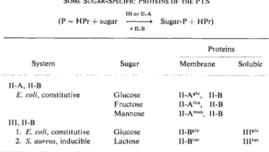
This discussion is restricted to a description of those sugar-specific proteins of the phosphotransferase system where at least one of a given pair of proteins has been isolated in homogeneous form. These proteins are listed in Table IV.
TABLE IV
SOME SUGAR-SPECIFIC PROTEINS OF THE PTS III or 1I-A
(P - HPr + sugar • Sugar-P + HPr)
+ ΙΙ-Β
Proteins
System Sugar Membrane Soluble
II-A, II-B
E. coli, constitutive Glucose II-Ag l c, II-B Fructose II-Af r u, II-B Mannose H.Am a n J [_β
III, II-B
1. E. coli, constitutive Glucose II-Bg , c l ng i c
2. S. aureus, inducible Lactose Il-B, a c mi a c
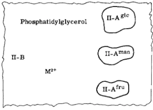
a. Constitutive Enzyme II Complex of E. coli. The first group of proteins to be considered are the constitutive enzymes of E. coli (and S. typhimurium). When E. coli is grown in a salts medium on glucose as the sole source of carbon, the membrane fraction isolated from such cells transfers phosphate from P-HPr to three sugars, glucose, mannose, and fructose (or to their analogs). Extraction of the membranes with a suitable combination of urea and w-butanol gives a protein fraction (II-A) and a pellet [28]. Both fractions are required for phosphorylation of the three sugars. Further purification of the soluble protein fraction leads to the isolation in homogeneous form of three II-A proteins, each specific for one of the sugars, i.e., ΙΙ-Α|£, II-A™n, and II-Af£™.
The proteins contain no detectable lipid. Fractionation of the pellet in the presence of detergent (deoxycholate) gives a protein designated II-B. The marked tendency of this protein to aggregate in the absence of detergent has thus far prohibited extensive physicochemical charac
terization. However, the isolated protein was free of lipid, and exhibited a single band when subjected to electrophoresis in polyacrylamide gel containing sodium dodecyl sulfate and urea; the rate of migration of
56 SAUL ROSEMAN
the single polypeptide band observed corresponded to a molecular weight of 36,000. A critical point remains to be resolved and hinges on the purity of the isolated II-B protein(s). If this fraction is indeed homogeneous, then the three specific II-A proteins utilize the same II-B protein, and II-B represents 10% of the total membrane protein.
The isolation of lipid-free proteins from the membrane permitted a study of lipid requirements. A minor E. coli membrane lipid, phos- phatidylglycerol, was found to be the active component. This lipid was not itself phosphorylated during the overall transfer of phosphate to sugar, and a few other anionic lipids including detergents could partially substitute for phosphatidylglycerol. Phosphatidylglycerol formed an active complex with II-B and divalent cation (Ca2 + or
M g2 +) when mixed in the proper sequence, and the complex was sedimentable. The complex interacts with the soluble II-A proteins, and permits transfer of phosphate from phospho-HPr to one of the three sugars, depending upon which II-A is employed. The divalent cation requirement has not been studied extensively, but it is interesting to note that, while calcium appears to be very active in forming stable, active Enzyme II complexes, calcium does not substitute for magnesium in the transfer of phosphate from PEP to sugar in the complete system and is, in fact, a potent inhibitor. Thus, it seems likely that calcium is involved in stabilizing and forming the Enzyme II complex, i.e., in a structural role, whereas it cannot act as a cofactor for the enzyme system. The structure of the constitutive Enzymes II of E. coli is schem- atically presented in Fig. 5.
b. Constitutive Factor III of E. coli. In addition to the constitutive glucose II-A, II-B E. coli sugar-specific protein pair described above, recent work [34a] has demonstrated the existence of another consti- tutive glucose system. Again, two sugar-specific proteins are involved,
FIG. 5. Schematic structure of the E. coli constitutive Enzyme II complex.
3. CARBOHYDRATE TRANSPORT IN BACTERIAL CELLS 57 but, in this case, one is found in the soluble supernatant of crude extracts, while the other is found in the membrane fraction. The super
natant protein has been isolated in homogeneous form and is desig
nated Factor III!1,?. The membrane protein has not yet been purified to homogeneity, but fractionation experiments as well as studies with sulfhydryl reagents indicate that this membrane protein is different from the II-B described above; the new system is relatively specific, as indicated below, and, on this basis, the membrane protein is tentatively designated II-B!1,0. (Perhaps the more appropriate nomenclature for the II-B proteins derived from the membrane would be to designate them according to the proteins with which they interact. Thus, the II-B proteins thus far described might best be designated II-B""CA and
ii-Br
c.)
The isolated Factor IllfJ? exhibits a molecular weight of about 20,000 [34a] by chromatography on Sephadex, and appears to dis
sociate into subunits, which are smaller in size than the HPr protein.
One interesting facet of the purification of III!1® was that it was tightly associated with a phosphatase that was inhibited by fluoride; the phos
phatase showed greatest activity with glucose-6-P and the 6-phosphate ester of methyl α-glucoside. The separation of the phosphatase and III!1® proved to be extremely difficult, but has been accomplished.
The physiological function of the phosphatase remains to be established, but conceivably it plays an important role in a coupled transport system, where a sugar-P would be a transient intermediate and the free sugar the final product.
The Factor III, II-B"1/1' system transfers phosphate from P-HPr to glucose as does the II-A!1®, II-B system. However, the Km for glucose is an order of magnitude lower in the case of the Factor III, II-B system. In addition, the latter actively phosphorylates the galactoside analog methyl /?-thiogalactopyranoside (TMG), which is phosphoryl- ated by the II-A, II-B system at a negligible rate.
A variety of mutants have been isolated that cannot utilize a single sugar, and these mutants are defective in one of the sugar-specific proteins of the phosphotransferase system. No such mutants have been isolated for glucose. The existence of the two glucose systems described above may explain this result, as well as some recent detailed kinetic studies relating the phosphotransferase system to glucose transport [35].
Finally, before turning to the S. aureus proteins, it is important to place special emphasis on the III!1®, II-B"!81" system. Preliminary evidence has been obtained [36] that this system may be involved in the regulation of transport of several sugars that are not phosphoryl- ated by the phosphotransferase system (see below).
58 SAUL ROSEMAN
c. Lactose-Specific Phosphotransferase Proteins from S. aureus.
Lactose metabolism in S. aureus has been extensively studied by Morse and his co-workers [15,16,26]. As in E. coli, the genes required for lactose metabolism exist in an operon, one of these codes for the permease, while another codes for a jS-galactosidase. The operon also contains genes for Factor Illjfa and for Enzyme llsflc [36a]. Staphylo
coccus aureus differs from E. coli, however, in several respects, the major differences being that the galactosidase is a phospho-/?-galactosidase, which cleaves lactose-P and does not function with free lactose, and the inducer for the lactose operon is galactose-6-P. It was originally shown [15] that in S. aureus all sugars appeared to be transported by group translocation, and the derivatives formed during this process were later found to be the sugar phosphates. Thus, unlike E. coli, where lactose is actively transported per se [21], this disaccharide is phosphorylated during the transport process in S. aureus [16,26].
Staphylococcus aureus contains a PEP-dependent phosphotransferase system [37], and mutants defective in Enzyme I or HPr are incapable of utilizing a large number of sugars. The most extensive studies on the Factor III, II-B systems are those conducted with the lactose- specific proteins from S. aureus [38,39].
As shown in Table V, lactose-specific Factor III (111^°) and the membrane protein II-B^ are inducible in S. aureus, under conditions that do not affect the levels of Enzyme I and HPr. Ill J." has been isolated in homogeneous form and has been characterized by a variety of physical techniques. The protein exhibits a molecular weight of about 34,000 by several methods and is dissociable to three subunits
TABLE ν
INDUCTION OF ENZYME I IL A C AND FACTOR I I I
(S. aureus PTS)
Component
Specific activity in extracts"
Noninduced Induced
I 52 58
HPr 6.8 6.7
II 0.1 38
III 0.1 2.5
"Specific activity = /xmoles TMG-P formed ( x l 02) / m g protein in crude extracts; 30 minutes at 37°. Cells grown in bactopeptone ± galactose or gal-6-P.
3. CARBOHYDRATE TRANSPORT IN BACTERIAL CELLS 59 of molecular weight 11,300 [39]. All available data suggest that the three subunits are identical; for example, amino acid sequence studies of a mixture of these subunits reveals a single amino acid at each posi- tion as far as the sequence was established (about 25 residues).
IIIsaflc is a phosphate carrier, and in this respect is similar to HPr.
Extensive studies have shown [38] that the phosphoryl residue is transferred from P-HPr to IIIsf l c and that the process does not require any additional protein fractions. Hydrolysis studies on the isolated P-III suggest it is similar to P-HPr, and it therefore appears that the phosphoryl residue is linked to histidine in this protein as it is in HPr.
The more interesting observation is that III^0 accepts three phosphoryl residues according to the equation
3 P-HPr + IIIia a c , P3-IIIia f l c + 3 HPr
In view of the fact that 111^° possesses subunits of approximately the same molecular weight as the HPr protein, two additional possibili- ties were tested [38]. The phosphoryl group could be transferred from one protein to the other, or a subunit interchange could occur, with P-HPr replacing one of the subunits of IIIsf l c. The experiments clearly showed that there was a direct transfer of the phosphoryl residue and not of the protein units.
Despite extensive efforts, the 5. aureus II-Bl a c protein has not yet been solubilized. The membrane preparation exhibits II-Bl a c activity after successive treatments with a variety of agents including detergents and alkali. These reagents were useful, however, in that they succeeded in partially purifying the activity by removing 90 % of the protein and essentially all of the lipid and cell wall polymers. II-Bsaflc is required for the transfer of phosphate from P-IIIsaflc to lactose or its analogs, such as TMG. The kinetics of this phosphorylation reaction suggested that both IIIsaac and the sugar substrate bind to II-Bsaac. Binding experiments showed that both exhaustively washed membrane preparations and partially purified II-B^aac formed complexes with lactose and its analogs (Table VI). Whereas membranes from wild type cells grown under noninducing conditions do not bind lactose to a significant extent, those from induced cells do. Binding was also observed with membranes from a mutant that constitutively synthesized the proteins of the lactose operon. Finally, mutants defective in II-B^0 showed little or no binding. The II-B^ protein remains to be isolated in homogeneous form, but this is an example of a binding protein from a bacterial system the function of which is known both physiologically (see below) and biochemically.
60 SAUL ROSEMAN TABLE VI
LACTOSE BINDING BY S. aureus l ll a c
1 4C-Lactose + I Il a c , ( I I, a c · 1 4C-lactose)f l
1 4C-Lactose bound Strain" Genotype (/xjLtmoles/mgm protein)
C22 i~ l a c+ + 500
F8, F16,
F18, F20 i~ lac~ — 10-17
F6, F l l , F15 i" lac~ — 158-236
5601 i+ l a c+ — 28
5601 (induced) + 520
a [Lactose] = 5 χ 1 0_ 7Λ / at 0.5 saturation; 25°.
b Strain 5601 is the wild type (inducible); strain C22 is a constitutive mutant of 5601. All other strains were derived from C22.
c By enzymatic (phosphorylation) assay.
d. Functional Relationships of Sugar-Specific Phosphotransferase Proteins. As discussed above, the lactose-specific proteins of S. aureus have been studied in detail and the results lead to the conclusion shown in Fig. 4. Phosphate is transferred from PEP to sugar by a sequential transfer to Enzyme I, HPr, Ills" and finally to the sugar in the presence of II-Bse. This sequence is established up to the point of the HPr protein in all systems studied thus far, but it is not known whether the E. coli, S. typhimurium, and other S. aureus sugar-specific proteins are phosphorylated. In other words, it is not known whether the II-A and
IIIg l c constitutive proteins of E. coli and S. typhimurium perform the
same function as does the IIIl a c protein of S. aureus. If the II-A proteins are phosphorylated as intermediates in the overall reaction, then these proteins simply represent membrane components that act in the same manner as the soluble cytoplasmic IIIl a c of S. aureus.
C. Role of the Phosphotransferase System in Sugar Transport
The phosphotransferase system has thus far been described in purely biochemical terms as illustrated in Fig. 4. This scheme does not take into account the fact that the II-B proteins are located in the plasma membranes. If this additional parameter is incorporated into the scheme of Fig. 4, then the schematic representation shown in Fig. 6 results.
The important point here is that not only is a sugar phosphorylated by the phosphotransferase system but also that the sugar is simultaneously translocated across the cell membrane. This schematic representation
3. CARBOHYDRATE TRANSPORT IN BACTERIAL CELLS 61
P E P ^*Y»-Enzyme I >γ ^Ρ ~ HPr -γ - ΙΠ
P y r u v a t e ^ * - Ρ ~ I HPr
Membrane Out
Sugar
FIG. 6. Phosphate transfer and sugar translocation via the phosphotransferase system.
Abbreviations are those used in Fig. 4.
depicts our current thinking on the role of the phosphotransferase system in the process of group translocation.
A considerable body of evidence has accumulated that supports this conclusion [9]. Some recent data on the physiological behavior of mutant cells are presented here since they emphasize the earlier state
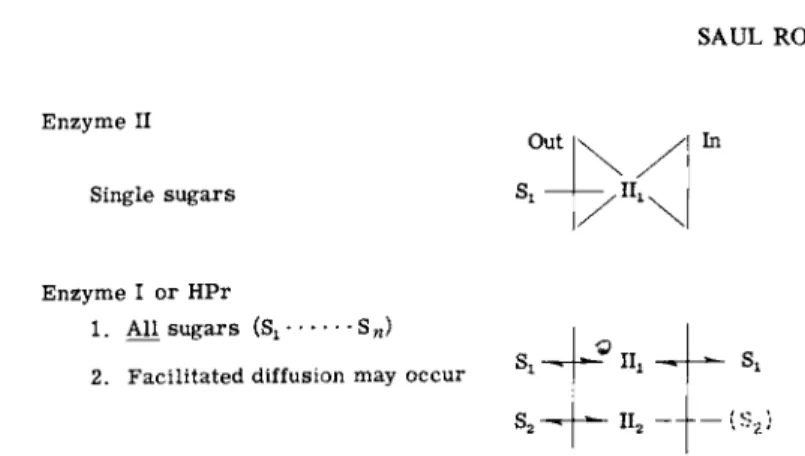
ments, i.e., the use of genetics is a powerful tool in unraveling the mechanism of transport. The basic approach is simple and based on the ideas illustrated in Figs. 7 and 8. In these schema, II represents the combination of two sugar-specific proteins, III + II-B or II-A + II-B.
If the mechanism for group translocation is that shown in Fig. 7, then the sugar-specific proteins translocate only their respective substrates, while the general proteins Enzyme I and HPr are required for all sugars transported by the phosphotransferase system. On this basis, simple predictions can be made concerning the behavior of mutants defective
Active transport
HPr + P E P
FIG. 7. Sugar transport via the phosphotransferase system. S ! , S2, S„, and S* signify different sugars. The corresponding sugar-specific protein pairs of the phosphotransferase system (Factor III + II-B, or II-A + II-B) are shown as Uu I I2, etc.
62 SAUL ROSEMAN Enzyme II
Single sugars
Out Si - . " i s
In
Enzyme I or HPr
1. All sugars (Sj · •s«)
2 . Facilitated diffusion may occur Si II2 - + - ( S2i
FIG. 8. Predicted transport defects in phosphotransferase protein mutants. See text for explanation.
in one of these proteins. These predictions are shown in Fig. 8. Mutants lacking or defective in a sugar-specific protein (11^ will be unable to transport that particular sugar but should be able to transport all other sugars normally. Similarly, mutants defective in Enzyme I or HPr should be unable to transport all sugars utilized by this system.
In the latter case, the membranes contain functional Enzymes II.
Facilitated diffusion is therefore possible and may or may not occur depending on the precise molecular mechanism for the group translo
cation process. These predictions correspond precisely to the behavior of a variety of mutants from different organisms isolated in several laboratories [2,9]. Only one example of the behavior of such mutants will be given.
Staphylococcus aureus is capable of fermenting a large number of sugars [15] including lactose, galactose (which in this organism is transported via the lactose permease), fructose, mannose, glucose, etc.
The same behavior is observed in a strain of S. aureus that has been made constitutive for the lactose operon. This constitutive strain was employed as the parent strain for selected mutants to avoid the problem of catabolite repression. The mutants isolated from the constitutive lactose strain were shown to be defective in the following proteins and unable to ferment the indicated sugars: phospho-/?-galactosidase, lactose; II-B^0, lactose and galactose; IIIsa a c, lactose and galactose; and Enzyme I, all sugars (glucose at high concentrations was slowly fer
mented). The mutants were also characterized with respect to all the other proteins enumerated above. For example, the II-Bl a c mutants contained normal levels of β-galactosidase, Enzyme I, HPr, and IIIsaflc.
Furthermore, several mutants of each class were independently isolated and showed the same properties.
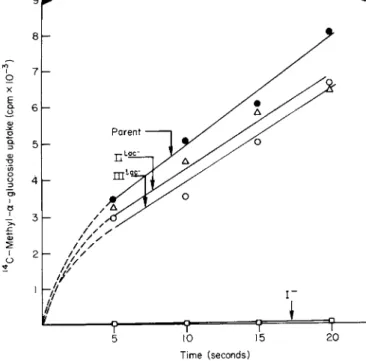
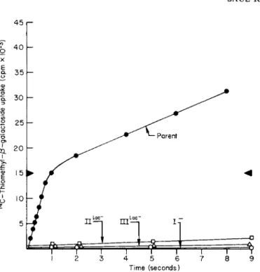
3. CARBOHYDRATE TRANSPORT IN BACTERIAL CELLS 63 The transport properties of the Enzyme I, II-Bl a c, and IIIl a c mutants are compared with the wild type as shown in Figs. 9 and 10. Methyl α-glucoside is used as an example of sugars not transported via the lactose permease, and, as can be seen, the transport of this sugar by the II-Bl a c and IIIl a c mutants compared favorably with the parent strain. On the other hand, there was essentially no uptake by the Enzyme I mutant. Similar experiments conducted with the lactose analog TMG are shown in Fig. 10. All three mutants were incapable of transporting TMG at a significant rate compared to the parent strain and, in fact, were unable to equilibrate the sugar with the medium even after prolonged periods of exposure. Thus, the mutants were incapable of conducting facilitated diffusion at a significant rate; this explains why mutants defective in the sugar-specific proteins exhibit the cryptic behavior described earlier. These types of experiments, therefore, meet the predictions made earlier and support the conclusion that the phos
photransferase and permease systems are identical.
ο X Ε Ρ- 6\-
Time (seconds)
FIG. 9. Methyl α-glucoside transport in S. aureus. The mutants employed were I Il a c~ (defective in the 1I-B protein specific for lactose phosphorylation), I I I, a c~ (defective in the cytoplasmic Factor III specific for lactose phosphorylation), Enzyme I" (defective in Enzyme I of the phosphotransferase system).
64 SAUL ROSEMAN
FIG. 10. Thiomethyl β-galactoside transport in S. aureus. The cells employed for these experiments were aliquots of suspensions used in Fig. 9. The level corresponding to equili
bration but no accumulation of solute by the cells is shown as dark triangles on the ordinates.
D. Occurrence of the Phosphotransferase System
The phosphotransferase system is responsible for the group trans
location of a variety of sugars across bacterial membranes [9]. How
ever, many questions have not yet been answered. Why is the system so complex i.e., why is group translocation not catalyzed by a simple membrane-bound kinase which can utilize ATP or PEP ? Is this system responsible for the active transport and facilitated diffusion of carbohy
drates in bacteria? Does it occur in all bacterial cells and in other organisms? Is it responsible for the transport of solutes other than sugars ?
Only a few answers can be given to these questions. The phospho
transferase system was detected in vertebrate intestine [40], but no further reports on this subject have appeared. Experiments with ex
tracts of fungi and yeast have thus far given negative results [41]. In addition, the system does not occur in all bacteria. It appears to be absent from P. aeruginosa, but this organism may transport sugars
3. CARBOHYDRATE TRANSPORT IN BACTERIAL CELLS 65 only by the process of facilitated diffusion [17]. In a more extensive survey (which has been criticized [42]), it has been reported to be present in facultative but not in obligate aerobes [43], and is generally present in anaerobic organisms. The latter makes teleological sense from the point of view of energetics, a topic that has been considered earlier [9].
As has been repeatedly emphasized in this discussion, E. coli and S. typhimurium differ from S. aureus, which may transport all carbo
hydrates via the phosphotransferase system. The gram-negative organ
isms appear to take up the following monosaccharides by the process of group translocation through the phosphotransferase system: glucose, mannose, fructose, galactose (?), 7V-acetylglucosamine [44], glucosamine, N-acetylmannosamine, mannosamine, β-glucosides, and the hexitols, sorbitol and mannitol.
In gram-negative organisms the following sugars are not phosphoryl
ated by the phosphotransferase system and are not taken up by group translocation: disaccharides (lactose, melibiose, maltose), pentoses, hexose 6-phosphates, and glycerol. With the exception of glycerol, which is taken up by facilitated diffusion [2], the other sugars in this group are actively transported. In earlier work, Enzyme I and HPr mutants were found to be incapable of utilizing or transporting these carbohydrates, and the results were interpreted to mean that the intact phosphotransferase system was required for normal transport of these sugars as well as for those which are group translocated. Recent studies (see below) have, however, shown that Enzyme I and HPr mutants are particularly sensitive to repression of enzyme synthesis and are unable to normally synthesize inducible levels of the permeases and catabolic enzymes required for utilization of this class of sugars. Thus, while the mutants were indeed transport-defective, it was an indirect consequence of the Enzyme I or HPr mutation that caused this phenomenon. How, then, are these sugars transported?
One possibility, which stems from the work on the phosphotrans
ferase system, is that organic solutes (sugars, amino acids, etc.) are converted to transient derivatives during the transport process across the membrane, and these derivatives are then reconverted to the original solute by an enzyme on the inner face of the membrane. For example, in the case of sugars, the transient derivatives could be the sugar phosphate esters, and the enzymes on the inner face of the membrane could be specific phosphatases of the type referred to earlier. Membrane- bound, sugar-specific phosphatases have been reported [45]. Another possibility is that the membrane-bound protein "carriers" are phos
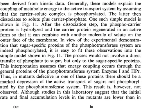
phorylated. Many models proposed to explain active transport have
66 SAUL ROSEMAN
been derived from kinetic data. Generally, these models explain the coupling of metabolic energy to the active transport system by assuming that the carrier-solute complex is phosphorylated. The latter then dissociates to solute plus carrier-phosphate. One such simple model is shown in Fig. 11. After the dissociation step, the phospho-carrier protein is hydrolyzed and the carrier protein regenerated in an active form so that it can combine with another molecule of solute on the outer face of the membrane. In view of the experimental demonstra
tion that sugar-specific proteins of the phosphotransferase system are indeed phosphorylated, it is easy to fit these observations into the simple model shown in Fig. 11. The process would not involve a direct transfer of phosphate to sugar, but only to the sugar-specific proteins.
This interpretation assumes that energy coupling occurs through the general proteins of the phosphotransferase system Enzyme I and HPr.
Thus, in mutants defective in one of these proteins there should be a marked depression of the active transport of sugars not phosphoryl
ated by the phosphotransferase system. This result is, however, not observed. Although studies in this laboratory suggest that the initial rate and final accumulation levels in the mutants are lower than in
Out In
T - P T - P
S
Active transport
Τ S - T - P
S-T S-T
Τ Τ
Facilitated diffusion
S-T S-T
FIG. 11. Models for active transport and facilitated diffusion. S = the solute, Τ = the permease or "carrier," T - P = the phosphorylated form of the carrier.
3. CARBOHYDRATE TRANSPORT IN BACTERIAL CELLS 67 wild type cells, the effect is not nearly so pronounced as observed with sugars that are phosphorylated.
Thus far, the emphasis has been that phosphorylation always pro- ceeds from PEP through Enzyme I and HPr to the sugar-specific proteins. However, if the latter can be phosphorylated by some other mechanism involving other pathways (for example, ATP), then the results obtained with the Enzyme I and HPr mutants would be more readily explicable. This idea would also at least partially explain the surprising complexity of the phosphotransferase system. In other words, the system has evolved into a high order of complexity which by its very nature and molecular mechanism permits considerable variation.
E. Cation-Dependent Sugar Transport
Until very recently, bacterial transport systems appeared to differ substantially from those of animal tissues with respect to active transport of solutes. Active transport in bacterial cells apparently functioned by way of so-called "primary" active transport system [4]; in this type of system, the solute is transported against a gradient as illustrated in Fig. 1, independently of the transport of other solutes. In sharp con- trast, active transport in animal cells generally occurs by coupling of the organic solute transport system to cation transport [4]. The vast majority of such transport systems in animal cells is mediated by cotrans- port with N a+ [14]. The model for transport systems of this type is illustrated in Fig. 12. The solute-specific membrane protein transports the solute with N a+ into the cell, the N a+ being transported down its electrochemical gradient, thereby providing energy for transporting the solute (S) up its concentration gradient. The N a+ concentration gradient across the plasma membrane is maintained by the N a+ pump (Fig. 12), which is thus the ultimate source of energy for driving the solute (S) into the cell against the gradient.
Earlier reports showed that halophilic bacteria required N a+ for growth, while growth of some other bacteria was markedly stimulated by this ion [46]. In addition, several laboratories reported that cations including N a+ affected the transport of certain solutes [47-50]. The most extensively studied system was amino acid transport in a marine pseudomonad [51], which was dependent on N a+, but the effect was interpreted to be independent of cotransport.
In a recent report [52] from this laboratory, the transport of melibiose by S. typhimurium was examined. This disaccharide is actively trans- ported by a permease system in E. coli and S. typhimurium designated
68 SAUL ROSEMAN
N a+ C o t r a n s p o r t
S ^ ^
N a+
^ N a
N a+ /
E n e r g y
K+ C o u n t e r t r a n s p o r t
E n e r g y
FIG. 12. Models for co- and counter-transport. In the case shown, sodium ion is co- transported with the solute, S. The external concentration of sodium ion is high relative to the internal concentration, whereas the reverse is true for the solute. The sodium gradient is maintained by a sodium pump which utilizes metabolic energy. In the model shown for countertransport, using potassium ion as an example, the solute S is pumped into the cell where it is present at higher concentration at the expense of potassium ion, which simul- taneously moves out of the cell. The potassium concentration difference is maintained by a potassium pump, PK.
TMG permease II [19,53]. As in the case of the analogous disaccharide lactose, the structural genes for melibiose exist within an operon and consist of at least a permease gene and one for the enzyme a-galactosi- dase. It was found that the melibiose permease system showed all of the properties of a Na+-dependent sugar cotransport system and exhibited kinetics completely analogous to those observed with intest- inal preparations [14]. At subsaturating levels of sugar, the rate of uptake was markedly influenced by N a+ (Figs. 13 and 14). The Fm a x of the system was not affected by N a+, but the Km was lowered by an order of magnitude. In addition to Na+-dependent sugar uptake, the cells showed a reciprocal relationship, i.e., N a+ was taken up in the presence of sugar by induced cells, but not by uninduced cells. Further, the N a+ accumulated during sugar transport was rapidly pumped out of the cell (Fig. 15).
The lactose system may be much more complex. It has been suggested that protons are involved in lactose transport [54,55] and also that K+ may be involved [21]. The precise relationships between these ions and lactose transport have not yet been established.
The finding of a N a+ sugar cotransport system leads to the possibility that bacterial cells may utilize ion gradients to transport solutes that are not phosphorylated by the phosphotransferase system. Whether or