Amino Acid Transport in Microorganisms
Dale L Oxender
I. Introduction 133 II. General Considerations 135
A. Endogenous Amino Acids 135
B. Transport Rates 139 C. Temperature and pH Effects 142
D. Kinetics 143 E. Transport Schemes or Models 148
III. Transport Systems 151 A. Glycine, Alanine, and Serine 151
B. Proline 154 C. Valine, Leucine, and Isoleucine 155
D. Phenylalanine, Tyrosine, and Tryptophan 159
E. Lysine and Arginine 163
F. Histidine 166 G. Methionine 167 H. Glutamic and Aspartic Acids 168
IV. Alkali Metal and Ammonium Ion Effects 170
V. Isolation of Transport Systems 172 A. General Approach 172 B. Protoplasts, Spheroplasts, and Membrane Preparations 173
C. Mutant Selection 174 D. Transport Proteins 176
References 180
I. I N T R O D U C T I ON
A number of more or less specific transport systems for amino acids have been shown to operate in the cellular membranes of bacteria. It appears likely that the bacterial cell has developed transport systems within the membrane that are reactive with most of the metabolizable compounds found within the cell. Metabolizability is, however, not necessary for an effective fit with transport receptor sites; hence these
133
134 DALE L. OXENDER
often function in the transport of substrate analogs not related to cell metabolism. This property has proved useful in describing the structural features of the reactive site of certain transport systems and for the selection of analog-resistant mutants. Early support for the presence of specific transport systems in bacteria came from three types of findings:
(a) the accumulation of high levels of solutes internally; (b) the property of crypticity, i.e., the inability to utilize metabolizable substrates even though the cell can be shown to contain the necessary enzymes; and more recently (c) the isolation from the membrane of macromolecular com- ponents bearing receptor sites.
The now classic work of Gale and his associates [1-3] gave the first clear indication that microorganisms were able to accumulate free amino acids. They found that, when Staphylococcus aureus and Streptococcus faecalis cells were grown in the presence of casein hydrolyzate, large amounts of glutamic acid and lysine could be extracted by water from broken cells. These studies have been summarized by Gale [4,5].
Chesbro and Evans [6] later showed that lysine transport was energy dependent. The binding of lysine to the cell wall studied by Britt and Gerhardt [7] probably contributed to the earlier failure to observe energy dependence. Free amino acids were later also found in gram- negative organisms [8-14].
Some of the first examples of crypticity have been described by Doudoroff [15,16]. A mutant of Escherichia coli was isolated that could not use glucose for growth, although externally added maltose could serve as a source of internal glucose which was then metabolized. The membranes of these cells proved to be permeable to maltose but not glucose, a finding that supported the concept of specific transport systems. The inhibition of the growth of some auxotrophic mutants produced by adding amino acid analogs can be overcome by adding peptides of the amino acid [17-21]. In certain cases the peptides serve to promote growth better than the corresponding free amino acid. Even though some of these earlier observations of antagonisms were not interpreted at the time as permeability problems (possibly because of the preoccupation at the time with the theory of transpeptidation for pro- tein synthesis), it later became clear that cells contain separate transport systems for peptides which usually do not serve for free amino acids.
Endogenous peptidase activity normally prevents the accumulation of the peptide. As pointed out in a 1956 review of membrane transport by Davis [22], "the evidence points strongly to the conclusion that the specific sites responsible for concentrating amino acids in the cell are catalytic ones in a membrane rather than stoichiometric ones within the cell."
Recent success with the isolation of macromolecular components of certain transport systems in bacteria has furnished the strongest evi- dence for specific systems within the cell membrane [23-26]. A review of the properties of the binding proteins has been made by Pardee [27].
The studies mentioned above have firmly established the presence of highly specific transport systems for amino acids located within the cytoplasmic membrane of various bacteria.
This chapter attempts to review some of the most recent studies and to focus attention on some of the current problems in the field of amino acid transport in microorganisms.
Additional reviews in the area of amino acid transport may be found in articles by Mitchell [28], Cohen and Monod [29], Christensen [30], Britten and McClure [31], Kepes and Cohen [32], Holden [33], and Kaback [33a].
II. GENERAL CONSIDERATIONS A. Endogenous Amino Acids
Bacteria have the ability to synthesize essentially all of the needed amino acids and to maintain them at high internal levels. The term
"amino acid pool" has been used to refer to the extractable amino acid content of the bacterial cell. It must be used with the reservation that it does not imply a morphological significance and probably over- simplifies the internal organization of the cell and metabolic turnover with respect to endogenous amino acids.
As indicated earlier Taylor [8] showed that eleven different gram-neg- ative organisms did not concentrate glutamate and lysine, whereas six- teen gram-positive bacteria did. The belief that the gram-negative bacteria did not concentrate amino acids persisted for some time. The lack of ana- lytical methods for the comparatively small concentrations of amino acids found in bacteria hampered progress in this area. Early studies used de- carboxylases as analytical tools [1,2] and were therefore limited to a few amino acids. The magnitude of the free amino acid content of the bac- teria became apparent with the development and widespread application of paper chromatography. Later the development of the amino acid analyzer greatly increased the available information on endogenous amino acids. In 1962 Holden put together an exhaustive review of the composition of the amino acid content of microorganisms to which the reader is referred for earlier work [34]. Many methods of extraction [35] have been found effective, boiling water or warm 80% ethanol
136 DALE L. OXENDER
probably being most widely used. Because of differences in cellular structure among microorganisms the suitability of a given extraction method cannot be generalized.
We have examined the endogenous amino acid levels of E. coli K12 grown on minimal medium with glucose as the carbon source [36]. For the analysis we extracted exponentially growing cells by boiling for 5 minutes with 5 volumes of 3 % sulfosalicylic acid. This extract can be placed directly on the resin column of the amino acid analyzer. The results are shown in Table I.
Table II presents the composition of the amino acid pools reported for some other microorganisms [35,37-39].
The free amino acid content of gram-positive bacteria may be ten times greater than that of gram-negative bacteria. The pool size can vary with growth conditions and washing procedures. Gram-negative bacteria seem to be more susceptible to these variations than are the gram- positive strains. Repeated washing in low osmotic strength solutions usually has little effect on the free amino acid composition of gram- positive bacteria, yeast, and fungi but can cause serious losses of amino acids from gram-negative bacteria. The temperature of the washing solution is important to the maintenance of the amino acids of E. coli.
Before the widespread use of the Millipore filter it was common practice
TABLE I
ENDOGENOUS AMINO ACID LEVELS IN Escherichia coli K12 [36]
Amino acid
Concentration (mmoles/100 gm dry wt.) Alanine
Glycine Serine Proline Valine Leucine Isoleucine Methionine Phenylalanine Tyrosine Aspartate Threonine
Asparagine plus glutamine Glutamate
1.02 0.41 0.02 0.06 0.11 0.11 0.15 0.08 0.42 0.38 0.05 0.045 0.10 1.53
AMINO A C I D COMPOSITION OF VARIOUS MICROORGANISMS
Gram-positive Gram-negative
bacteria, bacteria, Yeast, Fungi, Type of Staphylococcus Pseudomonas Saccharomyces Neurospora organism aureus aeruginosa cerevisiae crassa
Growth phase Late log Log Late log 5 days
Reference [35] [37] [38] [39]
(mmoles/100 gm (mmoles/kg (mmoles/100 gm (mmoles/100 gm dry wt.) dry wt.) dry wt.) dry wt.)
Glutamic acid 4.0 0.58 6.5 0.7
Aspartic acid 3.8 — 2.2 0.09
Glutamine — 0.07 9.5 0.5
Asparagine —
—
9.8 —Alanine 0.81 0.32 4.5 1.85
Glycine 0.28 0.15 2.9 0.12
Threonine 0.10 0.07 4.1 0.29
Serine 0.34 0.31 2.3 1.25
Lysine 0.22
—
2.8 0.32Arginine 0.22
—
4.9 —Histidine 0.17
—
0.2 —Leucine 0.26 0.022 1.1 0.68e
Isoleucine 0.85 0.022 — —
Valine — 0.05 1.9 0.48
Methionine 0.67
—
1.0 —Proline 1.7 — b —
Hydroxyproline — — — —
Tyrosine 0.24 0.05 b 0.09
Phenylalanine 0.13 — b
Tryptophan 0.13 — b
Cysteine 0.55 —
a Reported as leucine-phenylalanine.
* Denotes that it was detected but not quantitated.
to terminate incubations by dilution of the mixture with ice-cold buffer.
It has been found that up to 80% of the leucine that had been accumu- lated at 37°C is released when cells are washed with 10 ml of buffer at 4°C. Table III shows the effect temperature of the washing medium has on the loss of accumulated amino acid. A critical temperature associated with the loss appears to lie somewhere between 8.5° and 3°C. The cells can regain the lost amino acids completely on incubation at 37°C. Table III also shows that increasing the sodium ion in the washing buffer
TABLE II
138 D A L E L . O X E N D E R
TABLE III
EFFECT OF TEMPERATURE AND COMPOSITION OF WASHING MEDIUM ON RETENTION OF ACCUMULATED AMINO ACIDS BY Escherichia coli [36]
Temperature of wash Uptake
Experiment ( ° C ) (mmoles/30 sec/kg, wet wt.)
1 37 1.57
23.4 1.61
18 1.58
8.5 1.39
3 0.44
2 ( N a+, buffer) 4 0.85
produced a sparing or protective effect. Calcium ion (at 10 mM) produced similar effects. The ion effects may be related to the effects of EDTA observed by Eagon and Asbell [40] and by Leive [41]. Pseudomonas aeruginosa also undergoes a " cold shock " phenomenon resulting in the loss of free amino acids [42,43].
Several studies have indicated that the cellular levels of free amino acids rise when the growth of gram-negative bacteria is interrupted.
Britten and McClure [31] found that, when E. coli were starved for glucose or nitrogen, the free amino acid content was maintained for several hours. Mandelstam [44] reported that the total amino acids of E. coli increased when a source of nitrogen was removed. Ames [45] has reported similar results for Salmonella typhimurium. These results may be accounted for in part by the fact that organisms like E. coli and S.
typhimurium do not effectively utilize amino acids for nitrogen sources, and therefore the increase in protein breakdown without a concomitant increase in synthesis leads to an increased free amino acid content.
Pseudomonas aeruginosa, an organism that can utilize amino acids as nitrogen sources, undergoes a rapid and almost complete loss of in- ternal amino acids when the external carbon or nitrogen source is exhausted [37]. Methionine seemed to be the only amino acid that was not depleted. A more detailed discussion of the effect of age of the culture and the composition of the growth medium on the free amino acids has been presented by Holden [33].
The relative mildness of the procedures that can cause the loss of the intracellular amino acids has been cited as evidence that the amino acids are free inside the cell. At low internal levels of amino acids, however, it is extremely difficult to eliminate the possibility of internal binding.
Lachs and Gros [46] have reported that up to 5% of the extractable
amino acids of E. coli may be combined with a soluble nucleic acid fraction. The swelling of protoplasts during accumulation of amino acids has been presented as evidence for the osmotic activity of the accumu
lated amino acids [47,48],
B. Transport Rates
The rates of accumulation of amino acids by bacteria are sufficiently high that termination of the incubation period usually requires the rapid removal of the cells by filtration. The membrane filter technique has come into wide use [31,49]. In reading Table III one should remem
ber that the temperature and composition of the wash solution used after filtration are important when gram-negative organisms are being studied. For our studies with E. coli the cells are washed on the filter with 5 ml of 0.01 Μ potassium phosphate buffer, pH 6.9, at room tem
perature.
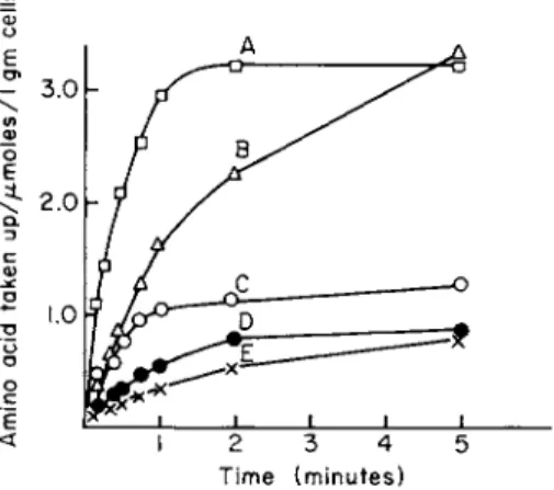
Figure 1 shows the time course of uptake of five amino acids into E.
coli Κ12. Chloramphenicol was added to the cell suspension to mini
mize the incorporation of the labeled amino acids into protein. The transport is sufficiently rapid that 15- or 30-second incubations are used to approximate initial rates. In these experiments the labeled amino
Time (minutes)
FIG. 1. Time course of amino acid uptake into Escherichia coli K12 cells. Exponentially growing cells were harvested, washed, and resuspended (about 1.0 mg cells/ml) in minimal salts medium containing 0.02% glucose and the appropriate labeled amino acid. The incubations were carried out at 37°C and terminated at the desired time interval by filtration on a Millipore filter, type HA. Cells were washed on the filter with 5 ml of minimal salts media at 25°C [36]. The final extracellular concentration of each amino acid was 11-17 μΜ.
A, L-leucine; B, glycine; C, L-phenylalanine; D, L-alanine; E, L-methionine. (Figure taken from Piperno and Oxender [36].)
140 DALE L. OXENDER
acids were added to the cell suspensions contained in test tubes, and at the desired time the incubation mixture was pipetted onto the filter.
Burrous and DeMoss [50] have used the technique of first depositing cells on the filter and then adding labeled amino acids to the cells to measure tryptophan uptake into E. coli.
If the endogenous level of an amino acid is very small because of rapid metabolic conversion to other products, it may be difficult to determine the initial rate of uptake. The L-serine level of E. coli is very small, and its metabolic conversion is extremely rapid so that an in
cubation time of 5 seconds is already too long to measure the initial rate of uptake [51]. From these observations it is questionable whether the uptake of serine can be adequately described kinetically. For some amino acids the rate of incorporation into protein may be greater than its rate of transport into the cell, and if other metabolic conversion is not significant the incorporation into protein can serve in such cases as a measure of transport. Histidine transport into S. typhimurium can be determined in this manner. When the carbon source is removed from the growth medium or protein synthesis is otherwise inhibited in S. typhim
urium, increased levels of endogenous histidine are formed which act as a
" sink " for the entering histidine and permit the initial rate of uptake to be measured [45].
In general it has been found that protein synthesis can be inhibited in bacteria without altering the initial rates of amino acid uptake, al
though there are enough exceptions to require that each case be inde
pendently tested. The effect of chloramphenicol on the 1-minute rate of
1 4C-proline accumulation by P. aeruginosa has been determined by Kay and Gronlund [52] and is shown in Fig. 2. The total uptake of proline remains constant when the chloramphenicol level is varied, but proline incorporation into protein is strongly inhibited when 80-100 μg of chloramphenicol/ml are added.
In contrast to these results Wiley and Matchett [53] found that tryp
tophan uptake into Neurospora crassa was inhibited by a few minutes treatment of the cells with 2 μg/ml of cycloheximide. Their results are shown in Fig. 3. Grenson et al. [54] have found a similar effect of cyclo
heximide on amino acid uptake into Saccharomyces cerevisiae. The interpretation of the results of these separate studies is based on the findings that, in both yeast and fungi, high internal amino acid levels inhibit the transport activity [53,54]. The latter authors suggest that the inhibitory effect of cycloheximide arises from an increased endogenous pool of amino acids that accumulate as a consequence of the inhibition of protein synthesis. Grenson et al. [54] have observed a marked loss in the transport activity for arginine, methionine, and leucine in yeast as
Chloramphenicol (ft.g/ml)
FIG. 2. Effect of chloramphenicol on the rate of 1 4C-proline uptake into cells and incorporation into protein of Pseudomonas aeruginosa. Chloramphenicol was added 30 minutes before addition of labeled proline. Incubations were carried out for 30 seconds at 30°C. The curves labeled pool and protein represent the trichloroactic acid-soluble and -insoluble 1 4C-proline, respectively. (Figure taken from Kay and Gronlund [117] with kind permission of A. F. Gronlund.)
1.0 — 0.8
£ 0.6
"θ
0.4
ο
0.2
10 20 30 40 Minutes
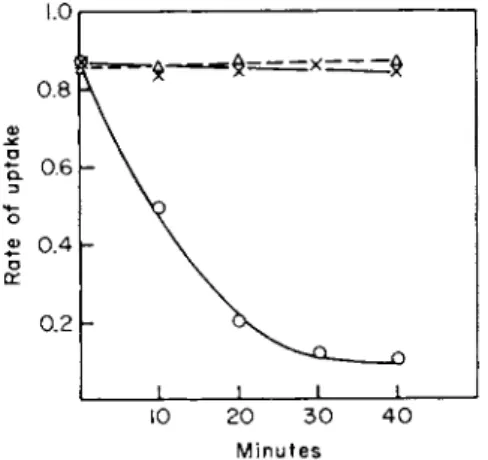
FIG. 3. Effect of cycloheximide on tryptophan transport activity in Neurospora crassa.
Cells were incubated at 30°C in the presence of cycloheximide (2 /xg/ml). Samples of the cells were removed by centrifugation at the indicated time intervals and the uptake of 1 χ 1 0 "5 Μ L-tryptophan-3-1 4C per gram dry weight of cells was measured in a 1-minute incubation at 30°C. O , treated with 2 fxg/ml cycloheximide; x , untreated control; Δ , treated with cycloheximide for 3 minutes at 30°C, and then the temperature was rapidly decreased to 4°. (Figure taken from Wiley and Matchett [53] with kind permission of W. R. Wiley.)
142 DALE L. OXENDER
a result of the increased endogenous amino acids produced by histidine starvation of a histidine-requiring mutant. The histidine transport re- mained constant, however, suggesting that the observed loss of uptake activity is not due to a general decrease in transport but a selective in- hibition of the transport system by accumulations of the particular amino acid that it transports. This feedback inhibition may account for the greater variability of the rates of transport of amino acids with the nutritional states of yeast and other fungi than is found for bacteria.
Transport rates for amino acids in gram-negative bacteria are relatively constant when the amino acid content is varied by inhibition of protein synthesis [52] or starvation [45,55]. We have used a leucine auxotroph of E. coli to vary the internal level of leucine by starving the cells and have found that the initial rate of leucine uptake appeared to be constant despite variation of internal leucine [51]. It should be pointed out that isoleucine and valine, amino acids that share the transport system with leucine, were presumably present during this test.
C. Temperature and pH Effects
The temperature dependency of amino acid accumulation in bacteria supports the conclusion that the transport process is a mediated entry rather than a simple diffusion process. Usually uptake is minimal at 0°- 4°C, although exchange between exogenous and endogenous amino acids of E. coli at such low temperatures has been reported [31,56].
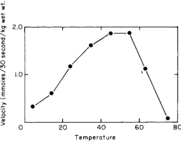
The uptake of L-leucine into E. coli K12 as a function of temperature is shown in Fig. 4 [36]. These results show a rather broad optimum with rapid uptake observed at temperatures as high as 45°C. Similar results have been observed for valine uptake into P. aeruginosa by Kay and Gronlund [37]. The high temperature coefficients reported for uptake of amino acids into bacteria are not surprising, where the process is con- centrative, in view of the requirement of metabolic energy for the ac- cumulation process; therefore this feature does not help elucidate the nature of the interactions occurring during transport.
The rapid loss of accumulated amino acids when the temperature of E. coli is decreased to about 0°C [31,36] has been discussed earlier in connection with the regulation of the amino acid pool. If bacteria are kept at 25-37°C after allowing them to accumulate amino acids, they exhibit a remarkable ability to retain their amino acids even if the medium is replaced by solutions free of amino acids. This property has been reported for Streptococcus faecalis and Staphylococcus aureus [3] for Lactobacillus arabinosus [33], Candida utilis [57] and Escherichia coli [36].
Ring [58] has described an increased permeability of Streptomyces
5 5
Temperature
FIG. 4. Temperature dependence of leucine uptake in Escherichia coli K12. The uptake of uniformly labeled L-leucine (27 μΜ) was measured as described in the legend of Fig. 1 except that the temperature of the incubation was varied. (Figure taken from Piperno and Oxender [36].)
hydrogenans when the cell suspension is cooled to 0°C. The increased penetration of molecules such as thiourea and ATP at 0°C compared to their entry at 30°C shows that the membranes were becoming leaky to other substances as well. Ring [58] suggested that a reversible temporary phase-transition within the membrane lipids may be responsible.
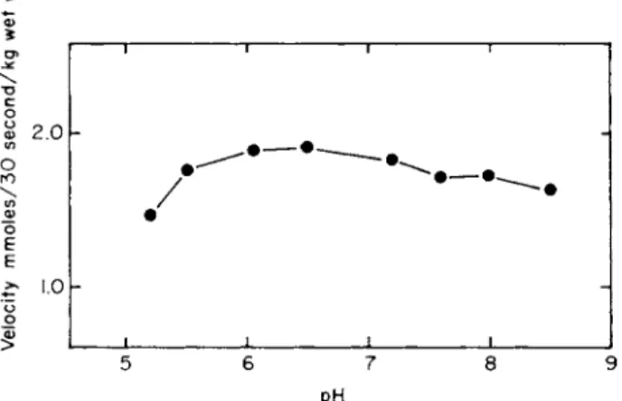
Since the pH optima are usually broad and represent a summation of effects of a number of ionizable groups, they have limited use for the identification of specific groups. Figure 5 shows the rather broad pH optimum obtained when the initial rate of L-isoleucine entry into E. coli Κ12 is measured at different extracellular pH values. The entry of glycine and L-alanine into E. coli seems to decrease slightly more than that of isoleucine at pH values below 6 (36). Gale [2] reported that the accumulation of lysine into S. faecalis was maximal around pH 9.5, a value close to the isoelectric point of lysine. Boezi and DeMoss [59]
found a pH optimum of 8.0-8.5 for tryptophan accumulation into E. coli using Tris buffer. We noted that the pH optimum for tryptophan uptake was much broader if phosphate buffer was used instead of Tris.
D. Kinetics
The criteria used to identify the various types of membrane transport by which a given solute may migrate have been discussed in another chapter of this volume and in greater detail in monographs by Stein [60]
and Christensen [61]. At low external levels of amino acids a small
144 DALE L. OXENDER
FIG. 5. pH dependence of isoleucine uptake into Escherichia coli K12 cells. The uptake of uniformly labeled L-isoleucine (14 μΜ) was measured as described in the legend to Fig. 1 except that the pH of the incubation medium was varied. (Figure taken from Piperno and Oxender [36].)
nonsaturable component of transport into microorgansims can usually be identified. Kinetically, this nonsaturable component resembles simple diffusion. Unlike simple diffusion, however, this process some
times exhibits stereospecificity, suggesting a type of mediated transport with a high Km. Although this transport is ordinarily a minor route of entry, nevertheless it becomes significant at elevated amino acid levels and can serve to support the growth of an auxotrophic organism even when this organism has lost low Λ^-transport systems for the amino acids.
This property has been exploited in selecting for so-called "permease"
mutants which will be discussed later. At low amino acid levels the major entry into microorganisms occurs by way of interactions with specific, low Km transport systems in the membrane. A study of the dependency of the initial rate of entry on the external concentration of amino acids shows that it obeys saturation kinetics and the unidirectional flux [J] is given by the Michaelis-Menten expression
V S
where S is the external concentration of amino acid, Km (sometimes KT) is the concentration of amino acid to produce one-half saturation of the maximal rate of transport, and Fm a x (sometimes Jmax) is the maximum velocity. The Km value for an amino acid is sometimes referred to as an affinity constant for the membrane mediator even before it has been
established for the system in question that the equilibrium assumption of Michaelis-Menten is being met. Only with the isolation of membrane components bearing receptor sites have direct measurements of the affinity constants for certain transport systems been possible for amino acids [25,62], sugars [62-64] inorganic sulfate [24] and phosphate [65].
The amino acids are rapidly accumulated by E. coli cells as illustrated in Fig. 1. Incubation times must be limited to 15-30 seconds in order to approximate initial rate measurements for the kinetic studies. The dependence of the initial rates of entry on the external concentration for several amino acids is shown for E. coli K12 in Fig. 6. Most of the amino acids studied did not produce an appreciable slope after the initial saturation; however, the results for methionine suggest some heterogeneity in the mode of uptake may exist.
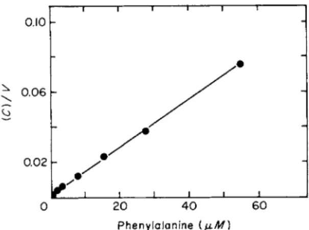
A double reciprocal plot of initial rate data for L-phenylalanine is presented in Fig. 7. From such plots the Km and Km a x values for amino acid entry have been obtained for E. coli (see Table IV). The Km values for most of the amino acids lie around 1.0 μΜ.
Not all of the kinetic plots for amino acid entry have been linear.
Data for valine entry into E. coli produce biphasic reciprocal plots from which two Km and Km a x values can be obtained [36] (see Table IV).
Halpern [66] had earlier found biphasic double reciprocal plots for glutamate uptake into various strains of E. coli when the cells were
FIG. 6. Concentration dependenceof amino acid uptake into Escherichia coli Κ12. The up
take was measured as described in the legend to Fig. 1 except that the concentration of the amino acid was varied as indicated above. A, L-isoleucine; B, glycine; C, L-phenylalanine;
D, L-methionine. (Figure taken from Piperno and Oxender [36].)
146 DALE L. OXENDER
0.10
^ 0.06
0.02
0 20 40 60 Phenylalanine (μΜ)
FIG. 7. Reciprocal plot of the concentration dependence of phenylalanine uptake. Up
take was measured as described in the legend to Fig. 1. The data in this figure correspond to curve C in Fig. 1. (Figure taken from Piperno and Oxender [36].)
T A B L E IV
KINETIC CONSTANTS FOR NEUTRAL AMINO A C I D UPTAKE INTO Escherichia coli K12 [36]
Km Y max
Amino acids (μΜ) (mmoles/30 sec/kg wet wt.)
L-Alanine 9.2 ± 1.7 (3)e 1.17
Glycine 3.8 ± 1.0 (2) 1.08
L-Isoleucine 1.22 ± 0 . 1 3 (2) 0.96 L-Leucine 1.07 ± 0 . 1 8 (2) 1.58
L-Valine 8.0 ± 5.7 (3) 1.41
0.70 ± 0 . 1 0 (3) 0.60 L-Methionine 2.27 ± 0.39 (3) 0.39 L-Phenylalanine 0.72 ± 0 . 1 7 (4) 0.75
L-Tryptophan 0.9 (1) 0.59
D-Alanine 8.3 (1) 0.36
a Number of determinations is indicated in parentheses.
grown on glucose, but linear plots if succinate was used as the carbon source. Attempts to obtain evidence for more than one transport system for valine have been uniformly unsuccessful, although leucine entry into E. coli is mediated by two transport systems for which separate binding proteins have been isolated [67]. It should be noted that one cannot read two meaningful sets of kinetic parameters directly from the intercepts of a biphasic Lineweaver-Burk curve. Instead, iterative procedures may be used.
Since mutants possessing an altered glutamate uptake showed higher activity than the parent strains in both parts of the biphasic reciprocal plots when grown on glucose, it was concluded that the biphasic nature of the plots was the result of a single transport system that was somehow subject to " allosteric" activation by glutamate. Holden [68] has found that the uptake of glutamate into the gram-positive orgaismL. arabinosus can be described by a single transport system that produces linear kinetics.
Alternatively nonlinear Lineweaver-Burk plots for amino acid up
takes can also be the result of the contributions of more than one trans
port system that serves for a single amino acid as found for histidine by Ames [45]. Figure 8 shows the Lineweaver-Burk plot for the uptake of histidine into S. typhimurium. Ames [45] found that histidine could enter these bacteria by a specific transport system and a system with much broader specificity that has been called the aromatic amino acid trans
port system. In this case the Km value for the specific histidine transport system (1.7 χ 10 ~7M) was sufficiently lower than that for the general aromatic amino acid transport system (1.1 χ 10"4M) so that a satisfac
tory resolution of the two values could easily be made. Phenylalanine and tyrosine also entered S. typhimurium by at least two transport systems, thereby producing nonlinear Lineweaver-Burk plots of initial rates of entry.
FIG. 8. Lineweaver-Burk plot for histidine uptake in starved cells of Salmonella typhi
murium. Cells were starved by incubation at 25°C without aeration. The indicated concen
tration of histidine-2-1 4C was then added and incubation continued at 25°C for 1 minute.
Incubations were terminated by filtration on a Millipore filter. Rates were expressed as jLtmoles per gram dry weight per minute. (Figure taken from Ames [45] with kind permission of G. F. Ames.)
148 DALE. L OXENDER
Nonlinear kinetic plots resulting from multiple transport systems have also been obtained for the uptake of some of the amino acids into Sac. cerevisiae, by Grenson [69].
The common finding of multiple routes of amino acid entry into microorganisms suggests that caution should be exercized in interpreting the sometimes complex kinetic data obtained.
E. Transport Schemes or Models
Models have been proposed in an attempt to give a molecular des- cription of the phenomenon of membrane transport. An adequate model should be constructed on the fewest and the simplest assumptions necessary to account for the experimental observations and should be testable.
I will describe the features of a scheme for transport in the context of the bacterial cell, although contributions have come from both bacterial and nonbacterial cases.
Models for amino acid transport into microorganisms need to account for the following kinds of experimental findings:
(a) Transport systems are located within the cytoplasmic membrane and can be studied in membrane preparations [70].
(b) A high degree of structural specificity is common.
(c) Accumulation can lead to internal concentrations from 500 [10] to 28,000 [31] times the external level.
(d) Metabolic inhibitors such as sodium azide and 2, 4-dinitrophenol prevent or cause the loss of the ability to accumulate amino acids but do not interfere with rapid exchange of external and internal amino acids [10,31,33,36].
(e) If bacteria are suspended in amino acid-free buifer at room tem- perature, they appear to be able to retain high internal concentrations of amino acids in the absence of externally added energy sources even though these amino acids can undergo rapid exchange if an amino acid is added externally [31,36].
(/) The rate of exit of amino acids is accelerated in the presence of metabolic inhibitors such as sodium azide [36,71] or by cooling to 0°C [36].
Several general models have been put forth to explain membrane transport. The " gate " model of Patlak [72] has been referred to as a very general and nonrestrictive model illustrating the concept of a receptor site in the membrane. We have used a similar model to describe our findings for amino acid entry into Ehrlich cells [73,74]. In these models the molecular entity in the membrane that bears the receptor site can
undergo reversible structural changes resulting in transfer of the receptor site from one phase to the opposite phase of the membrane. Cohen and Monod [29] have proposed a general model for transport in bacteria in which specific genetically determined proteins located within the mem- brane catalyze the transport of various solutes. They proposed that these proteins be called permeases and the entire membrane process for solute transport be called a permease system. Controversy arose over the use of the term permease since no enzymatic activity, only catalytic activity, was proposed [22, 31]. The limitations of the permease model as originally proposed by Cohen and Monod [29] are thoroughly discussed by Britten and McClure [31]. A modified and more detailed version of the permease model was described by Kepes and Cohen [32]. In this model a genetically identified, inducible, stereospecific protein component called permease combines with external substrate forming a complex in the rate-limiting step in transport. A second component in the membrane called the transporter, reacts with the permease-substrate complex and an energy donor to form a substrate-transporter complex that passively diffuses across the osmotic barrier of the cell, allowing the substrate to dissociate from the complex on either side. The latter steps are not rate- limiting. This model suggests that the interaction of the transport system with metabolic energy (ATP) occurs outside the osmotic barrier and also that the high energy form of the carrier is necessary for greatest binding activity. Neither of these features appears to be supported by experi- mental findings.
A general model that is rather widely used to explain the findings listed at the beginning of this section is shown in Fig. 9 [75]. This model, which is similar to other models [23,74,76], assumes a three-stage process. The first stage is a binding of a solute (A) to some macromole- cule (X) in the membrane, thus forming a complex (AX) as illustrated by the reversible reaction shown as 1 in the figure. Stage 2 involves a process in which the complex (AX) crosses the osmotic barrier of the cell. This step is believed to be a reversible conformational change within the macromolecule or membrane components with which it is associated.
These two steps alone give rise to a facilitated diffusion system. The third stage is the coupling of one of these reactions to metabolic energy, thus making it to some degree irreversible as illustrated in the figure as reaction 3. As can be seen by this scheme, the high energy form of the mediator (X*) has the lowest affinity for (A) and the solute is therefore mainly carried by the low energy form of the mediator (X). When sodium azide or low temperatures are used to inhibit step 3, most of the media- tor will be converted into the high affinity form, thus accelerating the exit of (A). In addition it is proposed that the mediator (X) finds greater
150 DALE L. OXENDER
FIG. 9. Transport model (see text for details). (Figure taken from Penrose et al. [75].)
difficulty in participating in step 2 than the mediator complex (AX), giving rise to an asymmetric distribution of the mediator on the two sides of the osmotic barrier. This property explains the increased initial rate of exit of labeled amino acid by exchange when unlabeled amino acid is added externally [36]. In the absence of external amino acid, much of the mediator would probably be at the external face of the membrane and that portion remaining inside would tend to be in the low affinity form (X*). This feature gives the appearance of a " retention mechanism."
Because experimental findings vary somewhat for different amino acid transport systems and especially for different microorganisms, variations of a general model for transport will probably be necessary to explain various transport systems. For example, we found that ex- ternal leucine and isoleucine cause the rapid loss of labeled leucine from E. coli while alanine and glycine produce very little effect on the loss of cellular alanine [36]. To account for the results we need to produce a variation of the general model by altering it so that the free (X) and combined mediator (AX) undergo step 2 at identical rates for the alanine transport system. Such a system will not show accelerative exchange [60].
Some permease models have included an enzyme which bears a receptor site that can catalyze the reaction shown by step 1, leading to
the attachment of the solute (A) to another receptor site on the mediator (X). It seems that a model with a single receptor side is less restrictive and still adequate at this time.
III. TRANSPORT SYSTEMS
Studies of amino acid transport into microorganisms reveal the presence of several distinct transport systems each of which serves for a limited number of structurally related amino acids. Some of the trans
port systems appear to serve only for a single amino acid; others serve for a rather large class of somewhat similar amino acids. The situation is similar to that seen in the animal cells, except that high specificity systems are more conspicuous in microorganisms. The following families of amino acids have been suggested by various kinetic and genetic analyses of amino acid transport.
A. Glycine, Alanine, and Serine
The growth of E. coli W (ATCC 9637) is strongly inhibited by DL- serine [77]. Davis and Maas [78,79] found that the L-serine did not relieve the inhibition by D-serine. D-Serine-resistant mutants of E. coli W were isolated and found to have an impaired ability to concentrate the D-serine, glycine, and L-alanine [80] but normal transport activity for other amino acids tested. These results suggest a common transport system for D-serine, alanine, and glycine. A more detailed study of the glycine uptake into whole cells of wild type E. coli W and the derived D-serine-resistant mutant has been reported by Kaback and Kostellow [81]. Although the kinetics were not linear, the initial rate of uptake tended to show saturation around 2.6 χ 10 "4M . Part of the problem may be related to the circumstance that 70 % of the glycine taken up by both strains was converted to other products some of which proved to be phospholipids. Kaback and Stadtman [82] studied glycine uptake in
to membrane preparations of E. coli W and the D-serine-resistant mutant with a glycine-defective transport system. The membrane preparations showed many of the same characteristics of transport into whole cells except that glycine entry into the internal pool was not concentrative and therefore could not be altered by metabolic inhibitors. DL-Serine, DL-alanine, and DL-threonine all showed competition for glycine entry and also participated in exchange with internal glycine. Levine and Simonds [83,84] have studied the uptake of glycine into two serine- glycine auxotrophs of E. coli K12. One auxotroph (S) that grows on
152 DALE L. OXENDER
glycine only after a lag period was found to be deficient in glycine trans- port, but it had a separate transport system for the dipeptide glycylgly- cine that could supply the glycine by intracellular hydrolysis. In other studies, peptides of glycine and of alanine were shown to be taken up by separate but specific transport systems in Lactobacillus casei by Leach and Snell [85], in Leuconostoc mesenteroides by Mayshak et al. [86], in Escherichia coli by Hirsch and Cohen [20] and Kessel and Lubin [87], in Streptococcus faecium by Brock and Wooley [88], and in Pediococcus cerevisiae by Florsheim et al. [89].
Mora and Snell [90] have shown that whole cells and protoplasts of S. faecalis both contain an active transport system serving for glycine, L-alanine, and D-alanine. The saturability of the transport system by the three amino acids was not altered by removal of the cell wall. Transport of these amino acids, into protoplasts only, was stimulated by K+ or high levels of pyridoxal phosphate and could be inhibited by Na + . D-Cycloserine (D-4-amino-3-isoazolidone), an antibiotic that inhibits cell wall synthesis [91], competitively inhibits the transport of both D- and L-alanine with a Kt value of 10" 5M. The L-isomer of the antibiotic was without effect. In similar studies Mora and Bojalil [92], using Mycobacterium acapulcensis, and Reitz et al. [93] using Streptococcus sp., found a common transport system for D-cycloserine, D-alanine, and L-alanine. The relative rate of transport of alanine was found to be about five or six times as fast as that of glycine or serine into Pseudo- monasaeruginosa [37].
The many studies on the mutual inhibitory actions of glycine, serine, and alanine seem to establish clearly a common transport system in most if not all microorganisms. We have examined more closely the inter- actions of glycine, alanine, and serine during transport into E. coli K12 [94] and found that, while mutual inhibition can be observed, quanti- tative measurements of the extent of the inhibitory actions suggest heterogeneity of mediation. We have obtained a D-serine-resistant mutant (PAD02) of E. coli W that shows no mediated transport of glycine, reduced transport of L-alanine, but apparently normal L-serine transport. An unexpected finding was that glycine is still a competitive inhibitor of L-alanine uptake in this mutant, even though glycine cannot be transported [51]. This latter finding suggests that some caution should be exercised when making conclusions based solely upon competitive interactions. It is still conceivable that a common system for glycine and alanine can be proposed if one assumes that the common receptor site in the mutant has been altered but still binds glycine although this glycine is not transported. More plausibly two or more transport systems serve for the transport of glycine, serine, and alanine. These results
suggest that there may be a rather specific transport system for L-alanine and a second common transport system that serves as a general trans
port system possibly for all three amino acids and their isomers [51].
The measurement of the transport of L-serine presents a special problem, since the endogenous pool of serine is very small (see Table I) and the metabolic modification is extremely rapid. We found that the approach to equilibrium was so rapid that it was not possible to measure the initial rate even with incubation times as short as 5 seconds. In parallel studies Wargel et al. [95] measured the competitive interactions of D-, L-alanine, glycine, and D-cycloserine in E. coli K12 and concluded that systems for D-alanine and glycine are related but separate from the accumulating system for L-alanine. D-Cycloserine appears to be trans
ported by the D-alanine system. The isolation of D-cycloserine-resistant mutants has provided support for these conclusions.
Still unanswered are the questions whether there are specific transport systems for each of the amino acids and what is the nature of the over
lap among the transport systems. The effect on transport of various carbon sources for E. coli suggests that the transport of all of the amino acids can be altered or perhaps controlled independently [95].
α-AMINOISOBUTYRIC A C I D
The model amino acid α-aminoisobutyric acid, which has been shown to be a competitive inhibitor of glycine, alanine, and serine in mammalian tissues such as the Ehrlich cell [96], is also actively accumulated by the gram-positive organisms Bacillus megaterium and Staphylococcus aureus [97]. Alanine and glycine competitively inhibit the entry of α-aminoiso
butyric acid into these bacteria. The amino acid seems to be blocked to metabolism in the gram-positive bacteria [97]. There are various Pseudomonas species, however, that can grow on α-aminoisobutyric acid as the sole source of carbon and nitrogen [98]. It has been reported [99]
that the gram-negative organism E. coli does not concentrate α-aminoiso
butyric acid. We have found that α-aminoisobutyric acid, however, is transported by E. coli K12 cells with a relatively high Km value of 4.2 mM [36]. Its transport can be competitively inhibited by low concen
trations of glycine, alanine, or serine. High levels of external α-amino
isobutyric acid inhibit the uptake of glycine and alanine. Drapeau, Matula, and MacLeod [100] and Wong, Thompson, and Macleod [101]
have studied the highly concentrative transport of α-aminoisobutyric acid into a marine Pseudomonadspecies andPhotobacterium fischeri. Na + was required for uptake and to prevent release. The effect of increasing N a+ or L i+ from 0 to 50 mM was to lower the Km 12-fold to a value of
154 DALE L. OXENDER
8.3 x 10"5 Μ while leaving the Vmax unchanged. At levels above 50 mM Na + , the Fm a x was increased. α-Aminoisobutyric acid is also actively accumulated in the bacterial strain S. hydrogenans [58]. N a+ depend
ency is a striking feature of the corresponding transport in animal cells [60]. In parallel with the effects of temperature on the loss of endogenous amino acids discussed earlier these bacteria also show a marked increase in the loss of accumulated amino acid when the temperature is lowered to 0°C.
B. Proline
In 1960, Lubin et al [102] isolated mutants from E. coli W and E. coli Β that were defective in the ability to accumulate histidine, proline, or glycine. From strain W6 (his"), a proline auxotroph [103], they were able to obtain mutant strains W 157 and W 408 that required 250-500 mg of proline/liter to produce the maximal rate of growth. They later carried out a detailed study of these mutants with a high proline requirement and showed that they lacked a specific proline transport system [104].
When the transport deficient mutants were allowed to revert to the pro
totrophic state, so that they no longer required proline for growth, the proline transport system was still missing. Exchange of external proline for intracellular proline, which can readily be demonstrated in the parent strain W6 even at 0°C, was not detectable in the mutant. Although the mutant W157 could not concentrate proline, it had a normal ability to concentrate glycine, phenylalanine, histidine, and lysine [104]. A study of the accumulation and exchange of proline in E. coli Β has been used to determine some of the mechanisms of pool formation and control [31]. Kaback and Stadtman [70] have shown that cytoplasmic membrane fractions prepared from E. coli W6 accumulate proline in an energy- dependent process. Membrane preparations from the proline transport mutant W157 were unable to accumulate proline. This transport system shows a high degree of specificity, hydroxyproline being the only other naturally occurring amino acid that competitively inhibits both the concentrative uptake at 37°C and the exchange at 0°C between intracel
lular and extracellualr proline. Three proline analogs, thiazolidine-4- carboxylic acid, 3, 4-dehydroproline, and azetidine-2-carboxylic acid, have, however, been shown to inhibit proline entry into E. coli [105].
Kaback and Deuel [106] have disrupted the membrane preparations by sonication or passage through a French pressure cell in unsuccessful attempts to separate and solubilize the proline transport system. Char
acteristics of proline transport into various microorganisms are sum
marized in reviews by Britten and McClure [31] and Britten [107]. Kay
and Gronlund [108] described an inducible proline transport system in P. aeruginosa. Behki [109] reports that the transport system for proline in Agrobacterium tumefaciens appears to be considerably less specific than has been found for other microorganisms. Results of a study of the entry and exit of proline in N. crassa by Zalokar have been interpreted as indicating intracellular compartmentalization of the amino acids in this organism [110].
Studies of proline transport in most microorganisms show that the system is highly stereospecific, and of the naturally occurring amino acids only the L-isomers of proline and hydroxyproline are transported to any extent. The lack of success with attempts to isolate the proline transport system from E. coli using disrupted membrane preparations [106] and osmotic shock techniques [25] indicates that it is firmly attached to the cytoplasmic membrane.
C. Valine, Leucine, and Isoleucine
A stereospecific transport system for leucine, isoleucine, and valine in E. coli K12 was first described by Cohen et al. [10,29,111]. Britten and McClure [31] have studied extensively the inhibitory interactions during accumulation, incorporation, and exchange of leucine, isoleucine, valine, and their structural analogs such as norleucine and norvaline.
In a kinetic analysis of amino acid transport in E. coli K12 we found that the Km values for the entry of the branched-chain amino acids were around 10" 6 Μ ([36]; see Table IV). The time course for leucine entry is shown in Fig. 1. Figure 6 presents the dependence of the initial rate of entry of isoleucine on the external concentration. Chloramphenicol was added to minimize protein synthesis; under these conditions 95% of the extractable amino acids could be accounted for unchanged. The overlap between this transport system and other systems is minimal, as shown by Fig. 10. This study shows that glycine and phenylalanine produce little inhibition of leucine uptake even at levels 100 times the concentration of leucine. In mammalian tissues, phenylalanine and leucine are usually found to share a common transport system [73].
When the E. coli cells were subjected to osmotic shock treatment, the accumulation of leucine and valine was reduced with no change in the transport of alanine and proline [25]. A binding protein (" LIV-binding protein") was then isolated from the shock fluid; it could bind leucine, isoleucine, and valine [25,75,112]. The dissociation constants for the binding protein were determined by equilibrium dialysis. These con
stants were found to be indistinguishable from the Km and Kt values for cellular uptake of leucine, isoleucine, and valine (Table V).
156 DALE L. OXENDER
100
8 0
6 0
4 0
20
100 3 0 0 5 0 0
Inhibitor concentration (Ι0'6Λ/)
FIG. 10. Inhibition of leucine uptake in Escherichia coli Κ12. Uptake of uniformly labeled L-leucine (5-10 μΜ) was measured as described in the legend to Fig. 1 in the presence of varying concentrations of the inhibitors indicated above. A, glycine; B, L-alanine; C , cycloleucine; D, L-phenylalanine; E, L-isoleucine. (Figure taken from Piperno and Oxender
[36].)
TABLE V
A COMPARISON OF KINETIC CONSTANTS FOR AMINO ACID-BINDING ACTIVITY OF BINDING PROTEIN WITH THOSE FOR
CELLULAR TRANSPORT BY Escherichia coli [25]
Transport studies Binding studies Half-maximal Dissociation inhibition of
Km constant binding
Amino acid (μΜ) (μΜ) (μΜ) (μΜ)
L-Leucine 1.1 0.5 (He) 1.1 2.1 (He)
L-Isoleucine 1.2 1.2 (Leu) 2.2 1.7 (Leu)
L-Valine 8.0 4.5 (Leu) 12.0 7.3 (Leu)
3.4 (He) 8.9 (He)
The presence of leucine in the growth medium represses the synthesis of the LIV-binding protein as well as the ability of the cells to transport leucine. Inui and Akedo [113] had noted repressiblility of this transport system by the model substrate 1 -aminocyclopentanecarboxylic acid and also by L-leucine or L-methionine. 1-Aminocyclopentanecarboxylic acid is actively transported in E. coli Κ10 by an energy-dependent transport
system which is presumably the same one that serves for the transport of leucine, isoleucine, and valine, since mutual competitive interactions are observed [113]. Another synthetic model amino acid, 2-aminobicyclo- [2.2.1.] heptane-2-carboxylic acid (BCH), whose uptake by E. coli is inhibited by leucine and which inhibits leucine uptake, provided the correct isomeric form is taken, is also bound to the LIV-binding protein and inhibits the binding of leucine [114]. This circumstance is an inter
esting one, because the same isomer serves as a specific model for a corresponding transport system seen in a variety of animal cells [114].
Furthermore the addition of BCH to the growth medium represses the synthesis of the LIV-binding protein. On the other hand, the synthesis of the LIV-binding protein is not derepressed in mutants* that have been derepressed for the biosynthetic enzymes for leucine, isoleucine, or valine, indicating that coding for synthesis of the binding protein does not occur in the various operons concerned with the synthesis of the branched-chain amino acids [51]. Additional support for a separation between the control of transport and biosynthesis was obtained by ex
amining a leucine-requiring mutant (leu 500)** of S. typhimurium which cannot make any of the biosynthetic enzymes for leucine. This mutant has normal leucine transport and LIV-binding protein.
The current evidence suggests that the coding for the binding proteins lies outside the operons that code for the biosynthetic enzymes but that both features can be coordinately repressed under certain conditions.
The LIV-binding protein isolated from E. coli K12 has been purified and crystallized and found to have a molecular weight of 36,000. It binds
1 mole of either leucine, isoleucine, or valine/mole of protein. It under
goes large and reversible conformational changes in the presence of urea [75]. In the presence of 6 Μ urea all nine tyrosine residues can be rapidly iodinated. An extensive search for substrate-induced conformational changes using ORD, CD measurements, and various fluorescent probes were uniformly unsuccessful [75]. Anraku [62] carried out a separate purification of the LIV-binding protein from another strain of E. coli and studied its properties, which are in agreement with the results from our laboratory. More recently Furlong and Weiner [67] have obtained evidence for a second transport system for leucine in whole cells and a corresponding second binding protein in the shock fluid. This second transport system and binding protein is specific for leucine and its analog trifluoroleucine and comprises 15-20% of the total uptake and
* These mutants (E. coli CU5001 and CU5002) were kindly sent to us by Dr. Η. E.
Umbarger.
** The mutant S. typhimurium (leu 500) was kindly sent to us by Dr. P. Margolin.
158 DALE L. OXENDER
binding activities present in E. coli strain H. It has been separated by DEAE-chromatography and crystallized. Its molecular weight is also 36,000. We have confirmed these results using E. coli K12 [51]. The specific binding protein for leucine accompanies the LIV-binding pro
tein during the various steps of purification but is eliminated by crys
tallization of the LIV-binding protein. A measurement of the extent of the isoleucine inhibition of either leucine transport or binding activity reveals the presence of an uninhibited component of leucine migration.
The kinetic constants of the leucine specific transport system can be determined by measuring leucine uptake in the presence of excess isoleucine which saturates the shared transport system. Under these conditions we found a Km of 0.2 μΜ for leucine entry. This value is slightly lower than that obtained in the absence of isoleucine. The Fm a x of the specific system represents about 15 % of the total leucine uptake.
The synthesis of both leucine-binding proteins is repressed when leu
cine is added to the growth medium. Both proteins react with the same antibody, showing a single precipitin band on diffusion plates.
Using kinetic studies, we have determined that various strains of E. coli contain different proportions of the binding proteins. About 50 % of the total leucine uptake into E. coli Β cells is specific for leucine; this suggests that the two transport systems occur in about equal amounts.
In support of this conclusion we found that the Vmax of leucine entry into E. coli Β was twice that for isoleucine entry. Almost the reverse situation was obtained for transport activity in E. coli W cells. The Vmax of isoleucine transport is about three times that found for leucine. About 50 % of the isoleucine entry into E. coli W cannot be inhibited by leucine, suggesting the presence of a second transport system that discriminates against leucine in addition to the shared transport system. Valine appears to share the same systems that serve for isoleucine. We have isolated two isoleucine-binding activities from E. coli using a hydroxylapatite column.
One of these binding proteins does not serve for leucine. These proteins also cross-react antigenically with antibody to the LIV-binding protein from E. coli Κ12. We can now say that E. coli K12 contains a common transport system for the branched-chain amino acids and a small amount of a specific leucine transport system. Escherichia coli Β contains about equal amounts of a leucine-specific system and the shared transport system. Escherichia coli W contains significant amounts of an isoleucine- valine transport system in addition to the shared transport system [51].
The three different proteins are now being isolated and characterized.
We have recently isolated a mutant of E. coli K12 that has derepressed levels of branched chain amino acid transport and LIV-binding protein (Rahmanian and Oxender [54a]). The mutant is more sensitive to the
leucine analog 4-azaleucine. We have isolated several azaleucine- resistant mutants that have a defective LIV-transport system. These mutants all have normal levels of the LIV-binding protein, indicating that some other component of the leucine-transport system is defective.
The transport of leucine and of valine has been studied in P. aeruginosa by Kay and Gronlund [55] and in yeast by Bussey and Umbarger [115].
The latter authors have studied leucine and trifluoroleucine uptake into Saccharomyces sp. and a trifluoroleucine-resistant mutant strain. The analog resistance was not a result of decreased uptake, since the mutant accumulated both amino acids to a greater extent than did the wild type.
The defect appeared to be a very low content of trifluoroleucine tRNA in the mutant. A pronounced overshoot in the accumulation of leucine was observed with as much as 60% of the leucine subsequently being returned to the medium. The wild-type yeasts, which do not normally show such an overshoot, could be made to do so by growing them on leucine as a sole source of nitrogen followed by depletion by nitrogen starvation. The ability to overshoot was rapidly recovered by reducing the leucine content a second time, suggesting a control of leucine uptake by feedback inhibition. Leucine-binding activity [115] was identified in extracts of yeast by the same authors, using an ion-exchange resin charged with leucine as an assay method modeled after that used by Pardee [24] to detect a sulfate-binding protein. Their purification of this protein has been hampered because of the complete loss of activity when the extract is passed over a Dowex-50 resin and the lack of a selective extraction procedure.
We can say in summary that most microorganisms studied have a common transport system for leucine, isoleucine, and valine, while some bacteria have in addition specific systems for leucine or for isoleucine and valine.
D. Phenylalanine, Tyrosine, and Tryptophan
A very elegant study of amino acid transport into wild-type and mutant strains of S. typhimurium by Ames [45] and Ames and Roth [116]
established a rather general pattern for transport systems in microor- ganisms. Ames found that histidine, phenylalanine, tyrosine, and tryp- tophan are each transported by a specific system and, in addition, a common system was shared by all four solutes. The apparent Km of histidine for the common system, however, is about 200-fold higher than the apparent Km of phenylalanine for this system; this suggests that the common system does not normally serve for histidine. Although Ames preferred to call the common system an aromatic permease, the
160 D A L E L . O X E N D E R
available preparations of leucine were also found inhibitory to it. The Km value of 10" 6 Μ for the specific system for phenylalanine was found to be higher by a factor of ten than that for the general system, making a kinetic resolution difficult. The kinetics of the specific system were determined by measuring phenylalanine entry in the presence of 5 χ 10 " 4 Μ tryptophan. A mutant (AZA-3), resistant to azaserine and 5-methyl- tryptophan, analogs of tryptophan that are transported by the general or aromatic system, proved to have a defective "aromatic" transport system.
We have obtained evidence for a common transport system appar
ently restricted to these three amino acids in E. coli K12 by measure
ments of the competitive interactions and their ability to participate in amino acid exchange reactions [36]. In another study using the gram- negative organism P. aeruginosa the characteristics of the transport of the three aromatic amino acids have been determined by Kay and Gronlund [37]. These authors have used 53 amino acid analogs to select for transport mutants in P. aeruginosa [117]. Table VI shows the
T A B L E V I
INHIBITION OF AMINO ACID TRANSPORT IN Pseudomonas aeruginosa CELLS GROWN IN THE PRESENCE OF
AMINO A C I D ANALOGS [117]
Inhibition'' Amino acid analog 1 4C-amino acida (%)
Canavanine Arginine 65.6
Lysine 21.5
Proline c
Dehydroproline Proline 94.4
Tyrosine —
Thioproline Proline 96.1
Tyrosine —
/7-Fluorophenylalanine Phenylalanine 96.3
Tyrosine 92.6
Proline —
m-Fluorotyrosine Tyrosine 93.8
Phenylalanine 95.1
Proline —
5-Fluorotryptophan Tryptophan 93.1
Proline —
a Uptake studies were performed with amino acid concen
trations of 2.5 χ 10~7 Μ and 0.1 mg (dry wt.) of cells/ml.
b Percent inhibition of normal transport rate.
c Within experimental error of no inhibition.
![Table II presents the composition of the amino acid pools reported for some other microorganisms [35,37-39]](https://thumb-eu.123doks.com/thumbv2/9dokorg/1152373.82866/4.664.156.499.599.871/table-presents-composition-amino-acid-pools-reported-microorganisms.webp)