Targeting of Calcineurin to an NFAT-like Docking Site Is Required for the Calcium-dependent Activation of the Background K ⴙ Channel, TRESK *
Received for publication, March 16, 2006 Published, JBC Papers in Press, March 28, 2006, DOI 10.1074/jbc.M602495200
Ga´bor Czirja´k1and Pe´ter Enyedi2
From the Department of Physiology, Semmelweis University, H-1444 Budapest, Hungary
The two-pore domain Kⴙchannel, TRESK (TWIK-related spi- nal cord Kⴙchannel) is activated in response to the calcium sig- nal by the calcium/calmodulin-dependent protein phosphatase, calcineurin. In the present study we report that calcineurin also interacts with TRESK via an NFAT-like docking site, in addition to its enzymatic action. In its intracellular loop, mouse TRESK pos- sesses the amino acid sequence, PQIVID, which is similar to the calcineurin binding consensus motif, PXIXIT (whereXdenotes any amino acids), necessary for NFAT (nuclear factor of activated T cells) activation and nuclear translocation. Mutations of the PQIVID sequence of TRESK to PQIVIA, PQIVAD, or PQAVAD increasingly deteriorated the calcium-dependent activation in the listed order and correspondingly reduced the benzocaine sensitivity (a property discriminating activated channels from resting ones), when it was measured after the calcium signal inXenopusoocytes.
Microinjection of VIVIT peptide, designed to inhibit the NFAT- calcineurin interaction specifically, also eliminated TRESK activa- tion. The intracellular loop of TRESK, expressed as a GST fusion protein, bound constitutively active calcineurinin vitro. PQAVAD mutation as well as addition of VIVIT peptide to the reaction abro- gated this calcineurin binding. Wild type calcineurin was recruited to GST-TRESK-loop in the presence of calcium and calmodulin.
These results indicate that the PQIVID sequence is a docking site for calcineurin, and its occupancy is required for the calcium-de- pendent regulation of TRESK. Immunosuppressive compounds, developed to target the NFAT binding site of calcineurin, are also expected to interfere with TRESK regulation, in addition to their desired effect on NFAT.
Two-pore domain potassium (2PK⫹)3channels give rise to back- ground (leak) potassium conductance and their diverse regulatory mechanisms control cellular function by adjusting both the resting membrane potential and excitability (for review, see Refs. 1 and 2).
TRESK (TWIK-related spinal cord K⫹channel) is uniquely regulated by the calcium signal among the 2PK⫹ channels. We have recently reported that TRESK, expressed heterologously inXenopusoocytes, is activated about 10-fold by the calcium/calmodulin-dependent protein
phosphatase, calcineurin (3). TRESK was cloned from human spinal cord (4) and mouse cerebellum (3), and its expression was also demon- strated by reverse transcription-PCR in cerebrum, brainstem, testis, pancreas, and placenta (5–7). A massive signal was detected by North- ern blot in rat thymus and spleen (7). Single channel activity of TRESK has recently been demonstrated in dorsal root ganglion neurons (8). In the absence of specific antibodies and inhibitors, TRESK has neither been identified at the protein level nor has it been detected as an endog- enous whole cell current. However, the robust activation of TRESK by the calcium signal implicates that it can influence substantially the func- tion of the native cells expressing the channel.
The calcineurin-mediated regulation suggests that TRESK activation is the target of the widely used immunosuppressive drugs, cyclosporine A and FK506, as it was, in fact, demonstrated inXenopusoocytes (3).
These drugs inhibit calcineurin (and consequently NFAT activation and interleukin-2 production of T lymphocytes) by forming inhibitory com- plexes with the ubiquitous immunophilin proteins (9, 10). However, general inhibition of calcineurin also causes several undesired effects.
Therefore, a novel direction of drug development focuses on the NFAT- docking site of calcineurin, which is considered to be a more specific target than the phosphatase activity (11, 12). The binding of calcineurin to the PXIXIT consensus motif (whereXdenotes any amino acid) is required for NFAT activation. Apart from NFAT, the only known mam- malian proteins possessing similar calcineurin binding sites are cal- cineurin inhibitors/modulators or anchoring proteins (13, 14). In the present study we report for the first time that an ion channel, TRESK, has an NFAT-like calcineurin binding consensus sequence and the binding of the phosphatase to this docking site is indispensable for the regulation of the channel. Therefore the inhibitors, designed to block the calcineurin-NFAT interaction specifically, may also interfere with TRESK activation.
EXPERIMENTAL PROCEDURES
Materials—VIVIT peptide (NFAT inhibitor) and calcineurin autoin- hibitory peptide (ITSFEEAKGLDRINERMPPRRDAMP) were pur- chased from Calbiochem, bovine calcineurin and calmodulin from Sigma. Enzymes and kits of molecular biology applications and all other chemicals of analytical grade were obtained from the companies listed elsewhere (3).
GST-TRESK Loop Fusion Proteins—To produce GST-TRESKloop protein, the cDNA encoding the intracellular loop of mouse TRESK (amino acids 164 –292) was PCR-amplified from our pEXO-TRESK plasmid (3), applying consecutively Loop-s1/Loop-a1 and Loop-s2/
Loop-a1 primer combinations (see the sequences and cloning sites in Table 1). The PCR product was ligated into the XhoI site of pGEX-4T-1 (Amersham Biosciences, Little Chalfont, UK). To obtain the longer GST-TRESKloop-TAPtag protein, TRESK loop was amplified with Loop-s1 and Loop-a2 oligonucleotides. The PCR product was cloned
*This work was supported in part by the Hungarian National Research Fund (OTKA T46954) and the Hungarian Medical Research Council (ETT-085/2003). The costs of publication of this article were defrayed in part by the payment of page charges. This article must therefore be hereby marked “advertisement” in accordance with 18 U.S.C.
Section 1734 solely to indicate this fact.
1Supported by the Ja´nos Bolyai fellowship of the Hungarian Academy of Sciences.
2To whom correspondence should be addressed: Dept. of Physiology, Semmelweis Uni- versity, P. O. Box 259, H-1444 Budapest, Hungary. Tel.: 36-1-266-2755 (or 4079); Fax:
36-1-266-6504; E-mail: enyedi@puskin.sote.hu.
3The abbreviations used are: 2PK⫹, Two-pore domain potassium channel; NFAT, nuclear factor of activated T cells; TRESK, TWIK-related spinal cord K⫹channel; GST, glutathi- oneS-transferase; ANOVA, analysis of variance.
into the pcDNA3-TAPtag plasmid (Se´raphin Laboratory, EMBL, Heidelberg, Germany). The fused TRESKloop-TAPtag coding region was amplified with the Loop-s2/TAP-a primer combination and ligated into the SalI site of pGEX-6P-3 (Amersham Biosciences).
QuikChangeTMin vitrosite-directed mutagenesis was performed as described previously (3).
The GST-TRESKloop fusion constructs were expressed in BL21 strain ofEscherichia coli. Bacteria were sonicated in buffer A, containing in mM: 50 NaCl, 50 KCl, 1 EDTA, 1 PMSF, 0.1 benzamidine, 1 dithio- threitol, 50 Tris-HCl (pH 7.6). The lysate was centrifuged and the GST fusion protein was affinity-purified from the supernatant by glutathi- one-agarose (Sigma).
Recombinant, Constitutively Active Calcineurin—The hexahistidine- tagged constitutively active calcineurin was produced by a modification of the methods reported previously (15). Coding regions of the trun- cated mouse calcineurin A (␣-isoform, amino acid residues 1–398) and human calcineurin B subunits were amplified from pBJ5-CnA-FL and pBJ5-CnB plasmids (16), applying the mCnA-s/mCnAdel-a and hCnB- s/hCnB-a primer combinations, respectively. The calcineurin A and B products were ligated into the same pET15b vector (Novagen), between the NdeI and BamHI sites. BL21 strain ofE. coliwas cotransformed with pBB131 (encoding myristoyl-CoA:proteinN-myristoyltransferase (17, 18), for modifying calcineurin B post-translationally) and the bicistronic plasmid encoding calcineurin A and B. Bacteria were grown in LB con- taining 50g/ml kanamycin and 130g/ml ampicillin and induced with isopropyl-D-thiogalactopyranoside (0.5 mM) in the presence of myr- istic acid (0.24 mM) for 3 h. Bacteria were sonicated in buffer B, contain- ing: 200 NaCl, 2 MgCl2, 1 EGTA, 5.5-mercaptoethanol, 30 phosphate (pH 7.8 with NaOH), supplemented with 1 PMSF, 0.2 benzamidine, 5 imidazole (in mM) and 30 soybean trypsin inhibitor (type IIS, Sigma), 16 leupeptin, 11 aprotinin (ing/ml). The lysate was centrifuged, and the protein was purified from the supernatant by nickel-nitrilotriacetic acid-agarose (Qiagen); after binding for 1 h the resin was washed six times with 14 ml of buffer B, containing 0, 20, and 50 mMimidazole, in the first, second, and third pair of washing steps, respectively. Cal- cineurin was eluted by 5⫻0.5 ml of 250 mMimidazole in buffer B and dialyzed against 140 mMKCl, 2 mMMgCl2, 1 mMEGTA, 0.1 mMdithi- othreitol, 20 mMTris-HCl (pH 7.3) and stored at⫺70 °C.
GST Pulldown Assay—Tenl of the 50% resin suspension of the appropriate GST fusion protein was incubated for 1 h with or without full-length bovine calcineurin (1.5 g), recombinant constitutively active calcineurin (5–10g), calmodulin (2.5g), VIVIT peptide (200
M), calcineurin autoinhibitory peptide (200M), CaCl2(0.25 mM), in the presence of 1% Triton X-100. The final volume was adjusted with a buffer containing (in mM): 50 NaCl, 50 KCl and 50 Tris-HCl (pH 7.6) (plus 1 mMEDTA in the reactions containing recombinant calcineurin).
The resin was washed twice with 150l of assay buffer, also containing 1% Triton X-100 in the first wash, and 0.3 mMCaCl2if full-length cal- cineurin was used. The bound proteins were analyzed on 12% polyacryl- amide gels stained with Coomassie Brilliant Blue or silver, according to Heukeshoven and Dernick (19).
Handling of Xenopus laevis Oocytes and Two-electrode Voltage Clamp Measurements—The oocytes were prepared, the cRNA was synthesized, and microinjected and two-electrode voltage clamp measurements were performed as described previously (3). The low [K⫹] solution con- tained (in mM): 95.4 NaCl, 2 KCl, 1.8 CaCl2, 5 HEPES (pH 7.5 with NaOH). The high [K⫹] solution contained 80 mMK⫹(78 mMNa⫹of the low [K⫹] solution was replaced with K⫹). The voltage protocol for measuring TRESK current and the calcium-activated chloride current simultaneously was as following:⫺100 mV for 300 ms, 0 mV for 250 ms, and⫹20 mV for 300 ms, applied every 3 s from a holding potential of 0 mV. TRESK current and the calcium-activated chloride current (ICl,Ca) were measured at the end of the voltage steps to⫺100 mV and⫹20 mV, respectively. As we reported previously (20, 21),ICl,Caat⫺100 mV in 80 mM [K⫹] was negligible compared with the robust K⫹ current of expressed 2PK⫹channels. ThusICl,Cawas estimated by applying voltage steps to⫹20 mV, which is close to the K⫹equilibrium potential. The initialICl,Cacould be measured independently from the K⫹ current, since TRESK activation was delayed compared with the onset ofICl,Ca. All treatments of the animals were conducted in accordance with state laws and institutional regulations. The experiments were approved by the Animal Care and Ethics Committee of the Semmel- weis University.
Statistics and Calculations—Data are expressed as means⫾S.E. Sta- tistical significance was estimated byttest for dependent or independ- ent samples, one-way ANOVA or two-way repeated measures ANOVA and Scheffe’s test for pairwise post hoc comparisons using the Statistica 6.0 program package (StatSoft, Tulsa, OK). The difference was consid- ered to be significant atp⬍0.05. Free [Ca2⫹] was calculated with Che- lator program (written by Theo J. M. Schoenmakers, University of Nijmegen, Nijmegen, The Netherlands). Some sequence operations (e.g.
automatic design of silent mutations to QuikChange primers for select- ing the mutant clones) were performed with the sequence-handling program “SeqHandler” developed in our laboratory.
RESULTS
The Local Anesthetic Benzocaine Distinguishes Activated TRESK from the Resting Channels—Reduced stimulation of a mutant TRESK chan- nel by the calcium signal may reflect the impaired mechanism of calcineurin-dependent activation. However, a constitutively active mutant channel, which is nearly maximally active even under basal con- ditions, is also expected to be unresponsive to the calcium signal. To
discriminate between these two mutant types, we searched for a phar- macological tool, which affects distinctly the basal and the calcineurin- activated currents.
TRESK current was measured in 80 mMextracellular K⫹concentra- tion at⫺100 mV with the two-electrode voltage clamp technique. The calcium signal evoked by ionomycin (0.5M) resulted in a 9.4⫾1-fold activation of TRESK (n⫽5). Benzocaine (1 mM) inhibited TRESK cur- rent significantly less before (13⫾2%) than after the application of ionomycin (51⫾1%,p⬍0.001, Student’sttest for dependent samples, Fig. 1). This indicates that benzocaine inhibits stimulated wild type TRESK preferentially and it can be applied to determine, whether a given macroscopic TRESK current is constituted by resting or activated channels.
We also tested the effect of benzocaine on S276A mutant TRESK, which had been reported to be a constitutively active form of the chan- nel, being unresponsive to the calcium signal (3). The resting current of S276A TRESK was inhibited by benzocaine (51⫾1%,n⫽6, Fig. 1B) identically to the current of the activated wild type channel. The strong inhibition of S276A mutant verified that benzocaine could be used for differentiating the two types of mutant TRESK channels, both of which are characterized by apparent insensitivity to the calcium signal. The constitutively active TRESK mutants are expected to be inhibited more effectively by the local anesthetic than the mutants of really impaired activation.
Mutations of the PQIVID Sequence of TRESK Prevent the Calcium- dependent Activation—In its intracellular loop, mouse TRESK pos- sesses the PQIVID sequence (amino acids 210 –215), which is highly similar to the calcineurin binding consensus motif (PXIXIT) of NFAT proteins. To examine the role of this motif in the calcium-dependent regulation, different mutants of TRESK channel were created by replac- ing distinct amino acid residues of the PQIVID sequence with alanines.
Since the mutation of a proline residue often results in major structural
changes and apart from the proline only the two isoleucines match directly to the PXIXIT consensus motif, we mutated one or both of these isoleucines (PQIVAD and PQAVAD mutants). In addition, we also examined the PQIVIA mutant to elucidate whether the aspartate resi- due, which does not correspond to the NFAT consensus motif, can substitute for the threonine and strengthen the binding to calcineurin.
Expression of all the mutants yielded functional channels inXenopus oocytes; however, their responses to the calcium signal, evoked by the calcium ionophore ionomycin, were affected (Fig. 2A). The response of the PQIVIA mutant was attenuated (4.1⫾0.3-fold activation (n⫽5) in contrast to the 7.5⫾0.6-fold (n⫽6) increase of the wild type), suggest- ing that the aspartate residue promoted the binding of calcineurin, and its mutation to alanine decreased the affinity to the phosphatase. Muta- tion of the second isoleucine of the motif (PQIVAD mutation) strongly reduced the ionomycin-evoked activation (only 1.6⫾0.1-fold increase, n⫽5). The mutation of both critical isoleucines (PQAVAD) had an even stronger effect; the calcium-dependent regulation was completely eliminated (1.1⫾0.1-fold current after the stimulation,n⫽5, Fig. 2A).
The reduced response of the mutant channels to the calcium signal suggests that the PQIVID sequence of TRESK, similarly to the PXIXIT motifs of NFAT proteins, is a functionally important calcineurin dock- ing site.
FIGURE 1.Differential inhibition of resting and activated TRESK channels by benzo- caine.A, currents of an oocyte expressing mouse TRESK were measured at the end of 300-ms voltage steps to⫺100 mV applied every 3 s from 0 mV holding potential. (The extracellular [K⫹] was changed from 2 to 80 mMand back, as indicated.) TRESK current was stimulated with ionomycin (0.5M,gray bar). Sensitivity of TRESK to benzocaine (1 mM) was tested with brief applications of the anesthetic, as indicated byblack bars.B, statistical representation of the inhibition of wild type TRESK evoked by benzocaine before and after ionomycin stimulation. The inhibition of the current of S276A constitu- tively active mutant of TRESK by benzocaine is also shown. (The current of the S276A mutant has not been stimulated with ionomycin.) Thenumbersin thebarsindicate the number of measured oocytes.
FIGURE 2.Mutations of the PQIVID motif reduce the activation induced by ionomy- cin, stimulation of M1muscarinic acetylcholine receptor, or microinjection of a sat- urated calcium buffer.A, TRESK currents of the oocytes expressing the wild type (wt, PQIVID), PQIVIA, PQIVAD, or PQAVAD mutant channels (as indicated on theright side) were stimulated with ionomycin (Iono., 0.5M,gray bar). After ionomycin application, the currents were inhibited with benzocaine (Benzo., 1 mM,black bar). TRESK currents were normalized to the resting value measured at 0 min. All the mutant channels were statistically different from the wild type in the respect of their ionomycin activation and benzocaine sensitivity (n⫽5– 6,p⬍0.001).B, wild type or the same mutant TRESK channels as inAwere coexpressed with M1muscarinic receptor. The oocytes were stim- ulated with carbachol (1M) as indicated by thegray bar. The activation of the PQIVAD and PQAVAD mutants was significantly smaller than that of the wild type (n⫽5 each,p⬍
0.01).C, the oocytes expressing the wild type or the mutant channels were microinjected (as indicated by thevertical arrow) with 50 nl of a saturated calcium buffer (50 mMEGTA, 50 mMCaCl2, 50 mMHEPES (pH 7.3 with KOH); calculated free [Ca2⫹]⬇30Mafter an estimated 10-fold dilution in the cytoplasm). The activation of the PQAVAD mutant was reduced significantly, compared with the wild type (n⫽5 each,p⬍0.05). (Statistical difference of the activation of the distinct channels was determined with ANOVA fol- lowed by pairwise comparisons with Scheffe’s test.)
the calcium signal.
To verify that the different levels of activation of TRESK mutants were not due to the difference in the calcium signal, we measured the endogenous calcium-activated chloride current (ICl,Ca), reflecting the cytoplasmic [Ca2⫹] of the oocytes. The amplitude ofICl,Ca, measured at
⫹20 mV in 80 mMextracellular [K⫹], 9 –15 s after the administration of ionomycin, was not significantly different in the oocytes expressing the different TRESK mutants (data not shown). This indicates that the reduced activation of TRESK mutants was not the consequence of the altered Ca2⫹homeostasis of the cell, but it was the inherent property of the mutant channels.
The Activation of the PQIVID Mutants Is Reduced Irrespective of the Mechanism of the Calcium Signal—The levels of activation of the wild type and mutant TRESK channels were also compared, when the cal- cium signal was induced in a more physiological manner, by stimulation of M1muscarinic ACh receptors, coexpressed with the channel. Whereas carbachol (1M) activated wild type TRESK efficiently, the response of the PQIVIA, PQIVAD, and PQAVAD mutants were attenuated, strongly reduced and eliminated, respectively (Fig. 2B), as in the case of stimula- tion with ionomycin. These results suggest that the receptor-mediated activation of TRESK also depends on the binding of calcineurin to the PQIVID motif.
In Xenopusoocytes, ionomycin increases the cytoplasmic [Ca2⫹] mainly by releasing the ion from intracellular stores (22), and the stim- ulation of M1 receptor also results in calcium release. To activate TRESK independently from the calcium stores, the cytoplasmic [Ca2⫹] was controlled directly by microinjecting a saturated calcium buffer of high (50 mMCa2⫹plus 50 mMEGTA) concentration. The order of responsiveness of wild type TRESK and the PQIVID mutants to the microinjection was the same as those obtained with ionomycin and carbachol (Fig. 2C). This indicates that the activation of TRESK may generally rely on the intact PQIVID motif in the case of calcium signals of different source and kinetics.
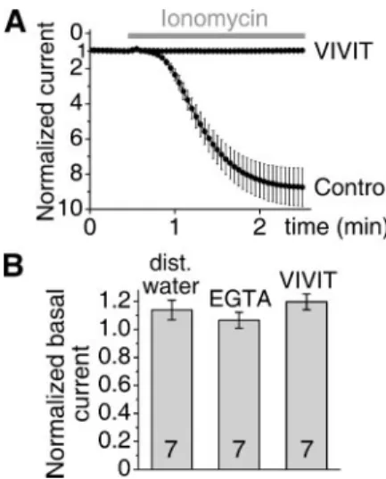
Microinjection of VIVIT Peptide Eliminates the Calcineurin-depend- ent Activation of TRESK—The sequence of VIVIT peptide (MAGPH- PVIVITGPHEE) was optimized for the highest affinity toward the NFAT binding site of calcineurin (23). To provide a furtherin vivo evidence for TRESK-calcineurin interaction in addition to TRESK mutants, we also attempted to block the interaction by saturating the NFAT binding site of calcineurin with VIVIT peptide.Xenopusoocytes expressing wild type TRESK were microinjected with the peptide and subsequently challenged with ionomycin. These oocytes failed to respond with TRESK activation to the stimulation (Fig. 3A). The effec- tive inhibition indicated that VIVIT peptide occupied the NFAT-dock- ing site of calcineurin and thus prevented the binding of the intracellular
loop of TRESK, which would have normally interacted with the same region of the phosphatase.
The dependence of TRESK regulation on calcineurin raised the question whether the basal K⫹current was also the result of the calcineurin activity in the resting cell. To suppress the resting cal- cineurin activity by reducing the cytoplasmic calcium concentration to subphysiological levels, EGTA was microinjected to the oocytes expressing TRESK, and the background K⫹current was measured before and after the microinjection. It was reported previously (3) that the 50-nl injection of a high (50 mM) concentration of EGTA was sufficient (even after the estimated 10-fold dilution of the che- lator in the cytoplasm) to eliminate the calcium-dependent TRESK activation. However, an identical EGTA injection failed to influence unstimulated TRESK (Fig. 3B), indicating that a calcium-dependent calcineurin activity did not contribute to the maintenance of the basal TRESK current. As an alternative approach to extinguish the possible basal calcineurin action on TRESK independently from cal- cium, the TRESK-interacting surface of calcineurin was blocked with VIVIT peptide. The microinjection of VIVIT peptide did not reduce the basal TRESK current (Fig. 3B), also confirming that calcineurin was not required for the resting TRESK activity.
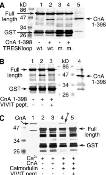
The PQIVID Motif of TRESK Binds Constitutively Active Calcineurin Permanently and Wild Type Calcineurin Calcium/Calmodulin-depen- dently—To demonstrate the direct binding of the PQIVID motif of TRESK to calcineurin, GST pulldown experiments were performed with a GST-TRESKloop-TAPtag fusion protein (see “Experimental Pro- cedures”), immobilized on glutathione-agarose beads. The constitu- tively active form of calcineurin (A subunit lacking the autoinhibitory domain and the calmodulin binding site but complexed with B subunit) was incubated with this resin. The characteristic band of the truncated calcineurin A could be easily detected with Coomassie Blue staining (comparelanes 1and2in Fig. 4A) if all proteins bound to the resin were
FIGURE 3.Microinjection of the VIVIT peptide eliminates the calcium-dependent activation of TRESK but does not influence the current of the resting channel.A, two groups of oocytes expressing wild type TRESK were microinjected with 50 nl of VIVIT peptide (10 mM,n⫽5) or distilled water (Control,n⫽5), respectively. The oocytes were stimulated with ionomycin (0.5M) 23–104 min after the microinjection, as indicated by thegray bar. TRESK currents were normalized to the resting value at the beginning of the measurement. The activation in the two groups was statistically different at the end of the measurement (p⬍0.0001, Student’sttest for independent samples).B, oocytes expressing wild type TRESK were microinjected with 50 nl of distilled water, high con- centration of calcium chelator (50 mMEGTA, 50 mMHEPES (pH 7.3 with KOH)) or VIVIT peptide (10 mM). TRESK currents of each oocyte were measured (at⫺100 mV by chang- ing EC [K⫹] from 2 to 80 mMand back) before and 54 –110 min after the microinjection.
The current measured after the injection was normalized to the initial value, and the averages of the normalized currents of seven cells were plotted for each microinjected solution. The effects of EGTA and VIVIT peptide microinjection on the basal TRESK cur- rent were not significantly different from that of distilled water (ANOVA).
analyzed on SDS-PAGE. To verify that calcineurin actually bound to the PQIVID sequence, the experiment was carried out with the PQAVAD mutant version of GST-TRESKloop-TAPtag under identical condi- tions. In good accordance with the results obtained in the oocyte exper- iments, the PQAVAD mutant GST-TRESKloop-TAPtag did not bind the constitutively active calcineurin at all (comparelanes 3and4in Fig.
4A). Moreover, the addition of VIVIT peptide (200M) to the binding reaction of the “wild type” (PQIVID) GST-TRESKloop-TAPtag also eliminated the association with the constitutively active calcineurin (Fig. 4B). These experiments indicate unequivocally that calcineurin binds directly and specifically to the PQIVID motif of TRESK.
To gain further insight into the regulation of TRESK-calcineurin inter- action, we also examined the binding of the full-length version of the phos- phatase to the PQIVID motif. Since the calcineurin purified from bovine brain has a larger molecular weight than its truncated recombinant coun- terpart, the smaller GST-TRESKloop protein could be used in this pull- down experiment. Apparently, full-length calcineurin bound to GST- TRESKloop more strongly in the presence of the calcium/calmodulin complex, and the specificity of this binding was also verified by the inhibi- tory effect of VIVIT peptide (Fig. 4C). Thus the affinity of wild type cal- cineurin to the PQIVID motif is enhanced by the activation of the phospha- tase, and calcineurin may be recruited to TRESK during the physiological activation by the calcium/calmodulin complex.
The Calcineurin Autoinhibitory Peptide Diminishes TRESK Activa- tion but Does Not Interfere with the Binding of Calcineurin to the PQIVID Motif—As the binding of calcineurin to the PQIVID motif was indispensable for TRESK activation, the question emerged whether this binding was sufficient on its own or whether the phosphatase activity of calcineurin was also required for the regulation. To suppress the phos- phatase activity without occluding the protein-protein interaction, a peptide corresponding to the autoinhibitory domain (467– 491 amino acids) of calcineurin was microinjected. This microinjection inhibited the ionomycin-induced activation of wild type TRESK (Fig. 5A), sug- gesting that the phosphatase activity of endogenous calcineurin was required for TRESK activation.
To demonstrate that the autoinhibitory peptide did not influence the binding of calcineurin to the PQIVID motif, recombinant constitutively active calcineurin was used. We reported previously that the coexpres- sion of the truncated calcineurin A subunit (1–398 amino acids, devoid of the autoinhibitory domain) and full-length calcineurin B with TRESK resulted in permanent activation of the channel (3). Inhibition of the phosphatase activity by the reintroduction of the autoinhibitory domain by peptide microinjection substantially reduced the otherwise perma- nently stimulated TRESK current (Fig. 5B). On the other hand, the autoinhibitory peptide, blocking only the active site of the phosphatase, did not influence the binding of the constitutively active calcineurin to the PQIVID motif in our GST pulldown experiments (comparelanes 3 and4in Fig. 5C). The reduction of TRESK current by the selective inhibition of the phosphatase activity supports the conclusion that dephosphorylation is also required for TRESK regulation in addition to the binding of calcineurin to the PQIVID motif.
FIGURE 4.Calcineurin directly binds to the PQIVID motif of TRESK.A, GST pulldown assays were performed with the wild type (PQIVID) GST-TRESKloop-TAPtag (wt.) or the PQAVAD mutant (m.) version of the fusion protein in the presence or absence of consti- tutively active calcineurin, as indicated at thebottomof the panel. In addition to TRESK loop fusion protein (Full length), the expression resulted in additional bands, represent- ing incomplete translation or bacterial protein degradation. One of these bands, slightly larger than GST itself, was especially prominent, and it was marked asGST. The truncated calcineurin A subunit (CnA1–398, 2.5g,lane 5) was also indicated by anarrow. Note the pulldown of calcineurin inlane 2but not inlane 4.B, binding of the constitutively active calcineurin to GST-TRESKloop-TAPtag was tested in the presence or absence of VIVIT peptide (pept.) (200M), as indicated at thebottomof the panel. Inlane 4, only constitu- tively active calcineurin (2.5g) was loaded. The same bands as inAare indicated with arrows. The lane of the markers was split, to introduce size labels. Note the pulldown of calcineurin inlane 2but not inlane 3.C, GST pulldown experiments were performed with the wild type (PQIVID) GST-TRESKloop fusion protein, in the presence of calcium and in the presence or absence of wild type calcineurin, calmodulin, and VIVIT peptide (200M), as indicated at thebottomof the panel. The full-length and incomplete fusion protein (GST) and wild type calcineurin A (CnA, 0.5g,lane 1) are marked witharrows. Note the binding of calcineurin inlane 4(also indicated with anarrowandasterisk). (The repre- sentative gels inAandBwere stained with Coomassie Blue and inCwith silver.)
FIGURE 5.The microinjection of calcineurin autoinhibitory peptide (autoinh. pept.) reduces TRESK activation, but the peptide does not prevent the binding of cal- cineurin to the PQIVID motif.A, two groups of oocytes expressing wild type TRESK were microinjected with 50 nl of calcineurin autoinhibitory peptide (10 mM,n⫽4) or distilled (dist.) water (Control,n⫽4), respectively. The oocytes were stimulated with ionomycin (0.5M, as indicated by thegray bar) 99 –158 min after the microinjection.
TRESK currents were normalized to the resting value at the beginning of the measure- ment. The activation in the two groups was statistically different at the end of the meas- urement (p⬍0.05, Student’sttest for independent samples).B, wild type TRESK was coexpressed with constitutively active calcineurin (truncated A subunit (amino acids 1–398) together with the full-length calcineurin B subunit; triple coexpression). Two groups of oocytes, hereby expressing permanently stimulated TRESK channels, were microinjected with 50 nl of calcineurin autoinhibitory peptide (10 mM,n⫽8) or distilled water (n⫽7), respectively. TRESK current was measured before and 51–101 min after the injection as detailed in Fig. 3B. The microinjection of the autoinhibitory peptide dimin- ished significantly the K⫹current, compared with the control water injection (p⬍10⫺6, Student’sttest for independent samples).C, binding of the constitutively active cal- cineurin (CnA1–398) to GST-TRESKloop-TAPtag was tested in the presence or absence of calcineurin autoinhibitory peptide (200M), as indicated at thebottomof the panel. In lane 1, only constitutively active calcineurin was loaded. The same bands as in Fig. 4Bare indicated witharrows. Note the pulldown of calcineurin in bothlanes 3and4.
ing motif does not resemble the sites dephosphorylated by calcineurin at all and it has been shown in the case of NFAT proteins that a surface of the phosphatase, other than the substrate binding region, connects to PXIXIT motifs (28). Interestingly, our GST pulldown experiments indi- cated that the regulation of calcineurin activity essentially influenced TRESK-calcineurin interaction. The constitutively active calcineurin devoid of the autoinhibitory domain bound to the PQIVID motif per- manently. In contrast, the binding of the wild type enzyme required (or at least it was enhanced drastically by) the presence of calcium/calmod- ulin complex. This substantial change of affinity may result in the shut- tling of calcineurin between the cytoplasm and the PQIVID site during the calcium signal.
Beyond thein vitroresults, we provided compelling evidence that the association of the two proteins is indispensable for the regulation of TRESK inXenopusoocytes. The channel was activated efficiently by endogenous calcineurin, and the activation evolved even if TRESK was expressed by injecting only a trace amount of cRNA (data not shown).
Thus TRESK-calcineurin interaction developed without the overex- pression of the interacting partners. Identification of the well defined NFAT-like motif also made it very unlikely that TRESK and calcineurin interacted only by chance in our heterologous expression system. The intracellular loops of the human and mouse TRESK orthologues show only limited similarity. Still, the PQIVID motif aligns exceptionally well to the PQIIIS sequence of human TRESK, and the two channels are activated identically by the calcium signal, indicating that the binding mechanism of calcineurin is evolutionary conserved.
In the present medical practice of organ transplantation, the phos- phatase activity of calcineurin is inhibited for immunosuppression. To eliminate the side effects of these treatments, significant effort has been made to develop compounds, which inhibit NFAT activation but not the phosphatase activity of calcineurin in general. In addition to VIVIT peptide (23), a family of small molecule inhibitors blocking the NFAT binding site of calcineurin has also been discovered (12). This novel class of inhibitors (and possibly future drugs) is also expected to prevent
REFERENCES
1. Kim, D. (2005)Curr. Pharm. Des11,2717–2736
2. Talley, E. M., Sirois, J. E., Lei, Q., and Bayliss, D. A. (2003)Neuroscientist9,46 –56 3. Czirja´k, G., To´th, Z. E., and Enyedi, P. (2004)J. Biol. Chem.279,18550 –18558 4. Sano, Y., Inamura, K., Miyake, A., Mochizuki, S., Kitada, C., Yokoi, H., Nozawa, K.,
Okada, H., Matsushime, H., and Furuichi, K. (2003)J. Biol. Chem.278,27406 –27412 5. Keshavaprasad, B., Liu, C., Au, J. D., Kindler, C. H., Cotten, J. F., and Yost, C. S. (2005)
Anesth. Analg.101,1042–1049 and table of contents
6. Liu, C., Au, J. D., Zou, H. L., Cotten, J. F., and Yost, C. S. (2004)Anesth. Analg.99, 1715–1722 and table of contents
7. Kang, D., Mariash, E., and Kim, D. (2004)J. Biol. Chem.279,28063–28070 8. Kang, D., and Kim, D. (2006)Am. J. Physiol., in press
9. Clipstone, N. A., Fiorentino, D. F., and Crabtree, G. R. (1994)J. Biol. Chem.269, 26431–26437
10. Snyder, S. H., and Sabatini, D. M. (1995)Nat. Med.1,32–37
11. Martinez-Martinez, S., and Redondo, J. M. (2004)Curr. Med. Chem.11,997–1007 12. Roehrl, M. H., Wang, J. Y., and Wagner, G. (2004)Biochemistry43,16067–16075 13. Liu, J. O. (2003)Biochem. Biophys. Res. Commun.311,1103–1109
14. Dodge, K. L., and Scott, J. D. (2003)Biochem. Biophys. Res. Commun.311,1111–1115 15. Mondragon, A., Griffith, E. C., Sun, L., Xiong, F., Armstrong, C., and Liu, J. O. (1997)
Biochemistry36,4934 – 4942
16. Clipstone, N. A., and Crabtree, G. R. (1992)Nature357,695– 697
17. Farazi, T. A., Manchester, J. K., and Gordon, J. I. (2000) Biochemistry 39, 15807–15816
18. Rudnick, D. A., McWherter, C. A., Adams, S. P., Ropson, I. J., Duronio, R. J., and Gordon, J. I. (1990)J. Biol. Chem.265,13370 –13378
19. Heukeshoven, J., and Dernick, R. (1988)Electrophoresis9,28 –32
20. Czirja´k, G., Fischer, T., Spa¨t, A., Lesage, F., and Enyedi, P. (2000)Mol. Endocrinol.14, 863– 874
21. Czirja´k, G., Petheo˝, G. L., Spa¨t, A., and Enyedi, P. (2001)Am. J. Physiol.281, C700 –C708
22. Yoshida, S., and Plant, S. (1992)J. Physiol.(Lond.)458,307–318
23. Aramburu, J., Yaffe, M. B., Lopez-Rodriguez, C., Cantley, L. C., Hogan, P. G., and Rao, A. (1999)Science285,2129 –2133
24. Klee, C. B., Ren, H., and Wang, X. (1998)J. Biol. Chem.273,13367–13370 25. Boustany, L. M., and Cyert, M. S. (2002)Genes Dev.16,608 – 619
26. Heath, V. L., Shaw, S. L., Roy, S., and Cyert, M. S. (2004)Eukaryot. Cell3,695–704 27. Kim, S. J., Ding, W., Albrecht, B., Green, P. L., and Lairmore, M. D. (2003)J. Biol.
Chem.278,15550 –15557
28. Li, H., Rao, A., and Hogan, P. G. (2004)J. Mol. Biol.342,1659 –1674