PHYSIOLOGICAL and
REPRODUCTIONAL ASPECTS OF ANIMAL PRODUCTION
Ferenc Husvéth
PHYSIOLOGICAL and REPRODUCTIONAL ASPECTS OF ANIMAL PRODUCTION
Ferenc Husvéth Publication date 2011
Table of Contents
Cover ... xi
1. NEUROENDOCRINE ADAPTATIONS IN DOMESTIC ANIMALS ... 1
1. Hypothalamo-pituitary axis ... 1
2. Hormones of the anterior pituitary ... 2
3. Posterior pituitary and its hormones ... 3
4. Thyroid as the endocrine gland for control of animal production and adaptation for cold environmental conditions ... 3
5. Adrenal gland as the organ important in adaptation to stress and maintain homeostasis ... 5
6. Pineal gland as the organ of the control of diurnal rhythm ... 7
7. Leptin ... 7
8. Self evaluation questions ... 8
2. COMPARATIVE STRUCTURE AND FUNCTION OF THE GASTROINTESTINAL TRACT OF ANIMALS ... 9
1. Self evaluation questions ... 11
3. DIGESTION IN MONOGASTRIC MAMMALS ... 12
1. Regulation of feed intake ... 12
2. Role of the salivary glands ... 12
3. Digestion in the monogastric stomach ... 13
4. Physiology of the small intestine, exocrine pancreas and liver ... 16
5. Digestive processes in the large intestine ... 18
6. Defecation ... 19
7. Self evaluation questions ... 19
4. DIGESTION IN THE RUMINANT STOMACH ... 20
1. Development of the ruminant stomach ... 21
2. Microbial digestion in the forestomachs of ruminants ... 22
3. Gases ... 26
4. Forestomach motility ... 26
5. Abomasal function ... 27
6. Self evaluation questions ... 27
5. PHYSIOLOGY OF AVIAN DIGESTION ... 28
1. Anatomy of the alimentary canal ... 28
2. Regulation of food intake ... 30
3. Secretion and digestion ... 30
4. Gastrointestinal cycle ... 32
5. Rate of passage ... 32
6. Self evaluation questions ... 32
6. GASTROINTESTINAL ABSORPTION AND INTERMEDIARY METABOLISM OF CARBOHYDRATES ... 34
1. Absorption in the small intestine ... 34
2. Absorption and metabolism of carbohydrates ... 35
3. Self evaluation questions ... 38
7. INTESTINAL ABSORPTION AND INTERMEDIARY METABOLISM OF PROTEINS ... 39
1. Absorption in the small intestine ... 39
2. Protein metabolism ... 39
3. Self evaluation questions ... 43
8. INTESTINAL ABSORPTION AND INTERMEDIARY METABOLISM OF FATS ... 44
1. Absorption of lipids ... 44
2. Transport of lipids via the circulatory system ... 45
3. Lipid metabolism ... 45
4. Self evaluation questions ... 47
9. GASTRIC ABSORPTION AND INTERMEDIARY METABOLISM OF SHORT CHAIN (VOLATILE) FATTY ACIDS AND AMMONIA IN RUMINANTS ... 48
1. Absorptive surface of the forestomachs in ruminants ... 48
2. Short chain fatty acids ... 49
3. Ammonia ... 50
4. Relationships between short chain fatty acids and glucose in ruminants ... 50
PHYSIOLOGICAL and REPRODUCTIONAL ASPECTS
OF ANIMAL PRODUCTION
5. Hypoglycemyc ketosis ... 51
6. Self evaluation questions ... 52
10. CONTROL AND MAND MANIPULATION OF ANIMAL GROWTH ... 53
1. Growth hormone and growth in meat producing animals ... 53
2. Self evaluation questions ... 59
11. REGULATION OF REPRODUCTION IN MALE MAMMALS ... 60
1. Endocrine control ... 60
2. Spermatogenesis ... 61
3. Release of spermatozoa into the lumen of seminiferous tubules ... 62
4. Daily sperm production ... 63
5. Testicular size ... 64
6. Spermatozoal viability ... 64
7. Ejaculation ... 65
8. Reproductive behaviour ... 66
9. Role of olfactory and vomeronasal system in reproductive behaviour ... 67
10. Self evaluation questions ... 67
12. REGULATION OF REPRODUCTION IN FEMALE MAMMALS ... 69
1. Reproductive cyclicity ... 69
2. Phases of the estrous cycle ... 69
3. Hormonal control and follicular development ... 72
4. Ovulation ... 75
5. Corpus luteum ... 77
6. PREGNANCY ... 78
7. Fertilization ... 78
8. Implantation and placentation ... 82
9. Hormones of pregnancy ... 84
10. Pregnancy diagnosis ... 85
11. Self evaluation questions ... 86
13. REPRODUCTION IN BIRDS (PHYSIOLOGY OF EGG PRODUCTION) ... 87
1. Female reproductive system ... 87
2. Egg formation and oviposition ... 88
3. Reproduction and Photoperiods ... 90
4. Self evaluation questions ... 90
14. MAMMARY GLAND AND LACTATION ... 91
1. Functional anatomy of the mammary gland ... 91
2. PHYSIOLOGY OF LACTATION ... 93
3. Lactation performance ... 98
4. Composition of milk ... 99
5. Milk secretion ... 101
6. Milk ejection or let down ... 102
7. Colostrum ... 103
8. Self evaluation questions ... 104
15. REFERENCES ... 105
List of Figures
1.1. Fig. 1.1. The connections between the hypothalamus and hypophysis (Husveth, 2000). A:
Neurosecretory products from hypothalamic nuclei are transported to the hypophysis and from there transferred to the general circulation. B: Neurosecretory products are transported from axons of
hypothalamic neurosecretory cells to the adenohypophysis via a portal circulation. (1) Optic chiasma, (2)
hypothalamic nuclei, (3) hypophysial stalk, (4) neurohypophysis, (5) adenohypophysis. ... 1
1.2. Figure 1.2.: Thyroid gland of domestic animals; A) horse, B) cattle, C) pork, D) dog (Fehér, 1980) 3 1.3. Figure 1.3.: Thyroid hormone response to TRH. Lactating cows were injected with TRH (25µg/100 kg body weight) and blood collected to monitor changes in T3 and T4. Secretion of both hormones was reduced in winter (Perera et al., 1985). ... 4
1.4. Figur1.5..: Cross section of the adrenal gland from the horse ... 5
1.5. Figure 1.6.: Concentrations (µg/ml) of adrenaline and noradrenalin in a racing horse (Marlin and Nankervis (2008) ... 6
1.6. Figure 1.7.: Processes of stress reaction in animals ... 6
2.1. Figure 2.1.: Gastro-intestinal tract of two carnivores.(Reece, 2004) ... 9
2.2. Figure 2.2.: Gastro-intestinal length of the sheep and horse (Reece, 2004). ... 10
3.1. Fig. 3.1.: Different glandular regions in the gastric mucosa in the pig. ... 13
3.2. Figure 3.2.: Gastric glands. The histological section of a gastric gland shows the gastric pit, the entrance to the gastric gland. Within the gland area neck cells that secret, mucous, parietal cells that secrete HCl, chief cells that secrete pepsinogen, and enteroendocrine cells that secrete hormone. G cells, which secrete gastrin, are an example of enteroendocrine cells. Once released into the stomach, pepsinogen is converted to pepsin by the action of HCl and pepsin. (Akers and Denbow, 2008). ... 14
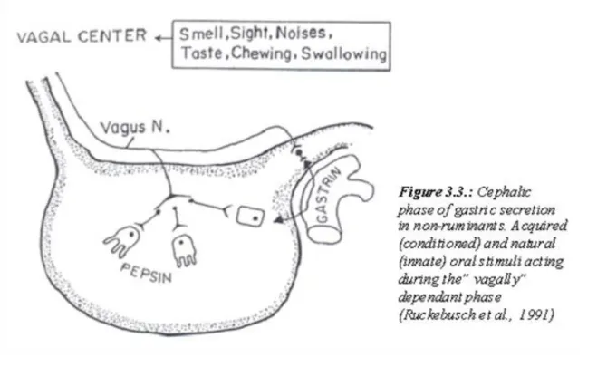
3.3. Figure 3.3.: Cephalic phase of gastric secretion in non-ruminants. Acquired (conditioned) and natural (innate) oral stimuli acting during the” vagally” dependant phase (Ruckebusch et al., 1991) ... 14
3.4. Figure 3.4.: Gastric phase of gastric juice secretion in non-ruminants. Distension of the stomach and ingesta directly stimulate cholinergic-mediated receptors in the oxyntic and pyloric areas and also include a” vagovagal” reflex. These short and long reflexes increase the output of gastrin by G cells, which reinforces the stimulation of the oxyntic cells. Histamine and acetylcholine share the same effect. Ruckebusch, et al., 1991) ... 15
3.5. Figure 3.5.: Intestinal phase of gastric secretion in non-ruminants. Stimulation of oxyntic cells by gastrin and peptic cells by secretin, both from the duodenum. Inhibition of gastrin effect is brought by other endocrines (GIP, CCK, secretin) and by excessive acidity or osmolality of the chime emptied into the duodenum. (Ruckebusch et al., 1991) ... 15
3.6. Figure 3.6.: Acinar cells of the pancreaton secrete enzymes, zymogens, and electrolytes. Proximal ductule cells produce most of the bicarbonate. At low secretion rates, bicarbonate is exchanged for chloride in the distal duct cells. (Ruckebusch, 1991) ... 16
3.7. Figure 3.7.: Neural and hormonal events during intestinal phase of exocrine pancreatic secretion (Ruckebusch, 1991). ... 17
4.1. Figure 4.1.: A schematic diagram of a ruminant stomach, viewed from the right side. 1) Dorsal sac of the rumen, 2) ventral sac of the rumen, 3) reticulum, 4) omasum, 5) abomasums, 6) esophagus, 7)ruminoreticular fold, 8) cranial sac of the rumen, 9) right accessory longitudinal groove, 10) longitudinal groove, 11) islet on the right longitudinal fold, 12) caudodorsal blind sac, 13) caudoventral blind sac, 14) incision (border) between cranial and caudal blind sacs, 15)dorsal-, and 16) ventral coronary pillar, 17) greater-, and 18) lesser curvature of abomasums, 19) pyloric region. ... 20
4.2. Figure 4.2.: Mucosa layer in the rumino-reticulum in calfs. A) fed only on milk, B ) grazing calf. 22 4.3. Figure 4.3.: Scanning electron micrograph of Diplodinium anisacanthum protozoa with attached bacteria (Ogimoto and Imai, 1981). (Rumen is essentially an open ecosystem with great diversity of microbes) ... 22
4.4. Figure 4.4.: Complex food web of diverse bacterial species involved in carbohydrate fermentation. H: an electron plus a proton or electrons from reduced-pyridine nucleotides; A: carbohydrate-fermenting species; B: methanogen species; C: lactate-fermenting species, which often also ferment carbohydrates (Reece et al., 2004). ... 23
4.5. Figure 4.5.: Transformation of nitrogenous substances in the rumen. Ammonia is produced during the microbial metabolism of diverse substrates and is a major source of the nitrogen used for the biosynthesis of microbial proteins. ... 24
PHYSIOLOGICAL and REPRODUCTIONAL ASPECTS
OF ANIMAL PRODUCTION
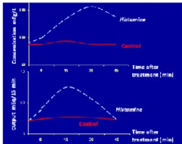
5.1. Figure 5.1.: Stomach and intestinal canal of the fowl and pigeon. a., oesophagus; b., proventriculus;
c., gizzard; d. and d‟., descending and ascending limbs of the duodenum; e., jejunum with the
supraduodenal loop;(e‟), e”., Meckels diverticulum; f., ileum; g., and g‟., left and right caeca; g”., cervical part, g‟‟‟., main part and gIV., tip of the caecum; h., colon; i., cloaca; i‟. anus; k., oviduct; l., ureter; m., spleen; n., dorsal and n‟., ventral lobes and n‟‟., splenic lobe of the pancreas. 1., left and 1‟. right ductus hepatoentericus (pigeon); 1‟‟., ductus hepatoentericus and 1‟‟‟., ductus cysticoentericus (fowl); 2., ventral pancreatic ducts; 2‟., dorsal pancreatic duct; 3., lig. ... 28 5.2. Figure 5.2.: Transverse section through proventriculus showing surface epithelium and glandular alveoli (Bell and Freeman, 1971) ... 28 5.3. Figure 5.3.: Average acid output and concentration in gastric secretion collected from control and histamine stimulated conscious chickens. (Long , 1967) ... 31 6.1. Figure 6.1.: Schematic structure of the intestinal wall with the absorptive surface (villi). Ruckebusch (1991) ... 34 6.2. Figure 6.2.: Cumulative Percentage of Glucose and Starch Disappearance from the Intestine of 7- week-old Chicks. Glucose absorption from glucose monohydrate as the sole dietary carbohydrate source (o), starch digestion (∆), and absorption (●) in chicks fed starch as the sole carbohydrate source (Whittow, 2000). ... 35 7.1. Fig 7.1. General mechanism for macromolecules uptake and transport by the intestine (Reece, 2004) 39
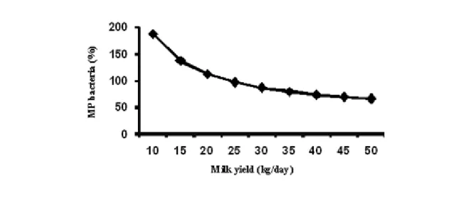
7.2. Fig. 7.2. Total amount of microbial protein (MP bacteria) synthesized in the rumen expressed as the
% of the requirement for milk production ... 40 7.3. Fig 7.3. When sheep are artificially nourishes (A): by intraabomasal infusion of enriched casein (BV=1.0), urinary nitrogen excretion is almost constant regardless of the quantity of protein administered;
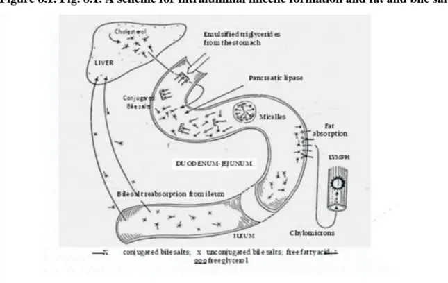
and by (B): intraabomasal infusion of gelatine (BV=0.07), urinary nitrogen excretion increases with the quantity of protein administered. ... 42 8.1. Fig. 8.1. A scheme for intraluminal micelle formation and fat and bile salts ... 44 8.2. Fig. 8.2. Molecular structure of chylomicron (Lehninger et al, 2000). ... 44 8.3. Fig.8.3. A scheme for the uptake of triglycerides from plasma chylomicrons and very-low-density lipoproteins by adipose tissue; FFA=free fatty acids; VLDL TG=very-low-density lipoprotein
triglycerides; ECF=extracellular fluid (Johnson and Davenport, 1971) ... 45 8.4. Figure 8.4:. Slices of liver showing fatty accumulation (fatty liver) in a dairy cow; (A) macroscopic (B) microscopic histology (showing large lipid vacuoles) ... 47 9.1. Figure 9.1.: Absorptive surface for short chain fatty acids (SCFA) in the rumen ... 48 9.2. Figure 9.2:. Absorption of acetate through the rumen wall and its effect on CO2 és HCO3- concentrations in rumen content ... 49 9.3. Figure 9.3.: Structural formula and physical properties of lactic acid (MP=melting point, BP=boiling point, DST=density) ... 50 9.4. Figure 9. 4. Major metabolic pathways in the ruminant liver (Reece, 2009) ... 51 9.5. Figure 9.5.: Synthesis of the common ketone bodies (acetoacetate, β-hydroxybutyrate, acetone);
Reece (2004) ... 51 10.1. Figure 10.1.: Hypophysectomy in twin calves at age d 21(Kemény, 1974). ... 53 10.2. Figure 10.2.: The amino acid sequence of bovine growth hormone (Wallis, 1978). ... 53 10.3. Figure 10.3.: The plasma GH profile of four young intact rams. Secretory episodes are seen in each profile (Davis et al., 1977) ... 55 10.4. Figure 10.4.: Live weight gains in lambs immunized against somatostatin (SRIF) (Spencer, 1986).
56
10.5. Figure 10.5.: Mean serum GH response of three lambs (45 kg) administered ovine GRF-44 by ... 56 10.6. Figure 10.6.: Construction of a recombined DNA-derived human growth hormone (Goeddel et al, 1979). ... 57 10.7. Figure 10.7.: Enhancement of bovine GH activity in dwarf mice by site directed antiserum. The growth response of hypopituitary dwarf mice to injected GH is associated with incorporation of 35-S labelled sulphate into costal cartilage (Aston et al., 1991). ... 58 10.8. Figure 10.8.: Enhancement of total carcass protein in lambs following passive and active vaccination against region 134-154 of GH (STH); Astonet al.(1991). ... 58 10.9. Figure 10.9.:The somatomedin hypothesis. Relationships between secretion of pituitary GH and liver IGF-I are illustrated by the solid black arrows. Dashed red arrows indicate direct effects of GH,
significance of local tissue production of IGF-I, and the role of IGF-I-binding proteins (IGFBP‟s) to control biological activity of IGF-I. ... 58
PHYSIOLOGICAL and REPRODUCTIONAL ASPECTS
OF ANIMAL PRODUCTION
10.10. Figure 10.10.: Effect of insulin and insulin-like growth factor (IGF) on protein synthesis in ovine embryonic myotub (structural compound of muscles) cultures; Hembree et al.(1991). ... 59 11.1. Figure 11.1: Relationships between GnRH, LH and FSH in Male (Senger, 2003). GnRH causes the release of LH and FSH. Episodes of all three hormones occur between 4 and 8 times in 24 hours. The lower FSH profile, when compared to LH, is due to inhibin secretion by Sertoli cells. Also, the greater duration of the FSH episode is probably due to its longer half-life (100 min) when compared to LH (30 min). ... 60 11.2. Figure 11.2: Scanning electron micrograph of testicular parenchyma in stallion. Senger (2003).
Seminiferous tubules (ST) containing developing germ cells (GS) are surrounded by basal membrane (BM). Flagella (F) from developing spermatids can be observed protruding into the lumen of some tubules. The interstitial compartment contains Leydig cells (LC), blood vessels (BV) and connective tissue (CT). ... 60 11.3. Figure 11.3: Interrelationships among hormones produced by Sertoli cells, Leydig cells, the hypothalamus and anterior lobe of pituitary. T = testosterone, E2 = estrogen, DHT = dihydrotestosterone (Senger, 2003). ... 61 11.4. Figure 11.4: Comparison of the spermatozoa of farm animals and other vertebrates (Frandson et al., 2009) ... 62 11.5. Figure 11.5: Associations of developing germ cells that represent various stages of the cycle of the seminiferous epithelium („gonia=spermatogonium, 1º cyte=primary spermatocyta, 2ºcyte=secondary spermatocyte, „Tid=immature spermatid, „Tid-m=mature spermatid). At any given cross-sectioned location along a seminiferous tubulus, one can observe different stages of the cycle of the seminiferous epithelium. In this example we can see three stages (I, IV, and VIII); (Senger, 2003). ... 63 11.6. Figure 11.6.: The cycle of seminiferous epithelium is analogue to a university (Senger, 2003). 63 11.7. Figure 11.7: Characteristics of copulation, site of seminal deposition and number of ejaculations to satiation and exhaustion in the ram, bull, stallion and boar (Senger, 2003). ... 65 11.8. Figure1.8. : Flehmen response in the stallion and bull and the vomeronasal pathway (Senger. 2003) 67
12.1. Figure 12.1.: Types of estrous cycles as reflected by annual estradiol E2 profiles (Senger, 2003).
69
12.2. Fig 12.2.: Stages of estrous cycle (E2: estradiol); Senger (2003) ... 70 12.3. Figure 12.3: Development of an ovarian follicle from its primordial form to a Graafian follicle (Reece, 2009) ... 71 12.4. Figure 12.4.: Hormone patterns during estrus in mare McKinnon et al. (2011) ... 72 12.5. Fig. 12.5:. Rrelationships between the hypothalamus, the pituitary, and the ovary during the
follicular phase (Senger, 2003). AL=Anterior Lobe, E2=Estradiol, OC= Optic Ciasma, PL=posterior Lobe ... 72 12.6. Figure 12.6: GnRH release from the hypothalamic and surge centre (Senger, 2003) ... 73 12.7. Figure 12.7.: The “two cell,2- gonadrothropin model” (Senger 2003). Arrows indicate the stimulatory effects of E2 on physiological functions ... 74 12.8. Figure 12.8.: Ovary of sow with mature tertiary follicles just before ovulation (Frandson et al.,2009).
a) follicle just before ovulation; b) a site where a follicle has just ovulated; g) a surface vessel. ... 75 12.9. Figure 12.9.: Preovulatory LH surge in the blood. ... 76 12.10. Figure 12.10.: Functional corpus luteum. The corpus luteum is now a mixture of large luteal cells,LLC (formerly granulosal cells) and many small luteal cells, SCL (formerly thecal cells). In some cases, there is a remnant of the follicular antrum that forms a small cavity in the centre of the corpus luteum. (Senger 2003) ... 77 12.11. Figure 12.11.: Typical concentrations of progesterone in the systemic circulation of mares during the estrous cycle and early pregnancy. Black bar represents estrus.(McKinnon et al., 2011) ... 77 12.12. Figure 12.12.: Major sequence of events following deposition of spermatozoa in female tract.
UTJ=uterotubular junction (Senger, 2003) ... 78 12.13. Figure 12.13.: Conceptual version of mammalian capacitation (Senger, 2003) ... 79 12.14. Figure 12.14.: Sequence of events from initial binding of spermatozoon to zona pellucida to fusion between plasma membranes of spermatozoon and oocyte: A, spermatozoa arrive at site of fertilization; B, initial binding of spermatozoon to zona pellucida; C, acrosome reaction; D and E, penetration of zona pellucida; F, fusion of plasma membrane of spermatozoon and oocytes; G, the cumulus matrix.(Frandson et al., 2009) ... 80 12.15. Figure 12.15.: Schematic illustration of preattachment embryo development (Senger, 2003). 81 12.16. Figure 12.16.: Transuterine migration of the equine conceptus (embryo). Each black sphere represents a “stopping spot” in which the conceptus will spend between 5 and 20 minutes. The migration
PHYSIOLOGICAL and REPRODUCTIONAL ASPECTS
OF ANIMAL PRODUCTION
of the conceptus probably distributes pregnancy factors (white lines) over a wide surface of the endometrium; (Senger, 2003). ... 82 12.17. Figure 12.17.: Equine fetus in the placenta. The chorioallantois is the outer allantois plus the chorion. The chorion is associated intimately with the endometrium. The inner allantois is fused with the amnion. (Frandson et al., 2009) ... 82 12.18. Figure 12.18.: Epitheliochorial type of placenta (sow, mare, ewe, and cow). 1. Endometrial capillary; 2. endometrial interstitium; 3. chorionic epithelium; 4. chorionic interstitium; 5. chorionic capillary (Senger, 2003). ... 83 12.19. Figure 12.19.: Placental types classified by distribution of sites of exchange. A) Diffuse placenta.
B) Cotyledonary placenta, C) Zonary placenta. D) Discoid placenta. (Frandson et al., 2009). ... 83 12.20. Figure 12.20.: Placental attachment of cow, ewe, and mare. Villi from chorioallantois (black) invaginates into crypts in maternal uterine epithelium (stippled) in caruncles in the cow and the ewe and in diffuse locations in the mare (Frandson et al,2009). ... 84 12.21. Figure 12.21.: Luteal progesterone output during the first half of gestation in the mare. CL=corpus luteum, P4=progesterone, eCG=equine chorionic gonadotropin (Senger, 2003). ... 84 12.22. Figure 12.22.: Production of equine chorion gonadropin (eCG). Production is closely related to the weight of the endometrial cups. (Senger 2003). Endometrial cups (EC) are seen here in a U-shaped configurtion. The fetus (F) is surrounded by the amnion (not visible). The membrane indicated by arrows is the allantochorion. This specimen was removed from a mare at 50 days of gestation). ... 85 13.1. Figure 13.1.: Structures of female chicken reproductive tract (Akers and Denbow, 2008). The ovary with multiple oocytes in various stages of development is apparent. After ovulation, the egg progresses through segments of the oviduct, infundibulum, magnum, and isthmus. In this illustration, a mature egg after albumin deposition (soft egg), but before shell formation, is shown. The mature egg is subsequently laid after passage into the cloaca ... 87 13.2. Figure 13.2.: Plasma concentrations of progesterone, testosterone, and luteinizing hormone (LH) relative to time of ovulation. Mean ± error for six ovulations from five hens. Reece et al. (2004) .. 88 13.3. Figure 13.3.: Diagram of a hen‟s egg (Nickel et al., 1997) a-e) sphere of yolk; a)double layered yolk membrane; b) germinal disc; c) latebra; d) yellow yolk; e) white yolk; f) chalaza;g-i)egg albumen; g) inner, less viscous layer; k) double-layered shell membrane; l) air cell (chamber); m) calcium shell with pores and cuticula. ... 89 13.4. Figure 13.4.: The role of light in avian reproduction (Etches, 1993) ... 90 14.1. Figure 14.1.: Diversity in anatomic position, number and teat morphology among mammals (Singer (2003) ... 91 14.2. Figure 14.2.: Duct system of the bovine mammary gland: 1.,: lobule; 2., intralobular duct; 2‟., interlobular duct; 3., lactiferous ducts of various diameter; 4., lactiferous sinus; 5., gland sinus; 6., teat sinus; 7., papillary duct; 8., teat sphincter; 9., teat orifice; 10., parenchyma of gland; 11., skin. Dyce et al.
(1996). ... 91 14.3. Figure 14.3.: Transverse section of the pelvic floor and caudal quarters of the cow‟s udder 1., Pelvic symphysis; 2., symphysial tendon; 3., lateral suspensory laminae; 4., mammary lymph node; 5. medial suspensory laminae; 6., branch of external pudendal vein. Dyce (1996) ... 92 14.4. Figure 14.4.: An alveolus surrounded by blood vessels and myoepithelial cells in the mammary gland (Reece, 2004). ... 92 14.5. Figure 14.5.: Blood circulation to and from the udder. RA=right atrium; LV=left ventricle; CA=
caudal aorta; CVC=caudal vena cava; EIV=external iliac vein; EIA=external iliac artery; EPA=external pudendal artery; EPV=external pudendal vein; CMA&V=caudal mammary artery and vein;
CrMA&V=cranial mammary artery and vein; SAV=subcutaneous abdominal vein. (Reece et al., 2004 93
14.6. Figure 14.6.: Proliferation index of epithelial cells of mammary ducts for heifers treated with estradiol(E2) or progesterone (P4) or a combined of the two. Estradiol clearly increased DNA synthesis (Woodvard et al., 1993) ... 94 14.7. Figure 14.7.: Mammary Development in a heifer (Smith, 1968). A. Udder of heifer during estrus cycle. B. Udder of heifer pregnant five months. C. Udder of heifer pregnant eight months. ... 95 14.8. Figure 14.8.: Effect of lactogenic hormones on secretion of α-lactalbuminin in bovine mammary tissue (Akers and Denbow, 2008). Panel A demonstrates a concentration dependent increase in α- Lactalbuminin in response to addition of bovine prolactin (Prl). Panel B shows the additive effect of cortisol (C ) on Prl-induced α-lactalbumin secretion. ... 96 14.9. Figure 14.9.: The surge of prolactin accompanying parturition. Peripartal prolactin surge initiates lactation (Akers and Denbow, 2008). ... 97 14.10. Figure 14.10.: Lactation curves for typical lactating Holstein cows (A), a Holstein production record holder (B). Akers (2002 ... 98
PHYSIOLOGICAL and REPRODUCTIONAL ASPECTS
OF ANIMAL PRODUCTION
14.11. Figure 14.11.: Lactation curve in sows. Diagram illustrates milk production in sows that were machine milked 4 times per day. (Ackers and Denbow, 2008) ... 99 14.12. Figure 14.12.: Alveolus (left) showing cells partially filled with milk and (right) after the discharge of the milk (Smith, 1968) ... 101 14.13. Figure 14.13.: Secretion of milk lipids, milk proteins and lactose by epithelial cells lining the alveoli of the mammary glands. Proteins and lactose are together in secretory vesicles that are released by exocytosis (arrows). Frandson et al. (2009). ... 101 14.14. Figure 14.14.: The anatomy and physiology of milk ejection (Senger, 2003). The milk ejection mechanism is initiated by suckling (1). The teat contains sensory neurons and impulses from these neurons travel through afferent nerves (2) to the hypothalamus. Nerves in the paraventricular nuclei are stimulated, and the terminals in the posterior lobe of the pituitary (3) release oxytocin. Oxytocin then enters the blood and is delivered to the mammary gland (4). The target cells for oxytocin are the
myoepithelial cells that surround the alveolus. Contraction of the myoepithelial cells (5) causes milk to be
„squeezed” out of each individual alveolus into small ducts and then into lager ducts. The net effect of simultaneous contraction of the myoepithelial cells throughout the entire mammary gland is to deliver milk to the large ducts and the gland cistern so that it is available for removal by the neonate. .... 102
List of Tables
1. ... xi
Cover
PHYSIOLOGICAL and REPRODUCTIONAL ASPECTS OF ANIMAL PRODUCTION Author:
Ferenc Husvéth
Az Agrármérnöki MSc szak tananyagfejlesztése TÁMOP-4.1.2-08/1/A-2009-0010 projekt
Table 1.
Chapter 1. NEUROENDOCRINE ADAPTATIONS IN DOMESTIC ANIMALS
1. Hypothalamo-pituitary axis
For many years, the pituitary gland was considered the master endocrine gland this was because of the large numbers of hormones that it produces and their widespread physiological effects throughout the body. However, since negative feedback loops and secretions of hypothalamic hormones ultimately regulate secretion of the pituitary hormones, the question of master and servant is a real one. Regardless, the pituitary hormones are essential and critical important in control of animal function directly related to animal agriculture productivity, i.e., rate of growth, muscle development, reproduction, lactation and environmental adaptation. Secretion of the hormones of the anterior pituitary is tightly related to secretion of other hormones that are produced by cells located in the nuclei of the hypothalamus. Although these releasing hormones (or releasing inhibiting hormones in some case;) are produced only in very small amount, they are able to impact the activity of cells of the pars distalis because of a unique arrangement of blood vessels between hypothalamus and the anterior pituitary. This is called the hypothalamic-hypophyseal portal blood system. Simple stated, venous blood that drains from the hypothalamus mixes with arterial blood and passes to anterior pituitary before it goes into the general venous circulation. The importance of this special anatomic relationship was confirmed by animal experiments in the 1960s and 1970s that showed that placing a foil barrier between the hypothalamus and pituitary markedly inhibited the secretion of all of the anterior pituitary hormones except prolactin. Of course the pituitary gland must also receive oxygenated arterial blood. Arterial branches of the circle of Willis supply most of this blood.
The pituitary gland, or hypophysis (Fig. 1.1.), is located at the very base of the brain, in a depression of the sphenoid bone. It is divided into three divisions or lobes. The largest is the adenohypophysis or anterior pituitary. Much of the anterior lobe contains cords of closely compacted epithelial cells, which secrete many hormones of the more familiar pituitary hormones, i.e., STH (GH), Prl, FSH, etc. A smaller region of tissue, the pars intermediate lob, is sandwiched between the anterior pituitary and the second largest division called the posterior, pituitary or pars nervosa. This later region of the pituitary gland has a very different cellular structure than the anterior lobe. The cells of the region are, in fact, neurosecretory cell nerve endings and associated supporting cells. The hormones of the posterior pituitary are actually synthesized by cell bodies of the hypothalamus but released from the neural cells that populate the posterior pituitary.
Figure 1.1. Fig. 1.1. The connections between the hypothalamus and hypophysis (Husveth, 2000). A: Neurosecretory products from hypothalamic nuclei are transported to the hypophysis and from there transferred to the general circulation. B:
Neurosecretory products are transported from axons of hypothalamic neurosecretory cells to the adenohypophysis via a portal circulation. (1) Optic chiasma, (2) hypothalamic nuclei, (3) hypophysial stalk, (4) neurohypophysis, (5) adenohypophysis.
Negative feedback loops Secretion of most of the anterior pituitary hormones is controlled at multiple levels.
Control begins in the hypothalamus with the synthesis and secretion of the hypothalamic hormones (releasing hormones, RH; Table 1.) into the hypophyseal portal blood supply.
NEUROENDOCRINE ADAPTATIONS IN DOMESTIC
ANIMALS
Because these agents reach their target cells in the pars distalis with minimal dilution, they are very effective.
One means of regulations is to alter the rate at which these hypothalamic hormones are made. Changes in higher brain function that impact the hypothalamus also alter production many of these agents and thereby activity of the pituitary. The hypothalamus functions as a crucial interface between the nervous and endocrine system, where secondary information is integrated and used to regulate the endocrine output of the pituitary gland. Much of these information is related the status of the internal environment in question (e.g., extracellular fluid osmolality, blood glucose concentration, body temperature and metabolic rate). Release of anterior pituitary hormones can also be regulated more different negative feedback loops based on the blood concentrations of the hormones involved. The hormone produced by the target endocrine gland of a specific trophic hormone can act on (1) the hypothalamus to reduce the production of its releasing hormone and (2) anterior pituitary to reduce its release of the trophic hormones. The tropic hormones of the anterior pituitary may also reduce hypothalamic releasing hormone via a short negative feedback loop (acting between hypothalamus and anterior pituitary).
2. Hormones of the anterior pituitary
All of the pituitary hormones are peptide or protein hormones based on their chemical structure. Several hormones stimulate distant endocrine glands to increase production of their own hormones. These stimulatory hormones anterior pituitary hormones are often called trophic or tropic hormones. Given the importance of these hormones in regulation of growth, development, lactation reproduction and adaptation to various environmental conditions, it is apparent that these agents are critical to understanding and improving animal agriculture. Following hormones are secreted in the anterior pituitary: growth hormone (GH; also known as somatotropic hormone; STH), adenocorticotropic hormone (ACTH), thyroid-stimulating hormone (TSH), gonadotropich hormones (FSH and LH) and prolactin (Prl). As the regulatory roles of GH will be discussed in Chapter 10, and those of gonadotropic hormones and Prl in Chapters 11 and 12, here the physiological role of only TSH and ACTH will be discussed.
Thyroid-Stimulating Hormone (TSH)
TSH is a glycoprotein that shares a structure having an α and a β chain. This hormone TSH ultimately binds to receptors in the thyroid to promote the synthesis of colloid by thyroid gland cells and stimulates the release of thyroid hormones. Associated with these functions are the accumulation of iodine, organic binding of iodine, and formation of thyroxine with the thyroid gland.
Adrenocorticotropic hormone (ACTH)
ACTH is a 39-amino-acid peptide processed from a larger precursor molecule called pro-opiomelanocortin (POMC) In the corticotrophs of the anterior pituitary, the mRNA from the POMC gene directs the synthesis and processing of the transcript ti yield at least eight biologically active fragments. The ACTH primarily stimulates the secretion of glucocorticoids from the cortex of the adrenal gland. Two of the fragments are derived from ACTH. the first 13 amino acids of ACTH are β-MSH and the ACTH 18-19 corticotropin-like intermediate lobe peptide (CLIP). The larger precursor POMC appears in the intermediate lobe of the pituitary in many species and is associated with secretion of melanocyte-stimulating hormone (MSH). Biological effects of adrenal steroids will be considered in a subsequent section.
Although ACTH is the focus, a family of diverse peptides is derived from POMC. Secretion of ACTH is greatly impacted by neural factors and hormones. These agents ultimately modify the secretion of CRH, which, like GnRH, is secreted in an episodic pattern. In many species there is also a diurnal rhytm in the ACTH secretion pattern and therefore a diurnal secretion of adrenal steroids. Negative feedback loops, involving cortisol, are important in control of ACTH. One mechanism is sensitive to the rate of change in circulating glucocorticoids
NEUROENDOCRINE ADAPTATIONS IN DOMESTIC
ANIMALS
(fast feedback). These effects are mediated at both the pituitary and hypothalamus. Furthermore, a host of stress (pain, hypoxia, cold and heat exposure, etc.) can override the usual rhythmic secretion of ACTH. This is classically thought of as the part of the fight-or-flight reactions and the need for nutrient mobilization, and it is associated with more prolonged secretion of glucocorticoids.
3. Posterior pituitary and its hormones
The posterior lobe is an outgrowth of the hypothalamus and contains the terminal axons from two pairs of nuclei (supraoptic nucleus and paraventricular nucleus) located in the hypothalamus. These two nuclei synthesized antidiuretic hormone and oxytocin (neurosecretion), respectively, which are transported to the axon terminals in the posterior pituitary, where they are stored in secretory granules until released. An action potential, generated by the need for each of stored hormones, causes the release of the hormone and subsequent absorption in the blood, where it is distributed to the receptor cells. The hormones of the posterior pituitary are of peptide class, specifically nonapeptides (they contain nine amino acids).
Antidiuretic hormone
When an animal is given an overload of water, a period of dieresis ((increased output of dilute urine) occurs.
Diuresis can be prevented by the administration of antidiuretic hormone (ADH), also known as vasopressin.
If dehydration occurs (osmoconcentration), osmorecptors respond to the increased concentration by stimulating greater output of ADH by the axon terminals in the posterior pituitary. The target cells of the secreted ADH are the collecting tubulus and collecting ducts of the kidney. The presence of ADH renders the cells of the collecting tubules and ducts more permeable to water, and more water is absorbed from the tubular fluid so that the plasma osmolality decrease (Na+ concentration returns to normal) and the urine volume decreases (becomes more concentrated). ADH is therefore important for water conservation by animals. Other stimulators of ADH secretion include reduced blood volume, trauma, pain, and anxiety.
Oxytocin
The functional activity of oxitocin is related to the reproductive process, which include lactation (see Chapters 12-14). Oxytocin is released from the posterior pituitary as a result of neuroendocrine reflex. The act of suckling or similar teat stimulation causes release of oxytocin and subsequent milk letdown (ejection). Similarly, an estrogen-dominated myometrium such as is found at ovulation and at parturition, is more responsive to oxitocin, and greater contraction of the uterus results. Oxytocine release at these times is associated with appropriate stimuli and subsequent myometrial contraction, which assists in the transport of sperm to the oviduct at copulation and in the expulsion of the fetus at parturition.
4. Thyroid as the endocrine gland for control of animal production and adaptation for cold
environmental conditions
Thyroid gland (Fig. 1.2.) is composed of clusters of follicles whose internal surfaces are lined by a layer of simple cuboidal epithelial cells. Triiodothyronine (T3) and thyroxyne (T4) is synthesized by the follicular cells and stored as part of a larger protein (thyroglobulin within the luminal spaces is called colloid. The critical elements required for thyroid hormone synthesis are amino acid tyrosine and iodine. Thyroid hormones are the major regulators of basal metabolism increasing oxygen consumption and therefore heat production, because they
Figure 1.2. Figure 1.2.: Thyroid gland of domestic animals; A) horse, B) cattle, C) pork,
D) dog (Fehér, 1980)
NEUROENDOCRINE ADAPTATIONS IN DOMESTIC
ANIMALS
stimulate oxidative phosphorilation. This is called a calorigenic effect, a response especially useful when animals are exposed to cold stressful situations. Of course, increasing demands require nutrient fuel. Small amounts of thyroid hormones promote glycogen storage, but glycogenolysis is stimulated as concentrations rise.
Other effects include increased absorption of glucose and promotion of glucose uptake by cells. Thyroid hormones also impact lipid metabolism but especially lipolysis. Fitting their ability to increase metabolism, the thyroid hormones also enhance the effects of sympathetic nervous system stimulation. This is believed to be occurring by thyroid hormone stimulation of synthesis of β-adrenergic receptors in tissues that are targets for epinephrine (adrenalin) and norepinephrine (noradrenalin).
Other effects of thyroid hormones are evident during growth and development. For example, classic experiments showed that thyroxine causes differentiation of tadpoles into frogs. The effect is less drastic in mammals, but thyroide hormones are nonetheless essentially for normal development of nervous system.
Hypothyroidism or deficiency slows metabolic processes. In young animals development and growth is impaired, and in primates serious permanent failure of neural development produces mental retardation. Among domestic species, lactating dairy cows are typically hypothyroid so that peripherial deiodination of T4 to produce the more potent T3 is especially important to maintain function of many target cells tissues.
A common functional test is to measure concentrations of plasma T3 and T4 in response to inject of TSH or TRH. In correspondence with variations in metabolic rate with season, several data are available (Perera et al., 1985) showing secretion of T3 and T4 following TRH injections in cows during winter compared with summer.
These data indicate that there is an evident seasonal variation in the response to TRH in cows. Mean concentration of T3 and T4 before administration of TRH were lower in cows sampled during winter and response to TRH was reduced in the study mentioned above. These reductions likely reflect greater utilization of thyroid hormones to enhance thermogenesis during the winter (Fig 1.3.). Concentrations of T4 were about fiftyfold greater than T3. This is similar to other domestic animals and suggests that substantial T3 formation occurs outside of the thyroid gland by the deiodination of T. Tissues with high levels of deiodination enzymes include the liver and kidney. The mammary gland also expresses a deiodinase that increases with the onset of lactation and in response to other hormones known to stimulate milk production. This provides an enhanced local tissue concentration of mammary T3 available to stimulate metabolic activity to support high levels of milk production despite the fact that lactating cows are typically in hypothyroid state.
Given the importance of thyroid hormones in regulation of metabolism, it was only natural for animal scientists to consider whether administration of thyroid hormones might be used to improve metabolic rate to support enhanced growth or development. For example, involvement of the thyroid gland in maintenance of lactation has been known since reports in the early 1900s showing that milk yield was reduced in thyreoidectomized goats. By the 1930s it was shown that thyroidectomy of dairy cows reduced milk yield; and conversely that treatment with T4 increased milk yield by approximately 20%. Because T4 is also efficacious when fed, these reports stimulated much interest in the practical utilization of the hormone to increase milk production in cattle.
This became economically feasible by the relatively low cost manufacture of thyroxine and other thyroactive iodinated proteins. However, results of multiple studies showed that while feeding thyroxine (or iodinated proteins) increased milk production by 10-40%, the galactopoietic effect was of variable duration and milk production returned to normal or below-normal levels despite continued treatment.
The galactopoietic effect of thyroxine supplementation depends on a general increase in body metabolism. Thus it is not effective when cows are in early lactation (negative energy balance) and already mobilizing body reserves to meet the energy demands of lactation. A general increase in body metabolism at this time would be counterproductive
Figure 1.3. Figure 1.3.: Thyroid hormone response to TRH. Lactating cows were
injected with TRH (25µg/100 kg body weight) and blood collected to monitor changes in
T3 and T4. Secretion of both hormones was reduced in winter (Perera et al., 1985).
NEUROENDOCRINE ADAPTATIONS IN DOMESTIC
ANIMALS
to meeting the nutrient demands of lactation. It was concluded that thyroxine treatment should not be initiated before midlactation and that energy density of the diet should be increased during treatment because feed intake does not increase in proportion to increased energy utilization. Furthermore, upon withdrawal of treatment, a hypothyroid condition ensues that exacerbates the usual decline in milk yield in late lactation. Despite the initial interest in thyroid hormone supplementation to increase milk yield, the temporary nature of milk yield response and frequent undershoot below normal production afterward led to the conclusion that its adaptation would be of minimal value.
Although T4 is the predominant thyroid hormone in the circulation, it essentially serves as a prohormone because it has little if any biological activity. The most metabolically active thyroid hormone, T3, is produced by enzymatic 5‟-deiodination of T4 within the thyroid activity of thyroxine-5-deiodinase (5‟-D) alter localized T3 availability. Activity of the enzyme also varies with physiological state. For example, with onset of lactation of rodents and ruminants, there is an increase in 5‟-D in mammary gland and a decrease in liver. These changes are believed to maintain a euthyroid state in the lactating mammary gland despite the fact that the body is hypothyroid as a whole. Maintenance of a euthyroid state in lactating mammary gland in the midst of a functional hypothyroid condition is consistent with increasing the metabolic priority of the mammary gland and providing T3 to heighten the effect of other galactopoietic hormones. For example, this occurs in response to treatment of cows with exogenous bSTH (Capucoet al., 1989).
The relationship between GH and thyroid hormones is not limited to GH-induced alterations in 5‟-D during lactation and galactopoiesis. There is a close relationship between thyroid hormones, thyroid hormone metabolism, and GH and IGF-I synthesis. Mechanistically, T3 can alter hepatic GH receptor binding and thus enhance GH stimulation of IGF-I synthesis. Alternatively T3 can also increase IGF-I synthesis in the absence of GH. It is worth noting that in those situations when GH does not stimulate IGF synthesis (e.g. during food restriction, fetal development, sex-linked dwarfism, and hypothyroidism) there is evidence for T3 deficiency. In addition, T3 serves as a regulator of GH synthesis by the pituitary. Conversely, GH can alter synthesis of 5‟-D and therefore peripheral production of T3 (Capucoet al., 1989).
5. Adrenal gland as the organ important in adaptation to stress and maintain homeostasis
Despite the small size the adrenal glands (Fig. 1.4.), located at the superior pole of each kidney, are critical regulators of metabolism. The outer portion of each gland, the cortex, is responsible for the production of two broad classes of steroid hormones, the mineralocorticoids of which aldosterone is a primary example and glucocorticoids represented by cortisol in mammals or corticosterone in birds. The central of the gland, the medulla, is derived from the neural tissue. It is essentially postganglionic tissue that is part of the sympathetic division of the autonomic nervous system. When stimulated it secrets catecholamines, epinephrine , and its structural cousin norepinephrine as neurotransmitters. Since both of these compounds are found in the adrenal medulla, they are also called noradrenalin and adrenalin, respectively. Both medulla and cortex of adrenal gland are important in adaptation necessary to respond to stress and the maintain homeostasis.
As both the postganglionic tissue of the sympathetic division of autonomic nervous system and medulla of adrenal gland secrets the same compounds with similar physiological effects these organs together are called sympathetic-adrenal system.
Figure 1.4. Figur1.5..: Cross section of the adrenal gland from the horse
NEUROENDOCRINE ADAPTATIONS IN DOMESTIC
ANIMALS
With the secretion of catecholamines this system allows that body to respond to emergency situations resulting from sudden changes in the internal or external environment. It mediates an increase in alertness, heart rate, blood pressure, metabolism, respiration rate, sweating, piloerection, and mobilization of energy within the body.
Simultaneously, it decreases activity of the digestive, urinary, and immune systems. It causes an increase in blood flow to the skeletal muscle with decreasing blood flow to the visceral organs. In other words, sympathetic-adrenal system activates those systems and animal needs in order to fight while inhibiting those systems not needed for fighting. Many of the response to anaerobe exercise (e.g., in racing horse) are exactly the same as the “fight or flight” response seen in animals in the wild and are brought about by both direct stimulation of the sympathetic-adrenal system and an increase in circulating adrenaline (Fig. 1. 6.).
Adrenal cortex is organised into three zones (zz. glomerulosa, fasciculataand reticularis). Synthesis of aldosterone (main mineralocorticoid hormone of z. glomeruosa) is largely controlled by the rennin-angiotensin system of the kidney. Decreases in renal blood pressure lead to the production of angiotensin II, which among other actions stimulates the secretion of aldosterone. This hormone then acts to increase resorption of sodium by the distal convoluted epithelial cells of the kidney nephrons. Increased recovery of sodium from the urinary filtrate, allows the companion recovery of more water (via osmosis) so that interstitialfluid volume and subsequent blood volume is increased. This returns blood pressure to normal, thus shutting off trigger for increased aldosterone secretion in the first place.
Figure 1.5. Figure 1.6.: Concentrations (µg/ml) of adrenaline and noradrenalin in a racing horse (Marlin and Nankervis (2008)
When animals are under prolonged stress it is not uncommon that the adrenal glands become enlarged.
However, under extreme situations the capacity to respond can be lost, resulting in exhaustion. I the 1930s Sir Hans Selye was among the first to focus on the role of the adrenal to combat stress. His hypothesis for stress consisted of three phases: 1) alarm reaction, 2) stage of resistance, 3) stage of exhaustion. This can be envisioned by considering the pathways responsible for glucocorticoid secretion and biological responses to glucocorticoid release. First, stress induces neural stimulation leading to hypothalamic secretion of CRH. This results in secretion of ACTH, which produces glucocorticoid secretion. An early effect of glucocorticoid release is mobilization of glycogen reserves to increase circulating glucose (Fig1.7.). Other tissues are progressively catabolised to provide fatty acids or amino acids for energy production. If these actions provide the necessary nutrients to respond to stress alarm, repairs are made and conditions return to normal. When stress continues but manageable, a new “set point,” or stage resistance is achieved. However, with more extreme or prolonged stress, a new balance cannot be achieved so that signals for more glucocorticoid release cannot be answered by the cortex. This is the stage of exhaustion.
As concerns with animal health and welfare and perceived problems attributed to stress have emerged in recent years, tools to quantitative measure stress have been sought. Certainly there are behaviour attributes and production-related measures (absence of chronic disease, rate of gain, milk production, etc.) that can be linked with disruption of homeostasis tied to stress, but many of these are poorly defined. It is also true that some level of stress is necessary, even desirable, for normal physiological response and health. During short-term stress secretion of glucocorticoids and epinephrine (adrenaline) allow mobilization of nutrients necessary for homeostasis. The problems arise with severe chronic stress and consequences of prolonged secretion of glucocorticoids, ie., immunosupression and atrophy of tissues. However, measuring changes in circulating concentrations of glucocorticoids, responses to an ACTH challenge, and/or secretion of epinephrine (adrenalin) provide a generally accepted quantifiable stress index for animals.
Figure 1.6. Figure 1.7.: Processes of stress reaction in animals
NEUROENDOCRINE ADAPTATIONS IN DOMESTIC
ANIMALS
generally accepted quantifiable stress index for animals. Paradoxically, it is possible that the process (handling, needle stick, and restraint) of taking frequent blood samples can be stressful itself. This has led to development of remote blood sampling devices or sampling of other body fluids (saliva or urine), but these samples may also require confinement or handling that can confounds results. Möstl and Palme (2002) described the assay of metabolites of cortisol in feces as a tool to noninvasively monitor secretion of glucocorticoids as a possible stress index. The concentrations of these cortisol metabolites in feces reflect reflected a kind of “average”
glucocorticoid production of hours that is likely species-specific.
6. Pineal gland as the organ of the control of diurnal rhythm
The pineal gland (epiphysis cerebri) is a middle structure on the dorsocaudal aspect of the diencephalon (forebrain). In fish, amphibians and some reptiles, it possesses photoreceptors, and is literally a third eye, the function of which is thought to involve setting day and yearly biological cycles based on photiperiod. In mammals, the pineal has no photoreceptors, and is located deep inside the braincase renders. It is incapable of detected photoperiods directly. The pineal gland nonetheless does receive information about light and dark cycles indirectly from a nucleus of the hypothalamus. The cells of pineal, although neuronal by lineage, are secretory, and they are supported by neuroglia and receive axonal input. These specialised cells are called pinealocytes.
The pinealocytes manufacture serotonin and an enzyme that converts this peptide to melatonin, a hormone.
Manufacture of melatonin exhibits a profound diurnal rhythm, with releases into the blood peaking during darkness. It is likely, therefore, to be intimately involved in a regulation sleep-wake cycles. It appears, too, to be linked to reproduction, inasmuch as the onset of puberty is associated with a profound fall in production of melatonin. Melatonin stimulates the release of GnRH and thus initiates reproductive cyclicity in animals of short-day breeders like ewe, doe, elk, nanny.
7. Leptin
Specifically, leptin is proposed to prevent obesity by reducing feed intake and increasing thermogenesis by affecting the hypothalamus. This hormone is a 16 kDa protein primarily produced in adipose cells, and circulates both in a free form and bound to other proteins in circulation. Energy stores influence expression of the leptin gene, as shown by increased adipose tissue leptin mRNA and serum concentrations in obese mammals. There is also a positive correlation between body fat stores and leptin concentrations in blood, and secretion occurs with a circoradian rhythm and may show episodic secretion.
Unfortunately, studies in domestic animals and especially dairy animals is are limited. However, fasting increases the expression of the leptin receptor (Ob-RL) in the sheep. Interestingly, leptin is also increased in the serum of animals fed high-energy diets, which may be related to decreased mammary development that can occur in these animals. Moreover, leptin appears in milk and is present in cultured bovine mammary epithelial cells. The cells also express mRNA for leptin and were impacted by addition of insulin and IGF-I, both of which are known mediators of mammary function. This suggests that leptin may be an autocrine- or paracrine- signaling molecule in the mammary gland (Smith an Sheffield, 2002).
Leptin may also be involved in regulation of onset of puberty in heifers and ewes. It is well known that age of puberty, within limits, is affected by dietary energy intake, rate of growth, and accumulation of adipose tissue in the body. Given the role of leptin in adipose tissue metabolism it is attractive to suggest that leptin is also important in this process. After puberty leptin may also stimulate “neuropeptid Y” or directly stimulate GnRH neurons in the hypothalamus. This way leptin is an indicator of metabolic status for the regulation of reproductive processes in mammals.
NEUROENDOCRINE ADAPTATIONS IN DOMESTIC
ANIMALS
8. Self evaluation questions
Define the hypothalamus-pituitary-dependent system! Which hormones are included in this system?
Describe the effect of thyroid hormones in the metabolic processes, especially in those playing important role in the adaptation of animals to cold weather conditions.
What is meant by the hypothalamic – hypophyseal axis, which hormones are included in this physiological process? Explain the negative feedback regulation of glucocorticoids!
What is the physiological effect of catecholamines? Summarise the physiological role of sympathetic –adrenal system.
Chapter 2. COMPARATIVE
STRUCTURE AND FUNCTION OF THE GASTROINTESTINAL TRACT OF
ANIMALS
The functions of the gastrointestinal tract and its accessory organs are to provide for the digestion and absorption of nutrients essential to an animal‟s survival. The energy requiring processes that mediate these actions are motor activity; enzyme and bile salt secretion; electrolyte transport mechanisms that control the secretion and absorption of electrolytes, nutrients, and water; and the barrier function of the gastrointestinal mucosa. The latter is essential for the maintenance for a functional epithelium capable of assimilating the products of digestion and excluding all other harmful components.
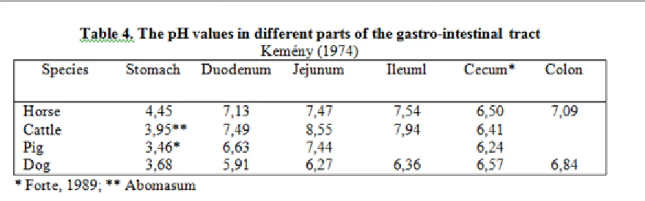
The gross structures of the gastrointestinal tract of four domestic species (dog, pig, horse and sheep) typify the extreme variations found in the mammalian digestive system (Table 2 and 3.). In carnivores, which obtain most of their food by eating other animals, digestion is mainly enzymatic and microbial digestion is minimal.
Therefore, gross digestive structure is relatively short and simple (Fig. 2.1.). In contrast, the domesticated herbivores fall into two groups: (1) the ruminants, such as cattle, sheep, and goats, in which extensive microbial fermentation of the plant diet occurs in a specialized region of the tract proximal to gastric digestion (pregastric fermentation); and (2) those with simple stomach, such as horse, in which microbial fermentation takes place in the distal digestive tract (postgastric fermentation).
Figure 2.1. Figure 2.1.: Gastro-intestinal tract of two carnivores.(Reece, 2004)
These sites of microbial digestion require a large digestive-fermentation organ in which transit can be delayed to provide the time necessary for fermentation, especially cellulose. Thus, the complex forestomach of ruminants and the large intestine of horses contain at least 10 to 15 percent of the animals‟ total body water volume at any given time (Fig. 2.2). Both enzymatic and postgastric fermentation are of importance in the intermediate structure, characteristic of the omnivore.
COMPARATIVE STRUCTURE AND FUNCTION OF THE GASTROINTESTINAL TRACT OF
ANIMALS
Simple-stomached carnivores may consume occasional meals of high-energy content that are followed by a period of relative quiescence. Plant material, on the other hand, is low in energy content, and the herbivore must consume a large quantity to satisfy its energy requirements. The actual time spent eating is much greater in herbivores than in carnivores and grazing by ruminants may occur 8 or more hours in the 24.hour period.
Rumination may occupy an equally long time. This type of feeding behavior is associated with essentially continuous activity of the secretory glands and the musculature of the tract. For example, the ruminant
Figure 2.2. Figure 2.2.: Gastro-intestinal length of the sheep and horse (Reece, 2004).
abomasum, “true stomach”, normally secrets acid gastric juice continuously because the flow of ingesta from the reticulorumen into the abomasums never ceases. Conversely, animals fed a concentrated meal once or twice daily display cyclic gastrointestinal secretory and motor activity.
Postgastric fermenters will spend as much time eating roughage as ruminants spend eating and ruminating. Food material is moved rapidly through the stomach and small intestine and delayed in the large intestine. The passage rate is more rapid through the small intestine in horse than in dogs. Total gastrointestinal transit through horses is also faster than in ruminants owing to a marked delay in passage through the latter‟s reticulo-omasal orifice. This marked delay in the forestomach allows more efficient microbial digestion of insoluble fiber (cellulose) in ruminants, a substance that cannot be digested by mammalian enzymes. In ruminants, both soluble (e.g., starch) and insoluble material is fermented by microorganisms before it reaches the site of enzymatic digestion; accordingly the principle end products of carbohydrate digestion are the short-chain fatty acids (SCFAs) to maintain the blood glucose concentration. Fermentation and SCFA metabolism are less efficient metabolic processes than direct absorption of glucose from the gut.
Dietary protein is also digested by microorganisms in the forestomach. However, the ammonia and peptides that are formed can be utilized by the microorganisms to form microbial protein, which is then digested in the small intestine by mammalian enzymes. The advantage of this process is that the microorganisms can utilize non protein nitrogen sources such as urea, together with a carbon source, to synthesize microbial proteins.
Accordingly, a waste product that would otherwise be excreted by the kidney is recycled into microbial protein in the forestomach.
Conversely, the horse has adopted the strategy of increasing the total intake and output and extracting the more digestible material prior to fermentation. This allows for more efficient digestion of soluble material in the small intestine by mammalian enzymes, yielding glucose as a major product of carbohydrate digestion.
Fiber and undigested starch will be fermented in the large intestine. However, the relative faster passage through the equine large intestine than through the bovine forestomach renders the horse less efficient in fiber digestion.
Differences between the structures of digestive tract also influence feeding behavior of animals. Carnivores tends to eat rapidly and may consume enormous meals. For instance, a mail Labrador retriever (type of dog) may consume 10 per cent of its body weight in canned food at once. Dogs and cats may use their forelimbs to hold food, which is passed into the mouth largely by head and jaw movements without much grinding. Cats and dogs convey fluids into the mouth by rapid extension and retraction of the tongue, which has a free mobile end
COMPARATIVE STRUCTURE AND FUNCTION OF THE GASTROINTESTINAL TRACT OF
ANIMALS
into a ladle. Prehension of food (feed intake) differs among herbivorous animals. In some species, the main prehensile structure is the lips (upper or lower). Others use mainly their tongues, and some rely mostly on their incisor teeth. In the horse, the sensitive, mobile upper and lower lips are the main prehensile structures used during feeding. During grazing, the lips are drown back to allow the incisor teeth to sever the grass at its base.
Under natural conditions, the pig digs up the ground with its snout (rooting), and grass is carried into the mouth largely by the action of the pointed lower lip. In other common grazing herbivores, the lips possess only limited movements.
In ruminants, lips tongue, lower incisor teeth, and the dental pad at the rostral end of the hard palate acts as organs for prehension of food. Grass and roughage eaters, the traditional grazers (cattle, sheep), have a small mouth opening and short tongue around mainly monocotyledonous forage (grass, hay), which is then drown between the lower incisor teeth and the upper dental pad and served by a movement of the head. In sheep, a cleft in the upper lip permits very close grazing. Concentrate selectors, the traditional browser ruminants (deer, moose) have a large mouth opening, which permits them to use their lower incisor teeth to strip twigs or to gnaw on fruits and flowers of dicotyledonous plants. Intermediary opportunistic mixed feeder ruminants (goat) can adapt to either feeding tipe but prefer the low-fiber, dicotyledonous plants. They will even climb suitable trees to get their preferred foods.
Domestic animals have two types of teeth: low-crowned (brachydont) and high-crowned (hypsodont) Low- crowned teeth are simple teeth, as found in man, carnivores, pigs, ruminant incisors, and horse deciduous incisors. They consist of a crown, neck, and root. The crown is the part projecting above the gum line, and is covered with enamel. The neck is the constriction between the crown and root, and it is located at the gum line.
The root is the part below the gum line. High-crowned teeth, which have no distinct neck, are found in all permanent horse teeth, ruminant cheek teeth (i.e. premolars and molars), and the tasks of pigs.
In herbivores , mastication, or chewing, refers to the mechanical breakdown of food in the mouth by deduction (opening) and occlusion (closing) of both sets of upper and lower teeth during eating and rumination. Grinding of plant tissues in horses and in ruminants occurs by lateral movements of the lower jaw (mandibule), which, along with others of pulsion (forward) and retropulsion (backward), are possible because of the flexibility of the temporomandibular joint.
Since the upper jaw (maxilla) is wider than the lower jaw, mastication occurs only on one side of the dental table at a time. Because of this lateral movement, the teeth wear with chisel-shaped grinding surfaces, the sharp edge of the lower teeth being innermost and that of the upper teeth outermost. In horses, these sharp inner and outer edges must be filed to prevent tongue and lip injuries and inadequate chewing of hay. The teeth of herbivores wear unevenly because they are made of substances with different degrees of hardness, but this characteristic increases the grinding efficiency of the dental tables.
1. Self evaluation questions
Why is the gastro-intestinal tract of carnivores simpler in structure than that of herbivores?
What is the difference between ruminants and horses with regard to the site of fermentation relative to the stomach?
Why are horses less efficient than ruminants in digesting fibre?
Chapter 3. DIGESTION IN MONOGASTRIC MAMMALS
1. Regulation of feed intake
Seeking, prehension, mastication salivation, and swallowing of food are under the direct control of the brain.
Some tumors of the hypothalamus result in excessive obesity. By studying lesions and by stimulating specific areas of the hypothalamus, an appetite centre and a satiety centre have been determined. Lesions of the lateral hypothalamic areas result in aphagia, conversely, continuous stimulation of these areas induces voracious eating in rats and other animals. The satiety centre is located in both ventromedial hypothalamic (VHM) nuclei.
Lesions of the VHM areas produce hyperphagia and obesity in adult rats and cats, as do microinjections of barbiturates. The onset of feeding is brought about by a reduction in negative signals from the satiety centre to the appetite centre.
The appetite centre is then free to operate and stimulate food seeking and intake. The end of a meal would be due to an increase in plasma glucose, amino acids, or peptides, hyperthermia, dehydration, or gut fill activating the satiety centre to halt the operation of the appetite centre. Initially, the glucostatic theory assumed that the level of excitability to food was related to the blood concentration of glucose, since intravenous infusion of glucose reduces the subjective feeding of hunger in man. Prolong injection of glucose in animals also reduces feed intake, however, appetite has not been reported to be inhibited by parenteral administration of glucose. The amino acid balance theory is supported by a lack of correlation between satiation and the arteriovenosus glucosemia differences and by the fact that maximal satiety follows ingestion of protein food or amino acid, even when arteriovenous glucosemia is low. Amino acids are considered as feed intake regulators in growing pigs. A homeostatic mechanism would control free amino acid levels in plasma and tissues by reducing the inflow of amino acids from blood when plasma amino acid concentrations exceed requirements. When energy- yielding substrates are more available than amino acids to built proteins, as in low-protein diets, food intake is also depressed. Conversely, intake of carbohydrate feed is lowered when diets are too rich in proteins. The amino acid balance theory and the glucostatic theory run into considerable difficulties in diabetes mellitus, during which hyperphagia occurs although the blood levels of amino acids and glucose are very high. On the other hand, in liver disease, appetite and amino academia are suppressed. The thermostatic theory suggests that food consumption has a certain dependence on the temperature of the body and of the environment. The basis of the thermostatic theory rests on an increase in the hypothalamic temperature during feeding and other activity and on the fact that heat stress depresses food intake in animals. The dehydration theory is found on the observation that satiation follows dehydration of the digestive tissues and that appetite is stimulated by hydration of the digestive tissues. Ingestion of food and secretion of digestive juices dehydrate the glandular tissues of digestion, which would signal inhibition of feed intake. Earlier observations on dogs reported that, during ingestion of feed, water passes gradually from the tissues into the digestive juices. Hyperosmolal chyme (digesta from the stomach or gizzard) draws water into the lumen of the small intestine to distend the duodenum and also contributes to digestive tissue dehydration in chickens.
2. Role of the salivary glands
Saliva is the fluid mixture produced by the serous and mucous salivary glands. The principal salivary glands (parotid; mandibular, or submaxillary; and sublingual) are paried and drain into the oral cavity; the others (palatine, buccal, and pharyngeal) line the mucus membranes of the mouth. In most animals, immediate preprandial and feeding stimuli excite a seromucinous salivary secretion, mostly facilitate mastication and deglutition. The universal functions of saliva are (1) to keep the buccal mucosa moist, (2) to physically (fluid) and chemically (lysozyme, bicarbonate, and mucin) prevent excessive resident microorganisms that produce lactic acid and neutralize the bacterial acids that would dissolve away tooth enamel and be detrimental to dental hygiene, (3) to facilitate the initial digestive motor events, mastication and deglutition, and (4) to solubilise substances that are able to stimulate gustatory and olfactory receptors. The saliva of some animals also contributes some pregastric enzymes for splitting of carbohydrates (amylase in pigs, rats, rabbits) or lipids (lipase in calves and lambs). Added to feeds these enzymes are operative in the stomach until they are inhibited by acid in gastric juice. In monogastric animals, saliva secreted at the basal rate is hypothonic to plasma but has the same pH as blood (7.4). When the secretion rate is faster, saliva becomes isotonic to plasma, K+
concentration changes but little, and the concentrations of Na+, Cl-, and HCO3 increase.