Cardiac consequences of metabolic derangements: role of mitochondrial
oxidative stress and autophagy
Ph.D. thesis
Gábor Koncsos, M.Sc.
Semmelweis University
Doctoral School of Pharmaceutical Sciences
Supervisor: Dr. Zoltán Giricz, Pharm.D., Ph.D.
Official reviewers: Dr. István Lekli, Pharm.D., Ph.D.
Dr. Gergő Szanda, M.D., Ph.D.
Head of the Final Examination Committee:
Dr. Éva Szökő, Pharm.D., Ph.D., D.Sc Members of the Final Examination Committee:
Dr. Zsuzsanna Miklós, M.D., Ph.D.
Dr. Árpád Márki, M.Sc., Ph.D.
Budapest
2018
1
1. Introduction
1.1. Metabolic diseases in cardiovascular system
Metabolic disorders including obesity, hyperlipidemia or diabetes can increase the incidence of cardiovascular diseases such as myocardial infarction or cardiomyopathies. Therefore, it is important to study the effect of metabolic diseases on the cardiovascular system to identify novel targets for future cardioprotective therapies.
It is known that cardioprotective effects of ischemic conditioning are attenuated in the presence of diabetes or hypercholesterolemia, however, it is still unclear which signaling pathways are responsible for the loss of cardioprotection in hypercholesterolemia. Furthermore, a period of prediabetic state (i.e., impaired glucose and insulin tolerance, insulin and leptin resistance, mild to moderate obesity) occurs, which may also promote structural and functional changes of the heart. Although cardiac pathophysiological alterations are relatively well characterized in fully developed diabetes (i.e., diabetic cardiomyopathy), consequences of prediabetes and their molecular mechanism is unknown in non-genetic prediabetic settings.
1.2. Molecular pathology of metabolic diseases in the heart
A disturbed metabolic status due to diabetes or hyperlipidemia may result in altered signaling pathways in cardiac cells.It is well-established that mechanistic target of rapamycin (mTOR) can be activated by various upstream regulators such as amino acids, elevated glucose and oxygen levels, however, the role of cardiac mTOR is still unclear in hypercholesterolemia and prediabetes.
Autophagy is a ubiquitous cellular housekeeping process, which is involved in protein quality control and cardiac cytoprotection, however its role in prediabetes and hypercholesterolemia is not well studied.
2
Insufficient autophagy can promote programmed cell death, for example apoptosis, which results in cardiac damage. It has been shown that activated apoptosis has an important role in diabetes, however, the role of cardiac apoptosis and necroptosis in hypercholesterolemia and prediabetes is still unclear.
1.3. Cardiac mitochondria in metabolic diseases
Mitochondria have pivotal role in these metabolic disorders including obesity and type 2 diabetes, however, their status and effect is less understood in prediabetes. Mitochondrial function is heavily influenced by mitochondrial dynamics. However, it is not well-established whether altered mitochondrial dynamics is involved in the mechanism of deteriorated cardiac functions in prediabetes.
There are three distinct fractions of cardiac and skeletal muscle mitochondria according to their subcellular localization: subsarcolemmal mitochondria (SSM), interfibrillar mitochondria (IFM), and perinuclear mitochondria (PNM). Moreover, the biology behind the proteomic differences is largely unknown, and a specific protein marker that allows the identification of mitochondrial subfractions has not been identified yet.
Differentiation between IFM and SSM in biochemical assays depends on the use of different isolation methods. Although the use of proteases is necessary for the isolation of IFM by the most widely accepted method, enzymatic digestion with bacterial proteases might influence the mitochondrial protein content and/or mitochondrial function, and it is unknown whether the nagarse-based method for the isolation of IFM affects the detection of mitochondrial proteins in cardiac mitochondrial subfractions.
3
2. Aims
To investigate the status of autophagy and mTOR and examine apoptosis and necroptosis pathways in a hypercholesterolemic rat model.
To characterize functional, morphological and molecular features of a diet-induced prediabetes model in rats.
To investigate the effect of prediabetes on the cardiovascular system.
To examine whether the nagarse-based method for the isolation of IFM affect the detection of mitochondrial proteins in cardiac mitochondrial subfractions.
3. Materials and methods
These studies conform to the Guide for the Care and Use of Laboratory Animals published by the US National Institutes of Health (NIH publication No. 85–23, revised 1996) and was approved by the animal ethics committee of the Semmelweis University, Budapest, Hungary (registration numbers:
XIV-I-001/450-6/2012) or animal welfare office of the Justus-Liebig University, Giessen, Germany.
3.1. Animal models and experimental designs
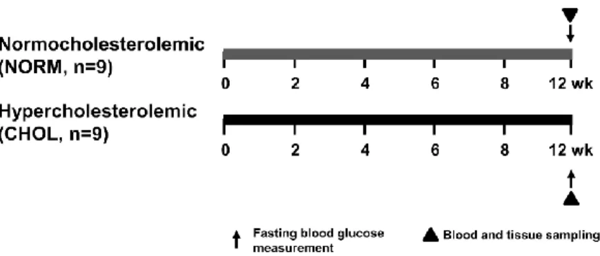
To investigate the effect of hypercholesterolemia, male Wistar rats were fed either normal (NORM) chow or with 2% cholesterol and 0.25% cholic acid-enriched diet (CHOL) for 12 weeks. Animals were anesthetized with diethyl ether and then they were sacrificed. Hearts were excised, briefly washed out, taken and snap-frozen for further experiments (Figure 1).
4
Figure 1. Experimental protocol for assessing the effect of hypercholesterolemia in vivo.
Fasting blood glucose, triglyceride and cholesterol were measured at week 12. Tissue sampling was performed after terminal procedures. NORM, normocholesterolemic;
CHOL, hypercholesterolemic
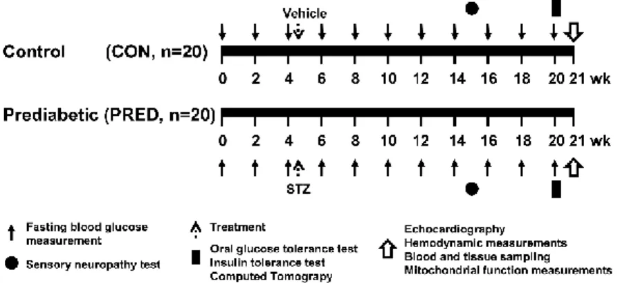
To characterize a prediabetic animal model, male Long-Evans rats were fed with either control (CON) or high-fat chow (PRED) for 21 weeks and treated with a single low dose (20 mg/kg) streptozotocin (STZ) at week 4 (Figure 2). Animals were allowed to food and water ad libitum. Body weights were measured weekly. Blood was taken and fasting blood glucose levels were measured from the saphenous vein every second week. To determine whether sensory neuropathy was developed, mechanical hind paw withdrawal thresholds were measured at week 15 of the diet. At the 20th week, oral glucose tolerance test (OGTT), insulin tolerance test (ITT) and computer tomography (CT) were performed. At 21th week, animals were anesthetized with pentobarbital. Before euthanasia, to measure cardiac function, echocardiography and hemodynamic measurement were performed. Then hearts were excised, shortly perfused with oxygenated Krebs-Henseleit buffer in Langendorff mode as described earlier and tissue weights were measured. Tissue samples were taken and snap-frozen for further experiments.
5
Figure 2. Experimental protocol for assessing the effect of prediabetes in vivo.
Long-Evans rats were fed with either CON diet for 21 weeks, or with high-fat diet and treated with 20 mg/kg STZ at week 4 to induce prediabetes (PRED). Body weights were measured weekly and blood samples were taken from the saphenic vein every second week. Sensory neuropathy was measured at week 15. OGTT, ITT and CT were performed at week 20. Echocardiography, hemodynamic analysis and parameters of mitochondrial function were measured at week 21 of diet. Tissue sampling was performed after terminal procedures. CON, control; PRED, prediabetic; STZ, streptozotocin; OGTT, oral glucose tolerance test; ITT, insulin tolerance test; CT, computed tomography.
To investigate the effect of nagarse on cardiac mitochondrial subfractions, male C57Bl6J mice and Wistar rats were used. Mice were anaesthetized with 5% isoflurane and sacrificed by cervical dislocation. Rats were anaesthetized with 4% isoflurane, subsequently hearts were removed, and cardiac tissue was collected.
6 3.2. Biochemical measurements
Serum cholesterol, high density lipoprotein (HDL) and triglyceride levels were measured in Long Evans rats and glucose, cholesterol and triglyceride levels were measured from plasma of NORM and CHOL Wistar rats by colorimetric assays. Plasma leptin was measured by enzyme-linked immunosorbent assay (ELISA) according to manufacturer’s instructions. To determine pancreatic insulin content, freeze clamped and pulverized pancreas samples were used according to the manufacturer’s instructions.
3.3. Histology
Heart, liver and pancreas samples were fixed in 4% neutral-buffered formalin and stained with hematoxylin-eosin and Masson’s trichrome staining. Nitrotyrosine immunostainig of left ventricular samples was also performed. Areas of SSM, IFM and lipid droplets were measured by transmission electron microscopy.
3.4. Quantitative RT-PCR
Total RNA isolation and cDNA synthesis were perfomed from left ventricular tissue according to the manufacturers’ instructions. Quantitative real-time PCR was performed with the StepOnePlus Real-Time PCR System (Thermo Fisher Scientific). Expression levels were calculated using the cycle threshold (CT) comparative method (2-ΔCT).
3.5. Isolation and measurements of cardiac mitochondria
SSM and IFM fractions from rat and mouse left ventricles were isolated as described by Palmer et al. in 1977 with minor modification and is referred to as the common protocol (Figure 3). Briefly, left ventricular tissues were minced with scissors in buffer A. Then, tissues were disrupted with a Polytron7
tissue homogenizer and the homogenates were centrifuged. The supernatants (containing SSM) were divided into 2 groups. One portion of SSM was used without nagarse and protease inhibitor treatment (SSM). Another portion of SSM was treated with of nagarse (SSM+N; Bacterial type XXIV) for 1 min.
Sediments of the first centrifugation (containing IFM) were resuspended in buffer A and were treated with nagarse for 1 min. Then, phenylmethylsulfonyl fluoride (PMSF) was added to one portion of IFM (IFM+N+I), whereas another portion was without PMSF (IFM+N). All mitochondrial samples were disrupted and were centrifuged. The resulting supernatants were centrifuged to collect the SSM and IFM. The mitochondria were resuspended in buffer A, and were centrifuged, and finally resuspended in buffer B. Then, two portions of isolated mitochondria fractions (SSM and IFM+N) were further used to investigate mitochondrial respiration, membrane potential, Ca2+ uptake and H2O2 formation from prediabetic rat hearts.
For Western blot analysis, mitochondria were further purified by a subsequent ultracentrifugation on 30% Percoll. The lower mitochondrial band was collected, washed twice in buffer B by centrifugation, and the purified mitochondria were stored at -80°C.
3.6. Western blot
Equal amount of protein (whole left ventricle, SSM and IFM samples) was loaded and separated in a Tris-glycine-SDS polyacrylamide gel. Proteins were transferred onto a polyvinylidene difluoride membrane. Membranes were blocked with bovine serum albumin or non-fat dry milk. Membranes were probed with primary antibodies, and with corresponding horseradish peroxidase-conjugated secondary antibodies. Signals were detected with an enhanced chemiluminescence kit.
8 3.7. Statistical analysis
Values are expressed as mean ± standard error of mean (SEM). Statistical analysis was performed between groups by unpaired two-tailed Student’s t- test or by Mann-Whitney U-test. Data on mitochondrial protein levels of MFN1, MFN2, DJ-1, p66shc, and Cx43 in mouse and rat SSM and IFM+N were normalized to VDAC and compared by unpaired two-tailed Student’s t- test or one-way ANOVA with LSD post hoc test. Mitochondrial oxygen consumption data was evaluated by one-way ANOVA with LSD post hoc test or by Kruskal-Wallis analysis with Dunn’s post hoc test in case of dataset with non-normal distribution. GraphPad Prism 6 software was used for statistical analyses. A p<0.05 value was considered significant.
4. Results
4.1. Effect of high-cholesterol diet in Wistar rats
In this study, CHOL rats exhibited a 41% increase in plasma total cholesterol level over that of NORM rats (4.09 mmol/L vs. 2.89 mmol/L) at the end of diet period. Moreover, our Western blot results showed that hearts of cholesterol-fed animals exhibited a decrease in LC3-II, Beclin-1, Rubicon, and RAB7, consistent with downregulation of autophagy initiation and vesicle traffic despite upregulation of the Class-III PI3K involved in autophagy initiation. No difference in the expression of markers of autophagic clearance NBR1 and SQSTM1/p62 proteins was detected between groups. There was no significant difference in the expressions or phosphorylations of Akt and mTOR protein between groups; however, phosphorylation of ribosomal S6, a surrogate marker of mTOR complex activity, was elevated in CHOL group compared to controls. Expression of apoptotic marker cleaved caspase-3 was significantly increased, while Bcl- 2/Bax protein expression ratio was unchanged. Furthermore, expression of RIP1, RIP3 and MLKL proteins, major markers of necroptosis, were
9
unchanged. These results suggest that autophagy is attenuated, mTOR and apoptosis are upregulated while necroptosis is unchanged in the heart of hypercholesterolemic rats compared to controls.
4.2. Effect of high-fat diet and streptozotocin treatment in Long-Evans rats
Body weights of the prediabetic animals were moderately but statistically significantly elevated from week 9 as compared to the control group, and that this difference reached 18% at the end of the diet period. Furthermore, at week 20, plasma leptin level was significantly increased in prediabetes, plasma cholesterol, HDL cholesterol and triglyceride levels, and parameters of liver and kidney function were unchanged. CT scan showed that body fat volume of prediabetic rats was substantially increased at the end of the diet.
Furthermore, electron microscopy showed an increased number of lipid droplets in the myocardium of prediabetic animals as compared to controls.
At week 20 of the diet, fasting blood glucose levels were slightly elevated in prediabetes from week 10, however, remained in the normoglycemic range. OGTT and ITT demonstrated impaired glucose tolerance and insulin resistance in the prediabetic group, however, there was no difference in pancreatic insulin content, or in pancreatic islet morphology between groups.
Accordingly, here we have found a decrease in the mechanical hind limb withdrawal threshold at week 15 of diet in the PRED, which indicates a moderate sensory neuropathy in this model of prediabetes.
To determine the cardiac effect of prediabetes, we measured morphological and functional parameters of the hearts. Heart weights, left ventricular mass, left ventricular anterior wall thickness, systolic and diastolic left ventricular posterior wall thickness were significantly increased, however, heart weight/body weight ratio was decreased in prediabetes, plausibly due to obesity. The slope of end-diastolic pressure-volume relationship (EDPVR), which is a very early and sensitive marker of diastolic
10
dysfunction, was significantly elevated in prediabetes, although other hemodynamic parameters, including blood pressure, were unchanged. On hematoxylin-eosin-stained left ventricular sections increased cardiomyocyte diameter was detected in prediabetes. To characterize components affecting diastolic function, we analyzed MHC expression. Interestingly, the gene expression of β-MHC was decreased, and α-MHC also showed a tendency of decrease (p=0.17), the ratio of which resulted in a strong tendency to decrease in prediabetes. Mitochondrial morphology and enzyme activity were analyzed from left ventricles of prediabetic rats. We have not seen any major difference in the number of IFM between the groups, however, area, perimeter and sphericity of IFM are decreased in PRED group. Furthermore, we have not seen any major difference in mitochondrial oxygen consumption, enzyme activities, Ca-uptake, or membrane potential. However, we have found that hydrogen-peroxide production was increased in the cardiac SSM fraction with glutamate-malate as a substrate, although, there was no difference when succinate was used as substrate. Interestingly, there was no increase in reactive oxygen species (ROS) production of the IFM isolated from left ventricles supported either with glutamate-malate or with succinate.
Nitrotyrosine immunohistology indicated that protein nitrosylation is increased in prediabetes. As CaMKII has been proposed to be activated in oxidative stress-associated conditions, we measured the levels of the active forms of the kinase which might affect the contractility and relaxation capacity of the heart. The phosphorylation of CaMKII and of its target PLB on Thr17 were not changed by prediabetes. Similarly, there was no change in the protein expression of SERCA2A in our model of prediabetes as compared to control animals. On the other hand, the level of p-Ser16-PLB showed a tendency for downregulation in prediabetes (p=0.08). Our results indicate that mitochondrial dynamics and autophagy/mitophagy were not modulated substantially by prediabetes, however, the upregulation of MFN2 (increased mitochondrial fusion, tethering to endoplasmic reticulum) and the downregulation of BNIP3 (decreased mitophagy) may implicate early changes in mitochondrial homeostasis, which might lead to the accumulation
11
of dysfunctional mitochondria. Our results showed no differences in the expression of HSP-60, HSP-70 and HSP-90 or in either phosphorylation or expression of HSP-27. Moreover, prediabetes did not affect the expression of pro-apoptotic caspase-3 and Bax in left ventricles. On the other hand, the anti- apoptotic Bcl-2 was downregulated in prediabetic animals. However, the Bcl-2/Bax ratio was unchanged.
4.3. Detection and quantification of proteins in cardiac mitochondrial subpopulations
To examine the amounts of proteins in cardiac SSM and IFM, inner (Cx43) and outer mitochondrial membrane (MFN1, MFN2, VDAC) proteins as well as p66shc as a marker for the intermembrane space were measured.
In Wistar rat and C57Bl6J mouse hearts, MFN2 and Cx43 protein signals were higher in SSM samples, while intensities of MFN1 and p66shc protein signals were not different between SSM and IFM+N, and DJ-1 protein signal was higher in IFM+N as compared to those in SSM. Nagarse treatment of SSM resulted in decreased signal intensities of MFN1, MFN2, and Cx43, as shown by Western blot analysis. In contrast, nagarse treatment had no effect on the amount of p66shc in SSM, whereas the level of DJ-1 slightly – but significantly – increased in SSM+N compared to SSM. Nagarse inhibition by PMSF, (IFM+N+I) had no influence on signals of Cx43, DJ-1, p66shc, and MFN1, whereas it significantly increased the signal intensity of MFN2 to that measured in SSM. Nagarse treatment had no effect on either basal or ADP- stimulated oxygen consumption by complex I or complex II compared to untreated SSM. However, a tendency towards a higher complex I-mediated oxygen consumption was observed after nagarse treatment in SSM (p=0.098). Mitochondrial respiration was similar in IFM+N and IFM+N+I.
12
5. Discussion
5.1. Hypercholesterolemia modulates cardiac mTOR and autophagy
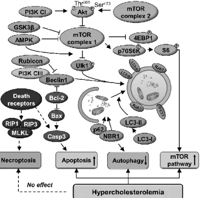
In this thesis, we show that hypercholesterolemia downregulates cardiac autophagy and activates mTOR pathway and apoptosis (for schematic representation see Figure 4).
We assessed an increased cleaved caspase-3 activation, however, the balance of pro- and anti-apoptotic Bcl-2 proteins was unaffected by hypercholesterolemia. We show the first time the status of necroptosis in vivo in hypercholesterolemia. Here we demonstrate that the expression of major markers of necroptosis are not modulated in the heart of cholesterol-fed rats.
Overall, our current findings demonstrate that complex metabolic disorders might modulate cardiac mTOR pathway and autophagy differentially depending on the animal model applied, and they also highlight that focused studies are necessary to decipher the role of specific metabolic pathways on autophagy.
13
Figure 4. Schematic representation of the cardiac effect of hypercholesterolemia on autophagy, apoptosis, necroptosis and mTOR pathways.
PI3K CI-III, class I-III phosphoinositide 3-kinase; NBR1, neighbor of BRCA1 gene 1; AMPKα, AMP-activated protein kinase α; GSK3β, glycogen synthase kinase- 3 beta; ULK1, unc-51-like kinase 1; LC3, microtubule-associated protein light chain 3; RAB7, ras-related protein Rab-7a; Rubicon, run domain Beclin-1-interacting and cysteine-rich domain-containing protein; SQSTM1/p62, sequestosome 1; mTOR, mechanistic target of rapamycin; P70S6K, ribosomal protein S6 kinase beta-1; S6, ribosomal S6 protein; 4EBP1; eIF-4E binding protein 1; Casp3, caspase 3; Bax, Bcl- 2-associated X protein; RIP1-3, receptor-interacting serine/threonine-protein kinase 1 and 3; MLKL, mixed lineage kinase domain-like protein.
14
5.2. Prediabetes induced diastolic dysfunction by mild mitochondrial oxidative stress and impaired mitophagy
Here we demonstrate that the deterioration of diastolic function and sensory neuropathy occurs well before overt diabetes develops, which is accompanied by early signs of cardiac hypertrophy.
Oxidative stress has a major role in the development of diabetic cardiomyopathy; however, it has not been well described whether it is responsible for the decreased cardiac function in prediabetes. Here we show an elevated hydrogen peroxide production in SSM, increased nitrotyrosine formation and an elevated cardiac expression of MFN2. However, in a previous study on diet-induced prediabetes, no sign of cardiac mitochondrial oxidative stress was evidenced in male Wistar rats after 16 weeks, which may implicate that mitochondrial oxidative stress might not be present in all models and stages of prediabetes and that it might not be the primary driving force of prediabetes-induced cardiac functional alterations.
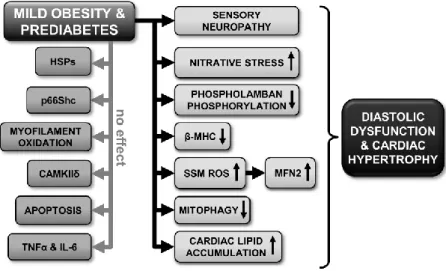
Our results evidence changes in several cellular processes, suggesting that hypertrophy and deteriorated diastolic function in prediabetes maybe consequences of numerous concurrent alterations in the cardiac homeostasis (see Figure 5). Characterizing active components of the contractile apparatus and Ca2+ homeostasis, here we observe a tendency to decrease in the Ser16 phosphorylation of PLB in prediabetes.
Apoptosis is considered to be one of the hallmarks of diabetic cardiomyopathy and it is induced by oxidative stress in diabetes. It has been described that experimental diabetes induces upregulation of pro-apoptotic, and downregulation of anti-apoptotic proteins. In this thesis, a modest downregulation of Bcl-2 was shown, however, no change in Bcl-2/Bax ratio, and in caspase-3 expression was detected in prediabetic animals.
Here we demonstrate an early dysregulation of mitochondrial fusion and mitophagy first in the literature, as evidenced by an elevated MFN2 and an attenuated BNIP3 expression in prediabetes; however, other canonical markers of autophagy, mitophagy and apoptosis were unaffected. Therefore,
15
we can assume that only major disturbances in glucose and lipid homeostasis, such as seen in untreated patients or in genetic models of diabetes, might be a powerful enough signal to extensively modulate cardiac autophagy, mitophagy, or mitochondrial dynamics, which might result in grossly deteriorated cardiac function.
Figure 5. Schematic representation of the cardiac effects of prediabetes.
SSM, subsarcolemmal mitochondria; ROS, reactive oxygen species; HSP, heat shock protein; CAMKIIδ, Ca2+/calmodulin-dependent protein kinase II; TNFα, tumor necrosis factor alpha; IL-6, interleukin-6; MFN2, mitofusin 2; β-MHC, beta-alpha- myosin heavy chain.
16
5.3. Detection of mitochondrial proteins is influenced by mitochondrial isolation with nagarse
Here we evidence that nagarse, a bacterial protease commonly used for isolation of IFM, significantly influences the detection of MFN2 in this mitochondrial subfraction. Differential protease sensitivity of SSM proteins was also evidenced: whereas detection of MFN1, MFN2, DJ-1 and Cx43 was influenced by nagarse, but that of p66shc was not. Furthermore, we also demonstrate that mitochondrial respiration was not influenced by nagarse treatment.
In this thesis, we assessed signal intensities of proteins from different mitochondrial compartments in control and nagarse-treated SSM. When a protease is added to intact mitochondria, it is reasonable to assume that first proteins of the outer mitochondrial membrane are digested, and indeed, the mitochondrial signals of MFN1 and MFN2 were reduced. However, VDAC signals were unaffected. The finding that the signal of p66shc – present in the intermembrane space - was unaffected by nagarse treatment of SSM implies that the extent of protein digestion by nagarse is not in the first instance determined by the submitochondrial localization of the protein. Furthermore, since the DJ-1 signal is higher in IFM+N than in both SSM and SSM+N, it is plausible that DJ-1 is present at higher levels in IFM, however, this finding needs further investigation.
We found that Cx43 was predominantly present within cardiac SSM.
Similarly to Cx43, MFN2 has been detected predominantly in SSM using the common protocol. Therefore, we recommend assessing mitochondrial proteins and function in IFM+N+I in studies comparing IFM and SSM and in case differential signal intensities of mitochondrial proteins are observed in SSM and IFM subfractions.
17
6. Conclusions
In this thesis, the following original findings were demonstrated:
Hypercholesterolemia activates cardiac mTOR and apoptosis but downregulates autophagy.
Prediabetes induces mild diastolic dysfunction and hypertrophy by elevated mitochondrial oxidative stress.
Nagarse treatment of cardiac subsarcolemmal and interfibrillar mitochondria may lead to the inaccurate quantification of certain mitochondrial proteins.
Therefore, before assessing the distribution of mitochondrial proteins, preliminary tests are recommended in case of proteins with unknown susceptibility to nagarse.
7. Own publications
7.1. Own publications involved in the current thesis
1. Koncsos, G., Varga, Z.V., Baranyai, T., Boengler, K., Rohrbach, S., Li, L., Schluter, K.D., Schreckenberg, R., Radovits, T., Olah, A., Matyas, C., Lux, A., Al-Khrasani, M., Komlodi, T., Bukosza, N., Mathe, D., Deres, L., Bartekova, M., Rajtik, T., Adameova, A., Szigeti, K., Hamar, P., Helyes, Z., Tretter, L., Pacher, P., Merkely, B., Giricz, Z., Schulz, R., Ferdinandy, P. Diastolic dysfunction in prediabetic male rats: Role of mitochondrial oxidative stress. American journal of physiology. Heart and circulatory physiology 311(4), H927-h943, 2016. (IF: 3.324) 2. Giricz, Z., Koncsos, G., Rajtik, T., Csonka, C., Szobi, A., Adameova, A.,
Gottlieb, R.A., Ferdinandy, P. Hypercholesterolemia downregulates autophagy in the rat heart. Lipids in health and disease 23; 16(1): 60, 2017. (IF: 2.137)
3.
Koncsos G., Varga, Z.V., Baranyai, T., Ferdinandy, P., Schulz, R., Giricz, Z., Boengler, K. Nagarse treatment of cardiac subsarcolemmal and interfibrillar mitochondria accounts for inaccurate quantification of18
proteins. Journal of Pharmacological and Toxicological Methods 31;91:50-58, 2018 (IF: 2.238)
7.2. Own publications not involved in the current thesis
4. Baranyai, T., Nagy, C.T., Koncsos, G., Onodi, Z., Karolyi-Szabo, M., Makkos, A., Varga, Z.V., Ferdinandy, P., Giricz, Z. Acute hyperglycemia abolishes cardioprotection by remote ischemic perconditioning.
Cardiovascular diabetology 14: 151, 2015. (IF: 4.015)
5. Baranyai, T,, Giricz, Z., Varga, Z.V., Koncsos, G., Lukovic, D., Makkos, A., Sárközy, M., Pávó, N., Jakab, A., Czimbalmos, C., Vágó, H., Ruzsa, Z., Tóth, L., Garamvölgyi, R., Merkely, B., Schulz, R., Gyöngyösi, G., Ferdinandy, P. In vivo MRI and ex vivo histological assessment of the cardioprotection induced by ischemic preconditioning, postconditioning and remote conditioning in a closed-chest porcine model of reperfused acute myocardial infarction: importance of microvasculature. Journal of translational medicine 15: 67, 2017. (IF: 3.991)
6. Giricz, Z., Varga, ZV., Koncsos, G., Nagy, C.T., Görbe, A., Mentzer, R.M. Jr., Gottlieb, R.A., Ferdinandy, P. Autophagosome formation is required for cardioprotection by chloramphenicol. Life Sciences 186:11- 16, 2017 (IF: 2.936)
7. Varga, ZV., Pipicz, M., Baán, JA., Baranyai, T., Koncsos, G., Leszek, P., Kuśmierczyk, M., Sánchez-Cabo, F., García-Pavía, P., Brenner, GJ., Giricz, Z., Csont, T., Mendler, L., Lara-Pezzi, E., Pacher, P., Ferdinandy, P. Alternative Splicing of NOX4 in the Failing Human Heart. Front Physiol. 8:935, 2017 (IF: 4.134)
8. Nagy, AM., Fekete, R., Horváth, G., Koncsos, G., Kriston, C., Sebestyén, A., Giricz, Z., Környei, Z., Madarász, E., Tretter, L.
Versatility of microglial bioenergetics machinery under starving conditions. Biochim Biophys Acta. S0005-2728(17)30193-7. 2017 (IF:
4.932)
19
8. Acknowledgements
The presented studies were supported by the following grants: European Foundation for the Study of Diabetes (EFSD) New Horizons Collaborative Research Initiative from European Association for the Study of Diabetes (EASD), the National Research, Development and Innovation Office of Hungary (NKFIA; NVKP-16-1-2016-0017 National Heart Program), the Hungarian Scientific Research Fund (OTKA K 109737 and OTKA PD 109051) and Semmelweis University (EFOP-3.6.3-VEKOP-16-2017- 00009). Furthermore, Pharmahungary Group also financed the experiments.
I greatly acknowledge to Prof. Dr. Péter Ferdinandy for providing me the possibility to work in the Cardiometabolic Research Group at the Department of Pharmacology and Pharmacotherapy. I am also grateful for the excellent laboratory environment that gave me the opportunity to create this thesis.
I am indebted to my supervisor, Dr. Zoltán Giricz who pushed my carrier with his helpful suggestions, scientific attitude and guidance. I am thankful for his constant motivation, support and patience.
I would like to express my enormous gratitude to Dr. Zoltán V. Varga, He always inspired my way of thinking and helped me with his encouragements during my PhD years. I am especially thankful to Melinda Károlyi-Szabó and Jenőné Benkes for their technical assistance. I thank Tamás Baranyai, Csilla Nagy, Gábor Brenner, András Makkos, Zsófia Onódy and all fellow PhD- students and all members of the Department of Pharmacology and Pharmacotherapy for the friendly support and environment. I am also grateful to my undergraduate students, Balázs Czakó, Sebestyén Tuza and Máté Ézsiás for their contribution to my work.
I am especially thankful to Tomáš Rajtík, Adriana Adameová, Monika Barteková, Prof. Rainer Schulz, Kerstin Boengler, Prof. Béla Merkely, Tamás Radovits, Attila Oláh, Tímea Komlódi, Prof. László Tretter and all my collaborators, who participated in the studies of this thesis.
Last, but not least, I am greatly thankful for my family to support me with their patient, belief and kindness.