THE USE OF INFLAMMATORY MARKERS AS A DIAGNOSTIC AND PROGNOSTIC APPROACH IN NEONATAL CALVES WITH
SEPTICAEMIA
Akın KIRBAS1*, Fatih Mehmet KANDEMIR2, Demet CELEBI3, Basak HANEDAN1 and Mehmet Ozkan TIMURKAN4
1Department of Internal Medicine, 2Department of Biochemistry, 3Department of Microbiology and 4Department of Virology, Faculty of Veterinary Medicine,
Ataturk University, 25240 Erzurum, Turkey (Received 30 March 2019; accepted 31 July 2019)
The objective of this study was to evaluate the usefulness of inflammatory markers as a diagnostic and prognostic approach in neonatal calves with septi- caemia. The study material consisted of 13 neonatal calves with septicaemia (sep- ticaemic calves, SC) and ten healthy neonatal calves (control calves, CC). Blood samples were collected for biochemical, haematological and microbiological analyses. In addition, faecal samples were collected for microbiological and viro- logical analyses. Three of neonatal calves with septicaemia were positive for E.
coli (E. coli O157 serotype) by microbiological examination, but all neonatal calves with septicaemia were negative for rota- and coronaviruses. By haemato- logical examination, there were no significant differences between SC and CC for white blood cell (WBC) and neutrophil (NEU) counts (P > 0.05). NEU counts were higher on day 0 than on day 15 in SC (P < 0.05). Red blood cell (RBC) counts and packed cell volume (PCV) values were higher on day 0 in the SC than in the CC (P < 0.05). By biochemical analyses, tumour necrosis factor-alpha (TNF-α), interleukin-6 (IL-6), procalcitonin (PCT), haptoglobin (Hp), and fibrin- ogen (Fb) concentrations were higher on day 0 in the SC than in the CC (P <
0.05). After treatment (on day 15), the serum IL-6, PCT, Hp, and Fb concentra- tions were significantly decreased in the SC compared to the CC (P < 0.05). The serum iron (Fe) concentrations were lower on day 0 in the SC than in the CC (P <
0.05), and were higher on day 15 than on day 0 in the SC (P < 0.05). The study revealed that inflammatory markers could be used for determining the diagnosis and prognosis in neonatal calves with septicaemia.
Key words: Acute phase response, calf, iron, neonatal septicaemia, pro- calcitonin, pro-inflammatory cytokines
Neonatal septicaemia means the systemic infection of newborn calves.
Escherichia coli (E. coli) has long been incriminated as the principal microbial
*Corresponding author; E-mail: akindahiliye55@yahoo.com; akirbas@atauni.edu.tr;
Phone: 0090 (442) 231-7164; Fax: 0090 (442) 231-7244
agent responsible for neonatal septicaemia in the bovine (Fecteau et al., 2009).
Colostrum-deprived calves are more susceptible to developing infections during the neonatal period (Basoglu et al., 1999; Basoglu et al., 2001; Kirecci et al., 2010). The early signs of septicaemia in neonatal calves are vague and nonspe- cific, and are often indistinguishable from signs of noninfectious diseases (Fec- teau et al., 1997; Lofstedt et al., 1999; Kirecci et al., 2010). Rapid and reliable culture with the subsequent identification of pathogens from the blood is vital so that effective antimicrobial therapy can be provided. A bacteriological culture of blood is necessary to confirm the diagnosis of septicaemia (Hariharan et al., 1992). The predominant pathogen cultured from calves with septicaemia is E.
coli, but other Gram-negative, Gram-positive and mixed bacterial infections have also been reported (Hariharan et al., 1992; Lofstedt et al., 1999). Positive blood cultures, less than 48 h old, are required in the definitive antemortem diagnosis of septicaemia (Aldridge et al., 1993). There is no single laboratory test that would be completely reliable for the early diagnosis of septicaemia in neonates of farm animal species (Aldridge et al., 1993; Lofstedt et al., 1999).
Cytokines are small cell-signalling glycoprotein molecules that play im- portant roles in local and systemic inflammatory reactions, including the regula- tion of the immune system and induction of the host organism’s reactions against antigens and microorganisms (Lohman and Baron, 2010). Pro-inflammatory cy- tokines are released in a cascade, tumor necrosis factor-alpha (TNF-α) being the initial cytokine (Gabay and Kushner, 1999). This cytokine stimulates the produc- tion of other cytokines such as interleukin-6 (IL-6). Plasma TNF-α levels are in- creased rapidly after infection and that is how TNF-α activates the inflammation cascade (Lohman and Baron, 2010). IL-6 is one of the initial cytokines released in inflammation. The diagnostic and prognostic accuracy of IL-6 may depend on the time and frequency of measurement and the severity of the underlying illness (Heinrich et al., 1990; Chiesa et al., 2003).
Procalcitonin (PCT) is a precursor of the hormone calcitonin and is syn- thesised physiologically by thyroid C cells. In bacterial infection PCT is synthe- sised in various extrathyroidal neuroendocrine tissues (Reinhart et al., 2000;
Reinhart et al., 2012). Systemic PCT secretion is a component of the inflammato- ry response that appears to be relatively specific to systemic bacterial infections.
In bacterial infections, serum PCT levels start to rise at 4 h after the onset of sys- temic infection, and peak at between 8 and 24 h (Reinhart et al., 2012).
Acute phase response (APR) occurs during infection and inflammation.
The aim of these reactions is to isolate and destroy the infectious agents, to pre- vent ongoing tissue damage and to restore the homeostasis. One of the main fea- tures of APR is the hepatic production of acute phase proteins (APPs). The secre- tion of APPs is regulated by pro-inflammatory cytokines such as TNF-α and IL-6.
The blood concentration of APPs generally increases within 8 h of stimulation, reaches the maximum level in 24–48 h and then gradually decreases to its normal
levels in 4–7 days relative to the inflammatory response (Gruys et al., 2005).
APPs may be used for the differentiation of bacterial and viral infections, for the differential diagnosis of clinical, subclinical, acute and chronic diseases, for de- termining the prognosis of sick animals and monitoring patients during treatment (Petersen et al., 2004; Gruys et al., 2005). The most important APPs for the de- termination of bovine diseases are haptoglobin (Hp) and fibrinogen (Fb) (Pe- tersen et al., 2004; Gruys et al., 2005; Ceciliani et al., 2012). Hp is an APP bind- ing free haemoglobin in the blood. Hp concentrations are increased during acute infection but decreased with treatment or chronicity (Petersen et al., 2004; Gruys et al., 2005). However, their level remains high in chronic cases if stimulation continues (Petersen et al., 2004). One of the most widely evaluated APPs in cat- tle is Fb. Plasma Fb concentration in cattle increases within 2 days after inflam- mation (Cole et al., 1997; Roussel et al., 1997). In cattle, Fb is used to evaluate inflammatory diseases and there is a marked increase in the synthesis of Fb in re- sponse to infection (Ceciliani et al., 2012). In veterinary medicine, in neonatal ruminant practice, there are not enough data showing the prognostic and diagnos- tic use of TNF-α, IL-6, PCT, Hp, Fb and iron (Fe) in bacterial septicaemia in ne- onatal calves. Therefore, the aim of the present study was to investigate the use- fulness of PCT, TNF-α and IL-6, Hp, Fb and Fe as a diagnostic and prognostic approach in neonatal calves with septicaemia.
Materials and methods
Animals
Thirteen Swiss Brown calves with septicaemia presented to the Large An- imal Clinic of Ataturk University, Faculty of Veterinary Medicine served as sep- ticaemic calves (SC). Ten clinically healthy Swiss Brown calves were obtained from the dairy farm of the Faculty of Veterinary Medicine and served as a con- trol calves (CC). All calves were 1 to 4 days old and weighed 35–40 kg. This study was approved by the Local Ethics Committee for Animal Experiments of Ataturk University (Protocol No.: 2013/81).
Clinical examination
Calves were examined for body temperature (RT), heart rate (HR) and respiratory rate (RR), condition of mucous membranes, degree of dehydration, suckling reflex and faeces features.
Treatment protocol
After the collection of blood and faecal samples before treatment (on day 0), antimicrobial treatment was applied to the calves with septicaemia for 7 days
(enrofloxacine, 5 mg/kg s.c. for 7 days, Baytril®, Bayer, Istanbul, Turkey; trime- thoprim-sulphadoxime, 30 mg/kg i.m. for 7 days, Animar®, Ceva, Istanbul, Tur- key). Intravenous fluid was administered to the sick calves according to the de- gree of dehydration (0.9% NaCl, PVC, 1000 ml, Eczacıbası Baxter, Istanbul, Turkey, and 8.4% NaHCO3, Bikarvil®, Vilsan, Ankara, Turkey). Furthermore, hyperimmune serum was administered at treatment dose [included K99, F41 and F(Y), Septicol®, 40 ml, s.c. Vetal, Adiyaman, Turkey]. Oral electrolyte drugs al- so were prescribed for the sick calves (Effydral® tablet, Zoetis, Istanbul, Turkey).
Blood and faecal sampling
On day 0, on day 1 and day 2 of treatment, and after treatment (on day 15), blood samples were taken from the jugular vein of each calf into tubes (BD, UK) with and without EDTA for haematological and biochemical analysis and tubes with heparin for bacteriological analysis. The blood samples were centrifuged at 750 × g for 15 min at 4 °C within a maximum period of 2 h after sampling and the sera were stored at −80 °C until biochemical analyses. Additionally, faecal samples were collected for bacteriological and virological analyses.
Bacteriological analyses
Collected blood samples were examined with a kit (catalog no.: 241300840) for Enterobacteriacea spp. using VITEK 2 Compact Bacterial Identification and Monitoring System (Biomérieux, Inc., Hazelwood, MO, USA). Escherichia coli was identified in three samples. Latex agglutination test was used for the identi- fication of E. coli O157 serotype (Dryspot E. coli O157, Oxoid, UK). In addi- tion, faecal samples were cultured in selective enriched broths and selective en- riched agars for E. coli O157.
Virological analyses
Faecal samples were tested for rotavirus and coronavirus. Specific primers were applied to the conserved regions in coronavirus and rotavirus genes (Cho et al., 2001; Gomora et al., 2002). An extraction kit of viral nucleic acid was used for extraction (Vivantis, Malaysia), and kit procedure was followed. After the ex- traction process, synthesis of complementary DNA (cDNA) was performed. For this purpose, revert aid cDNA synthesis kit (Thermo Scientific, Germany) was used. Thereafter, PCR process was followed. The processes of temperature con- ditions and PCR optimisation were made using the methods developed for coro- navirus by Cho et al. (2001) and for rotavirus by Gomora et al. (2002). After the PCR process, the amplicons were displayed in UV transilluminator (Vilber Loumart, France). The 406-bp and 379-bp amplicon sizes were evaluated for coronavirus and rotavirus positivity, respectively, for controls.
Haematological analyses
A cell counter (Abacus Junior Vet5, Hungary) was used to establish neu- trophil (NEU), red blood cell (RBC), platelet (PLT), and white blood cell (WBC) counts as well as haemoglobin concentration (HGB) and packed cell volume (PCV).
Biochemical analyses
The concentrations of IL-6 (Sunred Biological Technology, China) and TNF-α (Cusabio Biotech, China) were measured by highly sensitive ELISA kits, specific for bovine cytokines, according to the manufacturer’s instructions. The concentration of Hp was assessed using a commercial colorimetric kit (Tridelta Development Plc, Wicklow, Ireland) in microplates, based on Hp–haemoglobin binding and preservation of the peroxidase activity of the bound haemoglobin at a low pH. The optical densities were read on an Opsys MR automatic microplate reader (Dynex Technologies, USA) at 630 nm for Hp. Fb was measured by using a solid-phase sandwich ELISA (Sunred Biological Technology, China). The con- centrations of PCT were measured by highly sensitive ELISA kits, specific for bovine PCT (Cusabio Biotech, China), according to the manufacturer’s instruc- tions. Fe concentrations were determined using commercial test kits by a bio- chemistry autoanalyzer (Beckman Coulter, AU5800, USA).
Serum biochemistry
Serum albumin (ALB), glucose (GLU), total protein (TP), urea (UREA), and creatinine (Cr) concentrations were determined using commercial test kits by a biochemistry autoanalyzer (Beckman Coulter, AU5800, USA). The concentra- tion of total globulin (GLOB) was calculated by subtracting the ALB concentra- tion from the TP concentration (Roussel et al., 1997).
Statistical analysis
Statistical comparisons of values between the two groups were analysed using an independent t test. General Linear Model/Repeated Measures were used for intra-group comparisons (version 20.0 for Windows, SPSS Inc, Chicago).
Data were expressed as mean ± standard error of the mean (SEM). The level of statistical significance was set at P < 0.05.
Results
Clinical findings
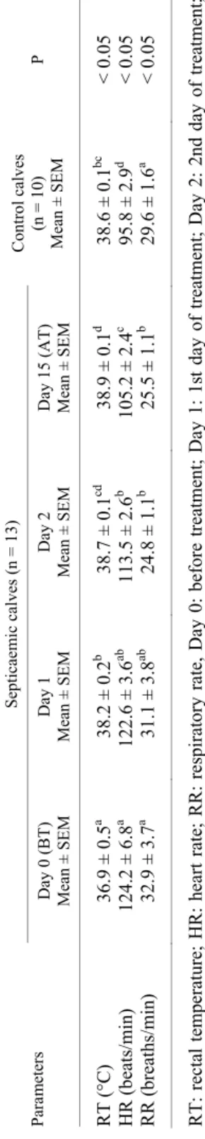
Data of clinical findings in neonatal calves with septicaemia and control calves are presented in Table 1. On day 0 in the SC compared to the CC, rectal
temperature was lower but respiratory and heart rates significantly higher. In parallel to the treatment, rectal temperature increased and respiratory and heart rates decreased (P < 0.05).
Bacteriological and virological findings
In the bacteriological analyses, E. coli O157 was identified in 3 out of 13 samples. In virological analyses, the faecal samples were negative for rota- and coronaviruses.
Haematological findings
Haematological parameters of neonatal calves with septicaemia and con- trol calves are shown in Table 2. There were no significant differences between SC and CC for WBC and NEU counts (P > 0.05). NEU numbers on day 0 were higher than on day 15 in the SC (P < 0.05). RBC counts and PCV values were higher on day 0 in the SC than the CC (P < 0.05).
Biochemical findings
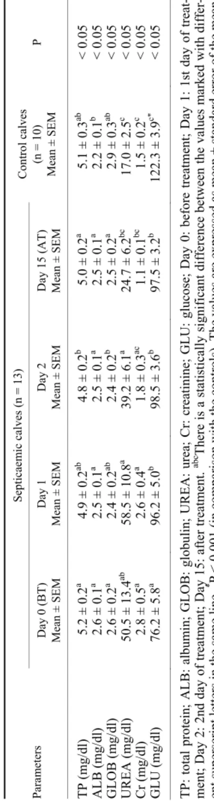
Inflammatory markers. Inflammatory markers of neonatal calves with sep- ticaemia and control calves are presented in Table 3. The serum TNF-α concen- trations were higher on day 0 in the SC than the CC (P < 0.05) and were lower on day 15 than on day 0 in the SC (P < 0.05). The serum IL-6, PCT, Hp, and Fb concentrations were higher on day 0 in the SG than the CC (P < 0.05), and the concentrations of these parameters were lower on day 15 than on day 0 in the SC (P < 0.001, P < 0.01). The serum Fe concentrations were lower on day 0 in the SC than the CC (P < 0.05), and were higher on day 15 in the SC than on day 0 in the SC (P < 0.05).
Serum biochemistry parameters. The serum biochemistry parameters of neonatal calves with septicaemia and control calves are shown in Table 4. The serum TP and GLOB concentrations were not statistically different in the SC compared to the CC (P > 0.05). The serum ALB concentrations were higher on day 0 in the SC than the CC (P < 0.05). The serum UREA and Cr concentrations were higher on day 0 in the SC than the CC (P < 0.05) and were not different on day 15 in the SC than in the CC (P > 0.05). The serum GLU concentrations were lower on day 0 in the SC than the CC (P < 0.001), and these concentrations in- creased gradually on the treatment days in the SC. However, these concentrations were still lower on day 15 in the SC than the CC (P < 0.001).
Discussion
Neonatal septicaemia is one of the most common causes of calf losses (House et al., 2015). For the early diagnosis of this important infection, culture
methods alone are not considered sufficient. Therefore, this study was carried out to determine the usability of PCT, TNF-α, IL-6, Hp, Fb and Fe measurements in the diagnosis and prognosis of septicaemia in neonatal calves.
In neonatal calves with septicaemia loss of suckling reflex, depression, hypothermia, tachycardia, tachypnoea, dehydration, hyperaemia of mucous membranes and scleral congestion are the most common clinical signs (Fecteau et al., 2009; House et al., 2015). Similarly, in the present study, the clinical ex- amination of the calves with septicaemia revealed fever, dehydration, loss of suckling reflex, increased cardiac and respiratory rates, hyperaemia in the mu- cous membranes, and scleral congestion. In addition, body temperatures in the SC were lower than in the CC (P < 0.05) and heart and respiratory rates in the SC were significantly higher than in the CC (P < 0.05). It was found that these pa- rameters returned to the reference values during treatment (Table 1).
TNF-α is used as proximal cytokine, and it is associated with most of the physiological disturbances that are characteristic of sepsis. TNF-α is a cytokine involved in systemic inflammation and a member of a group of cytokines that stimulate the APR. Later, these cytokines stimulate the production of distal cyto- kines, such as IL-6. Distal cytokines seem to intensify and perpetuate the in- flammatory response, and they are responsible for the modulation of lymphocyte function, activation of coagulation, and induction of hepatic APP synthesis (Blackwell and Christman, 1996). TNF-α concentrations have been revealed to significantly increase in calves with suspected septicaemia (Basoglu et al., 2004) and septicaemic colibacillosis (Ercan et al., 2016) compared to the control group.
Similarly, TNF-α concentration has been determined to significantly increase in endotoxaemic lambs (Kirbas et al., 2015a). In the present study, TNF-α concen- trations were higher on day 0 in the SC than the CC (P < 0.05) and were lower on day 15 than on day 0 in the SC (P < 0.05) (Table 3).
In adults and infants, an increased serum IL-6 concentration has been found to be a sensitive indicator of sepsis. In addition, IL-6 is considered the ma- jor inducer of hepatic protein synthesis (Bloos and Reinhart, 2014). Previous clinical studies of cytokines in neonatal foals have revealed that serum TNF ac- tivity is correlated with clinical criteria of sepsis and disease severity (Morris and Moore, 1991) and that IL-6 and TNF-α concentrations increase in foals that re- ceive an infusion of lipopolysaccharide (LPS) (Allen et al., 1993; Robinson et al., 1993). Kirbas et al. (2015a) have stated that IL-6 concentrations are in- creased at 12 and 24 h of endotoxaemia induced by LPS in lambs compared to the control group. In the present study, IL-6 concentrations were higher on day 0 in the SC than the CC (P < 0.05), and the concentration of this parameter was lower on day 15 than on day 0 in the SC (P < 0.001) (Table 3).
Table 1 Clinical examination findings in neonatal calves with septicaemia and in control calves ParametersSepticaemic calves (n = 13)Control calves (n = 10) Mean ± SEMP Day 0 (BT) Mean ± SEMDay 1 Mean ± SEMDay 2 Mean ± SEMDay 15 (AT) Mean ± SEM RT (°C)36.9 ± 0.5a 38.2 ± 0.2b 38.7 ± 0.1cd 38.9 ± 0.1d 38.6 ± 0.1bc < 0.05 HR (beats/min)124.2 ± 6.8a 122.6 ± 3.6ab 113.5 ± 2.6b 105.2 ± 2.4c 95.8 ± 2.9d < 0.05 RR (breaths/min) 32.9 ± 3.7a 31.1 ± 3.8ab 24.8 ± 1.1b 25.5 ± 1.1b 29.6 ± 1.6a < 0.05 RT: rectal temperature; HR: heart rate; RR: respiratory rate, Day 0: before treatment; Day 1: 1st day of treatment; Day 2: 2nd day of treatment; Day 15: after treatment. abcd There is a statistically significant difference between the values marked with different superscript letters in the same line. * P < 0.05 (in comparison with the controls). The values are expressed as mean ± standard error of the mean Table 2 Haematological findings in neonatal calves with septicaemia and in control calves ParametersSepticaemic calves (n = 13)Control calves (n = 10) Mean ± SEMP Day 0 (BT) Mean ± SEMDay 1 Mean ± SEMDay 2 Mean ± SEMDay 15 (AT) Mean ± SEM WBC (103 /μl) 10.5 ± 1.710.1 ± 1.79.2 ± 0.88.2 ± 0.69.6 ± 1.2> 0.05 NEU (103 /μl)6.1 ± 1.1a 5.5 ± 1.4ab 3.5 ± 0.7ab 3.3 ± 0.4b 4.8 ± 0.9ab < 0.05 RBC (106 /μl)10.4 ± 0.5a 8.9 ± 0.4b 9.0 ± 0.3b 8.8 ± 0.3b 8.3 ± 0.5b < 0.05 HGB (g/dl)14.6 ± 2.1a 10.5 ± 0.6ab 10.2 ± 0.5ab 9.9 ± 0.3b 10.3 ± 0.5ab < 0.05 PCV (%) 39.5 ± 1.9a 32.7 ± 1.7b 32.2 ± 1.7b 32.2 ± 1.0b 33.2 ± 1.6b < 0.05 PLT (103 /μl)305.0 ± 42.0ab 338.9 ± 34.0b 365.9 ± 37.8b 363.4 ± 24.8b 225.9 ± 21.8a < 0.05 WBC: white blood cells; NEU: neutrophils; RBC: red blood cells; HGB: haemoglobin; PCV: packed cell volume; PLT: platelets; Day 0: before treatment; Day 1: 1st day of treatment; Day 2: 2nd day of treatment; Day 15: after treatment. ab There is a statistically significant difference between the values marked with different superscript letters in the same line. * P < 0.001 (in comparison with the controls). The values are expressed as mean ± standard error of the mean
Table 3 Concentrations of inflammatory markers in neonatal calves with septicaemia and in control calves Parameters
Septicaemic calves (n = 13)Control calves (n = 10) Mean ± SEMP Day 0 (BT) Mean ± SEMDay 1 Mean ± SEMDay 2 Mean ± SEMDay 15 (AT) Mean ± SEM TNF-α (ng/ml)2.9±0.5a 1.8±0.4b 1.0±0.1c 0.8±0.1d 0.4±0.1e <0.05 IL-6 (ng/l)800.±31.8a 644.3±43.2b 548.5±34.96c 409.2±33.8d 598.8±47.8bc <0.001 PCT (pg/ml) 222.5±7.8a 51.2±1.4b 42.4±1.1c* 37.1±0.6d 37.3±1.7d <0.001 Hp (mg/ml) 0.6±0.1a* 0.1±0.0b 0.1±0.0b 0.1±0.0b 0.1±0.0b <0.01 Fb (mg/ml) 4.7±0.3a 3.3±0.2b 2.3±0.2c* 2.2±0.1c* 3.4±0.5b <0.01 Fe (μg/dl) 28.3±6.0a 51.8±9.8b 81.7±5.5c 118.6±9.7d 84.3±15.0bcd <0.05 TNF-α: tumour necrosis factor alpha; IL-6: interleukin-6; PCT: procalcitonin; Hp: haptoglobin; Fb: fibrinogen; Fe: iron; Day 0: before treatment; Day 1: 1st day of treatment; Day 2: 2nd day of treatment; Day 15: after treatment. abcd There is a statistically significant difference between the val- ues marked with different superscript letters in the same line.* P < 0.05 (in comparison with the controls). The values are expressed as mean ± standard error of the mean Table 4 The serum biochemistry findings in neonatal calves with septicaemia and in control calves ParametersSepticaemic calves (n = 13)Control calves (n = 10) Mean ± SEMP Day 0 (BT) Mean ± SEMDay 1 Mean ± SEMDay 2 Mean ± SEMDay 15 (AT) Mean ± SEM TP (mg/dl)5.2±0.2a 4.9±0.2ab 4.8±0.2b 5.0±0.2a 5.1±0.3ab <0.05 ALB (mg/dl)2.6±0.1a 2.5±0.1a 2.5±0.1a 2.5±0.1a 2.2±0.1b <0.05 GLOB (mg/dl) 2.6±0.2a 2.4±0.2ab 2.4±0.2b 2.5±0.2a 2.9±0.3ab <0.05 UREA (mg/dl) 50.5±13.4ab 58.5±10.8a 39.2±6.1a 24.7±6.2bc 17.0±2.5c <0.05 Cr (mg/dl)2.8±0.5a 2.6±0.4a 1.8±0.3ac 1.1±0.1bc 1.5±0.2c <0.05 GLU (mg/dl) 76.2±5.8a 96.2±5.0b 98.5±3.6b 97.5±3.2b 122.3±3.9c* <0.05 TP: total protein; ALB: albumin; GLOB: globulin; UREA: urea; Cr: creatinine; GLU: glucose; Day 0: before treatment; Day 1: 1st day of treat- ment; Day 2: 2nd day of treatment; Day 15: after treatment. abc There is a statistically significant difference between the values marked with differ- ent superscript letters in the same line. * P < 0.001 (in comparison with the controls). The values are expressed as mean ± standard error of the mean
PCT is known to be an effective marker used in early diagnosis of bacteri- al infections and is used in the differentiation of bacterial and viral infections. In systemic infections usually caused by bacterial infections, baseline PCT serum concentrations have been reported to increase 10- to 100-fold (Becker et al., 2004; Matur et al., 2017). PCT, having a long serum half-life (20–30 h), increas- es rapidly within a short time in bacterial diseases (Brunkhorst et al., 1999;
Becker et al., 2004; Shehabi and Seppelt, 2008; Matur et al., 2017). Ercan et al.
(2014) have stated that PCT concentrations in healthy neonatal calves were de- creased compared to healthy young and adult cattle but PCT concentrations were not different in healthy young and adult cattle. Bonelli et al. (2018) have found that the average PCT concentrations in calves with septic systemic inflammatory response syndrome (SIRS) were 166.5 pg/ml. Ercan et al. (2016) have reported that PCT concentrations in calves with septicaemic colibacillosis are approxi- mately 4-fold higher than in healthy controls, and that septicaemic colibacillosis in neonatal calves may be a beneficial biomarker. In the present study, PCT con- centrations were higher on day 0 in the SC than in the CC (P < 0.05), and the concentration of this parameter was lower on day 15 than on day 0 in the SC (P <
0.001) (Table 3). These results suggest that PCT is a useful marker for monitor- ing septicaemia in neonatal calves in compliance with the results of previous studies.
Acute phase proteins are important markers used in the diagnosis and fol- low-up of infection and inflammation. Over the past 20 years, they have been used to diagnose and monitor numerous diseases as prognostic markers. Many studies have indicated the significance of Hp as a clinically useful parameter for measuring the occurrence and severity of inflammatory responses in cattle with various diseases (Eckersall and Bell, 2010). Plasma Hp concentrations in healthy cattle have been reported to be less than 0.35 g/L. Hp increases within 24–48 h following inflammation and remains high for two weeks (Horadagoda et al., 1999). Prognosis is considered good when plasma Hp concentrations are about 0.1–1 g/L, but prognosis is poor and treatment is required when this concentra- tion is over 1 g/L (Eckersall and Bell, 2010). It was reported that Hp concentra- tions were decreased immediately after calving and they increased gradually 1 week after calving in neonatal calves (Alsemgeest et al., 1995; Orro et al., 2008;
Tothova et al., 2015). On the other hand, Hp concentration for anticipation of morbidity and mortality is determined to be 0.13 g/L in calves younger than 4 months (Murray et al., 2014). Hp concentrations have been reported to signifi- cantly increase in calves with pneumonia (Carter et al., 2002; Ganheim et al., 2003; Tothova et al., 2010; Tothova et al., 2012), enteritis (Balikci and Al, 2014), bovine respiratory disease (Joshi et al., 2018), pneumoenteritis (Ganheim et al., 2007), omphalophlebitis (Tothova et al., 2012), omphalitis (Bozukluhan et al., 2018), and neonatal diarrhoea (Pourjafar et al., 2011). In addition, Carter et al. (2002) have stated that Hp concentrations are increased before treatment in
the diseased calves compared to calves that received one or more than one treat- ment. Bozukluhan et al. (2018) have reported that Hp concentrations are in- creased before treatment and after treatment in calves with omphalitis. Similarly, in the present study, Hp concentrations were higher on day 0 in the SC than the CC (P < 0.05), and the Hp concentrations were lower on day 15 than on day 0 in the SC (P < 0.01) (Table 3).
During an inflammatory reaction, Fb concentrations can increase two- to threefold, which may significantly increase blood viscosity and cause red blood cell aggregation (Medcalf, 2007). In cattle, Fb has been used for many years to evaluate inflammatory and traumatic diseases, and is characterised by markedly increased synthesis in response to infection (Cole et al., 1997; Ceciliani et al., 2012; Kirbas et al., 2015b). It is reported that in neonatal calves, Fb concentra- tions are increased in the first two weeks of life but this increase lower and re- mains in the reference range in adults (Knowles et al., 2000). In diseased calves, Fb is used to determine whether anti-inflammatory treatment is required (Hum- blet et al., 2004). In calves with pneumonia and multisystemic infection, Fb con- centrations are increased compared to the control group but they did not change substantially in calves with omphalophlebitis (Tothova et al., 2012). Gokce et al.
(2015) have reported that calves with rotavirus enteritis had higher Fb concentra- tions than calves with enteritis caused by E. coli. A recent study in calves with omphalitis has revealed that Fb concentrations are significantly increased before and after treatment (Bozukluhan et al., 2018). Similarly, in the present study, Fb concentrations were higher on day 0 in the SC than in the CC (P < 0.05), and Fb concentrations were lower on day 15 than on day 0 in the SC (P < 0.01) (Table 3).
Iron has a role in the transportation of oxygen to the tissues in humans and animals. Fe is also important in the proliferation of numerous microorganisms (Cherayil, 2011; Constable et al., 2017). Fe deficiency caused by cytokines is re- ported during the inflammatory response (Walter et al., 1997). It is reported that in humans with sepsis and SIRS, Fe concentrations are decreased and the moni- toring of Fe concentrations may be beneficial (Shanbhogue and Paterson, 1990;
Ayoglu et al., 2016). It is reported that in horses (Borges et al., 2007), dogs (Tor- rente et al., 2015), adult cattle (Baydar and Dabak, 2014) and calves (Aydogdu et al., 2018), Fe concentrations are decreased during acute phase response due to in- flammatory reaction. It has been reported that in cattle with traumatic reticulop- eritonitis (TRP) and mastitis, serum Fe concentrations are decreased compared to the control group and serum Fe concentrations in those diseases may be a useful parameter in the determination of inflammation (Baydar and Dabak, 2014). In addition, it has been reported that decrease in the serum Fe concentrations in horses is a sensible marker for acute, subacute, and chronic systemic inflamma- tion, and alteration in serum Fe concentration may be a useful parameter in monitoring the response to treatment (Borges et al., 2007). It has been suggested that in dogs with SIRS, serum Fe concentration may be a beneficial parameter in
the determination of acute inflammation (Torrente et al., 2015). A recent study has revealed that in calves with SIRS, serum Fe concentration is significantly de- creased compared to the control group and in calves with SIRS, serum Fe con- centration may also be a beneficial parameter in the determination of inflamma- tory response (Aydogdu et al., 2018). In the present study, Fe concentrations were lower on day 0 in the SC than in the CC (P < 0.05), and were higher on day 15 in the SC than on day 0 in the SC (P < 0.05) (Table 3). Thus, it was found that in neonatal septicaemic calves, serum Fe concentration may be a useful parame- ter for monitoring the inflammatory process, in line with previous studies.
It has been reported that some haematological changes may occur in calves with septicaemia (Basoglu et al., 2001; Irmak and Guzelbektes, 2003;
Irmak et al., 2006; Ercan et al., 2016). Leukopenia or leukocytosis detected in the leukogram in calves with septicaemia is a diagnostic approach (Fecteau et al., 2009; Constable et al., 2017). Moreover, leukotic response, especially band neu- trophils and neutrophils with toxic changes, are important prognostic markers (Fecteau et al., 2009; House et al., 2015; Constable et al., 2017). WBC counts have been reported to significantly increase in calves with septicaemia (Basoglu et al., 2001), suspected septic shock (Irmak and Guzelbektes, 2003; Irmak et al., 2006), SIRS (Aydogdu et al., 2018) and septicaemic colibacillosis (Ercan et al., 2016) compared to the control group. In the present study, there were no signifi- cant differences between SC and CC in WBC and NEU counts (P > 0.05). NEU numbers on day 0 were higher than on day 15 in the SC (P < 0.05) (Table 2).
It is reported that there are no significant changes in RBC counts, HGB concentrations and PCV values of calves with SIRS (Aydogdu et al., 2018), sep- sis (Yildiz et al., 2018) and suspected septic shock (Irmak and Guzelbektes, 2003).
This is attributed to the mild diarrhoea in diseased calves. However, it has been reported that RBC counts and PCV values are significantly increased in calves with septicaemic colibacillosis compared to the control group (Ercan et al., 2016). In the present study, RBC counts and PCV values were higher on day 0 in the SC than the CC (P < 0.05) (Table 2). This was attributed to the severe diar- rhoea in the SC. It has been reported that thrombocytopenia may occur in calves with septicaemia (Irmak and Guzelbektes, 2003; Irmak et al., 2006), but in other studies, in calves with SIRS (Aydogdu et al., 2018) and sepsis, PLT counts have been determined to be physiological (Yildiz et al., 2018). In the present study, PLT counts were not different between the SC and the CC (P > 0.05) (Table 2).
It has been reported that important changes may occur in TP, GLU, UREA and Cr concentrations in neonatal septicaemic calves (Fecteau et al., 2009; House et al., 2015; Constable et al., 2017). TP and ALB concentrations may decrease due to failure of passive transfer (FPT) and may also increase due to dehydration. In- crease in the GLOB concentrations may occur due to infection (Fecteau et al., 2009; House et al., 2015; Constable et al., 2017). In the present study, TP and GLOB concentrations were not statistically different in the SC compared to the CC
(P > 0.05). ALB concentrations were higher on day 0 in the SC than in the CC (P <
0.05) (Table 4). Decrease in the serum TP, GLOB and ALB concentrations can be attributed to FPT in neonatal calves with septicaemia. Moreover, GLU concentra- tions may decrease in the early stage of septicaemia, but hyperglycaemia may be less likely to occur (Fecteau et al., 2009; House et al., 2015; Constable et al., 2017). In the present study, GLU concentrations were lower on day 0 in the SC than in the CC (P < 0.001), and these concentrations increased gradually on the treatment days in the SC. However, these concentrations were still lower on day 15 in the SC than in the CC (P < 0.001) (Table 4). The deterioration of renal perfusion as a result of dehydration in neonatal septicaemic calves has been stated to be the cause of increased serum urea and Cr concentrations (Constable et al., 2017). In the present study, UREA and Cr concentrations were higher on day 0 in the SC than in the CC (P < 0.05) and were not different on day 15 in the SC than in the CC (P > 0.05) (Table 4). Decrease in the serum UREA and Cr concentrations can be attributed to the loss of renal perfusion due to dehydration.
This study has revealed that the measurement of haematologic and serum biochemical parameters, as well as PCT, pro-infammatory cytokines (TNF-α and IL-6), acute phase proteins (Hp and Fb) and Fe concentrations could be useful markers for the diagnosis and prognosis of septicaemia in neonatal calves.
Acknowledgements
This study was financed by Ataturk University Scientific Research Office (PRJ2012/368). The abstract of the study was presented as a poster at the 3rd International VET Istanbul Group Congress held on 17–20 May 2016 in Sarajevo, Bosnia and Herze- govina.
References
Aldridge, B. M., Garry, F. B. and Adams, R. (1993): Neonatal septicemia in calves: 25 cases (1985–1990). J. Am. Vet. Med. Assoc. 203, 1324–1329.
Allen, G. K., Green, E. M., Robinson, J. A., Garner, H. E., Loch, W. E. and Walsh, D. M. (1993): Se- rum tumor necrosis factor alpha concentrations and clinical abnormalities in colostrum-fed and colostrum-deprived neonatal foals given endotoxin. Am. J. Vet. Res. 54, 1404–1410.
Alsemgeest, S. P. M., Jonker, F. H., Taverne, M. A. M., Kalsbeek, H. C., Wensing, T. and Gruys, E. (1995): Serum amyloid-A (SAA) and haptoglobin (Hp) plasma concentrations in new- born calves. Theriogenology 43, 381–387.
Aydogdu, U., Coskun, A., Yildiz, R., Guzelbektes, H. and Sen, I. (2018): Changes of hematologi- cal parameters and serum iron levels in calves with systemic inflammatory response syn- drome. Eurasian J. Vet. Sci. 34, 56–59.
Ayoglu, H., Sezer, U., Akin, M., Okyay, D., Ayoglu, F., Can, M., Kuucuukosman, G., Piskin, O., Aydin, B., Cimencan, M., Gur, A. and Turan, I. (2016): Selenium, copper, zinc, iron levels and mortality in patients with sepsis and systemic inflammatory response syndrome in Western Black Sea Region, Turkey. J. Pak. Med. Assoc. 66, 447–452.
Balikci, E. and Al, M. (2014): Some serum acute phase proteins and immunoglobulins concentra- tions in calves with rotavirus, coronavirus, E. coli F5 and Eimeria species. Iran J. Vet. Res.
15, 397–401.
Basoglu, A., Camkerten, I. and Sevinc, M. (1999): Serum immunoglobulin concentrations in diar- rheic calves and their measurement by single radial immunodiffusion. Isr. J. Vet. Med. 54, 9–10.
Basoglu, A., Sen, I., Sevinc, M. and Simsek, A. (2004): Serum concentrations of tumor necrosis factor-α in neonatal calves with presumed septicemia. J. Vet. Intern. Med. 18, 238–241.
Basoglu, A., Sevinc, M., Birdane, F. M. and Camkerten, I. (2001): Concentrations of immuno- globulin G and tumor necrosis factor in septicemic calves. TUBITAK Project number, VHAG-1338.
Baydar, E. and Dabak, M. (2014): Serum iron as an indicator of acute inflammation in cattle. J.
Dairy Sci. 97, 222–228.
Becker, K. L., Nylen, E. S., White, J. C., Muller, B. and Snider, R. H. (2004): Procalcitonin and the calcitonin gene family of peptides in inflammation, infection, and sepsis: a journey from calcitonin back to its precursors. J. Clin. Endocrinol. Metab. 89, 1512–1525.
Blackwell, T. S. and Christman, J. W. (1996): Sepsis and cytokines: current status. Br. J. Anaesth.
77, 110–117.
Bloos, F. and Reinhart, K. (2014): Rapid diagnosis of sepsis. Virulence 5, 154–160.
Bonelli, F., Meucci, V., Divers, T. J., Boccardo, A., Pravettoni, D., Meylan, M., Belloli, A. G. and Sgorbini, M. (2018): Plasma procalcitonin concentration in healthy calves and those with septic systemic inflammatory response syndrome. Vet. J. 234, 61–65.
Borges, A. S., Divers, T. J., Stokol, T. and Mohammed, O. H. (2007): Serum iron and plasma fi- brinogen concentrations as indicators of systemic inflammatory diseases in horses. J. Vet.
Intern. Med. 21, 489–494.
Bozukluhan, K., Merhan, O., Ogun, M., Kurt, B., Cihan, M., Erkilic, E. E., Gokce, G., Aydin, U.
and Ozcan, A. (2018): Investigation of haptoglobin, serum amyloid A, and some biochemi- cal parameters in calves with omphalitis. Vet. World 11, 1055–1058.
Brunkhorst, F. M., Eberhard, O. K. and Brunkhorst, R. (1999): Discrimination of infectious and noninfectious causes of early acute respiratory distress syndrome by procalcitonin. Crit.
Care Med. 27, 2172–2176.
Carter, J. N., Meredith, G. L. L., Montelongo, M., Gill, D. R., Krehbiel, C. R., Payton, M. E. and Confer, A. W. (2002): Relationship of vitamin E supplementation and antimicrobial treat- ment with acute-phase protein responses in cattle affected by naturally acquired respiratory tract disease. Am. J. Vet. Res. 63, 1111–1117.
Ceciliani, F., Ceron, J. J., Eckersall, P. D. and Sauerwein, H. (2012): Acute phase proteins in rumi- nants. J. Proteom. 275, 4207–4231.
Cherayil, B. J. (2011): The role of iron in the immune response to bacterial infection. Immunol.
Res. 50, 1–9.
Chiesa, C., Pellegrini, G., Panero, A., Osborn, J. F., Signore, F., Assumma, M. and Pacifico, L.
(2003): C-reactive protein, interleukin-6, and procalcitonin in the immediate postnatal pe- riod: influence of illness severity, risk status, antenatal and perinatal complications, and in- fection. Clin. Chem. 49, 60–68.
Cho, K. O., Hasoksuz, M., Nielsen, P. R., Chang, K. O., Lathrop, S. and Saif, L. J. (2001): Cross- protection studies between respiratory and calf diarrhea and winter dysentery coronavirus strains in calves and RT-PCR and nested PCR for their detection. Arch. Virol. 146, 2401–
2419.
Cole, D. J., Roussel, A. J. and Whitney, M. S. (1997): Interpreting a bovine CBC: Evaluating the leukon and acute-phase proteins. Vet. Med. 92, 470–478.
Constable, P. D., Hinckliff, K. W., Done, S. H. and Grunberg, W. (2017): Veterinary Medicine.
11th edition. W. B. Saunders Company, London.
Eckersall, P. D. and Bell, R. (2010): Acute phase proteins: Biomarkers of infection and inflamma- tion in veterinary medicine. Vet. J. 185, 23–27.
Ercan, N., Tuzcu, N., Basbug, O., Gok, K., Isidan, H. and Ograk, Y. Z. (2014): The evaluation of important biomarkers in healthy cattle. Kafkas Univ. Vet. Fak. Derg. 20, 749–755.
Ercan, N., Tuzcu, N., Basbug, O., Tuzcu, M. and Alim, A. (2016): Diagnostic value of serum pro- calcitonin, neopterin, and gamma interferon in neonatal calves with septicemic colibacillo- sis. J. Vet. Diagn. Invest. 28, 180–183.
Fecteau, G., Metre, D. C., Pare, J., Smith, B. P., Higgins, R., Holmberg, C. A., Jang, S. and Gu- terbock, W. (1997): Bacteriological culture of blood from critically ill neonatal calves.
Can. Vet. J. 38, 95–100.
Fecteau, G., Smith, B. P. and George, L. W. (2009): Septicemia and meningitis in the newborn calf. Vet. Clin. Food Anim. Pract. 25, 195–208.
Gabay, C. and Kushner, I. (1999): Acute-phase proteins and other systemic responses to inflamma- tion. N. Engl. J. Med. 340, 448–454.
Ganheim, C., Alenius, S. and Waller, K. P. (2007): Acute phase proteins as indicators of calf herd health. Vet. J. 173, 645–651.
Ganheim, C., Hulten, C., Carlsson, U., Kindahl, H., Niskanen, R. and Waller, K. P. (2003): The acute phase response in calves experimentally infected with bovine viral diarrhoea virus and/or Mannheimia haemolytica. J. Vet. Med. B 50, 183–190.
Gokce, E., Erdogan, H. M., Unver, A., Sozmen, M., Atakisi, O. and Karapehlivan, M. (2015): An investigation on the diagnostic and prognostic importance of some clinical, haematological and biochemical parameters in calves with neonatal diseases and septicemia. TUBITAK Project, Project Number: 111O476, 1-124, Kars.
Gomora, M. I., Wong, C., Blome, S., Desselberger, U. and Gray, J. (2002): Rotavirus subgroup characterisation by restriction endonuclease digestion of a cDNA fragment of the VP6 gene. J. Virol. Methods 105, 99–103.
Gruys, E., Toussiant, M. and Niewald, T. A. (2005): Acute phase reaction and acute phase proteins.
J. Zhejiang Univ. Sci. 11, 1045–1056.
Hariharan, H., Bryenton, J., St. Onge, J. and Heaney, S. (1992): Blood cultures from calves and foals. Can Vet. J. 33, 56–57.
Heinrich, P. C., Castell, J. V. and Andust, T. (1990): Interleukin-6 and the acute phase response.
Biochem. J. 265, 621–636.
Horadagoda, N. U., Knox, K. M. G., Gibbs, H. A., Reid, S. W. J., Horagoda, A., Edwards, S. E. R.
and Eckersall, P. D. (1999): Acute phase proteins in cattle: discrimination between acute and chronic inflammation. Vet. Rec. 144, 437–441.
House, J. K., Smith, G. W., McGuirk, S. M., Gunn, A. A. and Izzo, M. (2015): Manifestations and management of disease in neonatal ruminants. In: Smith, B. P. (ed) Large Animal Internal Medicine. 5th edition. Elsevier, USA.
Humblet, M. F., Coghe, J., Lekeux, P. and Godeau, J. M. (2004): Acute phase proteins assessment for an early selection of treatments in growing calves suffering from bronchopneumonia under field conditions. Res. Vet. Sci. 77, 41–47.
Irmak, K. and Guzelbektes, H. (2003): Alteration in some haematological and biochemical parame- ters in the calves with presumed septic shock. Kafkas Univ. Vet. Fak. Derg. 9, 53–57.
Irmak, K., Sen, I., Col, R., Birdane, F. M., Guzelbektes, H., Civelek, T., Yilmaz, A. and Turgut, K.
(2006): The evaluation of coagulation profiles in calves with suspected septic shock. Vet.
Res. Commun. 30, 497–503.
Joshi, V., Gupta, V. K., Bhanuprakash, A. G., Mandal, R. S. K., Dimri, U. and Ajith, Y. (2018):
Haptoglobin and serum amyloid A as putative biomarker candidates of naturally occurring bovine respiratory disease in dairy calves. Microb. Pathog. 116, 33–37.
Kirbas, A., Karakus, E., Ozkanlar, S., Gedikli, S., Hanedan, B., Topcu, A. and Bayraktutan, Z.
(2015a): Comparative efficacy of the steroids administered by inhalation and parenteral route in lambs with experimentally induced endotoxemia. Int. J. Vet. Sci. 4, 199–205.
Kirbas, A., Ozkanlar, Y., Aktas, M. S., Ozkanlar, S., Ulas, N. and Erol, H. S. (2015b): Acute phase biomarkers for inflammatory response in dairy cows with traumatic reticuloperitonitis. Isr.
J. Vet. Med. 70, 23–29.
Kirecci, E., Ozkanlar, Y., Aktas, M. S., Uyanik, M. H. and Yazgi, H. (2010): Isolation of pathogen- ic aerobic bacteria from the blood of septicaemic neonatal calves and the susceptibility of isolates to various antibiotics. J. S. Afr.Vet. Assoc. 81, 110–113.
Knowles, T. G., Edwards, J. E., Bazeley, K. J., Brown, S. N., Butterwoth, A. and Warriss, P. D.
(2000): Changes in the blood biochemical and haematological profile of neonatal calves with age. Vet. Rec. 147, 593–598.
Lofstedt, J., Dohoo, I. R. and Duizer, G. (1999): Model to predict septicemia in diarrheic calves. J.
Vet. Intern. Med. 13, 81–88.
Lohman, K. L. and Baron, M. H. (2010): Endotoxemia. In: Reed, S. M., Bayly, W. M. and Sellon, D. C. (eds) Equine Internal Medicine. 3rd edition. Saunders, Elsevier, USA.
Matur, E., Eraslan, E. and Cotelioglu, U. (2017): Biology of procalcitonin and its potential role in veterinary medicine J. Istanbul Vet. Sci. 2, 16–27.
Medcalf, R. L. (2007): Fibrinolysis, inflammation and regulation of the plasminogen activating system. J. Thromb. Haemost.5 (Suppl 1), 132–142.
Morris, D. D. and Moore, J. N. (1991): Tumor necrosis factor activity in serum from neonatal foals with presumed septicemia. J. Am. Vet. Med. Assoc. 199, 1584–1589.
Murray, C. F., Windeyer, M. C., Duffield, T. F., Haley, D. B., Pearl, D. L., Waalderbos, K. M. and Leslie, K. E. (2014): Associations of serum haptoglobin in newborn dairy calves with health, growth, and mortality up to 4 months of age. J. Dairy Sci. 97, 7844–7855.
Orro, T., Jacobsen, S., Lepage, J. P., Niewold, T., Alasuutari, S. and Soveri, T. (2008): Temporal changes in serum concentrations of acute phase proteins in newborn dairy calves. Vet. J.
176, 182–187.
Petersen, H. H., Nielsen, J. P. and Heegard, P. M. H. (2004): Application of acute phase protein measurements in veterinary clinical chemistry. Vet. Res. 35, 163–167.
Pourjafar, M., Badiei, K., Nazifi, S. and Naghib, S. M. (2011): Acute phase response in Holstein dairy calves affected with diarrhoea. Bulg. J. Vet. Med. 14, 142–149.
Reinhart, K., Bauer, M., Riedemann, N. C. and Hartoga, C. S. (2012): New approaches to sepsis:
molecular diagnostics and biomarkers. Clin. Microbiol. Rev. 25, 609–634.
Reinhart, K., Karzai, W. and Meisner, M. (2000): Procalcitonin as a systemic inflammatory re- sponse to infection. Intensive Care Med. 26, 1193–1200.
Robinson, J. A., Allen, G. K., Green, E. M., Garner, H. E., Loch, W. E. and Walsh, D. M. (1993):
Serum interleukin-6 concentrations in endotoxin-infused neonatal foals. Am. J. Vet. Res.
54, 1411–1414.
Roussel, A. J., Whitney, M. S. and Cole, D. (1997): Interpreting a bovine serum chemistry profile:
Part 1. Vet. Med. 92, 551–558.
Shanbhogue, L. K. and Paterson, N. (1990): Effect of sepsis and surgery on trace minerals. J.
Parenter. Enteral Nutr. 14, 287–289.
Shehabi, Y. and Seppelt, I. (2008): Pro/con debate: is procalcitonin useful for guiding antibiotic decision making in critically ill patients? Crit. Care 12, 211–216.
Torrente, C., Manzanilla, E. G., Bosch, L., Fresno, L., Rivera Del Alamo, M., Andaluz, A., Saco, Y. and Ruiz De Gopegui, R. (2015): Plasma iron, C-reactive protein, albumin, and plasma fibrinogen concentrations in dogs with systemic inflammatory response syndrome. J. Vet.
Emerg. Crit. Care 25, 611–619.
Tothova, Cs., Nagy, O., Nagyova, V. and Kovac, G. (2015): Changes in the concentrations of acute phase poteins in calves during the first month of life. Acta Vet. Beograd 65, 260–270.
Tothova, Cs., Nagy, O., Seidel, H. and Kovac, G. (2010): The effect of chronic respiratory diseases on acute phase proteins and selected blood parameters of protein metabolism in calves.
Berl. Munch. Tierarztl. Wochenschr. 123, 307–313.
Tothova, Cs., Nagy, O., Seidel, H. and Kovac, G. (2012): Acute phase proteins in relation to vari- ous inflammatory diseases of calves. Comp. Clin. Pathol. 21, 1037–1042.
Walter, T., Olivares, M., Pizarro, F. and Munoz, C. (1997): Iron, anemia, and infection. Nutr. Rev.
55, 111–124.
Yildiz, R., Beslek, M., Beydilli, Y., Ozcelik, M. and Bicici, O. (2018): Evaluation of platelet acti- vating factor in neonatal calves with sepsis. J. Turkish Vet. Med. Soc. 89, 66–73.