Influence of environmental distinctions on prooxidant properties of ascorbic acid and utilization of optical
imaging tools for assessment Ph.D. thesis
Pinar Avci
Doctoral School of Clinical Medicine Semmelweis University
Supervisor: Norbert Wikonkal, MD, PhD, DSc Official reviewers: Zsuzsanna Lengyel, MD, PhD
Miklós Csala, MD, PhD, DSc President of the Final Examination Committee:
Miklós Kellermayer, MD, PhD, DSc Members of the Final Examination Committee:
Gabriella Csík, PhD Török László, MD, PhD
Budapest 2019
Table of Contents
List of abbreviations……….……….3
1. Introduction ……….…………..………...5
1.1. Ascorbic acid – an overview……….………...5
1.1.1. A brief history ……….……….5
1.1.2. Chemistry and Biochemistry……….………...6
1.1.3. Absorption, Cellular Uptake and Excretion ….………...8
1.1.4. Functions ……….………10
1.2. Oxidative stress, antioxidants and prooxidants...………...14
1.2.1. Oxidative stress ……….14
1.2.2. Antioxidants ………...15
1.2.3. Prooxidant properties of antioxidants ………....18
1.2.3.4. Vitamin C as a prooxidant ……….…20
1.3. Candida albicans……….………29
1.3.1. Physiological, morphological and metabolic profile ……….…29
1.3.2. Treatment ………...………….…33
1.4. Basal Cell Carcinoma……….……..35
1.5. Two-Photon Excitation Fluorescence Microscopy………..40
1.5.1. Monitoring intracellular redox status by conventional confocal and two-photon excitation fluorescence microscopy ………....41
1.6. Second Harmonic Generation Microscopy………..…44
2. Objectives ……….46
3. Materials and methods ………...47
3.1. In vitro studies on Candida albicans………...…47
3.1.1. Materials ………..…………....47
3.1.2. Cell culture ………..…………...47
3.1.3. Experimental design ………48
3.1.4. Microscopy ………..………...48
3.1.4.1. Detection of hydroxyl radical generation ………...48
3.1.4.2. Assessment of intracellular redox status ………....49
3.1.4.3. Transmission electron microscopy ………...49
3.1.4.4. Statistics ………...49
3.2. Ex-vivo studies on basal cell carcinoma……….…50
3.2.1. Treatment protocol ………..………….…....50
3.2.2. Specimen collection and histopathology ……….……….….50
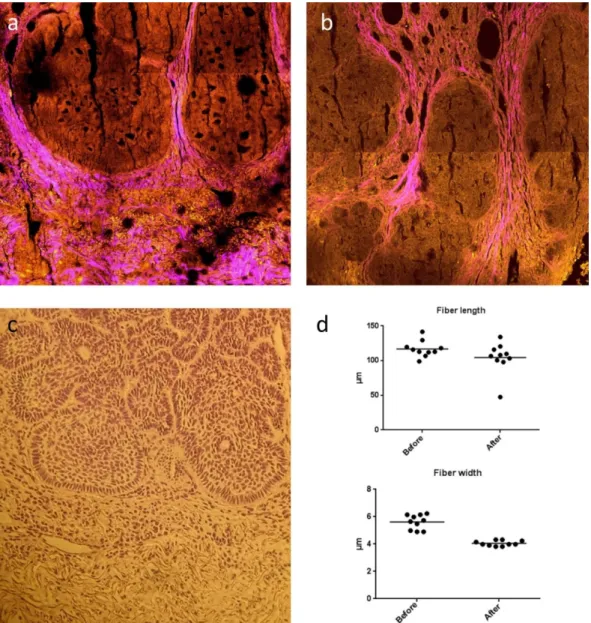
3.2.3. Assessment of tumor collagen environment by second harmonic generation and two-photon excitation fluorescence microscopy.....….51
4. Results ………....52
4.1. In vitro studies on Candida albicans ………..52
4.1.1. Ascorbate sensitivity of Candida albicans ……….52
4.1.1.1. Effect of different media and carbon source ………....52
4.1.1.2. Effect of oxygenation and temperature ……….54
4.1.1.3. Involvement of free iron ………55
4.1.1.4. Effect of growth history ……….…56
4.1.2. Imaging studies on C. albicans in different experimental conditions....57
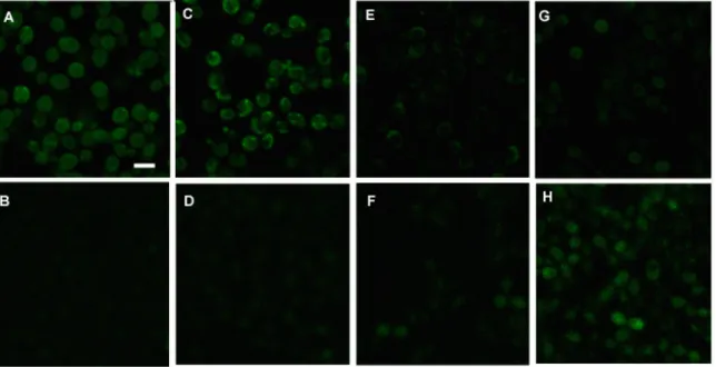
4.1.2.1. Detection of hydroxyl radical generation by conventional confocal microscopy……….57
4.1.2.2. Imaging of intracellular changes in redox state by combination of conventional confocal and two-photon excitation fluorescence microscopy ………...58
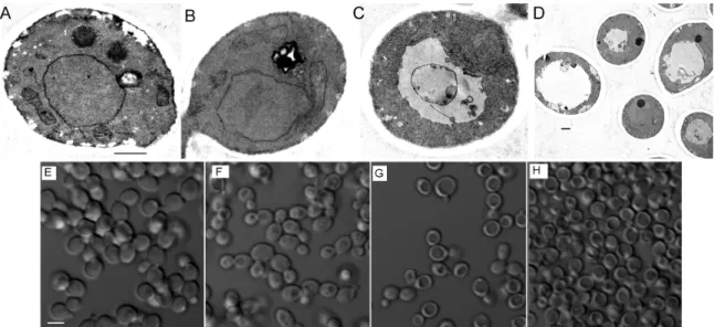
4.1.2.3. Tracking of morphological alterations at the cellular and subcellular level by brightfield and transmission electron microscopy ……….……60
4.2. Ex-vivo studies on basal cell carcinoma ……….……61
4.2.1. Label free imaging of tumor collagen environment to assess ascorbate’s effect on basal cell carcinoma ……….……….61
5. Discussion ………...63
6. Conclusion……….…….68
7.Summary ……….……69
8.Összefoglalás ……….…...70
9.References……….…..72
10. Acknowledgements ………..123
11. List of publications……….………..124
List of Abbreviations
ADP Adenosine diphosphate AMP Adenosine monophosphate
Asc•− Ascorbate (ascorbyl) radical, semidehydroascorbate Asc2− Ascorbate dianion
AscH• Neutral ascorbyl radical AscH− Ascorbate monoanion AscH2 Ascorbic acid
ATP Adenosine triphosphate BCC Basal cell carcinoma BCNS Basal-cell nevus syndrome CFU Colony forming units DHA Dehydroascorbic acid DMT1 Divalent metal transporter 1 DNA Deoxyribonucleic Acid ECM Extracellular matrix
EDTA Ethylenediaminetetra-acetic acid
FAD Flavin adenine dinucleotide (oxidized form) FADH2 Flavin adenine dinucleotide (reduced form) FDG 2-deoxy-2-[18F]fluoro-D-glucose
FMN Flavin mononucleotide
GAPDH Glyceraldehyde-3-phosphate dehydrogenase GLUT Facilitative glucose transporters
GSH Glutathione (reduced form)
GSSG Glutathione disulfide (oxidized form) H2O2 Hydrogen peroxide
HIF Hypoxia-inducible transcription factor HO• Hydroxyl radical
HPF 3′-(p-Hydroxyphenyl)-fluorescein Hsp90 Heat shock protein
IVA Intravenous pharmacologic ascorbic acid (as prooxidant) KRAS Kirsten rat sarcoma viral oncogene
MITF Melanocyte lineage-specification transcription factor mTOR Mammalian target of rapamycin
NAD(P)+ Nicotinamide adenine dinucleotide (phosphate) (oxidized form)
NAD(P)H Nicotinamide adenine dinucleotide (phosphate) (reduced form) Nd:YLF Neodymium-doped yttrium lithium fluoride
NNT Nicotinamide nucleotide transhydrogenase
NO Nitric oxide
NTBI Non-transferrin bound iron O2•− Superoxide
1O2 Singlet oxygen
P-Asc Pharmacologic ascorbic acid (as prooxidant) PARP Poly (ADP-ribose) polymerase
PBS Phosphate-buffered saline PET Positron emission tomography
PGC1 α Proliferator-activated receptor-gamma coactivator-1alpha
pKa Cologarithm logarithm of the dissociation constant (K) of an acid PTCH Patched
RNA Ribonucleic acid
RNS Reactive nitrogen species ROS Reactive oxygen species
SHG Second harmonic generation microscopy Shh Sonic hedgehog
Smo Smoothened
SVCT Na+-dependent Vitamin C transporters TCA Tricarboxylic acid
TEM Transmission electron microscopy TfR1 Transferrin receptor protein 1 TGF-β Transforming growth factor-β
TPEFM Two-photon excitation fluorescence microscopy UV Ultraviolet
VEGF Vascular endothelial cell growth factor VHL von Hippel-Lindau
YPD Yeast extract-peptone-dextrose YPG Yeast extract-peptone-glycerol
1. Introduction
1.1. Ascorbic acid – an overview 1.1.1. A brief history
During the Age of Discovery, which corresponds to years between 1500 and 1800, sailors spent at least 3 months continuously at the sea. Due to lack of access to fruits and vegetables for such long periods of time, more than 2 million sailors died of nutritional diseases such as beriberi, pellagra and scurvy, caused by thiamine (Vitamin B1), niacin (Vitamin B3) and ascorbic acid (Vitamin C) deficiency, respectively (1-3).
Among these diseases, scurvy was the most frequently encountered one that an English sea captain Sir Richard Hawkins described as the ‘’plague of the sea’’ (1). Although captains and naval surgeons were highly convinced that citrus fruits could cure scurvy, most physicians denied this theory for several years (4).
In 1907, two Norwegian physicians named Axel Holst and Theodor Frolich developed an interest in investigating the factors that led to a ship-related dietary disease then called ‘’shipboard beriberi’’ (5). In an attempt to accomplish this, they fed guinea pigs with a diet based on various types of grains. Although their attempts to develop beriberi failed; to their surprise, they observed the classical features of scurvy (5). Moreover, symptoms of scurvy were resolved upon addition of ‘’anti-scorbutics’’ such as fresh cabbage or lemon juice into their diet (5). This serendipitous discovery would open new horizons in Vitamin C research together with long lasting controversies over who deserved to be nominated for the Nobel Prize (6). In 1912, Casmir Funk, a Polish biochemist proposed that scurvy, beriberi, pellagra and rickets were due to dietary deficiencies of factors which he referred to as ‘vitamines’, a term that derived from
‘vita’: life and ‘amine’: nitrogen containing compound (7, 8). More than a decade later, in 1927, a Hungarian physician named Albert Szent-Gyorgyi discovered and isolated a substance called ‘’hexuronic acid’’ first from the plants at Cambridge University, then from the adrenal gland at Mayo Clinic in Rochester, USA (9-11). Upon his return to Hungary, he asked Joseph Svirbely, an American-born Hungarian post-doctoral fellow in his lab at the University of Szeged, to test whether this crystalline compound protected guinea pigs from scurvy (9, 12). Svirbely’s experiments demonstrated that
hexuronic acid was indeed an antiscorbutic factor, today known as Vitamin C. Around the same time, former mentor of Svirbely, Charles Glen King and William A. Waugh from the University of Pittsburgh, were working on isolation and crystallization of Vitamin C from the lemon juice. They have subsequently demonstrated that its daily administration protected guinea pigs from scurvy (13, 14). This generated a bitter dispute over priority, which would escalate in 1937 when Szent-Gyorgyi became the recipient of the Nobel Prize in Physiology or Medicine, in part, for isolating Vitamin C (9). Not long after, Szent-Gyorgyi succeeded to extract Vitamin C samples from paprika peppers and sent them to Walter Norman Haworth, who was at the time a Professor of Chemistry at the University of Birmingham. Haworth and his group determined the chemical structure of Vitamin C and due to its anti-scorbutic properties, together with Szent-Gyorgyi, they decided to rename it as L-ascorbic acid (15-20). Subsequently, Vitamin C was synthesized independently by both Tadeus Reichstein in Switzerland and Haworth’s group in England and from that point, it could be produced on a large scale for medical use (16, 21). This work earned Haworth the Nobel Prize for Chemistry the same year with Szent-Gyorgyi.
1.1.2. Chemistry and Biochemistry
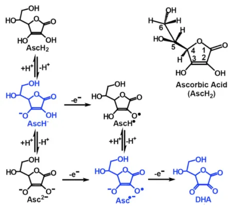
Ascorbic acid (L-ascorbic acid, AscH2, Vitamin C) is a ketolactone with a chemical formula of C6H8O6 and molecular mass of 176.12 g/mol. As a hexose sugar derivative, it shares a structural similarity to glucose. It is a water soluble, weak organic acid and has two pKa’s: pK1=4.2 and pK2=11.6 (22, 23). It is a strong reducing agent (antioxidant) by virtue of two enolic hydrogen atoms that it entails (24). Ascorbate monoanion (AscH−) is the most abundant form at physiological pH (22, 23). When AscH− donates a hydrogen atom (H+) or electron (e-) it forms ascorbate dianion (Asc2−) or neutral ascorbyl radical (AscH•), respectively (25). AscH• has a pKa of -0.86, and for this reason, it is present as an ascorbate radical (semidehydroascorbate, ascorbyl radical, Asc•−) at physiological conditions where pH is around 7.4 (25). Asc2− can also be transformed into Asc•− upon losing an e−. Asc•− is a relatively unreactive free radical and it can be converted back to AscH− by utilization of certain enzymes such as nicotinamide adenine dinucleotide phosphate (NADPH)-dependent thioredoxin reductase or nicotinamide adenine dinucleotide (NADH) dependent-cytochrome b5 reductase. (26-30). Alternatively, two Asc•− molecules form a dimmer that undergoes a
series of reactions, including a reversible disproportionation (31). This results in generation of AscH− and dehydroascorbic acid (DHA) (26, 27, 31) (Figure 1) (Net reacion: 2Asc•−+H+ ⟷ AscH−+DHA). DHA can be irreversibly hydrolysed to 2,3-diketogulonic acid but can also be recycled back to AscH− either directly by glutathione (GSH) itself or, by GSH or NADPH dependent enzymes such as thioredoxin reductase (26, 27, 32-35).
Figure 1: Ascorbic acid oxidation and recycling. Adapted and modified from (23).
Although oxidation of ascorbic acid is a slow process, its anionic forms have variable rates of oxidation (36). In general, rate of oxidation depends on the pH and the presence of catalytic metals (37, 38). At physiological pH, AscH2 and Asc2− coexist with AscH−, but the total concentration of AscH2 and Asc2− is less than 0.2% (23). An increase in pH, leads to an increase in the amount of Asc2−, which is in turn accompanied by an increased rate of oxidation resulting in generaton of Asc•− and superoxide (O2•−) (38).
Literature suggests that true autoxidation of AscH− is indeed very slow and according to
Williams and Yandell, Asc2− is the only Vitamin C species, which would go through a significant autoxidation (36, 38).
1.1.3. Absorption, Cellular Uptake and Excretion
Through evolution, humans lost their capacity to synthesize ascorbic acid de novo. This is due to a mutation in L-gulono-γ-lactone oxidase gene, which encodes L-gulono-γ - lactone oxidase enzyme that is required for the last step of ascorbic acid synthesis (39).
Hence, we are dependent on the dietary intake for obtaining adequate amounts of this essential vitamin. Upon ingestion, ascorbic acid is absorbed through the brush border cells of the small intestine. This takes place via two major mechanisms; such that ascorbate is taken up via Na+-dependent Vitamin C transporters (SVCT) and its two- electron-oxidized form DHA is taken up via facilitative glucose transporters (GLUT), mainly GLUT1 and GLUT 3 (40-43).
Two isoforms of SVCT mediate the transport of ascorbate; SVCT1 and SVCT2.
SVCT1, commonly called the ‘’bulk transporter’’, resides largely in apical brush border membranes of enterocytes and renal tubular cells, which are responsible for absorption and re-absorption, respectively (42, 44, 45). SVCT2 has a broader distribution and can be found in osteoblasts, platelets, cardiac, neural, neuroendocrine, exocrine and endothelial tissues (42, 45, 46). SVCT2’s activity seems to be crucial for protection against oxidative injury (47, 48). Unlike most tissues, epidermal cells express both SVCT1 and SVCT2 while dermal cells express SVCT2 only (49). Bioavailability of ascorbate is tightly controlled via regulation of SVCT expression. For example, when its levels are elevated in the intestinal lumen, SVCT1 is downregulated in enterocytes (50). Similarly, when intracellular ascorbate levels are reduced in lung epithelial cells, SVCT2 expression is increased (51). In vitro and in vivo studies have shown that hormones, paracrine factors, oxidative stress and intracellular signaling molecules also play a role in regulation of expression of SVCT (47, 52-54).
Affinity of GLUT to DHA is relatively low when compared to SVCT’s affinity for ascorbate (41, 55). Once DHA enters the cells, it is then immediately converted to ascorbate (41). However, there are exceptions to this case; for instance oxidative stress may hamper the accumulation of intracellular ascorbate from DHA (56). Presence of glucose may influence DHA uptake, as they compete for the same GLUT transporters,
different mechanism, Na+ dependent ascorbate transport was also shown to be modulated by glucose (55, 61). In addition to an endocrine regulated DHA transport, modulatory effects of intracellular signaling molecules and oxidative stress on DHA uptake have also been reported (53, 59, 62). However, it must be noted that SVCT and GLUT transporters may respond differently to the same hormone or cytokine. An intriguing example is the regulatory effects of transforming growth factor-β (TGF-β) on osteoblastic cells (63). While an increased rate of Na+-ascorbate cotransport activity is induced by TGF-β, no changes in DHA levels were observed in the same cells (63).
Amount of Vitamin C that is absorbed and excreted depends mainly on the intake, bioavailability, metabolism and the route of administration. In a study by Levine et al., a steep curve was observed between 30-100 mg daily doses whereas complete plasma saturation was reached at 1000 mg (64). Likewise, urinary excretion was only observed at and above 100 mg and, at a single dose of 500 mg or higher, although partially absorbed, Vitamin C was substantially excreted (64). Nevertheless, Padayatty et al.
demonstrated that, one can achieve a higher Vitamin C concentration in plasma, by delivering it intravenously rather than orally (65). In healthy adults, plasma concentration of ascorbic acid is usually in the range of 15-90 µmol (65-68). On the other hand, tissue concentrations vary depending on the tissue and cell type (68-72). For instance, average ascorbic acid contents of adrenal and pituitary gland were reported as 30-40 mg/100g and 40-50 mg/100g, respectively, while that of skeletal muscle was found to be only 3 mg/100g (69, 72).
Excretion of Vitamin C occurs via filtration and tubular reabsorption (73). However, when Vitamin C is presented to the tubules at a rate that is above the maximal rate of tubular reabsorption, excess Vitamin C is excreted in the urine (73). Pathologic conditions may alter stored and excreted levels of ascorbic acid (67, 74, 75). Spellberg and Keeton compared the levels of ascorbic acid excreted in healthy persons and cancer patients (75). They found that after a 400 mg daily ascorbic acid administration, first group excreted 56 to 80% of intake, whereas the latter excreted only 34 to 48% (75).
This may be due to metabolic differences in healthy and cancer tissues, which seem to be reflected by lower plasma levels of ascorbic acid in cancer patients (67, 76, 77).
Klimant and collegues also propose that a high level of Vitamin C consumption in certain pathological conditions is in part compensated by reduced rate of excretion by
As mentioned in the previous chapter, a fraction of DHA is irreversibly converted to 2,3-diketogulonic acid (27, 78). This metabolite can further be degraded into l- erythrulose and oxalate (79). It has been shown that oxalate excretion increases with high ascorbic acid intake (64). When further epidemiological evidence presented in the literature is taken into consideration, high levels of ascorbic acid seem to constitute a risk for oxalate nephropathy, especially when the renal function have been compromised (80-82).
1.1.4. Functions
Ascorbic acid is involved in several fundamental physiological and biochemical processes. Its major and probably the most important role lies in its property as an antioxidant (83). AscH− readily gives an electron to free radicals such as hydroxyl radical (HO•), O2•−, peroxyl radical, thiol radical, sulphur radicals and tocopheroxyl radical at the expense of generating an Asc•−.
AscH− + X•→ Asc•− + XH
Aside from its antioxidant activities, it is required as a co-factor in synthesis of norepinephrine, serotonin, tyrosine, homogentisic acid, carnitine, hydroxylysine and hydroxyproline. Moreover, it amidates peptides for hormone activation, mediates nitric oxide synthase and hypoxia-inducible transcription factor (HIF) activity, and assists iron absorption in the small intestine (26, 44, 78).
Two amino acids, proline and lysine are among the key components of collagen formation process. Proline needs to be hydroxylated to generate a more stable triple- helical structure of collagen (84). On the other hand, hydroxylysine not only acts as a precursor of the intra- and inter-molecular crosslinking process which gives collagen its tensile strength, but also facilitates the glycosylation process by serving as an attachment site for galactose and glucosylgalactose (85, 86). Hydroxylation of selective proline residues occurs by collagen prolyl-4-hydroxylase and prolyl-3-hydroxylase while lysine residues are hydroxylated by lysyl-hydroxylase (84). These three enzymes and γ-butyrobetaine dioxygenase and trimethylhydroxylase which catalyze the formation of L-carnitine, together with HIF prolyl-4- and asparaginyl- hydroxylases which suppress HIF-1 activity, belong to the family of 2-oxoglutarate and Fe2+-
dependent dioxygenases and require ascorbate either as a co-substrate or to recycle Fe3+
back to Fe2+ (23, 78, 87-90). Likewise, norepinephrine is synthesized by a copper- containing oxygenase, so called dopamine β-hydroxylase and it does require ascorbate as a co-factor (91).
Hormones and hormone-releasing factors such as gastrin, oxytocin, vasopressin, corticotropin, thyrotropin are initially synthesized as larger, inactive precursor molecules. They need to go through series of post-transitional modifications, to be converted to their active forms. The last step in this process is carboxyl-terminal α- amidation, which utilizes peptidyl glycine α-hydroxylating monooxygenase, an enzyme that is also dependent on O2, Cu+ and ascorbate (92, 93).
Tetrahydrobiopterin, a folic acid derivative, is a co-factor of several enzymes, including nitric oxide synthase, phenylalanine, tyrosine and tryptophan hydroxylase (94-97).
However, its plays a slightly different role for nitric oxide synthase in comparison with other enzymes (97). Binding of tetrahydrobiopterin to nitric oxide synthase, enables synthesis of nitric oxide (NO) (98). On the other hand, it gets rapidly oxidized to a short-lived intermediate, quinoid dihydrobiopterin, which then rearranges to dihydrobiopterin (98). As opposed to tetrahydrobiopterin, dihydrobiopterin inhibits NO formation and instead leads to O2•− generation (98). Ascorbate as a reducing agent and an antioxidant is able to maintain tetrahydrobiopterin in its reduced state (98, 99). In case of tyrosine, ascorbate is required for its catabolism (100). On the other hand, phenylalanine hydroxylase, an iron containing enzyme that catalyses the conversion of L-phenylalanine to L-tyrosine, requires, O2 and tetrahydrobiopterin as an electron carrier (101). During this process, tetrahydrobiopterin gets oxidized and an NADPH dependent enzyme so called dihydrobiopterin reductase recycles the oxidized form back to tetrahydrobiopterin. Stone and Townsley suggested that presence of ascorbate could also contribute to this recycling process (96).
Iron ingested from food presents in two forms; heme and nonheme iron. Heme, contains iron in ferrous (Fe2+) form, and it is derived from hemoglobin and myoglobin, found in meat, poultry and fish. Nonheme iron, which exists in ferric (Fe3+) state, is present in plant-based foods such as fruits and vegetables. It is known that dissociation of ferric compounds (eg. hydroxide, phosphates, complexes such as iron tannate) are much less than those of ferrous ones (102, 103). One of the key roles of ascorbate in iron
metabolism is that it promotes dietary nonheme iron absorption by reducing Fe3+ to Fe2+
together with duodenal cytochrome b reductase (103). An iron binding plasma glycoprotein, called transferrin, facilitates transport of iron through the bloodstream.
Although to a lesser extent, non-transferrin bound iron (NTBI) can also occur in the circulation (104). In order to bind transferrin, iron in ferrous form needs to be oxidized to Fe3+ by hephaestin (104).
Almost all cells acquire most of their iron from the serum iron-carrier protein transferrin, but they are also capable of importing it in the form of NTBI (104). The latter occurs through divalent metal transporter 1 (DMT1) and requires reduction of Fe3+
to Fe2+ (105, 106). This reduction occurs via release of ascorbate from the cytoplasm into the extracellular space (104-106). However, in case of transferrin dependent iron uptake, ascorbate can facilitate the uptake via an intracellular reductive mechanism, which follows a transferrin receptor dependent endocytosis of di-ferric transferrin complexes (104, 107). Once this complex is located inside the endosome, the endosome becomes acidified and enables release of Fe3+ from transferrin. A subsequent ferrireduction is followed by the release of iron, which then gets transported by DMT1 and/or Zip14 (104, 107). In addition to these properties, studies show that ascorbate is likely to further modulate iron metabolism by increasing the expression of the gene for the iron storage protein, ferritin, enhancing iron deposition, inhibiting lysosomal ferritin degradation and reducing iron efflux (104, 107).
Concentration of ascorbic acid in skin is relatively high when compared to other tissues (70, 108-110). In addition to dual expression of SVCT (SVCT1 and SVCT2) in the skin epidermis, there is also a 2 to 5 fold difference between the ascorbic acid content of the epidermis and dermis (49, 108-109). These findings suggest a high dependency on ascorbic acid, especially in the epidermis. There is growing evidence showing that ascorbic acid may play a role in differentiation of keratinocytes and formation of stratum corneum barrier lipids (111-113). In an in vitro study, Pasonen-Seppanen and collegues demonstrated that ascorbic acid improved stratum corneum structure, increased keratohyalin granules and the intercellular lipid lamellae present in the interstices of the stratum corneum (111). Extracellular matrix (ECM), which is an important component of connective tissue, entails two groups of biomolecules;
glycosaminoglycans and fibrous proteins such as collagen, elastin, fibronectin and
glycosaminoglycan synthesis, its deposition into the ECM and stimulate elastin (114, 115). Duarte et al. assessed the effect of ascorbic acid 2-phosphate, a more stable derivative of ascorbic acid, on gene expression in primary dermal fibroblasts and found an increase in expression of various genes that are involved in cell motility, matrix remodeling during wound healing, deoxyribonucleic acid (DNA) replication and repair (116). In agreement with these findings, several in vivo and clinical studies demonstrated that ascorbic acid plays a key role in wound healing (117-119). Protein and DNA damage induced by ultraviolet (UV) radiation is one of the leading causes of photoaging and photocarcinogenesis. Although cutaneous damage caused by UV radiation is a complex process, one of the proposed mechanisms of action for generation of UV damage is a possible reaction between UV induced hydrogen peroxide (H2O2) and metal ions that are already bound to DNA and, a subsequent generation of HO• (120, 121). A second proposed pathway is the lipid peroxidation of membranes caused by UV induced free radicals, which in turn may cause mutagenesis and cell death (121, 122). Ascorbic acid seems to ameliorate the damaging effects of UV both as a free radical scavenger and as an inducer of DNA repair and regeneration genes (122-128).
Increased consumption of ascorbic acid in such cases is likely to be compensated by an increased uptake by keratinocytes in an irradiation time and dose dependent manner (129). However, according to the current literature, in the context of modulation of UV induced skin damage, benefits of ascorbic acid alone is limited and satisfactory results can be achieved only when it is combined with two or more antioxidants (130-132).
1.2. Oxidative stress, antioxidants and prooxidants 1.2.1. Oxidative stress
A free radical is a reactive molecule that has an unpaired electron in its outer orbit. It may derive from endogenous metabolic processes, enzymatic systems or from external sources such as ionizing radiation, pathogens, drugs and chemicals. Free radicals tend to steal an electron from a surrounding molecule, which leaves the other molecule as a free radical with an unpaired electron. This may lead to a chain of reactions and result in cellular damage. Free radicals can bind to DNA bases and modify nucleotides, or cause DNA strand breaks via reacting with the 5-carbon sugar deoxyribose (121, 133). They can also oxidize thiol groups in cysteine residues and cause formation of a disulfide bond, which can alter the protein structure and function (134). Two hydrogen atoms (H+)that exist between the two double bonds of polyunsaturated fatty acids are highly prone to being targets of free radicals. Lipid peroxidation that is initiated by a reaction between one of these hydrogen atoms and an oxidizing agent such as HO• or iron- oxygen complex (perferryl or ferryl ion), leads to a cascade of events, resulting in formation of new radicals and a number of compounds such as; lipid alkyl radical, lipid peroxyl radical, lipid hydroperoxide, malondialdehyde and 4-hydroxy-2-noneal, that can in turn cause cellular damage (135, 136). Availablity of sufficient O2 is crucial during the propagation stage, as the newly generated lipid alkyl radical needs to react with O2
to form a lipid peroxyl radical, which will then steal a hydrogen atom to generate lipid hydroperoxide and a new lipid alkyl radical to continue the process. Among other enzymes, glutathione peroxidase seems to be the key enzyme that is shown to protect cells from the lipid peroxidation process and their effects (137, 138).
Molecular oxygen (O2) has two unpaired electrons and therefore it is a biradical. It readily accepts unpaired electrons and gives rise to a variety of partially reduced species named as reactive oxygen species (ROS). These include O2•−, HO•, peroxyl radical and alkoxyl radical. The cellular damage caused by ROS is termed oxidative stress. One- electron reduction of O2 to O2•− can occur by various processes such as mitochondrial electron transport by complex I and complex III, UVB induced type I photosensitizing reactions with internal chromophores, by NADPH oxidase, which gets activated by UVB exposure and found mainly in neutrophils, and by nitric oxide synthase as well as xanthine oxidase activity (139-142). O •− can react with another free radical, NO to form
peroxynitrite, a highly reactive intermediate which can damage a wide range of molecules in cells (143, 144). In an attempt to convert O2•− into less damaging species, superoxide dismutase catalyzes the dismutation of two molecules of O2•− into O2 and H2O2. Although H2O2 is not a free radical, it is a potent oxidant and it does contribute to generation of free radicals. This can occur via formation of hypochlorous acid (HOCl), a precursor of free radicals, by an enzyme called myeloperoxidase, by Fenton and Haber-Weiss reactions (Figure 2) or by various cyclical transition metal ion catalyzed redox reactions which all gives rise to HO•, the foremost reactive ROS (139, 145-147).
In an attempt to diminish potential harmful effects of H2O2, the heme-containing enzyme catalase and glutathione peroxidase participate in decomposition of H2O2 to H2O (148). In addition to HO• generation from H2O2, H2O exposed to ionizing radiation may also lead to formation of HO• and GSH plays an important role in its elimination (146).
2 O2•− + 2 H+ → H2O2 + O2
Fe3+ + O2•− ⇌ Fe2+ + O2
Fe2+ + H2O2 → Fe3+ + HO• + OH− Fenton Reaction O2•− + H2O2 → O2 + HO• + OH− Haber Weiss Reaction Figure 2: Fenton and Haber Weiss reactions (149-151).
1.2.2. Antioxidants
Normal physiological processes such as aerobic respiration continuously generate free radicals and oxidants and, when produced in moderate amounts, they play important roles in the regulation of intracellular signal transduction pathways, host defense system and immunity (152-157). Additionally, they are essential for a variety of catabolic and anabolic processes to take place. However, each cell maintains a homeostasis between prooxidant and antioxidant species and when there is an imbalance between the two, pathological processes ensue (139, 152, 158, 159).
Halliwell and Gutteridge first defined antioxidants in 1995 as ‘’any substance that, when present at low concentrations compared with those of an oxidizable substrate, significantly delays or prevents oxidation of that substrate’’ (160) but this definition
was later simplified and re-defined as ‘’any substance that delays, prevents or removes oxidative damage to a target molecule’’ (161, 162). Recently, Apak and collegues came up with a slightly different and more detailed definition and defined antioxidants as
‘’natural or synthetic substances that may prevent or delay oxidative cell damage caused by physiological oxidants having distinctly positive reduction potentials, covering ROS/reactive nitrogen species (RNS) and free radicals (i.e. unstable molecules or ions having unpaired electrons)’’ (163). However, a property that was not emphasized in these definitions is the ability of an antioxidant, which scavengers the radical, to generate a new radical which is stable through intramolecular hydrogen bonding upon further oxidation (152, 164). Moreover, substances which up-regulate antioxidant defenses may also qualify as antioxidants.
Antioxidant system is classified into two major categories; enzymatic and non- enzymatic antioxidants. Enzymatic antioxidants, such as superoxide dismutase, catalase and glutathione peroxidase prevent formation of ROS or reduce ones that have already been generated. For instance, glutathione peroxidase catalyses the reduction of H2O2 by two reduced glutathione (GSH) which leads to generation of oxidized glutathione (glutathione disulfide, GSSG) and two H2O (165). Regeneration of GSH from GSSG requires glutathione reductase as an enzyme and NADPH as an electron donor (165). In fact, many enzymes of the antioxidant system depend on NADPH for proper function and glucose-6-phosphate dehydrogenase acts as a major supplier for this intracellular reductant (166, 167). Therefore, both GSH/GSSG as well as NAD(P)H/NAD(P)+ ratios are considered to be important indicators of redox status of cells (166, 168, 169).
Non-enzymatic antioxidants are also categorized into two groups; endogenous antioxidants such as Vitamin A, Vitamin C and E, ubiquinol, carotenoids, urate, GSH, flavonoids, NAD(P)H, and synthetic antioxidants, which include butylated hydroxytoluene and butylated hydroxyanisole (152, 170).
α- and β-carotene, cryptoxanthin and lycopene, are the main carotenoids. Among these, β-carotene has a major role in Vitamin A formation by virtue of an enzyme called β, β- carotene-15, 15′ monooxygenase which catalyzes the centric cleavage of β-carotene to yield all-trans-retinal (171). All-trans-retinal can then be reduced to all-trans-retinol (Vitamin A) by retinol dehydrogenase (172, 173). Vitamin A and carotenoids’
antioxidant properties lie in their ability to quench singlet oxygen (1O2), neutralize thiyl
radicals, and combine with peroxyl radicals to protect cells from lipid peroxidation (173, 174). α-Tocopherol, the most dominant isoform of Vitamin E, exerts its main antioxidant effect by donating phenolic hydrogen to the peroxyl radicals, which in turn generate tocopheroxyl radicals (152, 175). This process protects cells from lipid peroxidation and consecutively maintains membrane integrity. In order to provide continuous supply and eliminate the newly generated radical, Vitamin E is recycled from its tocopheroxyl radical either by enzymes such as NADH-cytochrome b5
reductase or by nonenzymatic pathways, which utilize compounds such as AscH− and ubiquinol (176-180).
Glutathione is a tripeptide (cysteine, glycine, and glutamic acid) with a redox-active thiol group that generally exists in cells in its reduced state (GSH) (165). When GSH donates a hydrogen atom to a free radical intermediate, it is converted into a glutathiyl radical, which may react with a variety of species depending on the circumstances (181- 183). For instance, glutathiyl radical can enter an electron transfer reaction with AscH− to generate GS− and Asc•− (183). Glutathione also contributes to the detoxification process by conjugating with a plethora of reactive metabolites and reacting with electrophiles that are generated as a result of metabolic processes (165). Additionally, it facilitates recycling of Vitamins C and E from their oxidized forms and in turn increases availability of antioxidants (169).
Phenols are aromatic compounds that contain an –OH group attached to a benzene ring.
Phenols, which have more than two aromatic –OH groups, are termed as polyphenols.
Almost all phenols exert a degree of antioxidant activity as scavengers of reactive species such as peroxyl radical, HO• and HOCl. Some also serve as chelating agents by binding transition metal ions, which further reduces oxidative stress (184, 185).
Polyphenols are further classified according to the number of phenol rings they accommodate and the structures that bind these rings to one another. According to these properties, they are divided into four major categories; Phenolic acids, flavonoids, stilbenes, and lignans (186). Among these, more attention has been given to flavonoids.
Flavonoids consist of a fifteen-carbon skeleton that entails two benzene rings (A and B) linked by three carbon atoms that usually form a third oxygenated heterocyclic ring (C) (187). In majority of cases, B ring is attached to C ring in the 2-position but this may differ among different types of flavonoids such as isoflavones and neoflavonoids. Those
structural features of the C ring and include flavonols, flavones, flavanones, flavanols, flavanonols, catechins, anthocyanins (186). Flavonoids owe some of their antioxidant properties to the phenolic hydroxyl groups attached to ring structures, which can serve as reducing agents, hydrogen donors, O2•− scavengers and 1O2 quenchers (188-190).
Furthermore, specific ones serve as chelators of iron and copper, inhibitors of oxidases such as xhanthine oxidase and NADH oxidase and activators of detoxifying enzymes such as glutathione S-transferase (190, 191). Some can also replace antioxidant activity of α-tocopherol in the membrane, reduce α-tocopheryl radicals and regenerate α- tocopherol (190, 192-194).
Coenzyme Q is an endogenously synthesized lipid soluble substance that participates in the mitochondrial respiratory chain as an electron carrier (195). Ubiquinone and ubiquinol are the predominant oxidized and reduced forms of Coenzyme Q, respectively (196). Reduction of ubiquinone to ubiquinol occurs by a variety of oxidoreductases such as Complex I, Complex II, electron transfer flavoprotein-ubiquinone oxidoreductase, and non-proton pumping NADH dehydrogenases (in yeast) (196). In mammalian cells, reoxidation of ubiquinol to ubiquinone occurs only by Complex III, whereas in case of yeast, alternative oxidases also take part in the process (196). Ubiquinol is an effective antioxidant. Studies show that it prevents lipid peroxidation, takes part it regeneration of Vitamin E from the α-tocopheroxyl radical and halts oxidation of membrane proteins (180, 195-199).
1.2.3. Prooxidant properties of antioxidants
There is already substantial evidence that antioxidants play a major role in metabolic pathways and protect cells from the harmful effects of reactive species. However, some of the antioxidants can also exhibit prooxidant effects (200, 201). Whether an antioxidant behaves as a prooxidant seems to depend on multiple factors such as its concentration, cellular redox state, availability of other antioxidants and, the presence and amount of O2, catalytic metal ions and radicals. For instance, green tea polyphenol (-)-epigallocatechin-3-gallate was shown to generate HO• and O2•− and cause cleavage of DNA in the presence of Cu2+ (202). In a study by Palozza et al., β-carotene when tested at high concentrations, increased generation of ROS in leukemia cells (203).
Moreover, retardation of cell cycle progression and proapoptotic activity highly
concentrations were applied. The authors further assessed the variations in response to β-carotene in differentiated and undifferentiated leukemia cells and found that increase in ROS generation was observed at lower concentrations in undifferentiated cells when compared to differentiated ones. Likewise, post-treatment levels of GSH were higher in differentiated cells than those that are undifferentiated (203). Certain flavonoids, which act as powerful antioxidants, when tested at high concentrations, were shown to generate ROS, induce DNA strand breaks, oligonucleosomal fragmentation as well as caspase-3 activation (201, 204-206). However, sometimes it is challenging to distinguish an antioxidant dose range from a prooxidant dose range. For example, while fisetin, a plant polyphenol from the flavonoid group, at 22 µmol/L could protect cells from DNA strand breaks caused by H2O2, it could itself induce DNA strand breaks when H2O2 is absent (206). There is also some uncertainty about how flavonoids exert their cytotoxic and DNA damaging effects. Watjen et al. (206) found no indication for metal-catalysed oxidation, lipid peroxidation and ROS involvement whereas Sahu et al.
(207) demonstrated that in the presence of transition metal ions, ROS and mainly HO• was responsible for lipid peroxidation and the DNA damage caused by quercetin.
Duthie and colleagues observed DNA strand breaks as well as a decrease in GSH levels, however they found no evidence for oxidative DNA base damage (208). It might very well be possible that nucleus was protected from ROS at the expense of intracellular GSH (208). Agullo and co-workers argued that degree of quercetin cytotoxicity was dependent on cellular proliferative activity, and this selective cytotoxicity could be utilized to inhibit growth in tumor cells (209). The group observed a diminished lactate release in dividing cells, which was likely to be caused by its inhibitory action on lactate transporter (209, 210). Nevertheless, a dramatic reduction in levels of adenosine triphosphate (ATP) was observed for both exponentially growing and stationary growing human colonic carcinoma cells (209). DNA topoisomerases play a role in DNA replication, transcription, and recombination by introducing transient breaks in the DNA. These enzymes also act to regulate DNA supercoiling generated during transcriptional elongation (211). Evidence suggests that some flavonoids such as myricetin, quercetin and fisetin can inhibit topoisomerase II (212, 213). This may arrest cell cycle and induce apoptosis via a p53 dependent or independent pathway (201, 213, 214). Selected flavonoids can also exhibit antimicrobial activity (215). One example is quercetin, which was shown to demonstrate antibacterial activity by inhibiting gyrase
antioxidants exert their prooxidant effects through inhibition of cellular respiration.
Pavani et al. reported that nordihydroguaiaretic acid, a polyhydroxyphenolic antioxidant, acts as an inhibitor of mitochondrial electron transport, by interrupting the flow of electrons at the NADH-dehydrogenase-ubiquinone compartment of ascites tumor cells (217). A consequential decrease in ATP synthesis led to decreased cell viability and growth rates in the same cells (217). Several other phenols were also shown to inhibit mitochondrial electron flow at different sites and/or uncouple redox reactions from that of adenosine diphosphate (ADP) phosphorylation (218-221).
Capsinoids, which possess antioxidant properties mainly via forming complexes with the reduced metals and donating H+ to radical intermediates, can protect cells from iron- mediated lipid peroxidation and copper-dependent oxidation of low-density lipoproteins (222-224). Studies showed that some of its analogues may also exert selective prooxidant effects via preferentially inhibiting NADH oxidase activity in the plasma membrane of cancer cells. This inhibition was accompanied by an inhibition of growth and, apoptosis (225, 226). Furthermore, when HL-60 human promyelocytic leukemia cells were induced to differentiate, capsaicin’s effect on the plasma membrane NADH oxidase activity was much less (225). Morre and colleagues further suggested that in capsaicin resistant cases, selective inhibition of NADH oxidase activity and subsequent inhibition of growth could be achieved with co-administration of mild oxidizing agents such as H2O2 and t-butyl hydroperoxide (226). These findings indicate that modification of redox state may be considered as an alternative approach for cancer types that do not respond to therapy.
1.2.3.4. Vitamin C as a prooxidant
Ascorbic acid, as an electron donor, gained most of its popularity through its antioxidant effects (83). However it may also act as a prooxidant, at pharmacologic doses (P-Asc) when specific conditions [eg. Kirsten rat sarcoma viral oncogene (KRAS) positivity] are met (23, 227). Literature suggests that P-Asc can exert selective toxicity against some tumor cells and infectious microorganisms (23, 227-229). Nevertheless, these effects remain controversial for various reasons such as fundamental differences between in vitro and in vivo conditions, lack of understanding of the exact mechanism of action and scarce clinical data that supports its efficacy.
The most well accepted mechanism of action of P-Asc has been linked to its ability to generate H2O2 (228, 230, 231). It is already well known that AscH− reduces Fe3+ to Fe2+
at the expense of producing an Asc•− (229). A subsequent reaction of Fe2+ with O2
generates Fe3+ and O2•− (229). H2O2 is then formed via dismutation of two molecules of O2•−. A reaction between the newly generated H2O2 and Fe2+ leads to formation of HO• and Fe3+ (Fenton reaction) (Figure 1). AscH− can further reduce Fe3+ back to Fe2+ for the cycle to continue. Studies have demonstrated that levels of antioxidant enzymes differ across tissues and among certain cancer cells versus normal ones (232, 233). Low levels of catalase and glutathione peroxidase detected in a variety of cancer cells renders them especially vulnerable to H2O2, and indirectly to P-Asc(233). According to Doskey and colleagues, this vulnerability also varies among different tumor cell types because not all cell types possess same degree of catalase activity (230). Nevertheless, mechanisms for how H2O2 elicited by P-Asc, induces toxicity to tumor cells are still under investigation. Some studies suggest that poly (ADP-ribose) polymerase (PARP) activation through H2O2 induced DNA damage may lead to catabolization of NAD+, which could in turn deplete the substrate for NADH formation and hinder ATP synthesis (234-236). In the process of disposing H2O2 by an NADPH-dependent glutathione reductase/peroxidase system, NADPH is utilized to reduce GSSG back to GSH. In order to replenish NADPH that is consumed during this process, some of the glucose that is used for glycolysis could be diverted to the pentose phosphate pathway that in turn would result in reduction of ATP synthesis (234, 236). H2O2 induced inhibition of glycolysis by inactivation of glyceraldehyde-3-phosphate dehydrogenase (GAPDH) is assumed to further decrease NADH production and generation of ATP (236, 237). Also, at the level of mitochondria, inhibition of ATP synthase by H2O2
exposure appears to interrupt ADP phosphorylation and in turn ATP production (237, 238). However, findings by Du et al. were not in full alignment with some of these hypotheses (231). Although the group agrees that decreased cell viability caused by P- Asc is via a H2O2 mediated mechanism, they also indicate that PARP activation and depletion of ATP may not be involved in P-Asc induced cytotoxicity. Their experiments rather suggest that P-Asc’s effects are through a caspase-independent cell death mechanism that is associated with autophagy (231).
‘’Warburg Effect’’ first described by Otto Warburg refers to the phenomenon that, in tumor and other proliferating cells, there is an increased rate of glucose uptake and,
even in aerobic conditions, fermentation and subsequent production of lactate is preferred over oxidative phosphorylation (239-243). This preference towards a less efficient pathway for ATP synthesis was initially attributed to defective mitochondria (241). On the other hand, some more recent studies disputed this hypothesis and suggested that cancer cells heavily engage in both glycolysis and oxidative phosphorylation in order to be able to generate sufficient levels of ATP and NADPH but also to synthesize nucleotides and amino acids, which are all crucial for cell proliferation (244, 245). Lately, it has been further elaborated that under certain circumstances tumors can exert a metabolic plasticity to maintain growth and survival (246). One example is melanocyte lineage-specification transcription factor (MITF) upregulated proliferator-activated receptor-gamma coactivator-1alpha (PGC1α) positive melanomas, which are highly dependent on oxidative phosphorylation rather than glycolysis (247). Contrarily, an activated BRAF mutation leads to suppression of oxidative phosphorylation and induces a glycolytic phenotype (248). Moreover, evidence suggests that tumor microenvironment is heterogeneous (249). For instance, centers of solid tumors are generally poorly perfused lacking sufficient glucose and O2
supply. Therefore, metabolic activity may also vary within the tumor itself. On the other hand, some authors argue that when tumor microenvironment is hypoxic, HIF-1 gets activated which in turn induces enzymes involved in glycolysis, upregulate GLUT transporters, reduce mitochondrial function to save O2 and in turn reliance on glycolysis becomes more pronounced (239). This process is further enhanced via a positive feedback loop between glucose metabolites and HIF-1 (239, 250). Apart from hypoxia, HIF-1 can also be activated by other factors such as activation of Ras oncogene or loss of tumor suppressor von Hippel-Lindau (VHL) which all lead to a tendency of tumor cells to shift energy production towards glycolysis, even under normoxic conditions (239). Taken together, it seems that tumor cells tend to heavily depend on glycolysis despite the presence of a functional mitochondria but when the circumstances change, a metabolic switch is likely to occur. Consistent with this phenomenon, studies show that majority of tumors significantly upregulate GLUT to meet the glucose demands for increased glycolysis (251-253). Considering that specific GLUT channels are also responsible for transporting DHA across cellular membranes, one would expect a higher uptake of DHA by the tumor cells. Based on this assumption, Yun and collegues tested the effect of P-Asc on KRAS and BRAF mutant colorectal cancer cells that are
associated with upregulation of GLUT-1 transporter and elevated levels of oxidative stress caused by depletion of GSH during the conversion of DHA to its reduced form (236). Their study further revealed that GAPDH activity and consequently glycolysis were inhibited by ROS induced pentose posphate pathway activity as well as PARP activation, which leads to diminished level of NAD+, the substrate required for GAPDH.These chain of events resulted in depletion of ATP, cellular energy crisis and cell death (236). Spielholz et al. demonstrated that melanoma cell lines take up DHA at a rate that is at least 10 times greater than normal melanocytes (254). Subsequently, Corti and colleagues (255) documented that, in the presence of iron, gamma- glutamyltranspeptidase, a plasma membrane enzyme that is often highly expressed in human malignancies (256), could facilitate the oxidation of AscH− to DHA and promote its uptake through upregulated GLUT transporters in melanoma cells (254). Studies have shown that genetic variations in SVCT-2 are associated with risk of certain cancers such as lypmhoma, human papillomavirus type 16-associated head and neck cancer and gastric cancer (257, 258). On the other hand, Lv and collegues investigated whether level of expression of SVCT-2 would play a role in selective cytotoxicity of P-Asc in cancer stem cells of hepatocellular carcinoma (259). The results suggested that SVCT-2 expression was inversely associated with P-Asc concentrations needed to decrease hepatocellular carcinoma cells by 50%. Moreover, SVCT-2 expression was positively correlated with intracellular P-Asc concentrations and response to P-Asc. Wang et al.’s study further showed that P-Asc treatment of knockdown of SVCT-2 in cholangiocarcinoma cells resulted in less DNA damage, ATP depletion and mammalian target of rapamycin (mTOR) inhibition (260).
HIF-1 is a heterodimeric complex that plays an integral role in adaptive responses of the tumor cells to changes in O2 (261, 262). This involves not only a metabolic adaptation via channeling cells to a glycolytic pathway, but also transcriptional activation of various pro-angiogenic factors to increase O2 delivery (262). There is growing body of evidence, which suggests that AscH− by virtue of its role as a cofactor for HIF hydroxylases, may limit activation of HIF-1 (263). Two retrospective studies identified an inverse association between ascorbate levels in human endometrial (264) and colorectal tumor (265) tissues and the activation of HIF-1 pathway. Higher ascorbate content in tumor tissue was also associated with longer post-surgical disease free period (265). Another study demonstrated that in tumor-bearing Vitamin C dependent Gulo−/−
mice, increase in ascorbate intake alleviated levels of HIF-1α expression (266). On the other hand, in VHL-defective renal cancer cells, that already entail high levels of HIF- 1α activity, a higher uptake of DHA was observed through the HIF-1α upregulated GLUT-1 transporters (267). Given that these cells already rely on glycolysis through Warburg effect, P-Asc induced ROS generation and subsequent PARP activation led to NAD+ consumption and left very small amounts of NAD+ for glycolysis to proceed (267, 268). Consequently, significant reduction in cellular reserves of ATP promoted cell death (267). Whether P-Asc can selectively control tumor cell growth via inhibition of HIF-1 signaling or whether overexpression of HIF-1 serves, as an advantage for P- Asc’s selective cytotoxicity towards tumor cells, seems to depend on the tumor type, concentration of administered P-Asc, amount of O2 available in the tumor microenvironment and the factors that influence the activation of HIF-1 (267). For instance, HIF-1α in VHL-defective renal cancer cells are constitutively stabilized and hypoxia or HIF prolyl hydroxylase activity has little influence on their activation (267, 269). In such cases, inhibition of HIF via prolyl hydroxylase may not play a significant role in P-Asc’s anti-tumor activity. Vascular endothelial cell growth factor (VEGF) is a downstream gene product of HIF-1α and has been the focus of various targeted therapies (261, 270). Since HIF-1 activation could be potentially suppressed by P-Asc treatment, a possible concomitant decrease in VEGF was also investigated (264).
Human endometrial tumor samples revealed a strong inverse correlation between level of VEGF protein and ascorbate content (264). In a separate in vitro and in vivo study, P-Asc treated sarcoma 180 cancer cells had lower levels of VEGF and other two angiogenesis related proteins, but the authors did not elaborate on the mechanism of action (271). Wilkes and collegues noted that P-Asc treatment did reduce VEGF secretion which correlated with a decrease in HIF-1α expression but these effects were through a H2O2 mediated pathway rather than a O2 or prolyl hydroxylase-dependent inhibition of HIF-1α (272).
Iron plays a critical role for proliferation and metabolism of cancer cells and infectious microorganisms such as bacteria and fungi (273, 274). These rapidly dividing cells are highly dependent on presence of iron to carry out various cellular processes such as DNA synthesis, cell cycle regulation and oxidative phosphorylation (274, 275). In order to meet the increased iron demands, many type of cancer cells upregulate proteins that are involved in its uptake. Transferrin receptor 1 (TfR1) is one of these proteins that
became the target of antibody-mediated chemotherapeutic agents (276). Iron chelators such as desferrioxamine and 3-aminopyridine-2-carboxaldehyde thiosemicarbazone are also being considered as potential anti-cancer agents (277). On the other hand, iron’s ability to gain and lose electrons also enables it to participate in Haber-Weiss reaction, which leads to generation of free radicals. Therefore, in cells, which are rich in iron, a complementary strategy for anti-infective or anti-cancer therapy is to focus on agents that foster free radical generation. Cysteine, GSH and AscH− can slowly release iron from the iron storage protein ferritin (278). However, P-Asc by triggering an uncontrolled release of iron can generate a surplus, which in presence of O2, would lead to production of O2•−, HO• and H2O2 (229, 279, 280). ROS generated by P-Asc, can further increase the labile iron pool in part via H2O2 mediated disruption of iron-sulfur cluster proteins (280). This labile iron pool redox cycling can contribute to the P-Asc induced selective cytotoxicity.
Schoenfeld and collegues reported that P-Asc treatment exerted preferential killing against of glioblastoma and advanced-stage-non-small-cell lung cancer cells in vitro and in vivo (280). In their study, GLUT mediated DHA uptake did not play a role in P- Asc’s selective cytotoxicity but they argued that the effect was rather dependent on the increased redox-active labile iron present in cancer cells (280). Vilcheze et al. assessed the efficacy of P-Asc in treatment of Mycobacterium tuberculosis and their results also demonstrated that presence of high iron concentration played a critical role in its bactericidal effects (229). Kang et al. took a different view and proposed that P-Asc mediated death of melanoma cells were caused by P-Asc induced downregulation of TfR, which resulted in diminished iron uptake and subsequent apoptosis (281).
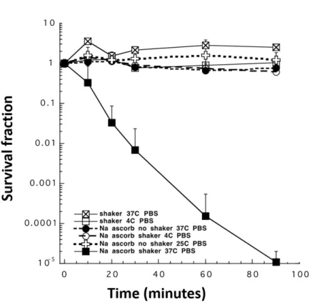
P-Asc has been the subject of various studies, which assessed its antibacterial, antifungal and antiviral properties (229, 282). Its inhibitory effects on the growth of various microorganisms such as Staphylococcus aureus, Helicobacter pylori, Mycobacterium tuberculosis, Bacillus cereus, and Candida have already been demonstrated (229, 283-287). However, studies, which elucidate its mode of action are limited and, the specific conditions required for P-Asc to exert its anti-microbial effects seem to vary depending on the infectious agent. For instance, while P-Asc can inhibit Helicobacter pylori in microaerophilic conditions, similar concentrations of P-Asc were shown to increase survival under aerophilic conditions (286). On the contrary, P-Asc’s
(229). Earlier studies suggested that the bactericidal effect of P-Asc was due to lowering of the pH (288, 289). In 1950, Slade and Knox challenged this hypothesis when they found a bacteriostatic effect for group A hemolytic streptococcus at near neutral pH (290). According to Ehrismann, while P-Asc stimulated growth in anaerobes, its effect was inhibitory in case of aerobes (288). In the process of exploring the role of O2 in bactericidal effects of P-Asc, Lwoff and Morel found that inhibition of Proteus vulgaris was halted by the presence of reducing agents and substances that catalyzed degradation of H2O2 (291). In 1954, Myrvik and Volk’s short-term growth experiments on Escheria coli (E. coli), was an attempt to reveal the chemical group that is responsible for the antibacterial properties of P-Asc. They reported that while enediol group of P-Asc had no antibacterial effect on E. coli, oxidized enediol (diketone) could exert immediate and strong antibacterial effects (284). Peloux and collegues proposed that P-Asc had little or no anti-viral activity in the absence of metal ions (282). Polio virus was completely inactivated when P-Asc was combined with 5 µM Cu2+ whereas, in the presence of ethylenediaminetetra-acetic acid (EDTA), P-Asc had no effect (282).
Interestingly, presence of various microorganisms such as Acinetobacter baumanii and Candida albicans (C. albicans) induce human neutrophils to uptake DHA rapidly and recycle it to AscH−, such that AscH− concentrations of activated neutrophils in vitro could increase up to 30 fold above the concentrations present in resting neutrophils (292, 293). Whether accumulation of AscH− in such high amounts is an attempt of the phagoctytotic cells’ to enhance ROS generation or the cells want to take the advantage of its antioxidant properties while undergoing an oxidative burst is still an enigma (294- 297).
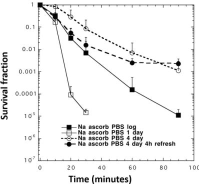
Literature on P-Asc’s antifungal effects on C. albicans is contradicting. P-Asc was shown in vitro to reduce virulence of C. albicans cells by lowering proteinase secretion (285). The same study also demonstrated that P-Asc could arrest cell growth and, induce concentration dependent cytotoxicity, which was potentiated when the cells were treated with H2O2 (285). In a separate study by the same group, P-Asc induced oxidative stress related enzyme activities in C. albicans were examined and reduced levels of GSH, decreased enzyme activity of catalase, glutathione reductase, glutathione peroxidase and glutathione S-transferase were reported (298). These effects are similar to those observed upon continuous exposure of yeast cells to H2O2, where cells
diminished, indicating inhibition of GSH synthesis (299). Surprisingly, some antioxidant effects were also demonstrated, such that with increasing concentrations of P-Asc, superoxide dismutase activity was increased and lipid peroxidation decreased (298). On the other hand, locally applied P-Asc in human subjects showed no antifungal activity against vaginal candidiasis or other antifungal infections (300). Only when P- Asc was applied upon a successful treatment of fungal infection, it was able to prevent reinfection (300). Brajtburg and collegues suggested that P-Asc potentiates the lethal action of amphotericin B on C. albicans and Cryptococcus neoformans cells (301).
Although in case of amphotericin B, P-Asc had such enhancing effects, P-Asc antagonized the effects of fluconazole both in vitro and in systemic murine candidiasis model (302). This inhibition was attributed to P-Asc’s reducing property which could diminish the oxidative effect of fluconazole induced ROS (302). Moreover, neither intragastrically nor intraperitoneally administered P-Asc had any effect on the survival of mice with systemic candidiasis (302). When Van Hauwenhuyse and colleagues further investigated the antagonistic effect of P-Asc on fluconazole, they found that only in the presence of Upc2 (a transcriptional regulator of Erg11 gene, which encodes an enzyme that is the target of azole antifungal drugs and is involved in ergosterol biosynthesis (303, 304)) P-Asc could exert its antagonistic effects (305). Their analysis also revealed that, P-Asc could restore the ergosterol levels and reverse elongated cell growth caused by inhibition or depletion of heat shock protein (Hsp90), and this activity was Upc2 dependent (305). Nevertheless, it is worth to mention that in normal cells, P- Asc lowered ergosterol levels and did not initiate elongated growth (305, 306).
In the 1970s, Ewan Cameron and colleagues conducted the initial clinical trials to test the effect of P-Asc on improving the survival of patients with terminal cancer (307- 309). Although their results were promising, subsequent double-blinded randomized studies in the Mayo Clinic demonstrated that orally administered P-Asc had no effect on patient survival (310, 311). It was later recognized that the route of administration might account for some of the discrepancy observed in, in vivo studies. Data suggested that intravenously administered P-Asc produced much higher plasma concentrations than the orally administered P-Asc due to the tightly controlled absorption process (64, 65, 312, 313). For instance, when 10 g of P-Asc was delivered via infusion, it was possible to achieve plasma concentrations up to 5 mM, whereas predicted peak plasma concentration for P-Asc given at the maximum tolerable oral dose, 3 g every 4 hours, is