Influence of glutathione-S-transferase (GST) inhibition on lung epithelial cell injury: role of oxidative stress and metabolism
Marianne E. Fletcher,1Piers R. Boshier,2Kenji Wakabayashi,1Hector C. Keun,3Ryszard T. Smolenski,4,9 Paul A. Kirkham,5,6Ian M. Adcock,5Paul J. Barton,5Masao Takata,1and Nandor Marczin1,7,8
1Anaesthetics, Pain Medicine and Intensive Care, Imperial College London, London, United Kingdom;2Biosurgery and Surgical Technology, Imperial College London, London, United Kingdom;3Biomolecular Medicine, Department of Surgery and Cancer, Imperial College London, London, United Kingdom;4Department of Biochemistry, Medical University of Gdansk, Gdansk, Poland;5National Heart and Lung Institute, Imperial College London, London, United Kingdom;
6Department of Biomedical Sciences, University of Wolverhampton, Wolverhampton, United Kingdom;7Department of Anaesthetics, Royal Brompton and Harefield NHS Foundation Trust, Harefield Hospital, Harefield, Middlesex, United Kingdom;8Department of Anaesthesia and Intensive Therapy, Semmelweis University, Budapest, Hungary;9Department of Surgery and Translational Medicine, University of Milano-Bicocca, Milano, Italy
Submitted 11 August 2014; accepted in final form 1 April 2015
Fletcher ME, Boshier PR, Wakabayashi K, Keun HC, Smolenski RT, Kirkham PA, Adcock IM, Barton PJ, Takata M, Marczin N.
Influence of glutathione-S-transferase (GST) inhibition on lung epi- thelial cell injury: role of oxidative stress and metabolism. Am J Physiol Lung Cell Mol Physiol 308: L1274 –L1285, 2015. First published April 10, 2015; doi:10.1152/ajplung.00220.2014.—Oxi- dant-mediated tissue injury is key to the pathogenesis of acute lung injury. Glutathione-S-transferases (GSTs) are important detoxifying enzymes that catalyze the conjugation of glutathione with toxic oxidant compounds and are associated with acute and chronic inflam- matory lung diseases. We hypothesized that attenuation of cellular GST enzymes would augment intracellular oxidative and metabolic stress and induce lung cell injury. Treatment of murine lung epithelial cells with GST inhibitors, ethacrynic acid (EA), and caffeic acid compromised lung epithelial cell viability in a concentration-depen- dent manner. These inhibitors also potentiated cell injury induced by hydrogen peroxide (H2O2), tert-butyl-hydroperoxide, and hypoxia and reoxygenation (HR). SiRNA-mediated attenuation of GST-but not GST- expression reduced cell viability and significantly enhanced stress (H2O2/HR)-induced injury. GST inhibitors also induced intra- cellular oxidative stress (measured by dihydrorhodamine 123 and dichlorofluorescein fluorescence), caused alterations in overall intra- cellular redox status (as evidenced by NAD⫹/NADH ratios), and increased protein carbonyl formation. Furthermore, the antioxidant N-acetylcysteine completely prevented EA-induced oxidative stress and cytotoxicity. Whereas EA had no effect on mitochondrial ener- getics, it significantly altered cellular metabolic profile. To explore the physiological impact of these cellular events, we used an ex vivo mouse-isolated perfused lung model. Supplementation of perfusate with EA markedly affected lung mechanics and significantly in- creased lung permeability. The results of our combined genetic, pharmacological, and metabolic studies on multiple platforms suggest the importance of GST enzymes, specifically GST-, in the cellular and whole lung response to acute oxidative and metabolic stress.
These may have important clinical implications.
N-acetylcysteine; ethacrynic acid; caffeic acid; reactive oxygen spe- cies; viability
ACUTE LUNG INJURY (ALI) and acute respiratory distress syn- drome (ARDS) are life-threatening inflammatory disorders,
which may result from both medical and surgical situations such as sepsis, heart surgery, and lung transplantation (41).
Among these, ischemia and reperfusion injury constitutes a unique entity, wherein metabolic and oxygen starvation and subsequent reoxygenation can lead to the release and accumu- lation of reactive oxygen species (ROS) (4). ROS can directly oxidize and damage DNA, protein, and lipid membranes, inactivate antioxidant enzymes, and enhance expression of proinflammatory genes, ultimately leading to cell injury and apoptosis (11).
The glutathione-S-transferase (GST) antioxidant enzymes form a multifunctional superfamily of intracellular isozymes that catalyze the conjugation of reduced glutathione (GSH) with a variety of toxic compounds, including xenobiotics and oxidative intermediates (such as lipid and DNA hydroperox- ides and aldehydes) (17, 18). This renders them less toxic and facilitates their removal from the cell (17, 18). Their key role in cellular detoxification protects macromolecules from attack by reactive electrophiles, such as ROS products, environmental carcinogens, and chemotherapeutic agents. Increasing evidence suggests that GST expression and function are important in both cancer progression (24, 39) and acute inflammatory con- ditions (23). For example, Luo et al. (23) demonstrated that treatment of mice with recombinant GSTP1 attenuated LPS- induced acute inflammation.
The clinical importance of diminished endogenous GST expression and activity is emerging. At least five of the eight GST genes in humans have functional polymorphisms where substitutions or deletions affect enzyme expression and activity and are present in up to 50% of the population (16). Such polymorphisms are believed to contribute to individual differ- ences in the clearance of xenobiotics and oxidative stress products and to susceptibility to various inflammatory and proliferative pathologies (16). For instance, individuals with homozygous GSTM1 gene deletions appear to be more sus- ceptible to DNA damage induced by GSTM1 substrates (32) and are more at risk of developing lung adenocarcinoma (7).
Preliminary clinical studies also suggest that attenuated GST expression may contribute to ALI (25). Moradi and colleagues (25) showed that GSTM1 polymorphisms were associated with increased mortality in critically ill patients with ALI/ARDS. In the setting of lung transplantation, donor GST polymorphisms
Address for reprint requests and other correspondence: N. Marczin, Section of Anaesthetics, Pain Medicine and Intensive Care, Imperial College London, Harefield Hospital, Harefield, Middlesex, UK. (e-mail: n.marczin@imperial.
ac.uk).
First published April 10, 2015; doi:10.1152/ajplung.00220.2014.
dramatically increased the development of primary graft dys- function (8, 14). Despite these suggestions, the exact role of GST attenuation in lung cell injury, the basic mechanisms involved, and the relative impact of different GST isozymes are not known.
We hypothesized that GST attenuation would lead to in- creased intracellular oxidative stress affecting mitochondrial energetics and cellular metabolism, resulting in lung cell in- jury. To test these ideas, we studied the influence of pharma- cological inhibitors ethacrynic acid (EA) and caffeic acid (CA) (1, 28, 29, 37, 49) and RNA silencing of GST isoforms on lung epithelial cell survival, ROS generation, and energetic/meta- bolic status in murine cell culture models of ALI. Finally, the physiological impact of GST inhibition was also explored using an ex vivo murine-isolated perfused lung (IPL) model by focusing on potential effects on lung mechanics and permea- bility.
MATERIALS AND METHODS
Cell culture. Murine lung epithelium (MLE)-12 cells (ATCC, Rockville, MD) were grown in Hanks medium (F12:MEM) supple- mented with 1% penicillin/streptomycin and 5% fetal calf serum at 37°C in humid conditions with 5% CO2-95% air. For hypoxia and reoxygenation (HR) experiments, we used deoxygenated culture me- dium (bubbled with 95% N2-5% CO2 gas mixture under sterile conditions) and a hypoxic chamber (Mini Galaxy A; Wolf Laborato- ries, York, UK). Cells were exposed to hypoxic conditions for 12 or 24 h. Subsequently, medium was exchanged for fresh, prewarmed, oxygenated medium, and cells were exposed to normoxic conditions for increasing periods of reoxygenation (2, 6, 12, or 24 h).
GST inhibition: method and efficacy.The pharmacological agents EA and CA were employed to attenuate activity of purified bovine liver GST (350 mU) or endogenous GSTs in cell lysates. The degree of inhibition was evaluated using the GST fluorometric activity assay kit (Biovision, Mountain View, CA). A linear increase in fluorescence at excitation/emission (ex/em) 380/460 nm was measured over the initial 10-min period and calculated as fluorescence U (FU)/min. A standard calibration curve was used to determine GST activity (mU) in unknown samples.
To complement the pharmacological data, we employed RNAi to specifically knockdown GST- or -isoforms. This was achieved using predesigned GST- and - ON-TARGETplus Smartpool siRNA (Dharmacon siRNA Technologies, Thermo Scientific, Glas- gow, UK). In brief, 20 pmol of scrambled control or GST-targeting siRNA was introduced into MLE cells using Lipofectamine 2000 (Invitrogen, Carlsbad, CA). After 5 h, the siRNA-Lipofectamine medium was replaced with fresh medium, and cells were incubated for an additional 48 h before further experimentation.
The extent of siRNA-mediated knockdown of GST expression was evaluated by RT-PCR. Total RNA was extracted from transfected cells using the RNAeasy mini kit (Qiagen, Valencia, CA) (13) and quantified spectrophotometrically (Biophotometer Plus; Eppendorf, Stevenage, UK). Single-stranded cDNA was synthesized from 2 g RNA with TaqMan RT-PCR reagents using GeneAmp PCR System 9700 (Applied Biosystems, Warrington, UK) (13). The cycling conditions were 25°C for 10 min, 48°C for 30 min, and 95°C for 5 min. cDNA was amplified using the TaqMan gene expression master mix (Applied Biosystems) and TaqMan probes specifically targeted against mouse GST- (Mm00833915_g1) and GST- (Mm00496606_m1) (Applied Biosystems). A standard nucleic acid template and water were used as positive and negative controls, respectively. Variations in RNA loading between reactions were normalized by measurement of 18S ribosomal RNA (12). PCR was performed under recommended thermal-cycling conditions: initially
50°C for 2 min and 95°C for 10 min (for enzyme activation), followed by 40 cycles of 95°C for 15 s (cDNA denaturation) and 60°C for 1 min (probe annealing and extension). Data were collected using the ABI PRISM 7700 and Sequence Detection System 1.9.1 (Applied Biosystems) and analyzed using the comparative cycle threshold (Ct) method (5).
Protein expression was evaluated by Western blotting. In brief, protein extracts (50 g) were separated on 10% SDS-PAGE, trans- ferred to nitrocellulose membranes, and incubated overnight with polyclonal anti-GST- (1:500) or anti-GST- (1:500) antibodies (Abcam, Cambridge, UK). Antibody binding was detected using peroxidase-conjugated anti-goat IgG (1:2,000; Cell Signaling Tech- nology, New England Biolaboratories, Hitchin, UK) and chemilumi- nescence detection reagents (Amersham Biosciences, Little Chalfont, UK). Equivalent loading of protein samples was confirmed by mouse monoclonal anti--actin or anti-␣-tubulin IgG (1:2,000; Sigma, St.
Louis, MO).
Analysis of lung cell viability.The effect of GST inhibition on cell viability was evaluated under control conditions and in combination with known oxidative stress conditions: hydrogen peroxide (H2O2), tert-butyl hydroperoxide (tBH), or HR. Cell viability was analyzed by the 2,5-diphenyltetrazolium bromide (MTT) reduction assay (26). The optical density was measured as an index of cell viability, at 595 nm using an MRX II plate reader (Magellan Biosciences, Surrey, UK).
To delineate the involvement of apoptosis vs. necrosis, we em- ployed the annexin V-FITC, propidium iodide (PI) apoptosis/necrosis detection kit (Sigma) and fluorescence-activated cell sorting (FACS)Calibur flow cytometer (Becton Dickinson, Franklin Lakes, NJ) (38). Har- vested cells were exposed to annexin V-FITC and PI in binding buffer for 10 min in the dark at room temperature. Ten thousand events were acquired and analyzed using the CellQuest program. Cellular debris (with low forward- and side-light scatter) was excluded from the analysis. FITC and PI fluorescence were detected at 525 nm and 620 nm, respectively. Quadrant statistics display annexin V and PI fluo- rescence on the x- andy-axis, respectively, to identify viable cells (negative for both annexin V and PI), apoptotic cells (positive for annexin V, negative for PI), and late apoptotic dead cells (labeled with both annexin V and PI). In most instances, apoptosis involves acti- vation of caspase cascades. To further evaluate the role of apoptosis, cells were treated in the presence or absence of 100 M zVAD (Sigma), a broad-spectrum caspase inhibitor, and cell viability was assessed by MTT assay.
Detection of oxidative stress.The influence of GST inhibition on intracellular ROS was quantified using the ROS-sensitive fluorescent dyes dihydrorhodamine (DHR) and 2=,7=-dihydrodichloro-fluorescein diacetate (H2DCF-DA) (Invitrogen). MLE cells were preincubated for 30 min with 5M DHR or 20M H2DCF-DA before exposure to the GST inhibitor EA for 1 h. Cells incubated in media alone or H2O2
served as negative and positive controls, respectively. Following treatment, cells were harvested and resuspended in FACS wash buffer (PBS supplemented with 5 mM EDTA, 2% FCS, and 0.1% sodium azide). Samples were analyzed by flow cytometry at ex/em 495/525 nm. ROS quantification is reported as mean fluorescence intensity (MFI). To evaluate the role of specific ROS in EA-induced cytotox- icity, cells were preincubated for 1 h before EA treatment with the ROS scavengers,N-acetylcysteine (NAC, 10 mM) or cell permeable polyethylene glycol (PEG)-conjugated catalase (5,000 U/ml), or in- hibitors of catalase (aminotriazole, ATZ, 10 mM), superoxide dismu- tase (diethyldithiocarbamate, DETC, 1 mM), NADPH oxidase (diphe- nyliodonium, DPI, 10M), and nitric oxide synthase (nitro-L-arginine methyl ester,L-NAME, 10 mM).
To evaluate the effect of GST inhibition on mitochondrial energet- ics (relative levels of ATP, ADP, and AMP) and redox status (re- flected by NAD and NADH ratios) (2, 21, 31), intracellular adenine nucleotides were extracted from control and experimental cells as described previously (47) and measured by reverse-phase high-per- formance liquid chromatography (HPLC) and UV detection at 254
nm. Briefly, samples were extracted with ice-cold perchloric acid (0.4 M) and neutralized with equimolar KOH. KClO4 was then precipi- tated by centrifugation, and the supernatant was retained. Levels of ATP, ADP, AMP, NAD⫹, and ADPR, a breakdown product of NADH following acid precipitation (21, 31), were analyzed using the Agilent HPLC 1100 system. The mobile phase was 0.1 M potassium dihydrogen phosphate (pH 6.0) with 0.15 M potassium chloride as phase A and 15% acetonitrile in A as phase B with the Hypersil BDS column and a flow rate 0.9 ml/min. The concentration of each nucleotide was expressed as nanomoles per milliliter of extract with reference to external standards used for calculation of concentrations with reference to cell protein.
Protein carbonyls were estimated using a 2,4-dinitrophenylhydra- zine (DNPH)-based procedure reported elsewhere (6, 33) with minor changes. Briefly, cell lysates containing 0.1 mg protein were incu- bated with 0.1% DNPH for 1 h at room temperature. Samples were precipitated by addition of 30% trichloroacetic acid. Following cen- trifugation, pellets were washed with ethanol:ethyl acetate (1:1 vol/
vol) and dissolved in 8 M guanidine-hydrochloride, 13 mM EDTA, and 133 mM Tris pH 7.4. Optical density at 365 nm was determined using a Hitachi U-2000 UV-VIS spectrophotometer. The carbonyl content was calculated using Beer’s Law and an extinction coefficient of 2.1⫻104M/cm. Results are reported as nanomoles carbonyl per milligram of protein.
Metabonomics. Global metabolic changes associated with GST inhibition and oxidative stress were detected by metabonomic analysis (3). Cellular extracts from control and experimental groups were prepared for nuclear magnetic resonance (NMR) spectroscopy anal- ysis. A Bruker DRX600 spectrometer (Bruker Biospin, Rheinstetten, Germany) was employed, operating at 600.13 MHz 1H NMR fre- quency and 283 K. Raw spectra were phased, baseline corrected, calibrated to the trimethylsilylpropionic acid (TMSP) internal stan- dard resonance (dH⫽0.00 ppm) and imported into Matlab (Student 2007; The MathWorks, Natick, MA). Spectra were normalized to protein. Isolated metabolite signals within spectra were defined through visual inspection, and their peak areas were determined.
Metabolite assignment was made using the Madison Metabolomics Consortium Database (9).
Ex vivo IPL model.To investigate the physiological relevance of our cellular findings, the direct impact of GST inhibitor EA on lung physiological parameters was explored using an IPL mouse model.
All procedures were approved by the United Kingdom Home Office in accordance with the Animals (Scientific Procedures) Act 1986, UK.
Male C57BL6 mice (Charles River Laboratories, Margate, UK) aged 8 –12 wk were used for the ex vivo IPL system (Hugo Sachs Elek- tronik; Harvard Apparatus, March-Hugstetten, Germany). In brief, mice were anaesthetized, tracheostomized, and ventilated with a custom-made mouse ventilator as previously described (35) at a respiratory rate of 80 breaths/min. After thoracotomy/laparotomy and heparinization, animals were exsanguinated, and the pulmonary artery and left atrium were cannulated. Lungs were perfused in a nonrecir- culating manner with prewarmed RPMI 1640 supplemented with 4%
BSA and 1.4 g/l NaCl (Sigma) to increase osmolarity, as described
previously (34). All experiments had an initial 15-min equilibration period, and low tidal volume (VT) of 8 ml/kg was applied throughout to minimize lung injury development. Peak inspiratory pressure (PIP), positive end-expiratory pressure, and gas flow were monitored con- tinually via transducers connected to a PowerLab data acquisition system (AD Instruments, Chalgrove, UK). Data were analyzed using Chart software (version 5.5.6, AD Instruments). Accurate measure- ments of VT, respiratory system compliance, and resistance were determined by end-inflation occlusion at 30-min intervals.
Lung permeability index.The pulmonary endothelial/epithelial per- meability to a fluorescence-labeled albumin was assessed, using an adaptation of a previously reported fluorescence-based method for measuring vascular leak into the alveolar space (45). Following a 2-h perfusion, 1 mg/ml Alexa Fluor 594-conjugated albumin (Invitrogen) was added to the perfusate in the recirculating circuit. After 30 min, the perfusate was collected from the reservoir and stored in the dark.
Thereafter, the circuit was switched to a nonrecirculating system for 5 min to wash out the residual fluorescence from the lung vasculature.
Bronchoalveolar lavage fluid (BALF) was obtained as described previously (44). Fluorescence levels of perfusate, BALF, and lung tissue were measured using a fluorescence plate reader (Flx-800;
BioTek Instruments, Potton, UK) at absorption 590 nm. An index of lung endothelial and epithelial permeability was then estimated by the ratio of lung tissue:perfusate and lavage fluid:perfusate fluorescent signal, respectively. Protein levels in BALF were also quantified as another index of pulmonary permeability.
Statistics.Data are reported as the means of repeated experiments⫾SD.
The majority of statistical analyses were performed using Prism software (version 4.0; GraphPad, La Jolla, CA). Metabonomics data were analyzed by SPSS 17 (Chicago, IL). Comparison of three or more variables was carried out by either one-way ANOVA or repeat- ed-measure ANOVA with multicomparisons/post hoc tests (including Bonferroni) where appropriate. Statistical significance of metabonom- ics data was assessed by Kruskal-Wallis test. Multiple comparisons of metabonomics data were controlled for using the Benjamini-Hoch- berg procedure, with accepted false discovery rate 0.05. A Pvalue
⬍0.05 was considered significant.
RESULTS
EA and CA inhibit GST activity and decrease lung cell viability.To study the efficiency of GST inhibition, pure GST and MLE cell lysates (prepared from 4 ⫻ 106 cells) were incubated with increasing concentrations of GST inhibitors at 37°C for 1 h. Both EA and CA effectively inhibited GST activity in pure GST as well as MLE cell lysates in a concen- tration-dependent manner with an IC50of⬃25M and 1 mM, respectively. Exposure of MLE cells to increasing concentra- tions of EA and CA revealed major morphological changes and a concentration-dependent reduction in cell viability, assessed by MTT assay. After 5-h exposure, 100 and 300 M EA reduced viability to 92⫾7% and 13⫾7% of control (Fig. 1A).
0.0 0.5 1.0 1.5 2.0 2.5 3.0
0 50 100 150 200 250 300
Cell viability [OD]
Ethacrynic acid [μM]
*
*
*
A
0.0 0.5 1.0 1.5 2.0 2.5 3.0
0 0.2 0.4 0.6 0.8 1 1.2 1.4
Cell viability [OD]
Caffeic acid [mM]
*
*
*
B
Fig. 1. Influence of increasing concentrations of glutathione-S-transferase (GST) inhibitors on murine lung epithelium (MLE) cell viabil- ity. Viability was assessed by 2,5-diphe- nyltetrazolium bromide (MTT) assay following 5-h ethacrynic acid (EA) exposure (A) and 24-h treatment with caffeic acid (CA) (B);n⫽4 – 6;
*P⬍0.05 vs. untreated control.
Similar findings were obtained following 24-h treatment with CA, which led to a 55⫾ 35% reduction in cell viability at 1 mM (Fig. 1B). To explore whether GST inhibitor-induced cytotoxicity was limited to epithelial cells or applicable to other cell types in the lung, we have explored the effect of EA on primary mouse lung endothelial cells available in our laboratory. Exposure of these cells to increasing concentrations of EA for 5 h showed that the endothelial cell viability was also compromised by 300M EA (74 ⫾7% of control).
Apoptotic nature of cell death. The nature of EA-induced cell death was investigated by staining MLE cells with annexin V and PI, and fluorescently labeled cells were identified using FACS (Fig. 2). Untreated cells showed 90% cell viability,
which decreased to 74% and 17% following 3- and 5-h incu- bation with 300M EA, respectively. This was associated with a significant rise in the necrotic cell population, i.e., those exhibiting PI fluorescence in the absence of annexin V label- ing. Moreover, after 5 h of treatment, the percentage of annexin V-fluorescent cells increased by ⬎10-fold, indicative of an apoptotic cell population. Cells exhibiting both annexin V and PI increased from 1 to 57% of total population.
To identify whether caspases are involved in EA-induced cell injury, MLE cells were incubated with zVAD (100 M) before treatment with 0 –300M EA (Fig. 3A). As a positive control, we evaluated whether zVAD preincubation influenced cell death induced by a combination of tumor necrosis factor-␣
PI intensitySide Scatter
Forward Scatter
Annexin V intensity
Untreated EA 300μμM, 3h EA 300μM, 5h
8.3 1.5
89.9 0.3 73.8
24.7
17.4 1.3
0.2
21.8 57.4
3.4
Fig. 2. Apoptotic/necrotic cells determined by annexin V (Ann V)/propidium iodide (PI) staining and flow cytometry. Cells were treated with 300M EA for the indicated times, and untreated cells were used as a negative control. A representative forward-scatter/side-scatter gating strategy is illustrated to exclude cell debris from the analysis. The gated cell population was then acquired for Ann V and PI staining. Viable: Ann V and PI negative. Apoptotic: Ann V positive and PI negative. Necrotic: PI positive. Apoptotic/Necrotic: Ann V and PI positive.
0.0 0.5 1.0 1.5 2.0 2.5
Control TNF + CHX
Cell viability [OD]
Vehicle zVAD
*
B
0.0 0.5 1.0 1.5 2.0 2.5 3.0
0 50 100 150 200 250 300
Cell viablility [OD]
Ethacrynic acid [μM]
Vehicle zVAD
A
Fig. 3. Influence of caspase inhibitor zVAD on stress-induced MLE cell death. MLE cells were exposed to stresses EA (A) and tumor necrosis factor-␣ (TNF-␣)/cycloheximide (CHX) (B) for 5 and 24 h, respectively;n⫽3– 6; *P⬍ 0.05 vs. vehicle control (i.e., in the absence of zVAD).
(TNF-␣)/cycloheximide treatment (Fig. 3B). Whereas zVAD preincubation attenuated cell death induced by TNF/cyclohex- imide (cell viability of 24% vs. 5%), it had no influence on EA-induced cell death.
Potentiation of stress-induced cytotoxicity by GST inhibitors.
H2O2 and tBH alone resulted in a concentration-dependent reduction in cell viability over 5 h. These stress-induced cytotoxic effects were significantly potentiated by subtoxic concentrations of GST inhibitors, namely 100M EA (Fig. 4, A and C) and 0.5 mM CA (Fig. 4B). For instance, cell viability was reduced from 64 ⫾ 24% after 0.5 mM H2O2
treatment to 26⫾14% and 31⫾5% in the presence of EA and CA, respectively, compared with untreated controls.
Similarly, EA exacerbated tBH-induced cell death. For instance, following 1 mM tBH treatment, the presence of GST inhibitor reduced viability from 71⫾4% to 23⫾14%
of untreated controls.
Before studying the influence of EA on MLE cell survival in the setting of HR, we explored the influence of increasing duration of HR on cell viability. Interestingly, hypoxia up to 24 h and reoxygenation up to 6 h did not appear to compromise MLE cell viability measured as MTT activity (Fig. 4D). Injury, however, was unmasked in the presence of EA. For instance, 25 M EA had no measurable effect during normoxia, but it reduced viability of hypoxic control by 19⫾9%. In addition, 50 M EA reduced cell viability by 35⫾ 11% in normoxic conditions but led to a 77⫾10% decrease in viability during HR conditions.
Oxidative stress is involved in EA-induced cytotoxicity. To test the hypothesis that EA-induced cell injury is mediated by an altered intracellular redox state (1, 37), we initially moni- tored oxidative stress with redox-sensitive fluorescent dyes.
Exposure of MLE cells to EA (300M) for 1 h increased the MFI of DHR123 and DCF by over 2- and 30-fold, respectively, compared with basal levels (Fig. 5, A andB). In the control experiment, EA had no direct effect on dye autofluorescence in the absence of MLE cells after 3 h (data not shown).
To assess global intracellular redox state, nucleotides NAD⫹ and ADPR (the breakdown product of NADH) were measured by HPLC and UV detection, and their ratio was calculated.
Figure 5C shows that redox state was altered in a concentra- tion-dependent manner by EA exposure alone, causing a de- crease in NAD⫹/NADH ratios to 11% of untreated control by 300M EA. Furthermore, H2O2also reduced NAD⫹/NADH ratios, and this effect was potentiated by EA (43 vs. 28% of untreated control following 3-h treatment with H2O2 in the absence or presence of 100 M EA, respectively, P⬍ 0.05) (Fig. 5D).
To further evaluate intracellular oxidative stress in the set- ting of GST inhibition, excess formation of carbonylated pro- tein was monitored spectrophotometrically. Exposure of cells to 100 M EA alone did not affect steady-state levels of protein carbonylation (Fig. 5E). H2O2alone increased protein carbon- yls from 1.2⫾0.2 to 2.6⫾0.3 nmol/mg, and EA significantly enhanced H2O2-induced increase in protein carbonyls from two- to fourfold, compared with untreated control after 3 h of treatment (Fig. 5E).
Inhibitors of H2O2and NO do not attenuate EA-induced cell injury.We have attempted to further characterize the reactive species involved in EA-mediated lung cell injury. Specifically, to test the hypothesis that increased generation of H2O2 is responsible for EA-induced cell injury (37), we investigated the effect of modulating catalase activity by preincubating cells with either cell-permeable (PEG-conjugated) catalase (5,000 U/ml) or the catalase inhibitor ATZ (20 mM). Although cata- lase completely abolished the cytotoxic effect of H2O2 (Fig.
6B), EA-induced cell injury was unaffected by PEG catalase (Fig. 6A). ATZ-induced catalase inhibition also had no influ- ence on EA cytotoxicity (Fig. 6C).
To further evaluate the role of intracellular oxidative stress in GST inhibitor-induced cytotoxicity, we preincubated cells with a variety of inhibitors, affecting well-known oxidative stress pathways and the broad-spectrum antioxidant NAC.
Exposure of cells toL-NAME and DPI, targeting ROS gener-
0.0 0.5 1.0 1.5 2.0 2.5
0 0.1 0.2 0.3 0.4 0.5
Cellviability[OD]
Hydrogen peroxide [mM]
Vehicle CA
B
*
* *
* *
0.0 0.5 1.0 1.5 2.0 2.5
0 0.1 0.2 0.3 0.4 0.5
Cellviability[OD]
Hydrogen peroxide [mM]
Vehicle EA
A
*
*
C
* *
* *
* * *
0.0 0.5 1.0 1.5 2.0 2.5
0 0.2 0.4 0.6 0.8 1 1.2 1.4
Cellviability[OD]
Tert-butyl hydroperoxide [mM]
Vehicle EA
D
*
*
*
† 0.0
0.5 1.0 1.5 2.0 2.5
Normoxia HR
Cellviability[OD]
Treatment
0μM EA 25μM EA 50μM EA Fig. 4. Impact of GST inhibition on MLE cell
response to oxidative stress conditions. Influ- ence of chemical stressors hydrogen peroxide (H2O2) (AandB) and tert-butyl hydroperoxide (tBH) (C) on cell viability, determined by MTT assay, in the absence (Œ) and presence of GST inhibitors EA (Œ) and CA ().D: expo- sure of cells to 24 h of either normoxic or hypoxia (24 h) followed by reoxygenation (6 h) (hypoxia/reoxygenation, HR) in the pres- ence of indicated concentrations of EA;n⫽5;
*P⬍0.05 vs. vehicle controls (without GST inhibitors); †P ⬍ 0.05 vs. appropriate nor- moxic control.
ators nitric oxide synthase and NADPH oxidase, respectively, had no impact on EA-induced cell death. However, cells preincubated with the ROS scavenger NAC (10 mM) fully protected cells from injury induced by 300M EA (92⫾6%
vs. 13⫾ 7% of untreated control,P⬍0.05) (Fig. 7C) and 1
mM CA (84 ⫾ 6% vs. 45⫾ 35% of untreated control,P ⬍ 0.05) (Fig. 7D). Furthermore, pretreatment with NAC com- pletely abolished EA-induced increased fluorescence (MFI) of ROS-sensitive dyes DHR123 and DCF (45⫾8% vs. 14⫾2%
and 352⫾ 49% vs. 9⫾1%,P⬍ 0.05) (Fig. 7,AandB).
A
0 20 40 60 80
MFI [495/525 nm]
Ethacrynic acid [μM] DHR 123
*
0 100 300
B
0 200 400 600 800
MFI [495/525 nm]
Ethacrynic acid [μM]
DCF *
*
C
0 10 20 30 40
NAD/NADH ratio
Ethacrynic acid [μM]
*
E
0 2 4 6 8 10
EA H2O2 EA +
H2O2 Protein carbonyls [nmoles/mg]
Treatment
*
Δ
Control
D
0 10 20 30 40
EA H2O2 EA +
H2O2
NAD/NADH ratio
*
*
Control
Treatment
0 100 300 0 100 200 300
Fig. 5. Influence of GST inhibitor EA on intracellular oxidative stress measured by fluorescence of redox-sensitive dyes dihydrorhodamine 123 (DHR123) (A) and dichlorofluorescein (DCF) (B) fluorescence, following 1-h incubation with indicated concentrations of EA;n⫽5. MFI, mean fluorescence intensity.
Intracellular redox status was assessed by NAD⫹/NADH (by measuring ADPR, a breakdown product of NADH under acid precipitation conditions) ratio following increasing concentrations of EA (over 1 h) (C) and EA (100M) treatment (D) with or without 0.8 mM H2O2stress over 3 h;n⫽3.E: influence of EA (100M) and/or H2O2(0.8 mM) on protein carbonyl levels after 3-h treatment;n⫽3; *P⬍0.05 vs. untreated control,oP⬍0.05 vs. cells exposed to H2O2.
C
0.0 0.5 1.0 1.5 2.0 2.5
0 50 100 150 200 250 300
Cell viability [OD]
Ethacrynic acid [μM]
vehicle catalase
A
0.0 0.5 1.0 1.5 2.0 2.5
0 0.2 0.4 0.6 0.8
Cell viability [OD]
Hydrogen peroxide [mM]
vehicle catalase
* * *
B
0.0 0.5 1.0 1.5 2.0 2.5
0 50 100 150 200 250 300
Cell viability [%]
Ethacrynic acid [μΜ] vehicle ATZ
Fig. 6. Influence of catalase on EA-induced (A) and H2O2-induced (B) MLE cell death.
Cells were treated for 5 h, and catalase groups are represented by the dashed line;n⫽4 – 6.
Influence of catalase inhibitor aminotriazole (ATZ) on EA-induced cytotoxicity is depicted inC. Cells were treated for 5 h, and ATZ is represented by dashed line;n⫽4.
GST inhibition has no influence on mitochondrial energetics.To investigate whether EA mediates cellular injury through oxi- dative stress affecting mitochondrial energetics, we determined ATP/ADP nucleotide ratios in extracts of cells exposed to EA, H2O2, or both treatments over 3 h and 5 h. In control cells, ATP levels were 27.6⫾1.7 nmol/mg protein, and ADP levels were 2.1⫾0.3 nmol/mg protein. ATP/ADP ratios were not affected by EA exposure alone, with ratios of 17 ⫾ 2, 15 ⫾ 1, and 20⫾4 following exposure to 100M, 200M, and 300M respectively for 3 h. By contrast exposure of cells to H2O2, used as a positive control, significantly reduced ATP/ADP ratio from 12⫾4 to 5⫾3 after 5 h (P⬍0.05). EA (100M) had no influence on H2O2-induced reduction in ATP/ADP ratio (5 ⫾3 vs. 3⫾ 2 nmol/mg protein,P⬎0.05) (Fig. 8).
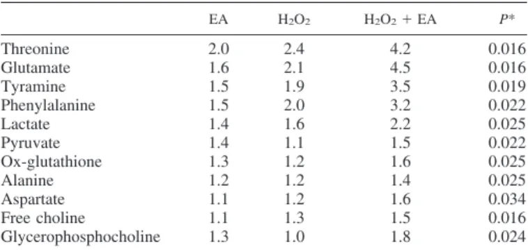
Changes in intracellular metabonomic profile.Metabonom- ics was employed to explore the impact of GST inhibitor EA and oxidative stress on global metabolic profile. 1H-NMR spectra acquired from control, EA-, H2O2-, and H2O2 ⫹ EA-treated MLE cells. Spectral regions that could be assigned to specific metabolites, which showed visual evidence of vari- ation in peak intensities, were subject to further analysis. Table 1 shows the fold change of assigned metabolites significantly affected by oxidative stress and EA. Changes in metabolite levels were enhanced after dual treatment with H2O2and EA
compared with treatment with EA or H2O2alone. Metabolites with the most prominent changes in response to treatment with H2O2and EA included glutamate (4.5-fold increase), threonine (4.2-fold increase), and tyramine (3.5-fold increase). Metabo- nomics also revealed a number of unassigned metabolites whose levels were altered by H2O2and EA exposure.
Genetic manipulation of GST-impacts on cell viability in control and stress conditions.To develop a cellular model of GSTM1-null and GSTP1 Ile105 polymorphisms, we investi- gated the impact of specific GST isoform knockdown by RNA interference. The efficiency of siRNA-mediated RNA knock- down was assessed by real-time PCR, 48 h following transfec- tion. Cells transfected with GST-and -siRNA showed 87⫾4%
and 90 ⫾ 3% knockdown of RNA expression, respectively, with no significant effect of control scrambled siRNA (Fig.
9A). Protein expression of GST- and - was evaluated by Western blotting 48 and 72 h after transfection. Whereas GST-protein expression was abundant in A549 cell line and expressed in whole mouse lung, the expression of this protein was low in MLE cells (Fig. 9C), and therefore the influence of GST knockdown could not be monitored by Western blotting. However, protein bands indicate that GST-pro- tein is absent in cells transfected with GST-siRNA at 48 and 72 h (Fig. 9B).
0.0 0.5 1.0 1.5 2.0 2.5 3.0
0 50 100 150 200 250 300
Cell viability [OD]
Ethacrynic acid [μM]
vehicle NAC
* * * *
C
0.0 0.5 1.0 1.5 2.0 2.5 3.0
0 0.2 0.4 0.6 0.8 1 1.2 1.4
Cell viability [OD]
Caffeic acid [mM]
vehicle NAC
* * *
D
0 10 20 30 40 50 60
0 300
MFI [495/525 nm]
Ethacrynic acid [μM] vehicle NAC
*
DHR 123
A
0 100 200 300 400 500
0 300
MFI [495/525 nm]
Ethacrynic acid [μM]
vehicle NAC
*
B
DCFFig. 7. Influence ofN-acetyl cysteine (NAC) on EA-induced increase in fluorescence of redox- sensitive dyes DHR123 (A) and DCF (B) over 1 h;n⫽4; *P⬍0.05 vs. vehicle control (in the absence of NAC).Cand D: influence of NAC (Œ) on EA-induced (Œ) (C) and CA- induced () (D) cell death, over 5 and 24 h, respectively, assessed by MTT assay;n⫽6.
*P⬍0.05 vs. vehicle control (in the absence of catalase or NAC).
0 10 20 30 40
0 100 200 300
Ethacrynic acid [μM]
0 5 10 15 20 25
Control EA H2O2 EA + H2O2
ATP/ADP ratio
ATP/ADP ratio
Treatment
*
*
A B
Fig. 8. Mitochondrial energetics was assessed by ATP/ADP ratio following increasing con- centrations of EA over 3 h (A) and EA (100
M) treatment and/or H2O2(0.8 mM) over 5 h (B);n⫽3; *P⬍0.05 vs. untreated control.
Oxidative stress was simulated by exposure of cells to H2O2
(3 h) or hypoxia (24 h) followed by reoxygenation for 6 h, and cell viability was assessed by MTT assay. GST- siRNA knockdown had no significant effect on cell viability alone or in response to H2O2or HR. Interestingly GST-knockdown resulted in a 16 ⫾11% reduction in viability of control cells (Fig. 10A). Moreover, the viability of GST- siRNA-trans- fected cells was significantly less than cells transfected with scrambled siRNA in the presence of 1 mM H2O2(28⫾ 10%
vs. 49⫾ 21%, P⬍ 0.05) (Fig. 10A). As in previous experi- mental series, HR did not compromise MLE cell viability in control conditions (scrambled siRNA). However, lack of GST-isoform resulted in a 20⫾ 18% reduction in viability (P⬍ 0.05) (Fig. 10B).
EA affects lung mechanics and permeability in the isolated perfused lung model.To begin to explore the physiological role of GST, we used an ex vivo IPL model and supplemented the perfusate with GST inhibitor EA. Pulmonary mechanics were
monitored at 15-min intervals over the 2-h perfusion period.
Physiological parameters remained stable and similar to con- trol values for ⬃90 min of perfusion. However, EA supple- mentation resulted in a significant increase in plateau pressure (34%,P⬍0.05) at 120 min (Fig. 11A). A similar increase in PIP was also observed (31%) at 120 min although this did not reach significance. We observed similar significant increases in pulmonary resistance (29⫾19,P⬍0.05) and elastance (33⫾ 24%,P⬍0.05) at 120 min in the presence of EA (Fig. 11,B andC).
Lung permeability was assessed via measurement of fluo- rescence-labeled albumin translocation from blood to alveolar space. BALF protein content was measured as an additional index of pulmonary edema. EA gave a threefold rise in the BALF:perfusate ratio, an index of epithelial barrier permeabil- ity (0.017⫾0.009 vs. 0.052⫾0.016,P⬍ 0.05) (Fig. 11D), and a sixfold increase in BALF protein levels (0.7⫾0.4 mg/ml vs. 4.3⫾ 2.4 mg/ml,P⬍ 0.05) (Fig. 11E).
DISCUSSION
There is increasing evidence from clinical literature that GST polymorphism and altered GST expression may impact on lung oxidative stress and outcomes of lung injury (7, 25, 32). Experimental data show that exogenous supplementation of recombinant GST enzymes impacts positively on oxidative events and inflammation (23), but the role of endogenous GSTs is less well known. The primary objective of this study was to explore the influence of attenuated endogenous GST activity in lung epithelial cells on cellular responses relevant to the setting of ischemia and reperfusion injury.
In the present study, we demonstrated that both pharmaco- logical inhibition of endogenous GSTs and genetic knockdown of GST isoforms affected epithelial cell injury. First, two well recognized GST inhibitors, EA and CA (28, 29, 49), compro- mised cell viability in a concentration- and time-dependent manner. Concentrations inducing such cytotoxicity were com- Table 1. Fold change of selected metabolites significantly
affected by EA and oxidative stress
EA H2O2 H2O2⫹EA P*
Threonine 2.0 2.4 4.2 0.016
Glutamate 1.6 2.1 4.5 0.016
Tyramine 1.5 1.9 3.5 0.019
Phenylalanine 1.5 2.0 3.2 0.022
Lactate 1.4 1.6 2.2 0.025
Pyruvate 1.4 1.1 1.5 0.022
Ox-glutathione 1.3 1.2 1.6 0.025
Alanine 1.2 1.2 1.4 0.025
Aspartate 1.1 1.2 1.6 0.034
Free choline 1.1 1.3 1.5 0.016
Glycerophosphocholine 1.3 1.0 1.8 0.024
Cells were exposed to either or both ethacrynic acid (100M) (EA) and/or oxidative stress (0.8 mM H2O2) over a 5-h period (n⫽3). *Kruskal-Wallis test. Multiple comparisons were controlled for using the Benjamini-Hochberg procedure, with accepted false discovery rate of 0.05.
Fig. 9. Efficiency of GST-and -siRNA and impact of GST isoform knockdown on MLE cells exposed to stress.A: GST-RNA expression measured by RT-PCR 48 h after cell transfection with scrambled, , and siRNA; n⫽ 4; *P ⬍ 0.05 vs. scrambled control. B: representative blot (n ⫽3) of GST-protein expression assessed by West- ern blotting 48 and 72 h after cells were nontransfected (NT), or transfected (Tx) with scrambled or GST-siRNA.C: West- ern blot analysis of GST-and -protein expression in MLE and A549 cell lysates and whole mouse lung homogenate (as ad- ditional controls);n⫽2.
parable to those inhibiting both pure GST and endogenous GST activity in MLE cell lysates. Although GST inhibitor- induced cell death was unexpected, such a response is not unprecedented, as similar observations were made in colon cancer cell lines and human leukemia cell lines (1, 37). In addition to causing cell injury themselves, subtoxic concentra- tions of EA and CA potentiated cytotoxicity induced by chem- ical stressors H2O2 and tBH. Although MLE cells tolerated long periods of hypoxia and subsequent reoxygenation, injury was unmasked in the presence of subtoxic concentrations of the GST inhibitor.
Genetic attenuation of GST-following siRNA transfection resulted in a decrease in viability of nonstressed cells and rendered cells more susceptible to H2O2-induced injury and HR conditions. Although the magnitude of this response was
less than those with pharmacological inhibition, this is remark- able considering the redundancy of intracellular GST enzymes.
Although our experiments may suggest a more specific involvement of GST-, as knockdown of GST-did not have similar effects in MLE cells, these results should be interpreted with caution. First, we only targeted one GSTM gene, and other genes in this class could have compensated for such effect. Second, the abundance of GST- in MLE cells was lower than GST-, and the effect of knockdown on cell viability, therefore, could have been related to protein abun- dance rather than specific activity. Nevertheless, our results suggest that endogenous GST-could be important for normal homeostasis and viability of lung epithelial cells and in the intrinsic survival mechanisms of these cells under oxidative stress conditions and HR.
Cellviability[OD]
0.0 0.5 1.0 1.5 2.0
0 0.5 1
Cellviability[OD]
A B
*
*
0.0 0.5 1.0 1.5 2.0
*
* Scrambled siRNA
GST mu siRNA GST pi siRNA
Hydrogen peroxide [mM]
Normoxia HR
Fig. 10. Impact of GST isoform knockdown on MLE cells exposed to stress. The influence of GST-and -attenuation on cellular response to chemical stress H2O2at the indicated concentrations (over 3 h) is shown inAand to 24-h hypoxia and 6-h reoxygenation (HR) inB;n⫽4 – 6; *P⬍0.05 vs. control cells transfected with scrambled siRNA.
0 0.02 0.04 0.06 0.08
Endothelial Epithelial
Permeability Index
Control EA
*
0 2 4 6 8
Control EA
Protein concentration [mg/ml]
*
E
-40 -20 0 20 40 60
0 30 60 90 120
%change
Time [minutes] -40
-20 0 20 40 60
0 30 60 90 120
%change
Time [minutes]
Pulmonary resistance Pulmonary elastance
* *
B C
5 10 15
0 30 60 90 120
cmH2O
Time [minutes]
D
*
Vehicle
A
Plateau pressure EAFig. 11. Effect of EA-supplemented perfusion on pulmonary mechanics of mouse isolated perfused lung (over 120 min). Plateau pressure (A), pulmonary resistance (B), and pulmonary elastance (C) were all monitored at 15-min intervals in murine lungs perfused with control (Œ) or EA-supplemented (100M) (Œ) perfusate. Influence of EA-supplemented perfusate on endothelial and epithelial barrier permeability (D) and bronchoalveolar lavage fluid protein content (E) are shown;n⫽3; *P⬍0.05 vs. vehicle control.
Apoptosis of lung epithelial cells represents a potentially important mechanism contributing to the development of ALI in various settings. Two previous studies evaluated the role of apoptosis in EA-induced cell death in cancer cell lines and reported conflicting results (1, 37). By using flow cytometry, we observed a slight increase in the percentage of purely apoptotic cells following EA exposure; however, the majority of cells exhibited both annexin V and PI fluorescence, repre- senting a late apoptotic/necrotic cell death. To further define the role of classical apoptotic events in EA-induced cell death, we investigated the involvement of caspases. EA-induced cell death was not affected by the caspase inhibitor, which in turn attenuated the response to another death stimulus (TNF in the presence of cycloheximide). Thus we concluded that EA- induced cytotoxicity is unlikely to be driven by a classical caspase-mediated apoptotic pathway. However, we cannot ex- clude the involvement of a caspase-independent apoptotic cascade, such as that mediated by apoptosis-inducing factor.
We have considered the possibility that the GST inhibitors increased intracellular oxidative stress, either through inhibi- tion of GST leading to an accumulation of intracellular ROS or through direct activation of ROS-producing pathways. In pre- vious studies, ROS accumulation has been implicated in EA- induced cell death (37); thus we investigated the influence of EA on various aspects of oxidative stress using complementary approaches. EA caused a significant increase in both DHR123 (2-fold) and DCF (30-fold) fluorescence in MLE cells. Al- though the exact mechanism responsible for the increased fluorescence and differential response with these probes re- mains unknown, the data provide evidence linking EA expo- sure to intracellular oxidative reactions. Although the limita- tion of DCF in detecting intracellular H2O2is well established (15, 40), it is possible that EA-induced redox events may involve H2O2-triggered downstream oxidative reactions. In- deed, one previous study concluded that EA-induced cytotox- icity was mediated by H2O2(37). In our experiments, however, H2O2 caused a dissimilar pattern of DCF/DHR123 increase compared with EA, and EA-induced cell injury was not pre- vented by PEG catalase or potentiated by endogenous catalase inhibition. A limitation of these experiments is that intracellu- lar catalase activity was not measured. Furthermore, the appli- cation of DETC may not have been specific, as it affects all copper-containing molecules, not just CuZnSOD, and would not be expected to affect MnSOD. The DHR response indicates the possible involvement of reactive nitrogen species; how- ever,L-NAME had no effect on EA responses. Therefore, we do not think that either H2O2or nitric oxide plays a significant role in GST inhibitor-induced intracellular oxidative stress and cell death.
To further substantiate the role of redox events, we deter- mined global intracellular redox state by measuring the prom- inent intracellular NAD⫹/NADH redox couples. Cellular and mitochondrial redox homeostasis is intricately connected and regulated by several redox-sensitive couples, including gluta- thione/glutathione disulphide and NAD⫹/NADH (2). We have shown the ability of EA to alter NAD⫹/NADH ratio and a tendency to potentiate H2O2-induced redox changes. Further- more, although low concentrations of EA did not cause protein carbonyl formation, EA significantly enhanced this reaction by H2O2. Taken together, these independent experiments suggest
an association between EA-induced intracellular oxidative stress and cellular injury.
Interestingly, preincubation of cells with NAC prevented both EA- and CA-induced increase in ROS, as well as the resulting cell death. There may be several explanations for this phenomenon. First, NAC may interfere with the GST inhibitors themselves in the extracellular space. Although we cannot exclude such possibility with certainty, the fact that NAC inhibited the actions of two chemically distinct pharmacolog- ical GST inhibitors makes this scenario less likely. Moreover, Aizawa et al. (1) also concluded that the influence of NAC on EA-induced responses in their experiments was not related to direct interaction with EA (1). The second explanation is that NAC could augment intracellular antioxidant capacity by en- tering the cells and may help scavenge reactive species respon- sible for fluorescent dye oxidation and cellular injury. Third, NAC as a precursor of glutathione may augment or replenish depleted glutathione levels. Indeed, DCF fluorescence is mod- ulated by intracellular glutathione, and glutathione is one of the major defense mechanisms guarding against intracellular oxi- dative stress. Finally, the effects of NAC may be unrelated to its antioxidant properties, as it is a very broad-spectrum reduc- ing agent.
The effects of GST inhibitors on mitochondrial energetics and its influence on cell injury and necrosis were considered.
As ATP/ADP ratio remained unchanged even by high concen- trations of EA, we have concluded that mitochondrial dysfunc- tion is an unlikely mediator of EA-induced cell death. Meta- bonomics profiling has provided a preliminary insight into the metabolic pathways affected in cells exposed to oxidant stress and/or GST inhibition. Initial multivariate analysis of acquired NMR spectral dataset separated treatment groups, suggesting that both oxidative stress and GST inhibition produce time- and treatment-specific changes in the global intracellular metabo- lome. Targeted spectral analysis identified specific changes in cellular metabolites in response to defined oxidative stress by H2O2, and many of these have close links to energy metabo- lism and cellular redox status and adaptation.
These metabolites have been previously reported to be associated with various ischemic, inflammatory, and oxidative conditions. For instance, glutamate acts as a neurotransmitter in various oxidative stress-related neurodegenerative condi- tions and has been linked to the development of lung injury in animal models of hyperoxia and sepsis (10, 30, 36). In addi- tion, threonine levels increase in response to hypoxic injury (42), and elevated levels of tyramine have been reported following myocardial infarction, acute cystitis, and smoking (22, 27, 43). Further experimental work and bioinformatics analysis is required to determine the precise role of these metabolites in ischemia, oxidative stress, and inflammation.
It is clear that multiple oxidative stress-induced changes were modified by cotreatment with the GST inhibitor. By detecting differences in a number of unassigned peaks of the metabonomic spectra, our findings also reveal potentially novel metabolic signatures of oxidative stress and GST inhibition.
In addition to the in vitro studies, experiments were per- formed to explore the physiological consequences of GST inhibition. A mouse ex vivo lung perfusion model documented the direct pulmonary influence of EA supplementation. EA caused time-dependent changes in multiple aspects of physi- ology, in particular, increased respiratory elastance, resistance,