1 The role of the inhibition of Glutathione S-transferase /GST/ in the protective mechanisms of ischemic postconditioning.
Borbála Balatonyi1, Balázs Gasz2, Viktória Kovács1, János Lantos1, Gábor Jancsó1, Nándor Marczin3, Erzsébet Rőth1
1Department of Surgical Research and Techniques, Medical Faculty, University of Pécs
2Department of Cardiac Surgery, Zala County Hospital, Hungary
3Faculty of Medicine, Imperial College of London, U.K.
Corresponding author:
Borbála Balatonyi, M.D.
Institute: Department of Surgical Research and Techniques, Medical Faculty, University of Pécs
Address:7624 Pécs, Kodály Z. St. 20.
Phone: +36-72-535-820 Fax: +36-72-535-821
E-mail: balatonyibori@gmail.com
2 Abstract
The antioxidant Glutathione-S-transferase (GST) is a crucial determinants of the development of ischaemic-reperfusion (I/R) injury and plays pivotal role in regulating mitogen activated protein kinase (MAPK) pathways involved in stress response and apoptosis. The aim of the study was to investigate whether inhibition of GST can abolish the benefit of ischaemic postconditioning (IPoC).
Neonatal rat cardiomyocytes cell culture was prepared and divided in six groups: control group (I) without treatment. Cells exposed to simulated I/R (II), sI/R with IPoC (III), EA alone (IV), sI/R with EA (V), sI/R and IPoC together with EA (VI). Viability of cells were measured by MTT assay, the amount of apoptotic cells were assessed by flow cytometry following annexin V-FITC/propidium iodide double staining. The activation of JNK, p38, ERK/p42-p44 MAPKs and GSK-3β protein kinase were determined by flow cytometric assay.
GST inhibition markedly increased the apoptosis and decreased the cell viability despite of IPoC. The protective effect of IPoC was lost in GST inhibited groups in case of all MAPKs and GSK-3β.
GST activity is required for survival of cultured cardiomyocytes under stress conditions. GST inhibition was associated with different activation of MAP and protein kinases regulating these pathways in the process of ischaemic postconditioning.
Keywords: primer cardiomyocyte cell culture, ischaemic postconditioning (IPoC), glutathione-S-transferase (GST), ethacrynic acid (EA), cell viability, apoptosis, mitogen activated protein kinase (MAPK)
3 Introduction
Oxidative stress can lead to apoptotic, necrotic disorders in cells, after ischaemic/reperfusion injury in any organs. Oxygen free radicals are highly reactive molecules with an unpaired electron, and are associated widely with ischemic/reperfusion injury (Kloner et al. 1989). In large quantities they overwhelm the endogenous antioxidant systems or, if the antioxidant system is insufficient or damaged, they accelerate the oxidative stress (Rőth et al. 2004.). Among numerous defence mechanism against oxidative stress and ischaemic/reperfusion injury, glutathion and the endogenous antioxidant enzyme glutathion-S transferase (GST) are crucially involved in cellular response to stress, apoptosis and proliferation.
Glutathione is the predominant low-molecular-weight thiol in cells. GSH is readily oxidized non-enzymatically to glutathione disulfide (GSSG) by electrophilic substances (e.g., free radicals and reactive oxygen/nitrogen species). Cellular GSH concentrations are reduced markedly in response to protein malnutrition, oxidative stress, and many pathological conditions (Kloner et al. 1989, Lu 2000, Bray and Taylor 1993, Ghosh et al. 2004).
Glutathione S-transferases (GSTs), are members of a multigene family of isoenzymes ubiquitously expressed in most living organisms. It was subsequently shown that these enzymes catalyze the conjugation of glutathione (GSH) to a variety of electrophilic compounds, thus establishing the now widely accepted role of GSTs as cell housekeepers involved in the detoxification of endogenous as well as exogenous substances (Tew 1994, Hayes and McLellan 1999).
The GSTs encompass three major families of proteins: (1) cytosolic, (2) mitochondrial, and (3) microsomal of which the cytosolic GSTs constitute the largest family (Habig et al. 1974a). Based on their amino acid sequence, seven classes of cytosolic GSTs
4 have been identified in mammals (mu, pi, theta, alpha, sigma, omega and zeta) (Habig et al 1974b, Hayes et. al. 2005, Mannervik et al. 1985, Meyer et al. 1991, Board et al. 1997). The human GST genes display functional polymorphisms according to the type and activity of these genes. These polymorphisms are presumably contribute the interindividual differences in response to xenobiotics and clearance of oxidative stress products and therefore may determine susceptibility to various inflammatory pathologies including cancer, cardiovascular, respiratory diseases and primary graft dysfunction following heart and lung transplantation (Habig et al 1974b, McIlwain et al. 2006, Christie et al. 2005).
More recently, isoenzymes from several GST classes have been shown to be associated with members of the mitogen activated protein kinase (MAPK) pathways involved in cell survival and death signaling. GSTs function to sequester the kinase in a complex, thus preventing it from acting on downstream targets. The result of this action is a regulation of pathways that control stress response to I/R injury, cell proliferation and apoptotic cell death (Davis 1999).
Our pilot study has been conducted to study the biological role of GST in cardiac myocytes under oxidative stress conditions. We found that that pharmacological inhibition of GST by EA augments the apoptosis as a result of oxidative injury and I/R. The study showed that GST inhibition was associated with increased activation of MAP kinases under stress condition (Rőth et al. 2011).
Principally, the aim of the study was to investigate whether inhibition of GST (by it potent inhibitor ethacrynic acid) can abolish the cellular mechanisms and benefit of ischaemic postconditioning (IPoC) in vitro ischaemic/reperfusion injury by assessing the cell viability and apoptosis in rat cardiomyocyte culture in addition on alteration of activities of mitogen activated protein (MAP) kinase pathways.
5 Materials and Methods
Cell culture
Primary culture of neonatal rat cardiomyocytes was prepared as described previously (Tokola et al. 1994, Luodonpa et al. 2001). Briefly, cells were obtained from ventricular myocytes of 2-4 day-old Wistar rats (Charles-River ltd., Hungary), using collagenase (Gibco™ Collagenase Type II, Invitrogen Corp., Carlsbad, CA, USA). Isolated cells were plated on collagen I-coated plates (Coll Typ 1 cellcoat, Greiner, Germany) at the density of 200 000 per cm2. Cells were incubated in DMEM/F12 medium (Sigma–Aldrich, USA) supplemented with 10 % of fetal bovine serum (Gibco, USA). The following day, when the cells attached to the plate firmly, the medium was replaced with complete serum free medium (CSFM) containing the following supplements: BSA (2.5 %, AlbuMax 1, Invitrogen), insulin (1 μM), transferring (5.64 μg/ml), selenium (32 nM) (insulin-transferrin-sodium-selenit media supplement, Sigma, Hungary), sodium pyruvate (2.8 mM, Sigma), 3,3’,5’-triiodo-L-thyronine sodium salt (1 nM, Sigma, Hungary), penicillin (100IU/ml) and streptomycin (0.1 mg/ml) (PS solution, Sigma, Hungary). Experiments started 24 hours after incubation with CSFM and the medium was changed every 24 hours.
Cultured cardiomyocytes were randomly assigned to one of six experimental groups:
Group I, control group of cells, incubated in CSFM without treatment; Group II, cell were treated with 150 μM ethacrynic acid (EA) alone; Group III, cells exposed to simulated ischemia-reperfusion (I/R); Group IV, cells treated with simulated ischemia-reperfusion together with ischaemic postconditioning (IPoC); Group V, cells exposed to simulated ischemia-reperfusion with 150 μM ethacrynic acid; Group VI, cells exposed to simulated ischemia-reperfusion with ischaemic postconditioning and 150 μM ethacrynic acid.
6 In groups receiving simulated I/R cells were exposed to 1,5 hours of ischemia using ischaemic buffer as described previously (Gordon et al. 2003) followed by 2,5 hours of reperfusion using normal CSFM. In group IV and VI (cells were exposed to both simulated I/R and EA) both ischemic buffer and reperfusion medium (CSFM) contained 150 μM of EA.
Based on our pilot experiments we chose to use a concentration of 150 μM and a treatment time of 4 hours.
Cells were exposed to mentioned concentration of chemicals for 4 hours. MTT assay evaluation of cell survival was performed immediately after termination of treatments.
Assessment of apoptotic signalling markers was also started after treatments until permeabilization, and samples were stored at -20 ºC until further processing according to the protocol supplied by the manufacturer. Experiments were repeated six times in duplicate wells.
Ischemic postconditioning of the myocardium
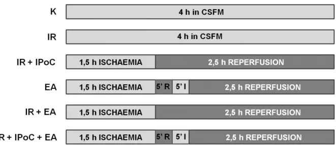
Primary cardiomyocytes were utilized in a well established cellular model of ischemic postconditioning. Briefly, cells were exposed to simulated ischemic (SI) buffer for 1,5 hours followed by 5 minutes reperfusion and then another 5 minutes ischemic insult before the 2,5 hours reperfusion. While cardiomyocytes were under hypoxic conditions in SI buffer, control cells were incubated in complete serum free medium (CSFM) such as during reperfusion period. (Fig. 1)
Assessment of cell viability and apoptotic signalling markers
Ratio of apoptosis was evaluated after double staining with fluorescein isothiocyanate (FITC)-labeled annexin V (BD Biosciences, Pharmingen, USA) and propidium iodide (BD Biosciences, Pharmingen, USA) using flow cytometry, as described previously (Vermes et al.
7 1995). First, the medium was discarded and wells were washed twice with phosphate buffered saline (PBS, Sigma). Cells were removed from plates using a mixture of 0.25 % trypsin (Sigma, Hungary), 0.2 % ethylene-diamin tetra-acetate (EDTA; Serva, Hungary), 0.296 % sodium citrate, 0.6 % sodium chloride in distillated water. This medium was applied for 5 minutes at 37 ºC. Removed cells were washed in cold PBS and were resuspended in binding buffer containing 10 mM Hepes NaOH, pH 7.4, 140 mM NaCl, 2.5 mM CaCl2. Cell-count was determined in Bürker’s chamber for achieving a dilution in which 1 ml of solution contains 106 cells. 100 μl of buffer (105 cells) was transferred into 5 ml round-bottom polystyrene tubes. Cells were incubated for 15 minutes with fluorescein-isotiocianate (FITC) conjugated annexin V molecules and propidium iodide (PI) according to instruction of manufacturers. After this period of incubation, 400 μl of Annexin-binding buffer (BD Biosciences, Pharmingen, USA) was added to the tubes as described by manufacturers. The samples were immediately measured by BD FacsCalibur flow cytometer (BD Biosciences, USA) and analysed with cellquest software. Cells in each category are expressed as percentage of the total number of stained cells counted.
Measurement of cell signaling markers
Phospho-JNK, phospho-p38, phospho-ERK/p42-44 and phospho-GSK-3β was quantified using intracellular staining for flow cytometry. Following the variant treatments in different groups, cells were harvested by trypsin–EDTA according to the protocol described at annexin V-PI staining. Cells were pelleted by centrifugation (175 g, 5 min) and then fixed in 2% formaldehyde in PBS for 10 min at 37 °C. After 1 min of chilling, cell suspensions were centrifuged again followed by permeabilization applying 90% methanol (Sigma, Hungary) for 30 min at 4 °C. Each tube of cells was rinsed twice with 0.5% BSA (Invitrogen, USA) and finally appropriate amounts of cells (0.5–5×10⁶) were resuspended in 0.5% BSA. Cells were
8 then incubated for 10 min at room temperature. Subsequent to blocking of cells in BSA, appropriate dilution of primary antibody was added to the solution and was incubated for 1 hour at room temperature. Antibodies against phospho-JNK, phospho-p38, phospho- ERK/p42-44 and phospho-GSK-3β were from Cell Signaling Technology Inc. (USA), and from R&D System (USA) and they were used at dilutions 1:100 (phospho-JNK), 1:25 (phospho-p38), 1:100 (phospho-ERK/p42-44), and 1:100 (phospho- GSK-3β). After centrifugation, supernatant was carefully aspirated and cells were resuspended in 100 μl 0.5%
BSA containing FITC conjugated secondary antibody (Goat anti-rabbit IgG; R&D System, USA) at a dilution of 1:20, and were incubated for 30 min. Fluorescent staining of samples was quantified by flow cytometric measurement of 5,000 cells. To determine the non-specific marking of cells, secondary antibody was applied for 30 min without primary antibody following permeabilization. Our results were analyzed by Cellquest software (BD Biosciences, USA), measuring the appearance of phospho-JNK, phospho-p38, phospho- ERK/p42-44, and phospho-GSK-3β in the cells as mean fluorescence intensity (MFI).
Statistics
All data are presented as mean ± standard error of the mean (S.E.M). Differences between groups were assessed with one-way ANOVA and Student’s t test and were considered significant when P-value was less than 0.05.
Results
Flow cytometry was performed to measure the percentage of living and apoptotic cells. With flow cytometry the control group had 83,54±2,31 % of intact, living (annexin V
9 and PI negative) cells and 7,8±2,05 % of cells in early phase of apoptosis (annexin V positive and PI negative) (Fig. 2,3). A significant increase of apoptotic cells was observed in both the simulated I/R and EA-treated groups with a lower number of living cells. When EA was added in simulated I/R groups the quantity of apoptotic cells was further increased with reduced amount of living cells (Fig. 2,3). Ischaemic postconditioning resulted significant increase in the percentage of living cells and a significant decrease in the ratio of apoptotic cells in simulated I/R group while we could not detected this protective effect in simulated I/R group when EA co-treatment was applied (Fig. 2,3). In ischaemic postconditioned group the percentage of living cells was significantly higher than in same group receiving EA administration (p=7,97E-11). Colorimetric MTT assay [3-(4,5-dimethylthiazol-2-yl)-2,5- diphenyl tetrazolium bromide, Sigma] was also performed to measure the absolute number of living cells in different groups. The assay is based on the reduction of MTT into a blue formazan dye by the functional mitochondria of viable cells. We found similar results and there was no statistically significant difference between the two methods (data not shown) so we chose only the flow cytometric assay results for presentation.
JNK activation increased markedly in simulated I/R group and upon administration of EA to cardiac myocytes (Fig. 4). EA administration resulted in further increase in JNK activation in cells exposed to simulated ischaemia and reperfusion (Fig. 4). Ischaemic postconditioning decreased the JNK phosphorylation significantly in simulated I/R group while this change could not be detected in EA-treated simulated I/R group (Fig. 4). Among the postconditioned groups the level of phosphorylated JNK was significantly higher when cardiomyocytes were treated with GST inhibitor EA.
Both, simulated I/R and GST inhibition led to significant increase in p38 activation related to non-treated cells. EA administration during simulated I/R resulted in further elevation in phosphorylation of p38. A significant decrease in p38 activation was detected in
10 ischaemic postconditioned group compared to I/R group (p=0,0037). However, we could not observe similar, significant decrease between these groups if the cells treated with EA (p=0,213) (Fig. 5). Ischaemic postconditioning reduced significantly the level of phospho-p38 compared to same group receiving EA administration (p=0,0072).
ERK phophorylation significantly increased in simulated I/R (p=2,52E-10) and GST inhibited (p=1,14E-08) groups. When cardiomyocytes were treated with both sI/R and EA we observed further elevation in the level of phosphorylated ERK/p42-44 compared to only GST inhibited group (p=0,039). The level of phosphorilated ERK/p42-44 of GST inhibited cells receiving simulated I/R was similar than the phopho-ERK level of cells undergone simulated I/R alone. Ischaemic postconditioning could significantly enhance the activation of ERK/p42-44 in simulated I/R group (p=0,0012) but we did not find significant change in case of double stress when simulated I/R combined with EA treatment using ischaemic postconditioning (p=0,53). (Fig. 6) The level of phosho-ERK/p42-44 was significantly higher in simple postconditioned group compared to the EA-treated group after postconditioning (p=0,0009).
The phosphorylation of GSK-3β was significantly lower in simulated I/R group compared to control group (p=0,0019). Significantly increased GSK-3β activation was observed in ischaemic postconditioned group compared to simulated I/R group (p=0,0393).
We measured significant decreasing in GSK-3β activation in EA-treated group compared to control (p=0,00047) and simulated I/R (p=0,0016) groups. Further reduction in the activation of GSK-3β could be detected in groups treated with double stress (simulated I/R and GST inhibitor EA) compared to simulated I/R (p=4,7E-07) or EA treatment (p=7,94E-05) alone.
(Fig. 7) We did not measure significantly higher phospho-GSK-3β level in postconditioned group treated with double stress compared to I/R+EA group (p=0,059). GSK-3β
11 phosphorylation was significantly lower in case of EA administration (p=0,0006) compared the postconditioned groups to each other. (Fig. 7)
Discussion
Present study showed that GST inhibition could markedly attenuate the protective effect of ischaemic postconditioning and resulted in increasing apoptosis in cardiomyocyte cell culture. GST inhibition was associated with different activation of MAP kinases and GSK3β protein kinase in the process of ischaemic postconditioning.
Myocardial reperfusion is the restoration of blood flow to an ischemic heart. Early reperfusion minimizes the extent of damage of heart muscle and preserves the pumping function of the heart. However, reperfusion after a prolonged period of ischemia produces a marked damage in myocardium rather than restoration of normal cardiac function. Thus, ischemia–reperfusion (I/R) injury could be defined as the damage to heart when blood supply is restored after a prolonged period of ischemia resulting in oxidative damage, inflammation and cardiac dysfunction (Balakumar et al. 2008)
Ischaemic preconditioning (IPC) is a well known strategy to protect heart against ischaemia and reperfusion injury (Murry et al. 1986). However, unpredictability of clinical acute myocardial infarction precludes the application of preconditioning.
Recently, Zhao et al. (Zhao et al. 2003) have reported that a short series of repetitive cycles of brief reperfusion and reocclusion of the coronary artery applied immediately at the onset of reperfusion, termed “postconditioning,” was as effective as preconditioning. They showed that the mechanisms involved in postconditioning protection take place within the
12 first minutes of reperfusion (Serviddio et al. 2005). It may have greater clinical potential than preconditioning (Yellon and Opie 2006).
A key determinant of the cellular response to oxidative stress relates to the level and molecular form of glutathione (Yin et al. 2000). A crucial factor that affects level of glutathione is its utilization by via GST (Griffith 1999, Sen 2000, Hayes and McLellan 1999).
GSTs function by conjugating reduced GSH and catalysing attack on foreign compound or oxidative stress products, generally forming less reactive materials that can be readily excreted. The ability of GST to alter levels of cellular glutathione in response to production of ROS has been implicated in protection of cells from ROS-inducing agents (Mannervik et al.
1992, Mannervik et al. 2005).
Present study utilised EA for pharmacological inhibition of GST. EA has been shown to be a substrate of majority of GST isozymes furthermore nonenzymatic GSH conjugation of EA also exists. Moreover it was shown that EA-SG was an inhibitor of the GSTs due to its greater affinity for the enzymes, whereas EA itself inhibits GST through reversible covalent interactions(Tirona and Pang 1999).
In our experiment administration of EA resulted in marked increase of apoptotic cells, principally when cells were co-treated with simulated I/R. While ischaemic postconditioning could decrease the ratio of apoptosis in simulated I/R group, this positive effect could not be detected in GST inhibited group receiving simulated I/R. The increased level of reactive oxygen species and more unfavourable glutathione state may exagravate the intensity of insult and may explain the increased amount of apoptotic cells in GST inhibited group during simulated I/R and IPoC (Rőth et al. 2011). Ferdinandy et al. has demonstrated that comorbidities and aging accompanying coronary disease modify responses to ischemia/reperfusion and the cardioprotection conferred by preconditioning and postconditioning. They reviewed that aging or age-associated alterations (such as hypertrophy
13 or remodeling) are associated with the loss or attenuation of cardioprotection by acute ischemic or pharmacological preconditioning as well as ischemic postconditioning. The majority of the studies showed that diabetes and some antidiabetic drugs interfere with cardioprotective mechanisms, attenuating the effectiveness of cardioprotective strategies (Ferdinandy et al. 2007).
GSTs associate with members of the mitogen activated protein kinase (MAPK) pathways involved in cell survival and death signaling (Laborde 2010). GSTπ was among the first isoenzymes found to inhibit c-Jun N-terminal kinase (JNK) through direct protein-protein interaction thus influencing cellular stress response and apoptosis (Yin et al. 2000). JNK is a proapoptotic MAP kinase mediates cytotoxicity in various conditions including I/R and oxidative, nitrosative stress, and involved in stress response, apoptosis, inflammation, and cellular differentiation and proliferation (Adler et al. 1999, Weston and Davis 2002). The phosphorylation of JNK activates c-Jun, resulting in subsequent activation of downstream effectors. In unstressed cells, low JNK catalytic activity is maintained through it sequestration within a protein complex that includes pi GST, JNK, and c-Jun thus it is presumed that the dimeric form of GSTp is responsible for the regulatory control of JNK (Laborde 2010). Under conditions of oxidative or nitrosative stress, however [during which all three of these proteins are S-glutathionylated (Adler et al. 1999, Townsend 2007)], the pi GST-JNK complex dissociates, so that JNK regain its activity by phophorylation and free to act on downstream gene targets, whereas the pi GST undergoes oligomerization (Davis et al. 2001). We have found that pharmacological inhibition of GST augments JNK activation by itself and abolish the protective effect of ischaemic postconditioning. This could be explained by elimination of sequestration of JNK within a protein complex with GST. On the other hand in unstressed cells effective inhibition of GST may cause JNK phophorylation as a result of oxidative injury due to hindered elimination of trivially developing oxidant and toxic materials. It has been
14 already described that GST knockout mice have high basal JNK activity furthermore treatment of cells with potent GST inhibitor causes activation of JNK (Ruscoe et al. 2001, Townsend et al. 2005).
The MAPK p38 is a signaling protein that plays a critical role in coordinating cellular responses to stress, including oxidative stress that is characterized by the accumulation of increased levels of reactive oxygen species (ROS) within the cell. Evidence from in vitro and in vivo models has shown that death kinases such as the p38 and JNK MAPKs linked to myocardial injury after ischaemia and reperfusion are also activated in response to stimuli present at reperfusion such as inflammatory cytokines and oxidants (Cicconi et al. 2003, Yue et al. 2000, Bogoyevitch et al. 1996). There is little information on the modulation of death kinases in postconditioning. In a preliminary report by Zhao et al. (Zhao et al. 2005) using isolated neonatal rat cardiomyocytes, intermittent reoxygenation and hypoxia (‘‘hypoxic postconditioning’’) inhibited the expression of p38 and JNK mitogen-activated protein kinases. According to Sun et al. attenuation in superoxide anion generation by postconditioning after hypoxia and reoxygenation, the expression and activation of JNK and p38 MAPKs are attenuated suggesting that modulation of MAPK signalling pathways are largely involved in postconditioning-induced protection (Sun et al. 2006). We found that simulated I/R treatment cause noticeable induction of p38 activation in cardiomyocytes, which was further increased by administration of EA. Consistent with the literature ischaemic postconditioning was able to decrease significantly the phosphorylation of p38 in I/R group while we observed similar but not significant decrease in sI/R group treated with GST inhibitor EA, accordingly the relationship is supposed between GST and p38 which led to abolished effect of IPoC.
In many cell types the ERK cascade appears to mediate specifically cell growth and survival signals. For instance, it has been shown that inhibition of ERK enhances ischaemia-
15 reperfusion induced apoptosis and that sustained activation of this kinase during simulated ischaemia mediates adaptive cytoprotection in cultured neonatal cardiomyocytes (Yue et al.
2000). According to our results ERK/p42-44 activation increased upon GST inhibition during reperfusion. It has been already described that immortalized fibroblast isolated from GST π 0 genotype expressed significantly elevated activity of ERK. Moreover treatment with potent GST inhibitor resulted in activation of ERK (Ruscoe et al. 2001). According to Lazou et al.
the cardioprotective effects of ischaemic preconditioning correlates with the activation of ERK/p42-44 during reperfusion (Lazou et al. 2006). On the other hand ERK/p42-44 plays a pivotal role not only in preconditioning but also in postconditioning. In 1999, Yellon and co- authors (Yellon and Baxter 1999) introduced the concept of a pro-survival reperfusion signalling pathway, which they subsequently termed the ’Reperfusion Injury Salvage Kinase’
(RISK) pathway.The pharmacological activation of pro-survival kinases, such ERK/p42-44, at the immediate onset of myocardial reperfusion reduced infarct size by 40–50% (Hausenloy and Yellon 2004, Hausenloy and Yellon 2007). Successive studies have also confirmed the role for ERK/p42-44 in the setting of postconditioning in both non-diseased animal hearts, as well as in post-infarct remodelling (Tsang et al. 2004, Zhu et al. 2006). Interestingly, obese mice have been reported to be resistant to IPoC protection, and this finding was associated with insufficient activation of the RISK pathway in the hearts harvested from obese animals compared to control ones (Bouhidel et al. 2008). In our experiment the level of antiapoptotic phospho-ERK/p42-44 was significantly higher as a result of IPoC – consistent with previous studies – but this change was not shown in the presence of GST inhibitor ethacrynic acid.
Although the activation of ERK/p42-44 increased due to GST inhibition or IPoC the elevation of this antiapoptotic MAPK was lost in case of double stress, probably cause of the extended stress.
16 Glycogen synthase kinase-3β (GSK-3β), a protein kinase linked to the regulation of a variety of cellular functions including glycogen metabolism, gene expression, and cellular survival, could either be considered as a specific downstream target of the RISK pathway or indeed as a component of the RISK pathway. Phosphorylation of GSK-3β causes the inhibition of this protein kinase itself. GSK-3β -inhibition prevents the opening of the mitochondrial permeability transition pore (mPTP). Inhibition of mPTP opening inhibits the release of cytochrome C from mitochondria, a mechanism preventing apoptotic cell death (Juhaszova et al. 2004, Wagner et al. 2008). It has already shown that mice containing a mutant form of GSK-3β, which cannot be phosphorylated and thereby inhibited, were resistant to the myocardial infarct-limiting effects of postconditioning, suggesting that GSK- 3b inactivation is required for postconditioning (Gomez et al. 2008). We detected significant reduction in activation of GSK-3β following simulated I/R treatment showing the harmful effect of I/R, but coincidently with the literature the activated form of GSK-3β increased in case of IPoC. In case of double stress (sI/R+EA) we observed significant reduction in the phosphorylation of GSK-3β, additionally IPoC was not able to significantly enhanced the activation of GSK-3β. This result may presume the association between GSK-3β and GST.
In sum, present study showed that inhibition of GST by EA augments the apoptosis as a result of simulated I/R furthermore abolish the protective effect of ischaemic postconditioning and this is presumably mediated by JNK, p38, ERK/p42-44 and maybe GSK-3β signaling pathways because the activities of these kinases change on this way during ischaemic postconditioning. GST activity is required for survival of cultured cardiomyocytes under stress conditions. These findings highlight the important role of GST in protection against oxidative stress likely not only in experimental conditions but in different pathological disorders in human beings thus serve as basis of (1) further studies investigating in vivo effect of GST inhibition, (2) clinical studies to investigate the role of GST on myocardial damage
17 under different pathological conditions and (3) whether GST 0 genotype is associated with susceptibility of reperfusion injury and also abolished mechanisms of ischaemic postconditioning.
ACKNOWLEDGEMENT
This work was supported by the Hungarian Science Research Fund OTKA- K78434 and the TÁMOP 4.2.2./B-10/1-2010-0029.
References
Adler V, Yin Z, Fuchs SY, Benezra M, Rosario L, Tew KD et al. Regulation of JNK signaling by GSTp. EMBO J 1999 Mar 1;18(5):1321-34.
Balakumar P, Rohilla A, Singh M. Pre-conditioning and postconditioning to limit ischemia–
reperfusion-induced myocardial injury: What could be the next footstep? Pharmacological Research 2008 Jun;57(6):403-12.
Board PG, Baker RT, Chelvanayagam G, Jermiin LS. Zeta, a novel class of glutathione transferases in a range of species from plants to humans. Biochem J 1997 Dec 15;328 ( Part 3):929-35.
18 Bogoyevitch MA, Gillespie-Brown J, Ketterman AJ, Fuller SJ, Ben-Levy R, Ashworth A, Marshall CJ, Sugden PH. Stimulation of the stress-activated mitogen-activated protein kinase subfamilies in perfused heart. p38/RK mitogen-activated protein kinases and c-Jun N-terminal kinases are activated by ischemia/reperfusion. Circ Res 1996 Aug;79(2):162-73.
Bouhidel O, Pons S, Souktani R, Zini R, Berdeaux A, Ghaleh B. Myocardial ischaemic postconditioning against ischaemia-reperfusion is impaired in ob/ob mice. Am J Physiol Heart Circ Physiol 2008 Oct;295(4):H1580-6.
Bray TM, Taylor CG. Tissue glutathione, nutrition, and oxidative stress. Canadian Journal of Physiology and Pharmacology, 1993 Sep;71(9):746-51.
Christie JD et al. Donor glutathione S-transferase genotype is associated with primary graft dysfunction following lung transplantation. The Journal of Heart and Lung Transplantation.
2005;Feb;24(2):S80-S80.
Cicconi S, Ventura N, Pastore D, Bonini P, Di Nardo P, Lauro R, Marlier LN.
Characterization of apoptosis signal transduction pathways in HL-5 cardiomyocytes exposed to ischemia/reperfusion oxidative stress model. J Cell Physiol 2003 Apr;195(1):27-37.
Davis RJ. Signal transduction by the c-Jun N-terminal kinase. Biochem. Soc. Symp., 1999;64:1–12.
Davis W Jr, Ronai Z, Tew KD. Cellular Thiols and Reactive Oxygen Species in Drug-Induced Apoptosis. J Pharmacol Exp Ther. 2001 Jan;296(1):1-6.
19 Ferdinandy P, Schulz R, Baxter GF. Interaction of Cardiovascular Risk Factors with Myocardial Ischemia/Reperfusion Injury, Preconditioning, and Postconditioning. Pharmacol Rev 2007;59:418–458.
Ghosh S, Ting S, Lau H, Pulinilkunnil T, An D, Qi D, Abrahani MA, Rodrigues B. Increased efflux of glutathione conjugate in acutely diabetic cardiomyocytes. Canadian Journal of Physiology and Pharmacology, 2004;82(10):879-887.
Gomez L, Paillard M, Thibault H, Derumeaux G, Ovize M. Inhibition of GSK3beta by postconditioning is required to prevent opening of the mitochondrial permeability transition pore during reperfusion. Circulation 2008 May 27;117(21):2761-8.
Gordon JM, Dusting GJ, Woodman OL, Ritchie RH. Cardioprotective action of CRF peptide urocortin against simulated ischemia in adult rat cardiomyocytes. Am J Physiol Heart Circ Physiol. 2003 Jan;284(1):330-6.
Griffith OW. Biologic and pharmacologic regulation of mammalian glutathione synthesis.
Free Radic. Biol. Med. 1999 Nov;27(9-10):922-35.
Habig WH, Pabst MJ, Jakoby WB. Glutathione S-transferases. The first enzymatic step in mercapturic acid formation. J Biol Chem 1974 Nov 25;249(22):7130-9 a
20 Habig WH, Pabst MJ, Fleischner G, Gatmaitan Z, Arias IM, Jakoby WB. The identity of glutathione S-transferase B with ligandin, a major binding protein of liver. Proc Natl Acad Sci USA 1974 Oct;71(10):3879-82 b
Hausenloy DJ, Yellon DM. New directions for protecting the heart against ischaemia/reperfusion injury: targeting the reperfusion injury salvage kinase (RISK)-pathway.
Cardiovasc Res 2004 Feb 15;61(3):448-60.
Hausenloy DJ, Yellon DM. Reperfusion injury salvage kinase signalling: taking a RISK for cardioprotection. Heart Fail Rev 2007 Dec;12(3-4):217-34.
Hayes JD, and McLellan LI. Glutathione and glutathione-dependent enzymes represent a co- ordinately regulated defence against oxidative stress. Free Radical Res. 1999;31:273–300.
Hayes JD, Flanagan JU, Jowsey IR. Glutathione transferases. Annu Rev Pharmacol Toxicol 2005;45:51–88.
Juhaszova M, Zorov DB, Kim SH, Pepe S, Fu Q, Fishbein KW, Ziman BD, Wang S, Ytrehus K, Antos CL, Olson EN, Sollott SJ. Glycogen synthase kinase-3beta mediates convergence of protection signaling to inhibit the mitochondrial permeability transition pore. J Clin Invest 2004 Jun;113(11):1535-49.
Kloner RA, Pryzyklenk K, Whittaker P. Deleterious effects of oxygen free radicals in ischemia/reperfusion. Circulation 1989;80:1115-27.
21 Laborde E. Review. Glutathione transferases as mediators of signaling pathways involved in cell proliferation and cell death. Cell Death and Differentiation 2010 Sep;17(9):1373-80.
Lazou A, Iliodromitis EK, Cieslak D, Voskarides K, Mousikos S, Bofilis E, Kremastinos DT.
Ischemic but not mechanical preconditioning attenuates ischemia/reperfusion induced myocardial apoptosis in anaesthetized rabbits: the role of Bcl-2 family proteins and ERK1/2.
Apoptosis. 2006 Dec;11(12):2195-204.
Lu SC. Regulation of glutathione synthesis. Curr. Top. Cell Regul. 2000;36:95–116.
Luodonpa M, Vuolteenaho O, Eskelinen S, Ruskoaho H. Effects of adrenomedullin on hypertrophic responses induced by angiotensin II, endothelin-1 and phenylephrine. Peptides 2001 Nov;22(11):1859-66.
Mannervik B, Alin P, Guthenberg C, Jensson H, Tahir MK, Warholm M et al. Identification of three classes of cytosolic glutathione transferase common to several mammalian species:
correlation between structural data and enzymatic properties. Proc Natl Acad Sci USA 1985;82:7202–7206.
Mannervik B, Awasthi YC, Board PG, Hayes JD, Di Ilio C, Ketterer B et al. Nomenclature for human glutathione transferases. Biochem J 1992 Feb 15;282(Part 1):305-6.
Mannervik B, Board PG, Hayes JD, Listowsky I, Pearson WR. Nomenclature for mammalian soluble glutathione transferases. Methods Enzymol 2005;401:1–8.
22 McIlwain CC, Townsend DM, Tew KD. Glutathione S-transferase polymorphisms: cancer incidence and therapy. Oncogene. 2006 Mar 13;25(11):1639-48.
Meyer DJ, Coles B, Pemble SE, Gilmore KS, Fraser GM, Ketterer B. Theta, a new class of glutathione transferases purified from rat and man. Biochem J 1991 Mar 1;274 ( Part 2):409- 14.
Murry CE, Jennings RB, Reimer KA. Preconditioning with ischaemia: a delay of lethal cell injury in ischaemic myocardium. Circulation 1986;74,1124-1136.
Rőth E, Hejjel L, Jaberansari MT, Jancsó G. The role of free radicals in endogenous adaptation and intracellular signals. Experimental review. Exp Clin Cardiol 2004.
Spring;9(1):13-6.
Rőth E, Marczin N, Balatonyi B, Ghosh S, Kovács V, Alotti N, Borsiczky B, Gasz B. Effect of a glutathione S-transferase inhibitor on oxidative stress and ischaemia-reperfusion-induced apoptotic signaling of cultured cardiomyocytes. Exp. Clin. Cardiol. 2011;16(3):92-96.
Ruscoe JE, Rosario LA, Wang T, Gaté L, Arifoglu P, Wolf CR, Henderson CJ, Ronai Z, Tew KD. Pharmacologic or genetic manipulation of glutathione S-transferase P1-1 (GSTpi) influences cell proliferation pathways. J Pharmacol Exp Ther. 2001 Jul;298(1):339-45.
Sen CK. Cellular thiols and redox-regulated signal transduction. Curr. Top. Cell Regul.
2000;36:1–30.
23 Serviddio G, Di Venosa N, Federici A, D'Agostino D, Rollo T, Prigigallo F, Altomare E, Fiore T, Vendemiale G. Brief hypoxia before normoxic reperfusion (postconditioning) protects the heart against ischemia-reperfusion injury by preventing mitochondria peroxyde production and glutathione depletion. FASEB J. 2005 Mar;19(3):354-61.
Sun HY, Wang NP, Halkos M, Kerendi F, Kin H, Guyton RA, Vinten-Johansen J, Zhao ZQ.
Postconditioning attenuates cardiomyocyte apoptosis via inhibition of JNK and p38 mitogen- activated protein kinase signaling pathways. Apoptosis. 2006 Sep;11(9):1583-93.
Tew KD. Glutathione-associated enzymes in anticancer drug resistance. Cancer Res., 1994;54:4313–4320.
Tirona RG, Pang KS. Bimolecular glutathione conjugation kinetics of ethacrynic acid in rat liver: in vitro and perfusion studies. J Pharmacol Exp Ther. 1999 Sep;290(3):1230-41.
Tokola H, Salo K, Vuolteenaho O, Ruskoaho H. Basal and acidic fibroblast growth factor- induced atrial natriuretic peptide gene expression and secretion is inhibited by staurosporine.
Eur J Pharm 1994 Apr 15;267(2):195-206.
Townsend DM, Findlay VL, Tew KD. Glutathione S-transferases as regulators of kinase pathways and anticancer drug targets. Methods Enzymol. 2005;401:287-307.
Townsend DM. Review. S-Glutathionylation: Indicator of Cell Stress and Regulator of the Unfolded Protein Response. Molecular Interventions 2007 Dec;7(6):313-24.
24 Tsang A, Hausenloy DJ, Mocanu MM, Yellon DM. Postconditioning: a form of ‘Modified Reperfusion’ protects the myocardium by activating the phosphatidylinositol
3-kinase-Akt pathway. Circ Res 2004 Aug 6;95(3):230-2.
Vermes I, Haanen C, Steffens-Nakken H, Reutelingsperger C. A novel assay for apoptosis.
Flow cytometric detection of phospatidylserine expression on early apoptotic cells using fluorescein labelled annexin V. J Immunol Methods 1995 Jul 17;184(1):39-51.
Wagner C, Kloeting I, Strasser RH, Weinbrenner C. Cardioprotection by postconditioning is lost in WOKW rats with metabolic syndrome: role of glycogen synthase kinase 3β. J Cardiovasc Pharmacol. 2008 Nov;52(5):430-7.
Weston CR, Davis RJ. The JNK signal transduction pathway. Curr Opin Genet Dev 2002 Feb;12(1):14-21.
Zhao ZQ, Corvera JS, Halkos ME, Kerendi F, Wang NP, Guyton RA, Vinten-Johansen J.
Inhibition of myocardial injury by ischemic postconditioning during reperfusion: comparison with ischemic preconditioning. Am J Physiol Heart Circ Physiol 2003 Aug;285(2):H579-88.
Zhao ZQ, Sun HY, Wang NP, Kin H, Guyton RA, Vinten-Johansen J. Hypoxic postconditioning attenuates cardiomyocyte apoptosis via inhibition of JNK and p38 kinases pathway. J Mol Cell Cardiol 2005;38:870 [Abstract].
25 Zhu M, Feng J, Lucchinetti E, Fischer G, Xu L, Pedrazzini T, Schaub MC, Zaugg M.
Ischemic postconditioning protects remodeled myocardium via the PI3K-PKB/Akt reperfusion injury salvage kinase pathway. Cardiovasc Res 2006 Oct 1;72(1):152-62.
Yellon DM, Baxter GF. Reperfusion injury revisited: is there a role for growth factor signalling in limiting lethal reperfusion injury? Trends Cardiovasc Med 1999 Nov;9(8):245-9.
Yellon DM, Opie LH. Inhibition of myocardial injury by ischemic postconditioning during reperfusion: comparison with ischemic preconditioning. The Lancet, Volume 367, Issue 9509, Pages 456 - 458, 11 February 2006
Yin Z, Ivanov VN, Habelhah H, Tew K, Ronai Z. Glutathione S-transferase p elicits protection against H2O2-induced cell death via coordinated regulation of stress kinases.
Cancer Res. 2000 Aug 1;60(15):4053-7.
Yue TL, Wang C, Gu JL, Ma XL, Kumar S, Lee JC, Feuerstein GZ, Thomas H, Maleeff B, Ohlstein EH. Inhibition of extracellular signal-regulated kinase enhances ischemia/reoxygenation-induced apoptosis in cultured cardiac myocytes and exaggerates reperfusion injury in isolated perfused heart. Circ Res 2000 Mar 31;86(6):692-9.
26 Figures
Figure 1. Experimental protocol and effects of postconditioning with simulated ischemia/reperfusion and the glutathione S-transferase (GST) inhibitor EA treatments on cardiomyocytes. Cardiomyocytes were incubated in complete serum-free medium (CSFM) or with EA followed by 1,5 hours ischaemia and brief period of ischemia before 2,5 hours long reperfusion (R).
27 Figure 2. The mean percentage of living cells determined by flow cytometry. Data expressed as mean percentage ± SEM. p#<0,05 compared with the control group. p*<0,05 and p**<0,01 compared the linked groups to each other. EA – Ethacrynic acid; I/R – Ischaemia and reperfusion; IPoC – Ischaemic postconditioning.
28 Figure 3. The mean percentage of apoptotic cells. Data expressed as mean percentage ± SEM.
p#<0,05 compared with the control group. p*<0,05 and p**<0,01 compared the linked groups to each other. EA – Ethacrynic acid; I/R – Ischaemia and reperfusion; IPoC – Ischaemic postconditioning.
29 Figure 4. Phosphorylation of c-Jun N-terminal kinase (JNK) is demonstrated in cultured cardiomyocytes. p#<0,05 compared with the control group. p***<0,001 compared the linked groups to each other. EA – Ethacrynic acid; I/R – Ischaemia and reperfusion; IPoC – Ischaemic postconditioning; MFI – Mean fluorescence intensity.
30 Figure 5. Phosphorylation of p38 (phospho-38) mitogen-activated protein kinase is demonstrated in cultured cardiomyocytes. p#<0,05 compared with the control group. p*<0,05 and p**<0,01 compared the linked groups to each other. EA – Ethacrynic acid; I/R – Ischaemia and reperfusion; IPoC – Ischaemic postconditioning; MFI – Mean fluorescence intensity.
31 Figure 6. Phosphorylation of extracellular signal-regulated kinase (phospho-ERK/p42-44) is demonstrated in cultured cardiomyocytes. p#<0,05 compared with the control group. p**<0,01 and p***<0,001 compared the linked groups to each other. EA – Ethacrynic acid; I/R – Ischaemia and reperfusion; IPoC – Ischaemic postconditioning; MFI – Mean fluorescence intensity.
32 Figure 7. Phosphorylation of Glycogen synthase kinase-3β (phospho-GSK-3β) is demonstrated in cultured cardiomyocytes. p#<0,05 compared with the control group. p**<0,01 and p***<0,001 compared the linked groups to each other. EA – Ethacrynic acid; I/R – Ischaemia and reperfusion; IPoC – Ischaemic postconditioning; MFI – Mean fluorescence intensity.