Journal of Proactive Medicine
The International Journal of cancer prevention, chemoprevention and epidemiology
Journal homepage: www.jproactive.hu
Original article
Influence of MAPK Inhibitors on the Oxidative Stress of Isolated Cardiomyocytes
Borbála Balatonyi1,*, Balázs Gasz2, Viktória Kovács1, János Lantos1, Gábor Jancsó1, Nándor Marczin3, Erzsébet Rőth1
1Department of Surgical Research and Techniques, Medical Faculty, University of Pécs
2Department of Cardiac Surgery, Zala County Hospital, Hungary
3Faculty of Medicine, Imperial College of London, U.K.
*Corresponding author. Borbála Balatonyi M.D.
E-mail address: balatonyibori@gmail.com
© Journal of Proactive Medicine All rights reserved.
Article Info
Accepted on 18 Feb. 2013 Available online 05 April 2013 Keywords:
cardiomyocytes, oxidative stress, glutathione S-transferase (GST), mitogen activated protein kinase (MAPK), apoptosis.
Abstract
Oxidative stress and ischemia-reperfusion (I/R) injury is crucially involved in the pathogenesis of cardiovascular diseases. Among numerous defense mechanisms against oxidative injury, the antioxidant glutathione S-transferase (GST) is responsible for metabolic inactivation of electrophilic compounds and plays crucial role in regulating mitogen activated protein kinase (MAPK) pathways involved in cellular response to stress, apoptosis and proliferation.
The main objective of this study was to identify the effect of MAPK inhibitors (JNK, p38 and ERK) on the viability and apoptosis of cardiomyocytes following various stress stimuli together with GST inhibition (with administration of ethacrynic acid). Primary culture of neonatal rat cardiomyocytes was prepared and divided into 6 groups, according to the different exposures:
ethacrynic acid (EA), hydrogen-peroxide (H2O2), simulated ischaemia- reperfusion or the combinations of these. We used JNK, p38 or ERK inhibitors in every group respectively. Viability of cells was measured by MTT assay, the ratio of apoptotis was assessed by flow cytometry following annexinV/propidium iodide double staining. Pharmacological inhibition of proapoptotic JNK and p38 MAPKs diminish apoptosis while the inhibition of antiapoptotic ERK/p42-44 MAPK augments apoptosis as a result of GST inhibition, oxidative injury and simulated I/R. The protective effect of the inhibiton of proapoptotic JNK and p38 MAPKs was lost in groups, exposed to double stress (GST inhibition together with oxidative stress or simulated I/R injury). The apoptotic signaling is presumably mediated by JNK, p38 and ERK/p42-44 MAPKs pathways under GST inhibition, oxidative stress and sI/R injury. GST activity is required for survival of cultured cardiomyocytes under stress conditions.
09
Introduction
It has been well investigated that oxidative stress is a major apoptotic stimulus in many cardiac diseases (1-3). Among numerous defence mechanism against oxidative stress and ischaemic/reperfusion injury, the endogenous antioxidant enzyme glutathione S-transferase (GST) are crucially involved in cellular response to stress, apoptosis and proliferation. GST is responsible for the high-capacity metabolic inactivation of electrophilic compounds and toxic substrates (4-6). More recently, isoenzymes from several GST classes have been shown to be associated with members of the mitogen activated protein kinase (MAPK) pathways involved in cell survival and death signaling. GSTs function to sequester the kinase in a complex, thus preventing it from acting on downstream targets. The result of this action is a regulation of pathways that control stress response to I/R injury, cell proliferation and apoptotic cell death (7).
Many studies have suggested that the mitogen activated protein kinases (MAPKs) may be important regulators of apoptosis in response to myocardial ischaemia/reperfusion. Three major MAPKs, namely c-Jun NH2-terminal protein kinase (JNK), p38 and extracellular signal- regulated protein kinase (ERK), play a pivotal role in the transmission of signals from cell surface receptors to the nucleus (8, 9). Although there are conflicting reports on the role of MAPKs in death or survival after stress (10), it is commonly agreed that JNK and p38 MAPKs appear to be pro- apoptotic in many cell types while ERKs are the modulators of cell survival after reperfusion. (11- 15). Our pilot study has been conducted to examine the biological role of GST in cardiac myocytes under oxidative stress conditions. We found that that pharmacological inhibition of GST by EA (ethacrynic acid) augments the apoptosis as a result of oxidative stress and simulated ischaemic-reperfusion (sI/R) injury. The study showed that GST inhibition was associated with increased activation of MAP kinases under stress condition. The main objective of this study was to verify our previous results and identify the effect of MAPK (JNK, p38 and ERK) inhibitors regarding GST inhibition (with administration of EA) on the viability and apoptosis of cardiomyocytes when cells are exposed to various stress components of ischaemia and reperfusion (I/R).
Materials and Methods
Cell culture from isolated neonatal cardiomyocytes
Primary culture of neonatal rat cardiomyocytes was prepared as described previously (16, 17).
Briefly, cells were obtained from ventricular myocytes of 2-4 day-old Wistar rats (Charles- River ltd., Hungary), using collagenase (Gibco™
Collagenase Type II, Invitrogen Corp., Carlsbad, CA, USA). Isolated cells were plated on collagen I-coated 24-well plates (BD Falcon) at the density of 2x10⁵ cells/ml. Cells were incubated in DMEM/F12 medium (Sigma-Aldrich, USA) supplemented with 10% of fetal bovine serum (Gibco, USA). The following day, when the cells attached to the plate firmly, the medium was replaced with complete serum free medium (CSFM) containing the following supplements:
BSA (2.5%, AlbuMax 1, Invitrogen), insulin (1 μM), transferring (5.64 μg/ml), selenium (32 nM)
(insulin-transferrin-sodium-selenit media supplement, Sigma, Hungary), sodium pyruvate
(2.8 mM, Sigma), 3,3’,5’-triiodo-L-thyronine sodium salt (1 nM, Sigma, Hungary), penicillin (100 IU/ml) and streptomycin (0.1 mg/ml) (PS solution, Sigma, Hungary). Experiments started 24 hours after incubation with CSFM and the medium was changed every 24 hours.
Inhibition of glutathione-S tranferase (GST)
Present study utilised EA for pharmacological inhibition of GST. EA has been shown to be a substrate of majority of GST isozymes furthermore nonenzymatic GSH conjugation of EA also exists. Moreover it was shown that EA-SG was an inhibitor of the GSTs due to its greater affinity for the enzymes, whereas EA itself inhibits GST through reversible covalent interactions (Figure 9) (18).
MAPK inhibitors
To determine the roles of c-Jun N-terminal kinase (JNK), p38 MAP kinase, and ERK/p42/p44 MAP kinase on the viability and apoptosis of cardiomyocytes under GST inhibition, myocytes were pretreated with SP600125 (10 μM), SB239063 (10 μM), and U0126 (1 μM).
JNK inhibitor: SP600125, Sigma-Aldrich (S5567) p38 inhibitor: SB239063 Sigma-Aldrich (S0569) ERK/p42-44 inhibitor: U0126 Sigma-Aldrich (U120)
Experimental protocol
Cultured cardiomyocytes were randomly assigned to one of six experimental groups according to different exposures: control group of cells incubated in CSFM without treatment. In Group II, cells incubated in medium containing 150 μM ethacrynic acid (EA) alone. In Group III, cells treated with 1mM H2O2 to simulate the oxidative stress. In group IV, cells exposed to 1mM H2O2 together with 150 μM ethacrynic acid.
In Group V, cells exposed to swapping ischemic buffer to CSFM to simulate ischaemic-reperfusion (sI/R) injury. In Group VI, cells were treated to both sI/R and ethacrynic acid (EA). To antagonize the effect of JNK, p38 and ERK/p42-44 MAPKs, 10
10
μM JNK inhibitor (SP600125), 10 μM p38 inhibitor (SB239063) and 1 μM ERK inhibitor (U0126) was added simultaneously in every group. (Table 1.) In groups receiving simulated I/R cells were exposed to 1,5 hours of ischemia using ischaemic buffer as described previously (19) followed by 2,5 hours of reperfusion using normal CSFM. In group VI (cells were exposed to both simulated I/R and EA) both ischemic buffer and reperfusion medium (CSFM) contained 150 μM of EA. Based on our pilot experiments we chose to use a concentration of 150 μM and a treatment time of 4 hours. Cells were exposed to mentioned concentration of chemicals for 4 hours. MTT assay evaluation of cell survival was performed immediately after termination of treatments.
Assessment of apoptotic signalling markers was also started after treatments until permeabilization, and samples were stored at -20 ºC until further processing according to the protocol supplied by the manufacturer.
Experiments were repeated six times in duplicate wells.
Cell viability test
Viability of cardiomyocytes was determined by colorimetric MTT assay [3-(4,5-dimethylthiazol-2- yl)-2,5-diphenyl tetrazolium bromide, Sigma]. The assay is based on the reduction of MTT into a blue formazan dye by the functional mitochondria of viable cells. The cells were seeded into 96-well plates at a density of 105 cell/well and cultured overnight before the experiment. At the end of the treatments, the medium was discarded from plates and the cells were subsequently washed twice with phosphate buffered saline (PBS, Sigma). Cells were then incubated with CSFM containing water-soluble yellow 0.5 mg/ml of mitochondrial dye MTT for 3 hours at 37ºC in an atmosphere of 5% CO2. The solution was aspirated carefully and 100 μl of dimethylsulfoxid (DMSO) was added to dissolve the blue-colored formazan particles. Absorbance of samples from duplicate wells was measured by an ELISA reader (Sirio microplate reader, Seac Corp.
Florence, Italy) at the wavelength of 570 nm representing the values in arbitrary unit (AU).
Results are expressed as percentage of control values.
Determine the ratio of apoptosis
Ratio of apoptosis was evaluated after double staining with fluorescein isothiocyanate (FITC)- labeled annexin V (BD Biosciences, Pharmingen, USA) and propidium iodide (BD Biosciences, Pharmingen, USA) using flow cytometry, as described previously (20). First, the medium was discarded and wells were washed twice with phosphate buffered saline (PBS, Sigma). Cells were removed from plates using a mixture of 0.25% trypsin (Sigma, Hungary), 0.2% ethylene-
diamin tetra-acetate (EDTA; Serva, Hungary), 0.296% sodium citrate, 0.6% sodium chloride in distilled water. This medium was applied for 5 minutes at 37 ºC. Removed cells were washed in cold PBS and were resuspended in binding buffer containing 10 mM Hepes NaOH, pH 7.4, 140 mM NaCl, 2.5 mM CaCl2. Cell-count was determined in Bürker’s chamber for achieving a dilution in which 1 ml of solution contains 106 cells. 100 μl of buffer (105 cells) was transferred into 5 ml round- bottom polystyrene tubes. Cells were incubated for 15 minutes with fluorescein-isotiocianate (FITC) conjugated annexin V molecules and propidium iodide (PI) according to instruction of manufacturers. After this period of incubation, 400 μl of Annexin-binding buffer (BD Biosciences, Pharmingen, USA) was added to the tubes as described by manufacturers. The samples were immediately measured by BD FacsCalibur flow cytometer (BD Biosciences, USA) and analysed with cellquest software. Cells in each category are expressed as percentage of the total number of stained cells counted. Cells that stain positive for Annexin V-FITC and negative for PI are undergoing apoptosis. Cells that stain positive for both Annexin V-FITC and PI are either in the end stage of apoptosis, undergoing necrosis, or are already dead. Cells that stain negative for both Annexin V-FITC and PI are alive and not undergoing measurable apoptosis (20, 21).
Statistical analysis
Up to four different cardiac myocyte preparations were studied. Data of the experiments are expressed as means±SE. Differences between the means were compared using two sample Student’s t-test for significance. p<0,05 was considered to be statistically significant.
Results
Cell viability (MTT assay)
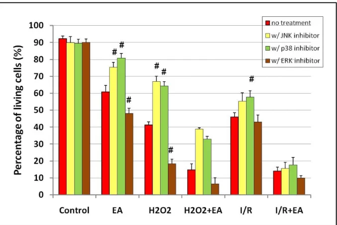
Viability of cardiomyocytes was determined by colorimetric MTT assay. In the control group without treatment, the amount of living cells was increased to 100%. JNK, p38 and ERK/p42-44 did not cause any significant changes in control groups without any stress effect. Ethacrynic acid, H2O2 and sI/R alone significantly reduced the ratio of living cells compared to control groups and only the p38 MAPK inhibition could significantly increase the viability of cells in H2O2-treated group. EA administration significantly enhanced the reduction of the viability of cells treated with H2O2 or exposed to sI/R compared to non-treated cells and MAPK inhibitors could not increase significantly the percentage of living cells (Figure 1.).
11
Apoptotic changes
The non-treated control group had 92,4±1,47% of intact, living cells (annexin V and PI negative) and 4.23±1,25% of cells in the early phase of apoptosis (annexin V positive and PI negative) (Figures 2 and 3). Control groups treated with different MAPK inhibitors had no significant differences compared to non-treated control group. EA, H2O2 administration and simulated I/R significantly decreased the amount of living cells (pEA=0,0001; pH2O2=5,72E-09; psI/R=2,65E-09) and increased the percentage of apoptotic cells (pEA=7,78E-05; pH2O2=1,68E-08; psI/R=1,12E- 08) in non-treated control group. The inhibition of proapoptotic JNK and p38 MAPKs significantly ameliorates the cell viability and attenuates the rate of apoptosis in EA- and H2O2-treated groups. On the other hand in these groups the inhibition of antiapoptotic ERK/p42-44 MAPK was significantly decreased the percentage of living cells (pEA=0,035; pH2O2=2,92E-05) and increased the amount of apoptotic cells (pEA=0,05; pH2O2=1,36E-05). In group, exposed to sI/R, only the p38 MAPK inhibitor was able to increase the percentage of viable cells (psI/R=0,022) and diminish the rate of apoptosis (psI/R=0,018) significantly. A significant increase in amount of apoptotic cells was observed in both groups, exposed to double stress, H2O2+EA or sI/R+EA, with a lower number of living cells compared to non-treated group or groups exposed to H2O2 or sI/R (Figures 2 and 3). The administration of JNK or p38 MAPK inhibitors elevated the level of living cells and reduced the rate of apoptosis but not significantly in groups treated with H2O2 or exposed to sI/R supplemented with EA. When the antiapoptotic ERK/p42-44 MAPK was inhibited in groups treated with double stress, the amount of living cells was further decreased and the quantity of apoptotic cells was further increased.
Discussion
The present study showed that pharmacological inhibition of proapoptotic JNK and p38 could significantly decrease the oxidative stress-induced apoptosis while the inhibition of antiapoptotic ERK/p42-44 markedly reduces the cell viability in cardiomyocytes. This effect of these MAPK inhibitors could not be observed in case of double stress (oxidative stress+GST inhibition).
GSTs play a regulatory role in the mitogen- activated protein (MAP) kinase pathway that participates in cellular survival and death signals via protein:protein interactions. Specifically, GSTp was shown to be an endogenous inhibitor of c- Jun N-terminal kinase 1 (JNK1), a kinase involved in stress response, apoptosis, and cellular proliferation (22, 23). In nonstressed cells, low JNK activity is maintained through the sequestration of the protein in a GSTp:JNK
complex. However, suppression of JNK activity is reversed by conditions of oxidative stress, resulting in the dissociation of the GSTp:JNK complex, activation the catalytic kinase activity, phosphorylation c-Jun, oligomerization of GSTp, and induction of apoptosis (22). To evaluate the significance of JNK, p38 and ERK/p42-44 and the possible link of GST to these MAPKs during oxidative stress, three specific inhibitors (SP600125, SB239063 and U0126) were used respectively. The SP600125 is a potent, cell- permeable, selective, and reversible inhibitor of JNK. SB239063 is a novel p38 inhibitor that exhibits improved kinase selectivity and in vivo activity compared with the other p38 inhibitors (24). To influence on ERK/p42-44 activity, we used U0126, which is a potent inhibitor of MEK1/2, an upstream regulator of the phosphorylation of ERK/p42-44 (25). According to Olli Tenhunen et all. administration of 1 μM U0126 significantly reduced the level of phospho- ERK/p42-44. On the other hand, treatment with 1μM U0126 had no effect on the levels of phosphorylated p38 kinase and, similarly, the administration of SB239063 did not affect the level of phospho-ERK/p42-44 (26). Difference between Figure 1 and 2 can be explained by different methodology. First of all, annexin V-PI staining can consider more subjective results than MTT assay. The ratio of living, apoptotic and necrotic cells depend slightly on settings. This method is able to compare treated groups to control rather than assess the number of living, etc. cells. During analysis of annexin staining quadrant plots were made. Living cells (low annexin V and PI staining) of control samples are highly separable and quadrants were placed to the border of cluster of living cells (see Figure 2).
This setting of quadrants was not changed further and data were measured using this setting. Van Engeland and coworkers (27) stated that it is feasible to use the annexin V and PI analysis for adhering cells in cultures. Nonetheless it is associated with several differences related to other techniques for assessing cellular viability and death. These arise from cell loss, which generally occurs during repeated centrifugations in sample preparation for flow cytometry (28).
MTT assay measures the reduction of tetrazolium salt to water-insoluble formazan. Because only living cells can reduce MTT, MTT reduction has been developed into one of the most widely used methods for measuring cell proliferation and viability and achieve cytotoxicity (29). With the understanding that MTT is endocytosed and that reduced MTT formazan is transported to cell surface through exocytosis, many of the published discrepancies between the MIT assay and other measures of cell growth and viability can be explained (30, 31). Despite the above described differences in methodology, there was no statistically significant difference between the
12
two methods. Oxidative stress-induced apoptotic cell death during reoxygenation in cultured cardiomyocytes and in vivo hearts during reperfusion has been linked to an increased expression in JNK and p38 MAPKs (11, 12, 13, 14, 15). We have found that pharmacological inhibition of the proapoptotic JNK and p38 MAPK significantly increase the cell viability and decrease the ratio of apoptosis in groups receiving GST inhibition or oxidative stress compared to groups without JNK and p38 inhibitors. On the other hand in groups treated with double stress (GST inhibition together with oxidative stress or I/R injury) this protective effect of JNK and p38 inhibitor was lost. These findings may represent the association between JNK, p38 MAPK and GST (32, 33, 34). In many cell types the ERK/p42-44 cascade appears to mediate specifically cell growth and survival signals. For instance, it has been shown that inhibition of ERK/p42-44 enhances ischaemia-reperfusion induced apoptosis and that sustained activation of this kinase during simulated ischaemia mediates adaptive cytoprotection in cultured neonatal cardiomyocytes (35). According to our results, inhibition of the antiapoptotic ERK/p42-44 significantly decrease the cell viability and increase the ratio of apoptosis in groups receiving GST inhibition or oxidative stress compared to groups for lack of ERK/p42-44 inhibitor. Although in our previous study we demonstrated that antiapoptotic ERK/p42-44 is activated on GST inhibition, in the presence of H2O2 administration or during reperfusion (36), in this present study we could not observed elevation in the percentage of living cells, likely because of the ERK/p42-44 inhibitor treatment. But among ERK/p42-44 inhibited groups exposed to GST inhibition, oxidative stress and simulated I/R injury, the ratio of living cells was the highest in case of GST inhibition. These findings may represent the association between ERK/p42-44 and GST. On the other hand in our preceding study we also demonstrated that the level of phosphorylated ERK/p42-44 of GST-inhibited cells receiving sI/R exceeded the ERK/p42-44 phosphorylation level
of cells that have undergone sI/R alone (36).
Accordingly in this present examinations presumably because of ERK/p42-44 inhibition, in groups treated with double stress (GST inhibition together with oxidative stress or simulated I/R injury) the cardiomyocyte cell viability was diminished and the ratio of apoptosis was elevated but not significantly compared to groups in absence of ERK/p42-44 inhibitor. However a significant increase in amount of apoptotic cells was observed in both groups, exposed to double stress, H2O2+EA or sI/R+EA, with a lower number of living cells compared to groups exposed to H2O2 or sI/R alone (32, 33, 34) The present study showed that pharmacological inhibition of proapoptotic JNK and p38 MAPKs diminish apoptosis while the inhibition of antiapoptotic ERK/p42-44 MAPK augments apoptosis as a result of GST inhibition even as oxidative injury and simulated I/R. Thus this apoptotic signalling is presumably mediated by JNK, p38 and ERK/p42-44 MAPKs pathways. The protective effect of the inhibiton of proapoptotic JNK and p38 MAPKs was lost in groups, exposed to double stress (GST inhibition together with oxidative stress or simulated I/R injury), so GST activity is required for survival of cultured cardiomyocytes under stress conditions. These findings highlight the importance of GST in protection against oxidative stress likely not only in experimental conditions but in different pathological disorders in human beings.
Therefore, further studies are needed to investigate the in vivo effect of GST inhibition on an animal model and the role of GST on myocardial damage under different pathological conditions in humans.
Funding sources
This work was supported by the Hungarian Science Research Fund OTKA-K78434 and TÁMOP 4.2.2./B-10/1-2010-0029.
References
01. Chen QM, Tu VC. Apoptosis and heart failure:
mechanisms and therapeutic implications. Am J Cardiovasc Drugs 2002, 2(1):43-57.
02. Kumar D, Jugdutt BI. Apoptosis and oxidants in the heart.
J Lab Clin Med 2003, 142(5):288-97.
03. Rőth E, Hejjel L. Oxygen free radicals in heart disease. In:
MK Pugsley, ed. Cardiac Drug Development Guide. Humana Press, 2003:47-66.
4. Townsend DM, Findlay VL, Tew KD. Glutathione S- transferases as regulators of kinase pathways and anticancer drug targets. Methods Enzymol 2005, 401:287-307.
5. Wu G, Fang YZ, Yang S, Lupton JR et al. Glutathione metabolism and its implications for health. J Nutr 2004, 134(3):489-92.
6. Tirona RG, Pang KS. Bimolecular glutathione conjugation kinetics of ethacrynic acid in rat liver: In vitro and perfusion studies. J Pharmacol Exp Ther 1999, 290(3):1230-41.
07. Davis RJ. Signal transduction by the c-Jun N-terminal kinase. Biochem Soc Symp 1999, 64:1-12.
08. Bogoyevitch MA, Gillespie-Brown J, Ketterman AJ et al.
Stimulation of the stress-activated mitogen-activated protein kinase subfamilies in perfused heart: p38/ERK mitogen- activated protein kinases and c-Jun N-terminal kinases are activated by ischemia/reperfusion. Circ Res 1996, 79:162–73.
13
09. Mansour SJ, Matten WT, Herman AS et al. Transformation of mammalian cells by constitutively active MAP kinase kinase. Science 1994, 265(5174):966-70.
10. Hahn RA, MacDonald BR, Morgan E et al. Evaluation of LY203647 on cardiovascular leukotriene D4 receptors and myocardial reperfusion injury. J Pharmacol Exp Ther 1992, 260(3):979-89.
11. Gao F, Yue TL, Shi DW et al. p38 MAPK inhibition reduces myocardial reperfusion injury via inhibition of endothelial adhesion molecule expression and blockade of PMN accumulation. Cardiovasc Res 2002, 53(2):414-22.
12. Clanachan AS, Jaswal JS, Gandhi M et al. Effects of inhibition of myocardial extracellular-responsive kinase and p38 mitogen-activated protein kinase on mechanical function of rat hearts after prolonged hypothermic ischemia.
Transplantation 2003, 75(2):173-80.
13. Cicconi S, Ventura N, Pastore D et al. Characterization of apoptosis signal transduction pathways in HL-5 cardiomyocytes exposed to ischemia/reperfusion oxidative stress model. J Cell Physiol 2003, 195(1):27-37.
14. Obata T, Brown GE, Yaffe MB. MAP kinase pathways activated by stress: the p38 MAPK pathway. Crit Care Med 2000, 28(Suppl 4):N67-77.
15. Park WH, Seol JG, Kim ES et al. Induction of apoptosis by vitamin D3 analogue EB1089 in NCIH929 myeloma cells via activation of caspase-3 and p38-MAPK. British J Haematol 2000 Jun, 109(3):576-83.
16. Tokola H, Salo K, Vuolteenaho O et al. Basal and acidic fibroblast growth factor-induced atrial natriuretic peptide gene expression and secretion is inhibited by staurosporine. Eur J Pharm 1994, 267(2):195-206.
17. Luodonpa M, Vuolteenaho O, Eskelinen S et al. Effects of adrenomedullin on hypertrophic responses induced by angiotensin II, endothelin-1 and phenylephrine. Peptides 2001, 22(11):1859-66.
18. Tirona RG, Pang KS. Bimolecular glutathione conjugation kinetics of ethacrynic acid in rat liver: in vitro and perfusion studies. J Pharmacol Exp Ther 1999, 290(3):1230-41.
19. Gordon JM, Dusting GJ, Woodman OL. Cardioprotective action of CRF peptide urocortin against simulated ischemia in adult rat cardiomyocytes. Am J Physiol Heart Circ Physiol 2003, 284(1):H330-6.
20. Vermes I, Haanen C, Steffens-Nakken H et al. A novel assay for apoptosis. Flow cytometric detection of phospatidylserine expression on early apoptotic cells using fluorescein labelled Annexin V. J Immunol Methods 1995, 184(1):39-51.
21. van Engeland M, Ramaekers FC, Schutte B et al. A novel assay to measure loss of plasma membrane asymmetry during apoptosis of adherent cells in culture. Cytometry 1996, 24(2):131-9.
22. Adler V, Yin Z, Fuchs SY et al. Regulation of JNK signaling by GSTp. EMBO J 1999, 18(5):1321-34.
23. Yin Z, Ivanov VN, Habelhah H et al. Glutathione S- transferase p elicits protection against H2O2-induced cell death via coordinated regulation of stress kinases. Cancer Res 2000, 60(15):4053-7.
24. Barone FC, Irving EA, Ray AM et al. SB 239063, a second- generation p38 mitogen-activated protein kinase inhibitor, reduces brain injury and neurological deficits in cerebral focal ischemia. J Pharmacol Exp Ther 2001, 296(2):312-21.
25. Favata MF, Horiuchi KY, Manos EJ et al. Identification of a novel inhibitor of mitogen-activated protein kinase kinase. J Biol Chem 1998, 273(29):18623-32.
26. Tenhunen O, Sármán B, Kerkelä R et al. Mitogen-activated protein kinases p38 and ERK 1/2 mediate the wall stress- induced activation of GATA-4 binding in adult heart. J Biol Chem 2004, 279(23):24852-60.
27. van Engeland M, Nieland LJ, Ramaekers FC. Annexin V- affinity assay: a review on an apoptosis detection system based on phosphatidylserine exposure. Cytometry 1998, 31(1):1-9.
28. Bedner E, Li X, Gorczyca W. Analysis of apoptosis by laser scanning cytometry. Cytometry 1999, 35(3):181-95.
29. Mosmann T. Rapid colorimetric assay for cellular growth and survival: Application to proliferation and cytotoxicity assays. J Immunol Methods 1983, 65(1-2):55-63.
30. Liu Y, Peterson DA, Kimura H et al. Mechanism of cellular 3-(4,5-dimethylthiazol-2-yl)-2,5-diphenyltetrazolium bromide (MTT) reduction. J Neurochem 1997, 69(2):581-93.
31. Liu Y. Understanding the biological activity of amyloid proteins in vitro: from inhibited cellular MTT reduction to altered cellular cholesterol homeostatis. Prog Neuropsychopharmacol Biol Psychiatry 1999, 23(3):377-95.
32. Balatonyi B, Gasz B, Subhamay G et al. A glutation S- transzferáz gátlásának hatása oxidatív stresszel károsított szívizomsejtekre. Cardiológia Hungarica 2011, 41(5):319-24.
Article in Hungarian.
33. Balatonyi B, Kovács V, Jávor Sz et al. Influence of MAPK inhibitors on the effect of GST enzyme inhibition on the viability and apoptosis of cardiomyocytes. 46th Congress of the European Society for Surgical Research, Aachen, Germany, 25-28. May 2011. British Journal of Surgery 2011, 98(S5):50- 69. Poster abstract.
34. Rőth E, Balatonyi B, Kovács V et al. How the inhibition of glutathione S-transferase can modulate stress response of cardiac myocytes. 75th Anniversary of Albert Szent-Györgyi’s Nobel Prize Award, Szeged 22-25. March 2012. Lecture abstract.
35. Yue TL, Wang C, Gu JL et al. Inhibition of extracellular signal-regulated kinase enhances Ischemia/Reoxygenation- induced apoptosis in cultured cardiac myocytes and exaggerates reperfusion injury in isolated perfused heart. Circ Res 2000, 86(6):692-9.
36. Rőth E, Marczin N, Balatonyi B et al. Effect of a glutathione S-transferase inhibitor on oxidative stress and ischemia- reperfusion-induced apoptotic signalling of cultured cardiomyocytes. Exp Clin Cardiol 2011, 16(3):92-96.
14
Figures and Tables
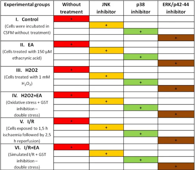
Table 1. Experimental groups. Cells treated with 150 μM EA for GST inhibition. Cells treated with 1mM H2O2 to simulate the oxidative stress. In groups receiving simulated I/R, cells were exposed to 1,5 hours of ischemia using ischaemic buffer followed by 2,5 hours of reperfusion using normal CSFM.
Cells were exposed to mentioned concentration of chemicals for 4 hours.
EA – Ethacrynic acid; H2O2 – Hydrogen-peroxide; I/R – Ischaemia and reperfusion.
15
Figure 1. Viability of cardiomyocytes as measured by MTT assay. p#<0,05 compared to the first column in every group without MAPK inhibitor.
EA – Ethacrynic acid; H2O2 – Hydrogen-peroxide; I/R – Ischaemia and reperfusion.
Figure 2. The mean percentage of living cells measured by flow cytometric assay.
Data expressed as mean percentage ± SEM. p#<0,05 compared to the first column in every group without MAPK inhibitor.
EA – Ethacrynic acid; H2O2 – Hydrogen-peroxide; I/R – Ischaemia and reperfusion
16
. Figure 3. The mean percentage of apoptotic cells measured by flow cytometric assay.
Data expressed as mean percentage ± SEM. p#<0,05 compared to the first column in every group without MAPK inhibitor.
EA – Ethacrynic acid; H2O2 – Hydrogen-peroxide; I/R – Ischaemia and reperfusion.
17