Plant Physiology and Biochemistry 169 (2021) 149–159
Available online 10 November 2021
0981-9428/© 2021 The Authors. Published by Elsevier Masson SAS. This is an open access article under the CC BY-NC-ND license
(http://creativecommons.org/licenses/by-nc-nd/4.0/).
Crosstalk between the redox signalling and the detoxification: GSTs under redox control?
Agnes Gall ´ ´ e
a,1, Krisztina Bela
a,1, Ad ´ ´ am Hajnal
a, N ´ ora Farag o ´
b,c,d, Edit Horv ´ ath
a, M ´ aty ´ as Horv ´ ath
a, L ´ aszl o Pusk ´ ´ as
b,c, Jol ´ an Csisz ´ ar
a,*aDepartment of Plant Biology, Faculty of Sciences, University of Szeged, K¨oz´ep fasor 52, 6726, Szeged, Hungary
bAvidin Ltd., Als´o Kik¨ot˝o sor 11/D, Szeged, 6726, Hungary
cLaboratory of Functional Genomics, Biological Research Centre, Temesv´ari k¨orút 62, Szeged, 6726, Hungary
dResearch Group for Cortical Microcircuits of the Hungarian Academy of Sciences, Department of Physiology, Anatomy and Neuroscience, University of Szeged, K¨oz´ep fasor 52, Szeged, 6726, Hungary
A R T I C L E I N F O Keywords:
Abiotic stress
Dehydroascorbate reductase Glutathione transferases Reactive oxygen species Redox homeostasis Salicylic acid Tomato
A B S T R A C T
Reactive oxygen species (ROS), antioxidants and their reduction-oxidation (redox) states all contribute to the redox homeostasis, but glutathione is considered to be the master regulator of it. We aimed to understand the relationship between the redox potential and the diverse glutathione transferase (GST) enzyme family by comparing the stress responses of two tomato cultivars (Solanum lycopersicum ‘Moneymaker’ and ‘Ailsa Craig’).
Four-week-old plants were treated by two concentrations of mannitol, NaCl and salicylic acid. The lower H2O2 and malondialdehyde contents indicated higher stress tolerance of ‘Moneymaker’. The redox status of roots was characterized by measuring the reduced and oxidized form of ascorbate and glutathione spectrophotometrically after 24 h. The redox potential of ‘Ailsa Craig’ was more oxidized compared to ‘Moneymaker’ even under control conditions and became more positive due to treatments. High-throughput quantitative real-time PCR revealed that besides overall higher expression levels, SlGSTs were activated more efficiently in ‘Moneymaker’ due to stresses, resulting in generally higher GST and glutathione peroxidase activities compared to ‘Ailsa Craig’. The expression level of SlGSTs correlated differently, however Pearson’s correlation analysis showed usually strong positive correlation between SlGST transcription and glutathione redox potential. The possible redox regulation of SlGST expressions was discussed.
1. Introduction
Reactive oxygen species (ROS), ROS-processing enzymes, antioxi- dants and their reduction - oxidation (redox) states all contribute to the redox homeostasis of plant cells (Potters et al., 2010). The generation of ROS, like superoxide radical (O2•-), hydrogen peroxide (H2O2), and hydroxyl radical (OH•) are natural byproducts of the normal aerobic metabolism, but their level typically elevates under abiotic and biotic stress conditions (Hasanuzzaman et al., 2020). Accumulation of ROS may cause detrimental damages by oxidation of different biological molecules, including lipids, proteins and nucleic acids (Foyer and Noc- tor, 2005; Mittler, 2002). Increased ROS production temporarily shifts the redox status to more oxidized values that will alter the operational controls of many redox-sensitive proteins, thus they play an important
role in regulation of growth and stress responses (Ding and Ding, 2020;
Foyer and Noctor, 2015; Mou et al., 2003; Schmidt and Schippers, 2015;
Tada et al., 2008; Wu et al., 2020).
Since ROS are in tight interaction with the main plant hormones, they are thought to be core components of the complex regulatory network by merging exogenous and endogenous signals, among them hormones (Mase and Tsukagoshi, 2021). It was found that the high level and proper distribution of ROS in the root tips are important to the normal growth and development (Dunand et al., 2007; Tognetti et al., 2017; Tsukagoshi et al., 2010). Large number of studies described different experimental design which aimed to elevate ROS production and alter redox potential to study transcriptional oxidative stress re- sponses (He et al., 2018). Each type of ROS has unique chemical prop- erties and targets a specific set of signalling routes (Bindoli and
* Corresponding author.
E-mail address: csiszar@bio.u-szeged.hu (J. Csisz´ar).
1 These authors contributed equally to this work.
Contents lists available at ScienceDirect
Plant Physiology and Biochemistry
journal homepage: www.elsevier.com/locate/plaphy
https://doi.org/10.1016/j.plaphy.2021.11.009
Received 13 August 2021; Received in revised form 24 October 2021; Accepted 8 November 2021
Rigobello, 2013; Møller et al., 2007), therefore activates specific set of genes (He et al., 2018). Molecular switches in the promoter of these ROS-activated genes are cis-regulatory elements called antioxidant-responsive elements (AREs), which are parts of the ROS signalling pathways.
A transcription factor-mediated gene expression network down- stream of ROS ensure the right response to the changing environmental conditions (Mase and Tsukagoshi, 2021). The redox balance may affect the functions of the key transcription factors, such as PLETHORA (PLT) (Licausi et al., 2013). The PLT1 and PLT2 APETALA-2 transcription factors are required for the proper root growth and differentiation by controlling distal cell division and stem cell maintenance (Aida et al., 2004). Recently, it was shown that salicylic acid (SA)-induced ROS accumulation affects stem cell niche through down-regulation of the PLT and WOX5 (WUSCHEL-related homeobox 5) transcription factors (Wang et al., 2021).
It was reported that, besides their contribution in ROS processing and fine-tuning of the redox homeostasis, Arabidopsis glutathione transferase AtGSTF8 and AtGSTU19 enzymes are also implicated in the determination of root meristem size (Horvath et al., 2019). Glutathione ´ transferases (EC 2.5.1.18) are a diverse group of antioxidant enzymes that catalyse a wide range of reactions. Their most known function is the detoxification of endogenous or exogenous harmful molecules by GSH-conjugation, but numerous GSTs also have glutathione peroxidase (GPOX) activity, thus reducing organic hydroperoxides. In last decades, several additional activities were described and rendered to them, such as glutathione-dependent isomerase, thiol transferase, deglutathionyla- tion or dehydroascorbate reductase (DHAR), furthermore they might possess non-catalytic binding properties as well, thus functioning as storage and transport proteins (Dixon et al., 2002a; Labrou et al., 2015).
GSTs are strongly induced in response to abiotic and biotic stresses, but they have diversified cellular roles in primary and secondary metabo- lisms, in development and in cell signalling even under normal physi- ological conditions (Gall´e et al., 2019; Gong et al., 2005; Gullner et al., 2018; Horv´ath et al., 2019; Labrou et al., 2015; Vaish et al., 2020).
GST proteins have been classified based on sequence similarity, genomic organization, kinetic and physiochemical properties, and immunological cross-reactivity (Dixon et al., 2002b; Edwards and Dixon, 2005; Lallement et al., 2014; Sylvestre-Gonon et al., 2019). In higher plants fourteen classes of GSTs can be found, among them the tau (U), phi (F), lambda (L), dehydroascorbate reductase (DHAR), hemer- ythrin (H) and iota (I) are regarded to be plant specific (Lallement et al., 2014), although similar sequences to GSTF isoenzymes were reported also to present in some fungi and bacteria (Wang et al., 2019). In Sola- num lycopersicum a total of 90 GST genes were identified (Csisz´ar et al., 2014; Islam et al., 2017). According to the detailed analysis of the SlGST proteins, tomato contains 57 GSTU, 7 GSTL, 6-6 GSTF and DHAR, 4 GSTT, 3 γ-subunit of the eukaryotic translation elongation factor 1 B (EF1Bγ), 2-2 zeta (GSTZ) and glutathionyl-hydroquinone reductase (GHR) and one tetrachlorohydroquinone dehalogenase (TCHQD) iso- enzymes (Islam et al., 2017). The expression of 30 tomato GST genes at different developmental stages and anatomical tissues was explored using microarray data available in Genevestigator database. It was found that several SlGSTs express on high level in roots, and some of them exhibited root-specific transcription. Moreover, the analysis of their 5’ regulatory regions revealed that most of them (73%) harboured hormone-related cis-regulatory elements, while one or more well-known stress-related regulator sequences, like HSE, TC-rich element, MYB-binding site, W-box motif were identified in all of them (Islam et al., 2017). Our earlier results showed that one-week-long treatment of 6-week-old Solanum lycopersicum ‘Rio Fuego’ plants with 100 mM NaCl mostly enhanced the GST and GPOX activities, and the expression level of 8 genes among the investigated 11 SlGSTs were elevated in shoots or/and roots. The results also indicated that the maintained GST, espe- cially GPOX and DHAR activities are important components of the sal- icylic acid (SA)-induced priming mechanism suggesting their significant
roles in redox homeostasis (Csiszar et al., 2014). ´
Key players of the redox processes are the non-enzymatic antioxidant ascorbate/dehydroascorbate (ASC/DHA) and glutathione (γ-Glu-Cys- Gly; reduced form: GSH, oxidized form: glutathione disulphide, GSSG) redox couples, but glutathione is the master regulator (Foyer and Noc- tor, 2011, 2013; Gill et al., 2013; Noctor et al., 2011). The crucial factor is the oxidation status of the Cys residue of glutathione. From the con- centration of GSH and GSSG the half-cell reduction potential (EGSH) can be calculated (Schafer and Buettner, 2001). This parameter was sug- gested to be a general marker of the overall intracellular „redox envi- ronment” and could be used to monitor stress-induced damages, moreover, may correlate with the level of stress tolerance (Kranner et al., 2006; Solt´esz et al., 2011; Szalai et al., 2009). The glutathione-related enzymes are usually considered to be accompanist of the main non-enzymatic antioxidative compounds, due to their involvement in biosynthesis (such as glutathione synthetases, GSH1, GSH2), reduction of the ascorbate - glutathione cycle (mono- dehydroascorbate reductase - MDHAR, DHAR, glutathione reductase - GR), or in ROS conversion (glutathione peroxidases - GPXs, GSTs). In recent years, the glutathione-related enzymes and their significance in redox processes received special attention. It has been proposed that members of glutathione peroxidases and GSTs can be even redox transducers (Laborde, 2010; Meyer et al., 2020; Miao et al., 2006; Pas- saia et al., 2014).
Our earlier results showed that AtGST isoenzymes belonging to the tau and phi classes with outstanding GSH-dependent peroxidase and transferase activities not only contributed to ROS processing but also in the determination of root meristem size (Horv´ath et al., 2019). Although the involvement of GSTs in the establishment of redox state in plants is described, the crosstalk between the redox signalling and GSTs with significant antioxidative activities is insufficiently investigated. Since AtGSTF8 and AtGSTU19 enzymes are implicated in the fine-tuning of the redox homeostasis, a tight interaction can be supposed between SlGSTs belonging to these large GST classes and the redox signalling.
In the present study we aimed to investigate the relationship among the glutathione redox status, the GST enzyme activities, and gene expression levels by comparing the stress responses of roots of two to- mato cultivars. Although both ‘Ailsa Craig’ and ‘Moneymaker’ has been cultivated for several decades and, as model plants, are investigated intensively by plant molecular research, there is only a few information about their relative stress sensitivities. Beside application of isoosmotic mannitol and NaCl, two concentrations of SA hormone were employed for 24 h to trigger redox changes. Generally, ‘Moneymaker’ had lower ROS and malondialdehyde (MDA) content, higher GST and GPOX ac- tivities than ‘Ailsa Craig’. The calculated EGSH revealed more oxidized redox state in ‘Ailsa Craig’ than in ‘Moneymaker’ even under control conditions, and it became more positive due to treatments. The expression level of SlGSTs involved especially in removing the toxic stress metabolites and re-reducing the main redox-active antioxidants was performed by high throughput quantitative real-time PCR (HT- qPCR).
2. Materials and methods
Two cultivars of tomato, Solanum lycopersicum ‘Moneymaker’ and
‘Ailsa Craig’, were used in our research. The plants were raised from seeds in moist vermiculite at a photon flux density of 200 μmol m−2 s−1 (12/12 day/night period), at a relative humidity of 70% and 21 ◦C in growth chamber (Fitoclima S 600 PLH, Aralab, Rio de Mouro, Portugal).
At the cotyledon stage, the plants were transferred into 10 ×15 ×8 cm containers filled with modified Hoagland solution (Csiszar et al., 2014). ´ Each boxes contained 12 seedlings. The hydroponic nutrient solution was changed twice a week. Four-week-old plants were treated hydro- ponically by adding 200 mM or 300 mM mannitol, 100 mM or 150 mM NaCl, 10−7 M or 10−4 M SA to the nutrient solution. The NaCl and SA concentrations were selected based on earlier experiments conducted on
tomato (Csisz´ar et al., 2014; Po´or et al., 2015; Szepesi et al., 2009), the mannitol treatments are isoosmotic with the chosen salt concentrations.
The samples were collected after 24 h, when in enzyme activities rela- tively remarkable changes could be detected.
The H2O2 content was measured according to the modified method of M´atai and Hideg (2017) using xylenol orange reagent. 100 mg of freshly harvested root tissues were homogenized on ice with 0.25 mL 5%
trichloroacetic acid (TCA), centrifuged at 15 000 g for 15 min at 4 ◦C, and collected supernatants were used immediately. 100 μL of superna- tant was added to 1 mL of chromophore solution (100:1 mixture of 0.125 mM xylenol orange solved in 100 mM sorbitol solution and 25 mM ammonium iron sulphate solved in 2.5 M sulphuric acid). After 30 min incubation on room temperature the absorbance was measured at 570 nm spectrophotometrically (Uvikon 930 spectrophotometer, Kontron AG, Eching, Germany). The amount of H2O2 was calculated using a standard curve prepared with 0.01–0.1 mM H2O2 concentrations.
To measure the MDA content of samples, the thiobarbituric acid method was applied (Bela et al., 2018). 100 mg tissue was homogenized with 1 mL 0.1% TCA. Also, 100 μL 4% butylhydroxytoluene was added to prevent further lipid peroxidation reactions. The absorbance was measured on 532 nm, accompanied by the non-specific absorbances, detected on 600 nm for correction. MDA concentration was calculated using an extinction coefficient of 155 mM−1 cm−1.
The contents of non-enzymatic antioxidants (ascorbate and gluta- thione) were determined as was published in Riyazuddin et al. (2019).
For sample preparation 300 mg root tissue was homogenized with 1.2 mL 5% TCA, and after centrifuge the supernatant was used for further measurements. By the method used for the determination of ascorbate contents, only the reduced ASC is detectable. To measure the total ascorbate content, 100 μL of the supernatant was pre-treated with 10 mM dithiothreitol (Sigma-Aldrich). Ascorbate concentrations were determined spectrophotometrically at 525 nm. DHA content was calculated as a difference between the concentration of total and reduced ASC. Total and oxidized glutathione contents were detected by measuring absorbance at 412 nm, using GR enzymatic assay (Carlberg and Mannervik, 1985). In case of GSSG measurement, part of the sam- ples was pre-treated with 4-vinylpyridine to mask reduced glutathione.
Reduced GSH content was calculated from the difference between the concentration of total glutathione and GSSG.
The half-cell reduction potential (Ehc) was calculated from the measured glutathione concentrations, using the formula of Schafer and Buettner (2001) based on the Nernst equation: Ehc = −240 − (59.1/2)log ([GSH]2/[GSSG]) mV; where − 240 mV is the standard reduction po- tential of glutathione on 25 ◦C, pH =7.0.
The enzyme activity measurements were performed as was published earlier (Csisz´ar et al., 2014). 250 mg tissue was homogenized with 1%
phenylmethylsulphonyl fluoride, 1 mM polyvinyl-polypirrolidone and pH 7.0 phosphate buffer on ice. After centrifugation, the supernatant was ready for the enzyme activity assays. The protein contents (Brad- ford, 1976) and enzymatic activities were measured spectrophotomet- rically. GST activity was determined by using an artificial substrate 1-chloro-2,4-dinitrobenzene. One U is the amount of the enzyme pro- ducing 1 nmol conjugated product in 1 min, ε340 =9.6 mM−1 cm−1. GPOX activity was measured by using cumene hydroperoxide (CHP) substrate, and the NADPH consumption was followed by monitoring the absorbance at 340 nm. The nonspecific NADPH decrease was corrected by using additional measurements without the CHP substrate. One U was equal to nmol converted NADPH in 1 min, ε340 =6.22 mM−1 cm−1. The expression rate of the selected genes in both Solanum lycopersi- cum cultivars was determined by high-throughput quantitative real-time PCR (HT-qPCR, Avidin Ltd.) in roots treated by 300 mM mannitol, 150 mM NaCl or 10−4 M SA. 50–100 mg of plant material was used for RNA purification following the manufacturer’s manual with Quick-RNA Miniprep Kit (Zymo Research). An additional DNase digestion (Thermo Scientific) and purification step was added to the protocol (RNA Clean & Concentrator-25 Kit, Zymo Research). Reverse
transcription was carried out with RevertAid reverse transcriptase (Thermo Scientific), using 1 μg total RNA and random hexamers. The used primers are available in the Supplementary material (Table S1) of this paper. The HT-qPCR technique allows concurrent running of 1536 reactions in one plate allowing to compare their relative transcript amount. After each HT-qPCR, melting curve analysis was performed which resulted in a single product-specific melting temperature peak in every case. In our experiments the tomato actin gene exhibited constant expression, thus it was used as internal control for the data normaliza- tion. Data from the HT-qPCR were calculated using the 2−(ΔΔCt) formula (Livak and Schmittgen, 2001). To represent the differences in the expression levels of the investigated genes, the relative transcript level in the control root samples from the ‘Ailsa Craig’ cultivar was considered to be 0 for each gene on a log2 scale, and the results were shown on a heat map.
To determine the relationship between the redox potential (Ehc) and the expression of selected genes (ΔCt values), we calculated the Pear- son’s correlation coefficients (Livak and Schmittgen, 2001). The values of the correlation coefficient varied between +1 and − 1. Values around the endpoints indicate close positive or negative relationship between the variables. As the correlation coefficient value approximates 0, the relationship between the two variables becomes weaker.
The in silico screening of cis-regulator sequences in the 5’ regulatory regions of the investigated genes (1500 bp up-stream from the start codon) found in the SGN (http://solgenomics.net) was performed using PlantCARE database (Lescot et al., 2002).
Experiments were carried out at least two times and the samples were examined in three biological replicates. Data presented here are the means ±SD of at least 3 measurements unless indicated otherwise.
Statistical analysis was carried out with Sigma Plot 11.0 (Systat Software Inc, USA), by Duncan’s test and differences were considered significant at P ≤0.05.
3. Results
3.1. Characterization of the stress sensitivity and the redox status of the two tomato cultivars
Although the 4-week-old ‘Moneymaker’ tomato plants grown in hydroponic cultivars seemed usually larger than ‘Ailsa Craig’ cultivar, their main growth parameters did not differ significantly (Table S2).
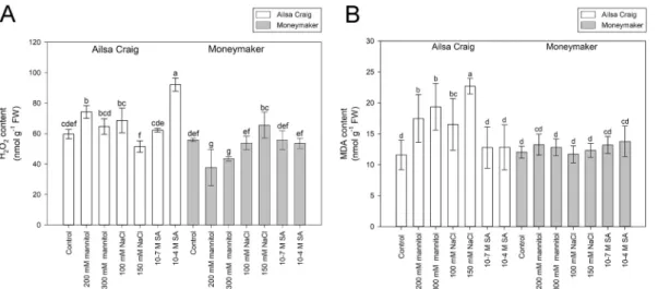
Except for the drastic effect of the 300 mM mannitol, the used treat- ments did not cause visible detrimental damages on roots after 24 h of treatments. Comparison of the H2O2 and the lipid peroxidation marker MDA contents in roots revealed that under control conditions there is no significant differences between the measured H2O2 and MDA levels of the two cultivars (Fig. 1). However, the ROS levels changed differently in the two genotypes after 24 h of mannitol, salt or SA treatments (Fig. 1A). In H2O2 content, the highest (154.5%) increase was detected in ‘Ailsa Craig’ after application of 10−4 M SA, and the lowest value (37.69 ±11.98 nmol g−1 FW) was measured in ‘Moneymaker’ after treatment by 200 mM mannitol. The MDA level was elevated substan- tially in ‘Ailsa Craig’ by osmotic and salt stresses, while in case of
‘Moneymaker’ no significant alteration was detected (Fig. 1B). Inter- estingly, only the lower concentration of mannitol caused significant increase in H2O2 content of ‘Ailsa Craig’, moreover the 150 mM NaCl treatment resulted in even lower ROS level than 100 mM NaCl, indi- cating that the higher stress might activated efficiently the ROS- processing mechanisms.
Determination of the ascorbate and glutathione levels showed that untreated ‘Ailsa Craig’ roots possess generally less non-enzymatic anti- oxidants than ‘Moneymaker’ (Fig. 2). The applied osmotic stress enhanced the total ascorbate content in both genotypes, but it still remained generally lower in ‘Ailsa Craig’ than in ‘Moneymaker’. In
‘Ailsa Craig’ the 100 mM NaCl resulted in the highest increase of the total ASC level (197.6%), but the 150 mM NaCl caused much smaller
changes. Except with relatively small elevation of DHA content in the presence of 10−7 M SA, SA had no effect on ascorbate content of ‘Ailsa Craig’ roots. Salicylic acid treatments had little or no effect also in
‘Moneymaker’, but the mannitol and the isoosmotic NaCl stresses enhanced the total and reduced ASC levels in a concentration-dependent manner (Fig. 2A). The GSH and GSSG content in ‘Ailsa Craig’ roots remained on control level after the 24 h treatments, but those in
‘Moneymaker’ were enhanced by the osmotic and salt stresses (Fig. 2B).
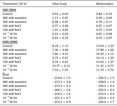
The ASC/DHA and GSH/GSSG ratio data strengthen that the redox state of these antioxidants differed substantially (Table 1). Calculation of the glutathione half-cell potential values resulted in − 214.6 mV in ‘Ailsa Craig’, while − 226.5 mV in ‘Moneymaker’. The most oxidized redox state (− 206.1 mV) in ‘Ailsa Craig’ was detected after applying 100 mM NaCl, while the most reduced state (− 221.4 mV) occurred in case of 10−7 M SA treatment. The mannitol and salt stresses shifted the gluta- thione redox state of ‘Moneymaker’ roots even towards more negative values (Table 1).
3.2. Glutathione transferase-related enzyme activities
Measuring the main glutathione transferase enzyme activities of the
4-week-old tomato roots revealed that under control conditions the specific GST and GPOX activities were higher by 13% and 49%, respectively, in ‘Moneymaker’ compared to ‘Ailsa Craig’ (Fig. 3A and B).
Significant increase of the GST activity was detected only in case of 10−4 M SA-treated ‘Moneymaker’, but GPOX activity was enhanced by 150 mM NaCl and 10−4 M SA in both cultivars, furthermore that of in ‘Ailsa Craig’ by 300 mM mannitol. Interestingly, except the application of 200 mM mannitol on ‘Moneymaker’, the DHAR worked at higher level in
‘Ailsa Craig’. In 10−4 M SA-treated ‘Moneymaker’ roots this activity was not detectable (Fig. 3C).
3.3. Expression pattern of SlGST genes in ‘Ailsa Craig’ and ‘Moneymaker’
roots under different stress treatments
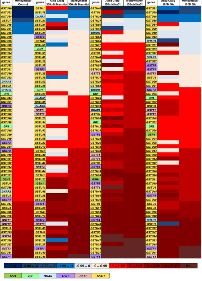
SlGSTs involved especially in removing the toxic stress metabolites and some other genes involved in reestablishment of the main redox- active antioxidants were selected to compare their expression in the two cultivars. The transcript level of 46 SlGSTU, five SlGSTF, four SlGSTT, three DHAR, two GR and one GSH1 gene sequences were detected by HT-qPCR after 24 h of 300 mM mannitol, 150 mM NaCl and 10−4 M SA treatments. To demonstrate the differences in the expression Fig. 1.Hydrogen peroxide (H2O2; A) and malondialdehyde (MDA; B) contents of four-week-old tomato (Solanum lycopersicum ‘Ailsa Craig’ and ‘Moneymaker’) roots after 24 h of mannitol, NaCl and salicylic acid (SA) treatments. Data are means ±SD. Columns with different letters are significantly different at p <0.05, determined by Duncan’s test.
Fig. 2. Reduced and oxidized ascorbate (A) and glutathione (B) contents (ASC, DHA, GSH and GSSG, respectively) of four-week-old tomato (Solanum lycopersicum
‘Ailsa Craig’ and ‘Moneymaker’) roots after 24 h of mannitol, NaCl and salicylic acid (SA) treatments. Data are means ±SD. Columns with different letters are significantly different at p <0.05, determined by Duncan’s test.
pattern of the cultivars, the expression of each gene was compared to the normalized average transcript amount detected in untreated ‘Ailsa Craig’ cultivar, then the log2 expression data were ranked according to the transcript amounts measured in ‘Moneymaker’ (Fig. 4).
The transcript level in the control ‘Ailsa Craig’ cultivar root samples was considered to be 0 for each gene. This representation demonstrates that almost half of the investigated genes expressed at higher level in
‘Moneymaker’ under control conditions, and only seven SlGSTs showed lower transcript abundance. In some cases, there was more than 100 times bigger differences in the 2(−ΔΔCT) values between the two cultivars (like in case of GSTF1, GSTU41 and GSTT3). Analysis of the expression pattern of the totally 58 SlGSTs after different stress treatments revealed that all the selected genes responded in roots to one or more stresses.
Except with a few examples (such as GSTU18), their expression was induced, but the changes were largely stress- and genotype specific.
Interestingly, several genes expressed in ‘Moneymaker’ on extremely high level even under control conditions (e.g., GSTU27, GSTU41, GSTF1), after induction by stress treatments worked at similar level also in ‘Ailsa Craig’ (Fig. 4).
To check the relation between the redox potential and the transcript levels of GSH, GR, DHAR and GST genes, we determined the Pearson’s correlation coefficients. Our results indicated that some genes (GR1, -2, GSTF1, GSTT2, -4, GSTU19, -20, -22, -23, -35, -37, -38, -47) have a very strong positive correlation with the redox potential in both cultivars (Fig. 5). As we used Ehc and ΔCt values for calculation (Table S3), strong positive correlation means that as the root tissues become more reduced, the expression level of a given gene increases, while in case of strong Table 1
Ratio of ascorbic acid and dehydroascorbate (ASC/DHA), reduced and oxidized glutathione (GSH/GSSG) and glutathione redox potential (EGSH) of hydroponi- cally grown 4-week-old Solanum lycopersicum ‘Ailsa Craig’ and ‘Moneymaker’
calculated from mean values presented on Figs. 1 and 2.
Treatments (24 h) Ailsa Craig Moneymaker
ASC/DHA
Control 0.64 ±0.03 0.82 ±0.10
200 mM mannitol 1.13 ±0.07 0.95 ±0.09
300 mM mannitol 0.48 ±0.07 0.76 ±0.11
100 mM NaCl 0.77 ±0.08 0.97 ±0.07
150 mM NaCl 1.01 ±0.05 0.70 ±0.03
10−7 M SA 0.53 ±0.05 0.81 ±0.08
10−4 M SA 0.62 ±0.10 0.76 ±0.07
GSH/GSSG
Control 8.18 ±1.15 12.63 ±1.33
200 mM mannitol 7.36 ±0.46 17.80 ±1.69
300 mM mannitol 7.85 ±0.21 16.43 ±1.12
100 mM NaCl 5.75 ±0.57 16.14 ±1.49
150 mM NaCl 9.18 ±0.63 14.65 ±1.27
10−7 M SA 10.77 ±2.15 11.41 ±0.77
10−4 M SA 7.52 ±1.01 11.70 ±0.72
EGSH
Control −214.6 ±1.0 −226.5 ±1.5
200 mM mannitol −213.5 ±2.8 −238.0 ±1.0
300 mM mannitol −214.4 ±0.2 −235.2 ±0.4
100 mM NaCl −206.1 ±2.8 −235.0 ±0.5
150 mM NaCl −218.5 ±1.6 −233.5 ±0.4
10−7 M SA −221.4 ±0.7 −225.6 ±0.4
10−4 M SA −214.2 ±0.9 −226.0 ±1.7
Fig. 3. Specific glutathione transferase (GST; A), glutathione peroxidase (GPOX; B), and dehydroascorbate reductase (DHAR; C) activity of four-week-old tomato (Solanum lycopersicum ‘Ailsa Craig’ and ‘Moneymaker’) roots after 24 h of mannitol, NaCl and salicylic acid (SA) treatments. Data are means ±SD. Columns with different letters are significantly different at p <0.05, determined by Duncan’s test.
Fig. 4. Heat map of 59 Solanum lycopersicum glutathione transferase (DHAR, GSTF, GSTT, GSTU), two glutathione reductase (GR1, GR2), and one glutathione synthetase (GSH1) gene expression levels. Relative transcript amounts of genes were determined in four-week-old tomato (‘Ailsa Craig’ and ‘Moneymaker’) roots by HT-qPCR under control conditions and after 24 h of 300 mM mannitol, 150 mM NaCl and 10−4 M salicylic acid (SA) treatments. For this, the expression of genes was normalized first by actin2 gene, and second to the average transcript amount of each gene in untreated ‘Ailsa Craig’ cultivar. Log2 of 2(-ΔΔCt) data were presented as a heat map. Blue colours show repression, while red colours show activation, as it is indicated on the upper colour scale bar. The name of genes belonging to different classes were coloured according to the lower scale bar. Genes were arranged according to the increasing expression level measured in ‘Moneymaker’. The presented data are from two biological replicates.
negative correlation the transcript level of a given gene increases as the root tissues become more oxidized (Table S3). The correlation analysis revealed that there are a lot of differences in the R values of ‘Ailsa Craig’
and ‘Moneymaker’. For example, in ‘Ailsa Craig’ strong positive corre- lation can be found between the redox potential and the expression of DHAR3 and -5 genes, while in ‘Moneymaker’ among DHAR genes the transcript level of DHAR2 has strong positive correlation with the redox potential. The transcription of GSTT3 relates differently to the redox state: while in ‘Ailsa Craig’ it shows positive correlation, in ‘Money- maker’ a very strong negative correlation was determined. Among the sequences belonging to GSTU group we found strong positive correla- tion values in the case of GSTU4, -5, -12, -28, -42, -44, -46, -53, -55 only in ‘Ailsa Craig’ plants, while in the case of GSTU3, -8, -24, -25, -34, -48, -52 only in ‘Moneymaker’ plants. Negatively correlated the expression level of GSTU18, -25, -40, -54 with the redox potential only in ‘Ailsa Craig’, and that of GSTU28, -39, -50 only in ‘Moneymaker’ (Fig. 5).
Hereafter we analysed the 5’ regulator regions of SlGSTs showing high positive correlation (SlGSTT2, SlGSTT4, SlGSTU19, SlGSTU20) or negative correlation (SlGSTU31, SlGSTU39, SlGSTU40, SlGSTU43) with the changes in redox potential of tomato roots. The in silico analysis revealed different occurrence of redox-responsible cis-elements, such as W-box in the promoter regions of the selected tomato genes (data not shown).
4. Discussion
4.1. The higher GSH and ASC contents of the ‘Moneymaker’ might be responsible for the more efficient defence against different abiotic stresses
The essential buffering mechanism which prevents excessive reduc- tion or oxidation is the cellular redox homeostasis that involves complex interaction of oxidants and ROS-processing mechanisms. It is well- established that elevation of endogenous ROS level and changes in redox status can fulfil signalling functions, playing a beneficial role in adaptation to changed environmental conditions (Choudhury et al., 2016; Das et al., 2015; Foyer and Noctor, 2005; Potters et al., 2010;
Schmidt and Schippers, 2015). Redox changes might be genetically determined or triggered by the environment. The viable and
proliferating cells possess a high GSH/GSSG ratio and a sufficiently negative redox potential, whereas higher redox potentials (more oxidative) are found in apoptotic and senescent cells (Schafer and Buettner, 2001). The spatial and temporal redox changes influence growth and development due to reprogramming of metabolism (Kocsy et al., 2013; Paciolla et al., 2016) and altering hormone (i.e., auxin) levels (Jiang et al., 2016). Using genetically encoded redox-sensitive green fluorescent proteins (roGFP1, roGFP2) as redox sensors it was demonstrated that enhanced ROS production can temporarily shift the redox potential of cells towards more oxidized values (Hanson et al., 2004; Jiang et al., 2006, 2016; Meyer et al., 2007; Schwarzl¨ander et al., 2008). Oxidation alters the operational controls of many redox-sensitive proteins and non-protein thiols (Foyer and Noctor, 2015; Jacques et al., 2015). It was reported that H2O2-triggered modulation of GSH content also could be perceived by the cells as a signal (Han et al., 2013).
Furthermore, Han et al. (2013) proved that GSH accumulation and redox status are closely linked to SA synthesis and signalling. In our experiments, the ASC and GSH levels of 4-week-old hydroponically grown tomato roots proved to be higher in ‘Moneymaker’ than in ‘Ailsa Craig’. The used SA concentrations, with one exception, did not affect the GSH and ASC levels of the plants after 24 h, similarly to the earlier investigations conducted on 5-week-old ‘Rio Fuego’ tomato cultivars (Tari et al., 2015). Since parallelly only the 10−4 M SA increased the H2O2 content in ‘Ailsa Craig’ roots, we may assume that distinct ROS-processing mechanisms were activated by SA to cope with excess ROS, such as superoxide dismutase, ascorbate peroxidase and GR en- zymes (Tari et al., 2015). The applied osmotic and salt stresses further elevated the levels of these non-enzymatic antioxidants in ‘Money- maker’, while in ‘Ailsa Craig’ only the ASC content was increased by the lower concentrations of mannitol and NaCl. The more MDA measured in
‘Ailsa Craig’ roots compared to ‘Moneymaker’ indicates that operation and/or activation of the antioxidant mechanisms in ‘Ailsa Craig’ due to stresses did not prevent the accumulation of ROS and lipid peroxides as efficiently as that in the ‘Moneymaker’ tomato cultivar (Figs. 1 and 2).
GSH has prominent role in the cellular redox control because GSH/
GSSG may reduce/oxidise or de/glutathionylate protein thiols, but be- sides GSH, all members of the ASC-GSH cycle have specific functions in the metabolism and thus in the growth and development (Foyer and Fig. 5.Correlation analysis on the basis of the redox potential (Ehc) and expression of selected genes (ΔCt values) measured under different conditions in the roots of
‘Ailsa Craig’ and ‘Moneymaker’ tomato plants. Green colour highlights show positive, red colour highlights show negative correlations.
Noctor, 2011, 2015; Kocsy et al., 2013). The redox control of growth can be observed not only at cellular, but at the tissue and organ levels too (De Tullio et al., 2010). GSH has essential function in roots, as it was shown in Arabidopsis (Koprivova et al., 2010; Schnaubelt et al., 2014). It was reported that low level of GSH significantly increased the redox potentials and caused arrest of the cell cycle in roots but not shoots. Root is a key organ, and it is crucial for plant performance and crop pro- ductivity even under normal and stress conditions when they are forced to adopt by structural and functional modifications (Vives-Peris et al., 2020). GSH is specifically required to activate and maintain the cell division cycle in root apical cells and development of the root apical meristem (Vernoux et al., 2000). Severe GSH depletion specifically inhibited, while low root GSH levels decreased lateral root densities (Cheng et al., 1995; Frendo et al., 2005; Schnaubelt et al., 2014; Vernoux et al., 2000). It is also evidenced that enhancement of GSH and ASC pools could help the adaptation to different types of stresses (Antoniou et al., 2016). In correlation with the GSH contents of the investigated cultivar’s roots, the transcription rate of the first rate-limiting enzyme of glutathione synthesis, SlGSH1 (gamma-glutamylcysteine synthetase1, also known as glutamate-cysteine ligase) was higher in ‘Moneymaker’ than in ‘Ailsa Craig’ both in control conditions and in the presence of 150 mM NaCl (Fig. 4.). Calculation of the glutathione half-cell redox potential values revealed ca. 12 mV differences between the EGSH of the untreated roots and, because of the dramatic increase of GSH content in
‘Moneymaker’, the difference was further increased under salt and os- motic stresses (Table 1). A relatively small global shift in the EGSH is associated with a very large change in expression of genes (Aller et al., 2013; Schnaubelt et al., 2014).
4.2. Many SlGSTs were expressed at higher level in ‘Moneymaker’ than in
‘Ailsa Craig’
Plant genomes contain dozens of GSTs encoding subunits that can form homodimers or heterodimers, leading to enormous diversity within GST protein families (Csisz´ar et al., 2019; Dixon and Edwards, 2010;
Islam et al., 2017). They possess high cytoprotective roles and have essential functions during oxidative conditions (Est´evez and Hern´andez, 2020, Horv´ath et al., 2015; Labrou et al., 2015; Ugalde et al., 2020).
Most plant GSTs are reported to be induced by abiotic and biotic stresses and have positive roles in defence mechanisms (Edwards et al., 2000;
Gullner et al., 2018). GSTs could act on a broad range of endogenous or exogenous substrates increasing their solubility and promoting their metabolism or sequestration into the vacuole (Dixon et al., 2002a, 2002b; Sousa et al., 2021).
Few exceptions aside, the GSTU class is the largest plant GST class, and together with the GSTFs they represent around 70% of all GST genes in tomato (Islam et al., 2017; Sylvestre-Gonon et al., 2019) and Arabi- dopsis (Dixon and Edwards, 2010). The main known functions of these isoenzymes are the GSH-dependent peroxidase (GPOX) and GSH trans- ferase activities (Edwards et al., 2000; Sylvestre-Gonon et al., 2019).
Due to the high specificity of their GSH-binding region, GSTUs and GSTFs are commonly considered in a conjugation context (Cummins et al., 2011; Est´evez and Hern´andez, 2020), although in some cases other, even signalling function was rendered to them (Laborde, 2010;
Meyer et al., 2020). In Arabidopsis, AtGSTU17 was reported exception- ally to be a negative component of the signalling pathway in response to drought and salt stress (Chen et al., 2012). Among the investigated SlGSTUs in our study, the expression of SlGSTU18 was generally downregulated by the used treatments in both cultivars, moreover its transcript amount was lower in ‘Moneymaker’ than in ‘Ailsa Craig’.
However, this gene, named earlier as LeGSTU2, was reported to be induced by salt-, osmotic-, and heat stresses, and its overexpression in Arabidopsis resulted in enhanced resistance to NaCl and mannitol (Xu et al., 2015).
In tomato, the SlGSTU24 was described as a very effective ROS scavenger, and later it was found that its expression in yeast significantly
enhanced the resistance to H2O2-induced stress and brought the total GSH levels back to normal (Kilili et al., 2004). This isoenzyme was isolated and marked as an enzyme involved in the protection against Bax-induced cell death (“Bax-inhibitor GST”) (Kampranis et al., 2000).
In our experiments, the SlGSTU24 expressed at extremely high level in both cultivars after applying SA treatments in accordance with the re- sults of in silico gene expression analysis performed by Islam et al. (2017) that indicated important function of SlGSTU24 in different biotic stresses and wounding. Although 150 mM NaCl induced its transcription in both genotypes, SlGSTU24 expressed on higher level in ‘Ailsa Craig’
than ‘Moneymaker’, and the osmotic stress activated it only in the ‘Ailsa Craig’.
However, our results showed in most cases that, besides the overall higher SlGST expression levels, these genes were activated due to stresses more efficiently in ‘Moneymaker’ resulting in generally higher GST and GPOX activities. Comparing the transcriptomes of ‘Money- maker’ and a salt tolerant wild tomato (Solanum pimpinellifolium
‘PI365967’) Sun et al. (2010) found that several GSTs were expressed at a higher level in the salt tolerant genotype but were induced by salt stress in the ‘Moneymaker’ cultivar. These authors concluded that those genes might have lost their high expression capacity during domesti- cation (Sun et al., 2010). Interestingly, most of the SlGSTs expressing on very high level in untreated ‘Moneymaker’, such as SlGSTU27, SlGSTU41, SlGSTF1, were strongly induced in ‘Ailsa Craig’ by different stress treatment. While SlGSTF1 transcription showed strong positive correlation with the calculated EGSH values in roots of both cultivars, stronger correlation was found between the expression of SlGSTU27 and SlGSTU41 and the redox potential changes in ‘Ailsa Craig’, than in
‘Moneymaker’ (Fig. 5). The stronger redox dependency of SlGST tran- scriptions observed in ‘Ailsa Craig’ at several cases may indicate that alteration of the redox status and/or ROS level is more important component of the SlGST expression induction in the ‘Ailsa Craig’ than in
‘Moneymaker’ cultivar. The positive correlation found at most of the cases between the more negative redox potential and induction of SlGST expression suggests that the relationship among the GSH redox potential and GST activity or gene expression is more complex than it was sup- posed earlier.
4.3. The higher expression of SlGSTs together with increased GSH indicates new way of relationship among the GSTs and redox signalling
GSTs are known also for their roles in maintaining the physiological redox state of the cell (Dixon et al., 2011). Several reports indicated that GST expression and activity are affected by GSH concentration and GSH/GSSG ratio. The more oxidized environment, the increased GSSG, the low GSH level all might induce transcription of GST genes (Schnaubelt et al., 2014). GST proteins might undergo post-translational modification connected to stress responses, such as disulphide formation or S-glutathionylation (Dixon and Edwards, 2010). Moreover, it was proved that oxidation of AtGSTs might improve the biochemical prop- erties, substrate binding capacity and activity of proteins (Tossounian et al., 2018, 2019). The co-expression of GSTs and genes involved in GSH biosynthesis (GSH1, 2) and re-reduction (GR) were also reported (e.g., Islam et al., 2017; Wei et al., 2019). Most of the known GST activity, like GST and GPOX uses GSH as co-substrate and might decrease the con- centration of the GSH in each compartment, which can explain this phenomenon. However, our present results highlight the importance of the high level of GSH and more negative redox potential in induction of SlGST expression. This implicates the operation of intensifying or feed- back mechanisms. Interestingly, similar pattern was observed in SlGSH1 expression (coding the first, rate-limiting enzyme of GSH biosynthesis) to one of the highlighted SlGST group: higher level of transcription in un-treated ‘Moneymaker’, that was further induced by abiotic stress treatment, while bigger elevation was detected in ‘Ailsa Craig’, espe- cially after applying 300 mM mannitol (Fig. 4).
Several cis-regulatory elements were connected to the upset of
cellular oxidative balance. This led to in silico analysis of the promoter region (− 1500 bp) of SlGSTU genes with the strongest positive and negative correlation to calculated redox potential (in both cultivars).
Differences were found in the number of AREs in the 5’ regulatory re- gion between the SlGSTs showing the strongest positive (SlGSTU19 and SlGSTU20) and negative (SlGSTU31, SlGSTU39, SlGSTU40 and SlGSTU43) correlation to redox potential. The promoter of the later four genes contained four or five W boxes. The negative correlation of the expressional changes of these genes to the redox potential reflects to their transcriptional induction with elevation of GSSG amounts in the cells. This relationship indicates that these GSH-utilising enzymes may have redox status-directed feedback in their transcriptional regulation.
Since W-box is among the PLT-regulated sequence elements, we may assume that in this way some GSTs can be part of the gene regulatory network controlled by PLT, thus might be the link between growth processes and antioxidant mechanisms. Whether these GSTs are under ROS/redox control through the PLT and/or other transcription factors that was explored in quiescent centre (Wang et al., 2021), needs further investigations.
5. Conclusions
As a summary, under control conditions the Solanum lycopersicum
‘Moneymaker’ possess higher ASC and GSH contents, more negative glutathione redox potential; several SlGST genes are expressed at higher levels and its GST and GPOX activities were enhanced compared to those in ‘Ailsa Craig’ cultivar. These features might eventuate the elevated stress tolerance of ‘Moneymaker’ cultivar. Nevertheless, the induction of non-enzymatic and enzymatic antioxidants may also contribute to the recovery of the redox potential during stresses. In ‘Moneymaker’, be- sides activation of sequences involved in recycling of ASC and GSH pools, large number of SlGSTs were upregulated, but at several cases higher correlation with redox changes was observed in the ‘Ailsa Craig’. The higher expression of SlGSTs together with elevated SlGSH1 expression and increased GSH level indicate unknown aspect of rela- tionship among the GSTs and redox signalling.
SlGSTs with high expression levels or showing strong positive or negative correlations with redox potentials presumably have important roles in stress responses. It is suggested that GSTs can be part of the transcriptional gene regulatory network controlled by main transcrip- tional factors in root, thus functioning as a link between antioxidant mechanisms and growth processes. Further investigations of their transcript amount in other genotypes and their molecular character- ization may lead to new insight in improving stress tolerance of tomato and other crops.
Author contributions
Conceptualization, A.G, J.Cs., L.P.; methodology, ´ A.G., ´ A.H., E.H., K. ´ B., N.F.; software, A.G., ´ A.H., E.H., K.B., N.F.; validation, ´ A.G., ´ A.H., N. ´ F.; formal analysis, A.H., J.Cs., K.B.; investigation, ´ A.G., ´ A.H., E.H., K.B., ´ M.H., N.F.; resources, J.Cs. and L.P.; data curation, A.G., ´ A.H., J.Cs., K. ´ B.; writing - original draft preparation, A.G. and K.B.; writing - review ´ and editing, A.H., E.H., J.Cs; visualization, ´ A.G., ´ A.H., K.B.; supervision, ´ J.Cs. and L.P.; project administration, J.Cs.; funding acquisition, E.H., J.
Cs., K.B.
Funding
This research was funded by the Hungarian National Research, Development and Innovation Fund, grant numbers are K 125265, PD 131909 and PD 131884. OA was supported by the University of Szeged Open Access Fund, grant number: 5432.
Declaration of competing interest
The authors declare that they have no known competing financial interests or personal relationships that could have appeared to influence the work reported in this paper.
Acknowledgments
The authors would like to thank Mrs. Erzs´ebet Porkol´ab for her excellent technical assistance.
Appendix A. Supplementary data
Supplementary data to this article can be found online at https://doi.
org/10.1016/j.plaphy.2021.11.009.
References
Aida, M., Beis, D., Heidstra, R., Willemsen, V., Blilou, I., Galinha, C., Nussaume, L., Noh, Y.S., Amasino, R., Scheres, B., 2004. The PLETHORA genes mediate patterning of the Arabidopsis root stem cell niche. Cell 119 (1), 109–120.
Aller, I., Rouhier, N., Meyer, A., 2013. Development of roGFP2-derived redox probes for measurement of the glutathione redox potential in the cytosol of severely glutathione-deficient rml1 seedlings. Front. Plant Sci. 4.
Antoniou, C., Savvides, A., Christou, A., Fotopoulos, V., 2016. Unravelling chemical priming machinery in plants: the role of reactive oxygen–nitrogen–sulfur species in abiotic stress tolerance enhancement. Curr. Opin. Plant Biol. 33, 101–107.
Bela, K., Riyazuddin, R., Horv´ath, E., Hurton, A., Gall´ ´e, ´A., Tak´acs, Z., Zsigmond, L., Szabados, L., Tari, I., Csisz´ar, J., 2018. Comprehensive analysis of antioxidant mechanisms in Arabidopsis glutathione peroxidase-like mutants under salt- and osmotic stress reveals organ-specific significance of the ATGPXL’s activities.
Environ. Exp. Bot. 150, 127–140.
Bindoli, A., Rigobello, M.P., 2013. Principles in redox signaling: from chemistry to functional significance. Antioxidants Redox Signal. 18 (13), 1557–1593.
Bradford, M., 1976. A rapid and sensitive method for the quantitation of microgram quantities of protein utilizing the principle of protein-dye binding. Anal. Biochem.
72 (1–2), 248–254.
Carlberg, I., Mannervik, B., 1985. Glutathione reductase. Glutamate, Glutamine, Glutathione, and Related Compounds 484–490.
Chen, J., Jiang, H., Hsieh, E., Chen, H., Chien, C., Hsieh, H., Lin, T., 2012. Drought and salt stress tolerance of an Arabidopsis glutathione s-transferase u17 knockout mutant are attributed to the combined effect of glutathione and abscisic acid. Plant Physiol.
158 (1), 340–351.
Cheng, J., Seeley, K., Sung, Z., 1995. Rml1 and rml2, Arabidopsis genes required for cell proliferation at the root tip. Plant Physiol. 107 (2), 365–376.
Choudhury, F., Rivero, R., Blumwald, E., Mittler, R., 2016. Reactive oxygen species, abiotic stress and stress combination. Plant J. 90 (5), 856–867.
Csisz´ar, J., Hecker, A., Labrou, N., Schr¨oder, P., Riechers, D., 2019. Editorial: plant glutathione transferases: diverse, multi-tasking enzymes with yet-to-be discovered functions. Front. Plant Sci. 10.
Csisz´ar, J., Horv´ath, E., V´ary, Z., Gall´e, A., Bela, K., Brunner, S., Tari, I., 2014. ´ Glutathione transferase supergene family in tomato: salt stress-regulated expression of representative genes from distinct GST classes in plants primed with salicylic acid.
Plant Physiol. Biochem. 78, 15–26.
Cummins, I., Dixon, D., Freitag-Pohl, S., Skipsey, M., Edwards, R., 2011. Multiple roles for plant glutathione transferases in xenobiotic detoxification. Drug Metabol. Rev. 43 (2), 266–280.
Das, P., Nutan, K.K., Singla-Pareek, S.L., Pareek, A., 2015. Oxidative environment and redox homeostasis in plants: dissecting out significant contribution of major cellular organelles. Frontiers in Environmental Science 2, 70.
De Tullio, M., Jiang, K., Feldman, L., 2010. Redox regulation of root apical meristem organization: connecting root development to its environment. Plant Physiol.
Biochem. 48 (5), 328–336.
Ding, P., Ding, Y., 2020. Stories of salicylic acid: a plant defense hormone. Trends Plant Sci. 25 (6), 549–565.
Dixon, D., Davis, B., Edwards, R., 2002a. Functional divergence in the glutathione transferase superfamily in plants. J. Biol. Chem. 277 (34), 30859–30869.
Dixon, D., Edwards, R., 2010. Glutathione transferases. Arabidopsis Book 8, e0131.
Dixon, D., Lapthorn, A., Edwards, R., 2002b. Plant glutathione transferases. Genome Biol. 3 (3) reviews3004.1.
Dixon, D.P., Steel, P.G., Edwards, R., 2011. Roles for glutathione transferases in antioxidant recycling. Plant Signal. Behav. 6 (8), 1223–1227.
Dunand, C., Cr`evecoeur, M., Penel, C., 2007. Distribution of superoxide and hydrogen peroxide in Arabidopsis root and their influence on root development: possible interaction with peroxidases. New Phytol. 174 (2), 332–341.
Edwards, R., Dixon, D., 2005. Plant glutathione transferases. Methods Enzymol.
169–186.
Edwards, R., Dixon, D., Walbot, V., 2000. Plant glutathione s -transferases: enzymes with multiple functions in sickness and in health. Trends Plant Sci. 5 (5), 193–198.
Est´evez, I.H., Hern´andez, M.R., 2020. Plant glutathione S-transferases: an overview.
Plant Gene 23, 100233.