International Journal of
Molecular Sciences
Article
Compensation of Mutation in Arabidopsis glutathione transferase (AtGSTU ) Genes under Control or Salt Stress Conditions
Edit Horváth1,2, Krisztina Bela2 ,Ágnes Gallé2, Riyazuddin Riyazuddin2,3 , Gábor Csomor2, Dorottya Csenki2and Jolán Csiszár2,*
1 Institute of Plant Biology, Biological Research Centre, Temesvári krt. 62., H-6726 Szeged, Hungary;
horvathedo@gmail.com
2 Department of Plant Biology, Faculty of Sciences and Informatics, University of Szeged, Közép fasor 52., H-6726 Szeged, Hungary; belakriszti88@gmail.com (K.B.); galleagnes@gmail.com (A.G.);
riyazkhan24992@gmail.com (R.R.); tsomyer@gmail.com (G.C.); csenkidorottyazsuzsanna@gmail.com (D.C.)
3 Doctoral School in Biology, Faculty of Science and Informatics, University of Szeged, H-6726 Szeged, Hungary
* Correspondence: csiszar@bio.u-szeged.hu; Tel.:+36 62 544 307
Received: 29 January 2020; Accepted: 26 March 2020; Published: 28 March 2020
Abstract: Glutathione transferases (GSTs) play a crucial role in detoxification processes due to the fact of their glutathione (GSH) conjugating activity, and through glutathione peroxidase or dehydroascorbate reductase (DHAR) activities, they influence the redox state of GSH and ascorbate (AsA). The plant-specific tau (GSTU) group is the largest class ofArabidopsisGSTs, and their members are involved in responses to different abiotic stresses. We investigated the effect of salt stress on two-week-old Arabidopsis thaliana wild-type (Col-0), Atgstu19 andAtgstu24 mutant plants after applying 150 mM NaCl for two days. TheAtgstu19seedlings had lower GST activity and vitality both under control conditions and after salt stress than the wild-type, but the level of total ROS was similar to the Col-0 plants. The GST activity of the knockoutAtgstu24mutant was even higher under control conditions compared to the Col-0 plants, while the ROS level and its vitality did not differ significantly from the wild-type. Analysis of theAtGSTUexpression pattern revealed that the mutation in a singleAtGSTUgene was accompanied by the up- and downregulation of several other AtGSTUs. Moreover, elevated AsA and GSH levels, an altered GSH redox potential and increased DHAR and glutathione reductase activities could help to compensate for the mutation ofAtGSTU genes. The observed changes in the mutants suggest that the investigated isoenzymes influence the redox homeostasis under control conditions and after NaCl treatment inArabidopsisseedlings.
These data indicate for the first time the more general role of a temporary shift of redox status as part of GST mechanisms and regulation.
Keywords: antioxidant enzyme activity; glutathione; glutathione reductase; glutathione transferase;
gene expression; NaCl; redox state
1. Introduction
Glutathione transferases (GSTs) belong to a very ancient protein superfamily that is thought to have evolved in response to the development of oxidative stress. Plant genomes contain dozens of GSTgenes and most of the proteins can be found in homodimer or heterodimer form, leading to enormous diversity within GST protein families [1]. Glutathione transferases function both as catalytic enzymes with activity towards various substrates as well as non-catalytic, binding and carrier proteins [2,3]. Plant GSTs are grouped into ten classes and among them, the most abundant
Int. J. Mol. Sci.2020,21, 2349; doi:10.3390/ijms21072349 www.mdpi.com/journal/ijms
Int. J. Mol. Sci.2020,21, 2349 2 of 16
tau (GSTU), phi (GSTF), dehydroascrobate reductase (DHAR) and lambda (GSTL) isoenzymes are plant-specific. Dehydroascrobate reductase and GSTL enzymes catalyse redox reactions and participate in the recycling of antioxidants, such as ascorbate (AsA) and flavonols [4]. Tau and phi GSTs are involved mainly in xenobiotic metabolism, which may be related to their high affinity toward a broad spectrum of harmful compounds, including xenobiotics and endogenous stress metabolites. They not only have detectable glutathione-conjugating activity towards standard xenobiotic GST substrates, but via their glutathione peroxidase (GPOX) activity, they may take part in reducing the levels of H2O2
and other peroxides [5]. Moreover, by using glutathione (GSH,γ-Glu-Cys-Gly) as a co-substrate, their catalytic activity may reduce the pool of this redox-active molecule.
It is widely accepted that GSTs orchestrate and fine tune plant adaptation and tolerance to environmental stresses, pathogenic attackers and the detoxification of toxic chemicals and metabolites [2,3,6–16]. Conducting genetic screens to identify components of salt stress tolerance in Arabidopsisrevealed thatAtGSTU19andAtGSTF9can be important cytoplasmic proteins in salt stress responses [17]. During the investigation of the hardening mechanism, we previously found that the maintained or even increased GST and GPOX activities are important for efficient adaptation to the subsequent salt stress [11,18]. Among the investigatedArabidopsis GSTgenes, induction ofAtGSTU19 andAtGSTU24genes and elevated GST activity was observed in the leaves and roots of hydroponically grown eight-week-old plants after one week of 100 mM NaCl treatment [11].
InArabidopsis, GSTUs include 28 members which can be the result of gene duplication events [19].
Tau classArabidopsisGSTs can be divided into three separate clades using phylogenetic methods [5], but the importance of this differentiation is still unknown. Several studies describe the role of GSTUs in detoxification processes [20–22] and plants overexpressingGSTUgenes have enhanced stress resistance [23–25]. Almost all of theArabidopsisGSTUs have been found to selectively bind fatty acid derivatives and some of the isoenzymes are induced by abiotic stresses or play a role in signalling [2]. Under control conditions, AtGSTU19 represents a significant percentage of the GST pool [26]. Analysis of the expression pattern ofAtGSTgenes based on micro-array data and using Genevestigator meta-analyser revealed thatAtGSTU19was expressed in almost all of the investigated tissues and had the highest relative transcription level among theAtGSTs[19]. Transgenic plants overexpressing theAtGSTU19gene had enhanced salt-, drought- or methyl viologen stress tolerance, and it was suggested that AtGSTU19 may act as a stress regulator by increasing the activities of antioxidant enzymes to strengthen the ROS scavenging activity or maintain ROS homeostasis [27].
Comparing the effect of 75 and 150 mM NaCl treatments on the roots of seven-day-old knockdown Atgstu19 mutants to the Col-0 wild-type, we previously found that theAtgstu19 roots were more sensitive to the salt stress than the wild-type.Atgstu19had lower vitality, a higher amount of H2O2
and application of the redox sensitive green fluorescent protein (roGFP2) [28] affirmed a more oxidized redox state in all root zones compared to the Col-0 roots after 3 h (h) of treatment [29].
Redox homeostasis is a fundamental cell property in which regulation includes the control of reactive oxygen species (ROS) generation, sensing and readjustment of the cellular redox state. It is widely accepted that the ratio of reduced and oxidized glutathione (GSH:GSSG) is an effective marker of cellular redox homeostasis [30,31]. From the concentration of GSH and GSSG, the glutathione half-cell reduction potential (EGSSG/2GSH) can be calculated [32]. A great deal of evidence underpins the importance of ROS and redox homeostasis in the acclimation of plants to salinity (reviewed in Reference [33]), and the salt tolerance in some plants appears to correlate with their ability to scavenge ROS [11,18,34–37]. In order to keep ROS levels tightly regulated, different non-enzymatic antioxidants and enzymatic systems have evolved in aerobic organisms [38,39]. Non-enzymatic scavengers of ROS include AsA, GSH, phenolic compounds, carotenoids, flavonoids and tocopherol.
Enzymatic ROS scavengers include superoxide dismutase (SOD), catalase (CAT), ascorbate peroxidase (APX), glutathione peroxidase-like (GPXL), DHAR and GST enzymes [40].
Although there is some information in the literature on the involvement of individualAtGSTUsin the salt stress responses of plants, their role is still poorly understood. Much evidence indicates that
Int. J. Mol. Sci.2020,21, 2349 3 of 16
GSTUs significantly contribute to adaptation and have a positive role in tolerance to salinity and other abiotic environmental stresses [6,11,18,21,39,41]. On the other hand, AtGSTU17 was reported to be a negative component of the stress-mediated signal transduction pathway in adaptive responses to drought and salt stress [42]. Measuring the enzyme activities of bacterially expressed recombinant ArabidopsisGSTs assayed with 1-chloro-2,4-dinitrobenzene (CDNB), benzyl isothiocyanate and cumene hydroperoxide (CHP) model substrates suggested that related sequences have broadly similar enzyme activity spectrums, and it is more likely that there is a substantial overlap of activities and functional redundancy within the superfamily [5].
In the present experiments, we aimed to investigate the particular role of AtGSTU19 and AtGSTU24 in salt stress responses induced by 150 mM NaCl using T-DNA insertion mutants. While the two isoenzymes show high amino acid identity [5] and our earlier results indicated that both of them play positive roles in salt stress responses, the changes in total extractable GST activity in the two mutants showed different tendencies. TheAtgstu19mutants had lower GST activity and vitality and higher dehydroascorbate (DHA) content than the other two investigated genotypes under control conditions, but the decreased ROS levels and maintained redox status indicate a successful induction of the antioxidant mechanisms. Here, we demonstrate that besides the alteredAtGSTUsexpression pattern, the changes in the redox-active antioxidant mechanism may contribute to the compensation for the effect of a mutation in anAtGSTUgene. The elevated DHAR and glutathione reductase (GR) activities in the untreatedAtgstu24seedlings suggest that AtGSTU24 may also contribute to the regulation of redox homeostasis. These observed physiological and molecular changes highlight the importance of the redox-related processes as common part of the mechanisms of GSTUs.
2. Results
2.1. The Atgstu19 Mutant had Lower GST Activity, While the Atgstu24 Mutant had Higher Activity, in Comparison to the Wild-Type
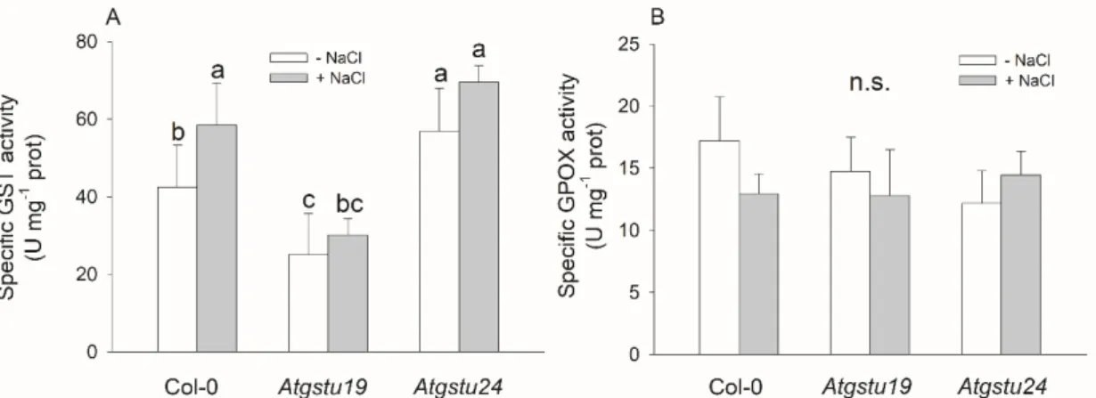
The 16 day old mutant lines did not show marked changes in their phenotype in comparison to the wild-type plants either in optimal conditions or under salt stress triggered by 150 mM NaCl. Measuring GST activity using the CDNB substrate revealed that knockdown mutation of theAtGSTU19gene resulted in decreased GST activity (38% lower than in wild-type controls) in seedlings under control conditions, in contrast to the mutation ofAtGSTU24,in which the total GST activity was even higher (by 35%) than that of Col-0 plants. Treatment of plants with 150 mM NaCl for 48 h elevated the GST activity in the wild-type andAtgstu24lines (136% and 166%, respectively), but not inAtgstu19plants (Figure1A). Glutathione peroxidase activity, measured with CHP substrate, did not differ significantly among the genotypes either under control conditions or after the NaCl treatment (Figure1B).
Int. J. Mol. Sci.2020,21, 2349 4 of 16
to be a negative component of the stress-mediated signal transduction pathway in adaptive responses to drought and salt stress [42]. Measuring the enzyme activities of bacterially expressed recombinant Arabidopsis GSTs assayed with 1-chloro-2,4-dinitrobenzene (CDNB), benzyl isothiocyanate and cumene hydroperoxide (CHP) model substrates suggested that related sequences have broadly similar enzyme activity spectrums, and it is more likely that there is a substantial overlap of activities and functional redundancy within the superfamily [5].
In the present experiments, we aimed to investigate the particular role of AtGSTU19 and AtGSTU24 in salt stress responses induced by 150 mM NaCl using T-DNA insertion mutants. While the two isoenzymes show high amino acid identity [5] and our earlier results indicated that both of them play positive roles in salt stress responses, the changes in total extractable GST activity in the two mutants showed different tendencies. The Atgstu19 mutants had lower GST activity and vitality and higher dehydroascorbate (DHA) content than the other two investigated genotypes under control conditions, but the decreased ROS levels and maintained redox status indicate a successful induction of the antioxidant mechanisms. Here, we demonstrate that besides the altered AtGSTUs expression pattern, the changes in the redox-active antioxidant mechanism may contribute to the compensation for the effect of a mutation in an AtGSTU gene. The elevated DHAR and glutathione reductase (GR) activities in the untreated Atgstu24 seedlings suggest that AtGSTU24 may also contribute to the regulation of redox homeostasis. These observed physiological and molecular changes highlight the importance of the redox-related processes as common part of the mechanisms of GSTUs.
2. Results
2.1. The Atgstu19 Mutant had Lower GST Activity, While the Atgstu24 Mutant had Higher Activity, in Comparison to the Wild-Type
The 16 day old mutant lines did not show marked changes in their phenotype in comparison to the wild-type plants either in optimal conditions or under salt stress triggered by 150 mM NaCl.
Measuring GST activity using the CDNB substrate revealed that knockdown mutation of the AtGSTU19 gene resulted in decreased GST activity (38% lower than in wild-type controls) in seedlings under control conditions, in contrast to the mutation of AtGSTU24, in which the total GST activity was even higher (by 35%) than that of Col-0 plants. Treatment of plants with 150 mM NaCl for 48 h elevated the GST activity in the wild-type and Atgstu24 lines (136% and 166%, respectively), but not in Atgstu19 plants (Figure 1A). Glutathione peroxidase activity, measured with CHP substrate, did not differ significantly among the genotypes either under control conditions or after the NaCl treatment (Figure 1B).
Figure 1. Main glutathione transferase activities of two-week-old wild-type (Col-0) and GSTU mutant (Atgstu19 and Atgstu24) Arabidopsis plants treated with 150 mM NaCl for 48 h. (A) Glutathione transferase (GST) activity, (B) glutathione peroxidase (GPOX) activity of seedlings. Means ± SD Figure 1.Main glutathione transferase activities of two-week-old wild-type (Col-0) andGSTUmutant (Atgstu19andAtgstu24)Arabidopsisplants treated with 150 mM NaCl for 48 h. (A) Glutathione transferase (GST) activity, (B) glutathione peroxidase (GPOX) activity of seedlings. Means±SD denoted by different letters indicate a significant difference between the treatments at ap<0.05 level according to Duncan’s test.
2.2. The AtGSTU Gene Expression Profiles Changed in the Mutants
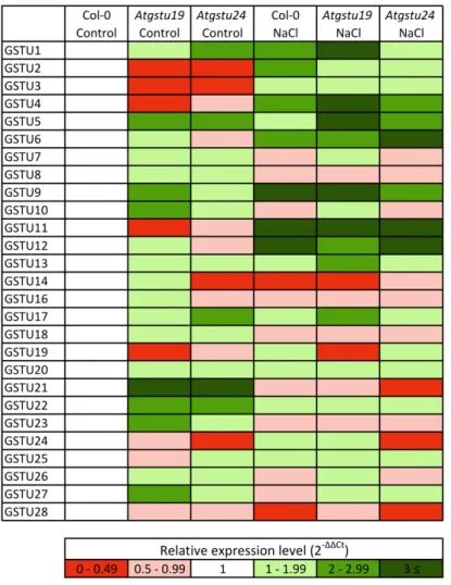
Determination of the transcript amounts of the genes belonging to the tau class of GSTs by quantitative real-time PCR (RT-qPCR) revealed thatGSTU19was expressed at the highest level among theAtGSTUsin the 16 day old Col-0 seedlings, while the expression level ofGSTU24would be ranked eighth (2-∆Ct data are shown in Table S1). To compare the expression pattern of AtGSTUsin the investigated genotypes, the transcript amount of eachAtGSTUgene detected in Col-0 control plants was taken as an arbitrary one. The transcript amount ofAtGSTU19inAtgstu19was approximately half of that in the wild-type, but that ofAtGSTU24in theAtgstu24mutant was at the level of detectability.
At the same time, the expression of several otherAtGSTUgenes was altered significantly in mutants (Figure2, Table S1). Under control conditions,AtGSTU1, AtGSTU5, AtGSTU21andAtGSTU22were upregulated (2–6 times higher gene expressions than in Col-0) in bothAtgstumutants. Additionally, AtGSTU9, AtGSTU10, AtGSTU23 andAtGSTU27were induced in theAtgstu19 mutant, while in Atgstu24,AtGSTU17showed elevated expression compared to the wild-type. However, some genes such asAtGSTU2andAtGSTU3in both mutants,AtGSTU4andAtGSTU11inAtgstu19andAtGSTU14 inAtgstu24showed repression.
Application of NaCl treatment to two-week-old seedlings induced the expression ofAtGSTU3-6, AtGSTU9, AtGSTU11andAtGSTU12in all investigated lines after two days andAtGSTU1andAtGSTU2 in Col-0 andAtgstu19plants. InAtgstu19mutants,AtGSTU13andAtGSTU17showed additional induction, furthermore the expression of theAtGSTU5gene was significantly higher than in Col-0 plants (Figure2). Along with Col-0 plants’ repression of theAtGSTU14gene inAtgstu19and of the AtGSTU28gene inAtgstu24,AtGSTU21had lower expression only inAtgstu24mutants.
Int. J. Mol. Sci.2020,21, 2349 5 of 16 denoted by different letters indicate a significant difference between the treatments at a p < 0.05 level according to Duncan’s test.
2.2. The AtGSTU Gene Expression Profiles Changed in the Mutants
Determination of the transcript amounts of the genes belonging to the tau class of GSTs by quantitative real-time PCR (RT-qPCR) revealed that GSTU19 was expressed at the highest level among the AtGSTUs in the 16 day old Col-0 seedlings, while the expression level of GSTU24 would be ranked eighth (2-∆Ct data are shown in Table S1). To compare the expression pattern of AtGSTUs in the investigated genotypes, the transcript amount of each AtGSTU gene detected in Col-0 control plants was taken as an arbitrary one. The transcript amount of AtGSTU19 in Atgstu19 was approximately half of that in the wild-type, but that of AtGSTU24 in the Atgstu24 mutant was at the level of detectability. At the same time, the expression of several other AtGSTU genes was altered significantly in mutants (Figure 2, Table S1). Under control conditions, AtGSTU1, AtGSTU5, AtGSTU21 and AtGSTU22 were upregulated (2–6 times higher gene expressions than in Col-0) in both Atgstu mutants. Additionally, AtGSTU9, AtGSTU10, AtGSTU23 and AtGSTU27 were induced in the Atgstu19 mutant, while in Atgstu24, AtGSTU17 showed elevated expression compared to the wild-type. However, some genes such as AtGSTU2 and AtGSTU3 in both mutants, AtGSTU4 and AtGSTU11 in Atgstu19 and AtGSTU14 in Atgstu24 showed repression.
Application of NaCl treatment to two-week-old seedlings induced the expression of AtGSTU3- 6, AtGSTU9, AtGSTU11 and AtGSTU12 in all investigated lines after two days and AtGSTU1 and AtGSTU2 in Col-0 and Atgstu19 plants. In Atgstu19 mutants, AtGSTU13 and AtGSTU17 showed additional induction, furthermore the expression of the AtGSTU5 gene was significantly higher than in Col-0 plants (Figure 2). Along with Col-0 plants’ repression of the AtGSTU14 gene in Atgstu19 and of the AtGSTU28 gene in Atgstu24, AtGSTU21 had lower expression only in Atgstu24 mutants.
Figure 2.Heat map of 27A. thalianatau group glutathione transferase (GSTU) gene expression levels.
Relative transcript amounts ofGSTUgenes were determined by RT-qPCR under control conditions (Control) and after 150 mM NaCl treatment (NaCl) in wild-type (Col-0) andGSTUmutant (Atgstu19 andAtgstu24)Arabidopsisplants. For this, the expression ofGSTgenes was normalized first by reference to the average transcript amount ofGAPDH2and actin2 genes, and second to the average transcript amount of eachGSTgenes in wild-type control plants. 2-∆∆Ctdata were presented as a heat map.
Red colours show repression, while green colours show activation, according to the colour scale bar.
The presented data are from one biological replicate.
2.3. The Vitality and ROS Levels of Seedlings Indicated that the Mutants were Able to Cope with the Applied Salt Stress to Some Extent after 48 h
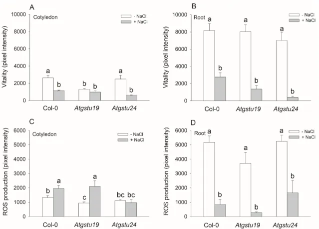
The vitality and the level of ROS were investigated in the cotyledons and roots of 16 day old seedlings using fluorescent microscopy with fluorescein diacetate (FDA) and 2’-7’-dichlorodihydrofluorescein diacetate dyes, respectively. While in control conditions, there were no significant differences in the vitality and ROS levels between the wild-type and mutant roots, the FDA pixel intensity ofAtgstu19 cotyledons was about half of that in the wild-type, showing the lower vitality of this insertional mutant (Figure3A,B). Interestingly, the ROS content was less in theAtgstu19 cotyledons than in the Col-0 plants (Figure3C). These parameters were used to assess the degree of stress after applying the salt treatment. The smallest decrease of vitality was in Col-0 plants and the highest was inAtgstu24cotyledons and roots after 150 mM NaCl treatment. The salt stress significantly enhanced the ROS content in the cotyledons of Col-0 andAtgstu19mutants, but inAtgstu24seedlings, it remained on the level of untreated controls. The ROS content of roots was decreased due to the salt stress in all investigated lines (Figure3A–D). The MDA content of the mutants did not differ from that
Int. J. Mol. Sci.2020,21, 2349 6 of 16
of the untreated or 150 mM NaCl-treated Col-0 plants, indicating that these mutants might successfully cope with the applied NaCl stress on a short-term scale (Table1).
Figure 2. Heat map of 27 A. thaliana tau group glutathione transferase (GSTU) gene expression levels.
Relative transcript amounts of GSTU genes were determined by RT-qPCR under control conditions (Control) and after 150 mM NaCl treatment (NaCl) in wild-type (Col-0) and GSTU mutant (Atgstu19 and Atgstu24) Arabidopsis plants. For this, the expression of GST genes was normalized first by reference to the average transcript amount of GAPDH2 and actin2 genes, and second to the average transcript amount of each GST genes in wild-type control plants. 2-ΔΔCt data were presented as a heat map. Red colours show repression, while green colours show activation, according to the colour scale bar. The presented data are from one biological replicate.
2.3. The Vitality and ROS Levels of Seedlings Indicated that the Mutants were Able to Cope with the Applied Salt Stress to Some Extent after 48 h
The vitality and the level of ROS were investigated in the cotyledons and roots of 16 day old seedlings using fluorescent microscopy with fluorescein diacetate (FDA) and 2’-7’- dichlorodihydrofluorescein diacetate dyes, respectively. While in control conditions, there were no significant differences in the vitality and ROS levels between the wild-type and mutant roots, the FDA pixel intensity of Atgstu19 cotyledons was about half of that in the wild-type, showing the lower vitality of this insertional mutant (Figure 3A,B). Interestingly, the ROS content was less in the Atgstu19 cotyledons than in the Col-0 plants (Figure 3C). These parameters were used to assess the degree of stress after applying the salt treatment. The smallest decrease of vitality was in Col-0 plants and the highest was in Atgstu24 cotyledons and roots after 150 mM NaCl treatment. The salt stress significantly enhanced the ROS content in the cotyledons of Col-0 and Atgstu19 mutants, but in Atgstu24 seedlings, it remained on the level of untreated controls. The ROS content of roots was decreased due to the salt stress in all investigated lines (Figure 3A–D). The MDA content of the mutants did not differ from that of the untreated or 150 mM NaCl-treated Col-0 plants, indicating that these mutants might successfully cope with the applied NaCl stress on a short-term scale (Table 1).
Figure 3. Cell vitality and level of reactive oxygen species (ROS) in cotyledons and root tips of two-week-old wild-type (Col-0) andGSTUmutant (Atgstu19andAtgstu24)Arabidopsisplants treated with 150 mM NaCl for 48 h. (A) Vitality of cotyledons and (B) root tips investigated by fluorescein diacetate. (C) ROS level in cotyledons detected by 2’-7’-dichlorodihydrofluorescein diacetate. (D) ROS level in root tips under the same conditions as described above. Means±SE,n=9 denoted by different letters indicate a significant difference between the treatments at ap<0.05 level according to Duncan’s test.
Table 1. Malondialdehyde (MDA) content of two-week-old wild-type (Col-0) and glutathione transferase (GSTU) mutant (Atgstu19andAtgstu24)Arabidopsisplants treated with 150 mM NaCl for 48 h.
MDA content (nmol g−1FW)
- NaCl +NaCl
Col-0 17.66±1.14n.s. 19.43±1.83n.s.
Atgstu19 20.22±2.89n.s. 16.11±3.08n.s.
Atgstu24 15.38±0.68n.s. 17.44±1.35n.s.
Means±SD; n.s.—not significant at ap<0.05 level according to Duncan’s test.
2.4. Elevated AsA and GSH Levels and Increased DHAR and GR Activities might Help to Compensate for the Mutation of AtGSTU Genes
The total ascorbate content was higher in theAtgstu19andAtsgtu24mutants (by 34% and 19%, respectively) than in wild-type plants under control conditions (Figure S1). Application of 150 mM NaCl increased the total ascorbate level of Col-0 andAtgstu24mutant plants by 46% and 31%, respectively, while at the same time, the changes were not significant in theAtgstu19seedlings (Figure S1). Analysis
Int. J. Mol. Sci.2020,21, 2349 7 of 16
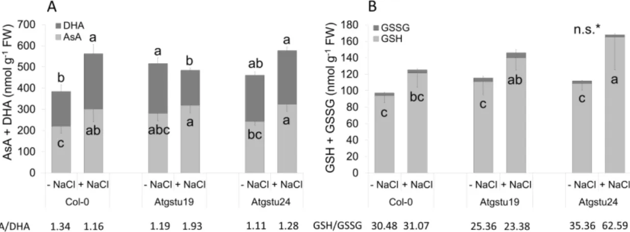
of the ascorbate redox state revealed that in the untreated seedlings, the amount of the oxidized form of ascorbate (DHA) was elevated significantly in theAtgstu19lines, but after 48 h of salt treatment, the DHA content increased only in Col-0 andAtgstu24plants. Interestingly, the AsA/DHA ratio was lower in bothAtgstumutants than in Col-0 plants under control conditions, but it increased after salt treatment (Figure4A).
Figure 3. Cell vitality and level of reactive oxygen species (ROS) in cotyledons and root tips of two- week-old wild-type (Col-0) and GSTU mutant (Atgstu19 and Atgstu24) Arabidopsis plants treated with 150 mM NaCl for 48 h. (A) Vitality of cotyledons and (B) root tips investigated by fluorescein diacetate. (C) ROS level in cotyledons detected by 2’-7’-dichlorodihydrofluorescein diacetate. (D) ROS level in root tips under the same conditions as described above. Means ± SE, n = 9 denoted by different letters indicate a significant difference between the treatments at a p < 0.05 level according to Duncan’s test.
Table 1. Malondialdehyde (MDA) content of two-week-old wild-type (Col-0) and glutathione transferase (GSTU) mutant (Atgstu19 and Atgstu24) Arabidopsis plants treated with 150 mM NaCl for 48 h.
MDA content (nmol g−1 FW)
- NaCl +NaCl
Col-0 17.66 ± 1.14 n.s. 19.43 ± 1.83 n.s.
Atgstu19 20.22 ± 2.89 n.s. 16.11 ± 3.08 n.s.
Atgstu24 15.38 ± 0.68 n.s. 17.44 ± 1.35 n.s.
Means ± SD; n.s.—not significant at a p < 0.05 level according to Duncan’s test.
2.4. Elevated AsA and GSH Levels and Increased DHAR and GR Activities might Help to Compensate for the Mutation of AtGSTU Genes
The total ascorbate content was higher in the Atgstu19 and Atsgtu24 mutants (by 34% and 19%, respectively) than in wild-type plants under control conditions (Figure S1). Application of 150 mM NaCl increased the total ascorbate level of Col-0 and Atgstu24 mutant plants by 46% and 31%, respectively, while at the same time, the changes were not significant in the Atgstu19 seedlings (Figure S1). Analysis of the ascorbate redox state revealed that in the untreated seedlings, the amount of the oxidized form of ascorbate (DHA) was elevated significantly in the Atgstu19 lines, but after 48 h of salt treatment, the DHA content increased only in Col-0 and Atgstu24 plants. Interestingly, the AsA/DHA ratio was lower in both Atgstu mutants than in Col-0 plants under control conditions, but it increased after salt treatment (Figure 4A).
Figure 4. Ascorbate and glutathione contents of two-week-old wild-type (Col-0) and GSTU mutant (Atgstu19 and Atgstu24) Arabidopsis plants treated with 150 mM NaCl for 48 h. (A) The reduced ascorbic acid (AsA) and the oxidized dehydroascorbate (DHA) contents in seedlings. (B) Reduced (GSH) and oxidized (GSSG) glutathione contents in seedlings. Means ± SD. Means denoted by different letters indicate a significant difference among the treatments at a p < 0.05 level according to Duncan’s test. * n.s.—not significant. There were no significant differences in GSSG content.
The reduced glutathione (GSH) level of mutants was slightly enhanced compared to the wild- type seedlings under control conditions. The 150 mM NaCl treatment increased the glutathione
Figure 4.Ascorbate and glutathione contents of two-week-old wild-type (Col-0) andGSTUmutant (Atgstu19andAtgstu24)Arabidopsisplants treated with 150 mM NaCl for 48 h. (A) The reduced ascorbic acid (AsA) and the oxidized dehydroascorbate (DHA) contents in seedlings. (B) Reduced (GSH) and oxidized (GSSG) glutathione contents in seedlings. Means±SD. Means denoted by different letters indicate a significant difference among the treatments at ap<0.05 level according to Duncan’s test.
* n.s.—not significant. There were no significant differences in GSSG content.
The reduced glutathione (GSH) level of mutants was slightly enhanced compared to the wild-type seedlings under control conditions. The 150 mM NaCl treatment increased the glutathione content in all lines, but the increment was about 1.5—1.7-fold higher in mutant plants than in Col-0 (Figure S1). Although the oxidized glutathione (GSSG) contents of plants were not significantly different, the GSH/GSSG ratio was lower inAtgstu19plants than in Col-0 both under control and salt stress conditions. The salt treatedAtgstu24seedlings had abundant amounts of total glutathione (GSH plus GSSG) and high GSH-to-GSSG ratios (Figure4B). The calculated reduction potential of the GSH/GSSG couple became more negative due to the salt stress in all genotypes, but the highest changes (11.65 mV) were detected in theAtgstu24mutant (Table2).
Table 2.The reduction potential of the GSH/GSSG couple (half-cell reduction potential; Ehc) determined with the Nernst equation in two-week-old wild-type (Col-0) andGSTUmutant (Atgstu19andAtgstu24) Arabidopsisplants treated with 150 mM NaCl for 48 h.
Ehc(mV)
- NaCl +NaCl
Col-0 −252.32±6.16n.s. -256.73±10.28n.s.
Atgstu19 −252.27±3.75n.s. -255.32±11.77n.s.
Atgstu24 −257.46±3.45n.s. -269.11±8.96n.s.
Means±SD; n.s.— not significant at ap<0.05 level according to Duncan’s test.
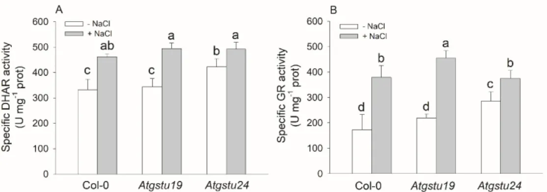
Measuring the activity of some enzymes connected to the re-reduction AsA–GSH pool revealed that theAtgstu24mutant plants had the highest DHAR and GR activities and possessed the most negative redox potential among the untreated genotypes. The salt treatment elevated both the DHAR and GR activities in all investigated lines and the highest GR activity (approximately two-fold increment) was measured in the salt treatedAtgstu19mutants (Figure5).
Int. J. Mol. Sci.2020,21, 2349 8 of 16 content in all lines, but the increment was about 1.5—1.7-fold higher in mutant plants than in Col-0 (Figure S1). Although the oxidized glutathione (GSSG) contents of plants were not significantly different, the GSH/GSSG ratio was lower in Atgstu19 plants than in Col-0 both under control and salt stress conditions. The salt treated Atgstu24 seedlings had abundant amounts of total glutathione (GSH plus GSSG) and high GSH-to-GSSG ratios (Figure 4B). The calculated reduction potential of the GSH/GSSG couple became more negative due to the salt stress in all genotypes, but the highest changes (11.65 mV) were detected in the Atgstu24 mutant (Table 2).
Table 2. The reduction potential of the GSH/GSSG couple (half-cell reduction potential; Ehc) determined with the Nernst equation in two-week-old wild-type (Col-0) and GSTU mutant (Atgstu19 and Atgstu24) Arabidopsis plants treated with 150 mM NaCl for 48 h.
Ehc (mV)
- NaCl +NaCl
Col-0 -252.32 ± 6.16 n.s. -256.73 ± 10.28 n.s.
Atgstu19 -252.27 ± 3.75 n.s. -255.32 ± 11.77 n.s.
Atgstu24 -257.46 ± 3.45 n.s. -269.11 ± 8.96 n.s.
Means ± SD; n.s.— not significant at a p < 0.05 level according to Duncan’s test.
Measuring the activity of some enzymes connected to the re-reduction AsA–GSH pool revealed that the Atgstu24 mutant plants had the highest DHAR and GR activities and possessed the most negative redox potential among the untreated genotypes. The salt treatment elevated both the DHAR and GR activities in all investigated lines and the highest GR activity (approximately two-fold increment) was measured in the salt treated Atgstu19 mutants (Figure 5).
Figure 5. Redox-related antioxidant enzyme activities of two-week-old wild-type (Col-0) and GSTU mutant (Atgstu19 and Atgstu24) Arabidopsis plants treated with 150 mM NaCl for 48 h. (A) Dehydroascorbate reductase (DHAR) activity of seedlings. (B) Glutathione reductase (GR) activity of plants. Means ± SD denoted by different letters indicate a significant difference between the treatments at a p < 0.05 level according to Duncan’s test.
3. Discussion
3.1. Arabidopsis Plants Compensated for the Decreased AtGSTU Transcripts with Altered AtGSTU Gene Expression and Elevated Antioxidant Activity
Glutathione transferases play a variety of functions in plant physiological processes under both normal and stress conditions. The members of the plant-specific tau GST class are stress inducible, have xenobiotic detoxification and glutathione peroxidase activities and play a role in the secondary metabolism during defence mechanisms [2,43].
Both AtGSTs investigated in this study, AtGSTU19 and AtGSTU24, belong to the third clade of the GSTUs, which consist of 10 members [2]. Their protein sequences show 71% amino acid identity Figure 5. Redox-related antioxidant enzyme activities of two-week-old wild-type (Col-0) and GSTU mutant (Atgstu19 and Atgstu24) Arabidopsis plants treated with 150 mM NaCl for 48 h.
(A) Dehydroascorbate reductase (DHAR) activity of seedlings. (B) Glutathione reductase (GR) activity of plants. Means±SD denoted by different letters indicate a significant difference between the treatments at ap<0.05 level according to Duncan’s test.
3. Discussion
3.1. Arabidopsis Plants Compensated for the Decreased AtGSTU Transcripts with Altered AtGSTU Gene Expression and Elevated Antioxidant Activity
Glutathione transferases play a variety of functions in plant physiological processes under both normal and stress conditions. The members of the plant-specific tau GST class are stress inducible, have xenobiotic detoxification and glutathione peroxidase activities and play a role in the secondary metabolism during defence mechanisms [2,43].
Both AtGSTs investigated in this study, AtGSTU19 and AtGSTU24, belong to the third clade of the GSTUs, which consist of 10 members [2]. Their protein sequences show 71% amino acid identity (Figure S2) [44]. The main cis-regulatory elements found in the 5’ up-regulatory region of the genes are rather different, although both genes contain several presumably abiotic stress- and hormone-inducible sequences (Table S2). The AtGSTU19 represents a significant percentage of the GST pool inArabidopsis[26], while AtGSTU24 came to the forefront especially according to its detoxification ability [9,45]. Both isoenzymes may have a role in salt stress responses and possess high activity toward the CDNB substrate [5,11,46]. The altered level of these proteins may significantly influence the total extractable GST activity of cells. Xu et al. [27] found that different transgenic lines overexpressing the AtGSTU19gene showed significantly enhanced GST activity toward CDNB compared to the wild-type.
In the search for clues to understand the role of AtGSTU19 and AtGSTU24 in defence against salt stress, we focused our attention on the changes after 48 h of the 150 mM NaCl treatment of two-week-oldA. thalianaplants. Correlating well with the results of earlier studies [26], we found that knockdown mutation of theAtGSTU19gene resulted in decreased GST activity and vitality, both under control conditions and after applying 150 mM NaCl (49% in roots compared to salt-treated Col-0), but the level of total ROS was even lower than in the wild-type. The ROS level of theAtgstu24mutant did not differ significantly from that of the Col-0 plants, and its vitality decreased only in the presence of NaCl (53% in cotyledons and 14% in roots compared to salt-treated Col-0), but interestingly, the GST activity was even higher under control conditions than in the wild-type. The GPOX activity or the level of lipid peroxidation marker MDA were not significantly altered in the investigated genotypes.
Plant GSTs seem to have a high degree of functional overlap and variability both within and between classes [13]. The expression of particular GST isoenzymes in different plant organs and tissues and in response to different stimuli (e.g., constitutive versus stress-inducible) have been proposed as reasons to explain this apparent redundancy [47,48]. In our study, investigation of theAtGSTU transcript levels revealed that the expression of severalAtGSTUgenes was altered significantly in Atgstu19 andAtgstu24 mutants even under control conditions compared to the wild-type plants.
Interestingly, fourAtGSTUgenes were induced and three were repressed in both mutants, respectively.
Int. J. Mol. Sci.2020,21, 2349 9 of 16
Additionally, four other genes were induced inAtgstu19, while one gene inAtgstu24seedlings and one further gene in eachAtgstumutant was repressed. These changes inGSTUexpression may explain the elevated GST activity in theAtgstu24mutant. It is possible that the induction of eightGSTgenes in theAtgstu19 mutant was not sufficient to compensate for the lack of this extremely important GST isoenzyme concerning the total extractable glutathione S-transferase activity. Isoenzymes with an enhanced gene expression level inAtgstu19mutants were still expressed at a much lower level than AtGSTU19in the wild-type (Table S1); thus, their induction might not compensate for the missing activity. One other possible explanation for our results could be that the newly induced isoenzymes may have lower activity toward CDNB than AtGSTU19 [5,46].
Monitoring the known or predicted interactions of these isoenzymes in the STRING database [49]
shed light on connections among the AtGSTs and DHARs (Figure S3 ). Dehydroascorbate reductases are responsible for the recycling of AsA and are also classified as belonging to the GST superfamily [2].
In our experiments, a single mutation ofAtGSTUgenes caused slightly increased total AsA inAtgstu19 andAtgstu24seedlings (by 34% and 19%, respectively), but because of the elevated DHA content of mutants, the AsA/DHA ratio was lower. According to the known and predicted interactions, both AtGSTU isoenzymes have a close connection with AtDHAR1 and AtDHAR2 (first shell of interactors, Figure S3). Furthermore, this analysis highlighted that both AtGSTU19 and AtGSTU24 can closely interact with GR (responsible for the reduction of GSSG) and glutathione synthetase (GSH2) enzymes. Glutathione synthetase, which catalyses the second step of GSH biosynthesis (addition of glycine toγ-glutamyl-cysteine), is regulated by several factors [30].
The total GSH content in the untreatedAtgstu19andAtgstu24mutants was enhanced by 18% and 13%, respectively (Figure S1). Rahantaniaina et al. [50] also found thatAtgstu24mutant seedlings had slightly higher GSH content under control conditions. After 3-aminotriazole treatment, the lack of this isoenzyme did not induce the bleaching of the seedlings, suggesting that the plants can compensate for the mutation ofGSTU24during oxidative stress [50]. InAtgstu17mutants, besides the elevated GSH levels, a higher GSH/GSSG ratio was measured [42,51]. In our previous experiments, we found that Atgstf9mutants also exhibited increased AsA and GSH pools, and their redox status was modified [10].
These results suggest that the increase in the GSH pool may be a common characteristic ofAtgst mutants, at least for the members of tau and phi classes.
3.2. Is the Altered GSH Level of Mutants Needed to Cope With Salt Stress?
Constitutively high levels of reduced GSH are advantageous as they act as a strong buffer against ROS but would make the system less responsive to changes in redox potential that may be needed to upregulate the inducible defence components [52]. Proteomic analysis ofArabidopsisroots subjected to the 150 mM NaCl treatment revealed an increase in the amount of important ROS-scavenging and detoxifying proteins, including APX, glutathione peroxidase, Class III peroxidases, SOD and GSTs [53].
In the present study, we found that the salt stress was accompanied by enhanced DHAR and GR activities. KnockoutAtgstu24also had higher DHAR activity and GSH content after salt treatment than wild-type plants and this could help to keep ROS under tight control and maintain a higher AsA/DHA and GSH/GSSG ratio. However, taking into account that theAtgstu24mutant had slightly lower vitality than the other two genotypes, it can be assumed that these changes were not suitable to allow the plant to cope successfully with salt stress. Although the lack of AtGSTU24 increased the total GST activity under control conditions and it was slightly elevated even after salt treatment, the induction of severalAtGSTUgenes (AtGSTU3-6,AtGSTU9,AtGSTU11andAtGSTU12) observed after salt treatment was in most cases lower than inAtgstu19or wild-type plants.
In theAtgstu19mutant, the GSH content was also increased, but the GSH to GSSG ratio was lower than in Col-0. Interestingly, the calculated redox potential from the measured data revealed similar redox status in this mutant than in the wild-type, both under control conditions and two days after application of the salt stress (Table2). By analysing the redox status ofArabidopsisroot tips using a roGFP1 redox sensor, Jiang et al. [54] demonstrated that the immersion of seedlings in 100 mM
Int. J. Mol. Sci.2020,21, 2349 10 of 16
of NaCl for 3–24 h shifted the redox potential of the entire root toward the more oxidized status at the beginning, but it was re-established after 6 h and more negative redox potentials were detected compared to control roots, especially in the case of 24 h long treatment. Generally, the redox potential of roots depended on the strength and duration of the applied stress: while it might remain more negative in the case of mild (50 mM NaCl) stress, it became more oxidized in the presence of a higher (150 mM) salt concentration. However, after a few days, the salt-induced changes in redox potentials decreased and the differences in the redox status of seedlings practically disappeared [54]. Investigation of the redox status of five-day-oldAtgstu19andAtgstf8mutants using a roGFP2 fluorescent probe revealed that theAtgstu19mutant had the most oxidized redox status in all root zones and under all investigated conditions [29]. Interestingly, the redox potential of the mutant roots showed smaller changes after applying 150 mM NaCl for 3 h than the wild-type, due to the fact of their already more positive value under control conditions. It was concluded that their increased salt sensitivity can be associated with the decreased redox potential response [29]. The lower GSH/GSSG ratio and increased GSH level observed in severalGSTmutants and overexpressing lines [41,55] and references above) suggest that altering the redox homeostasis can be part of a general mode of action in the mechanisms of GSTs.
The dynamic interaction of GSTs with other glutathione-related enzymes might be how temporary redox status changes allow the regulation of normal cellular physiology. A schematic model illustrating predicted physiological and regulatory events inAtgstumutants resulting in the observed changes in metabolite concentrations and enzyme activities under control conditions and after applying salt stress are summarized in Scheme1.
beginning, but it was re-established after 6 h and more negative redox potentials were detected compared to control roots, especially in the case of 24 h long treatment. Generally, the redox potential of roots depended on the strength and duration of the applied stress: while it might remain more negative in the case of mild (50 mM NaCl) stress, it became more oxidized in the presence of a higher (150 mM) salt concentration. However, after a few days, the salt-induced changes in redox potentials decreased and the differences in the redox status of seedlings practically disappeared [54].
Investigation of the redox status of five-day-old Atgstu19 and Atgstf8 mutants using a roGFP2 fluorescent probe revealed that the Atgstu19 mutant had the most oxidized redox status in all root zones and under all investigated conditions [29]. Interestingly, the redox potential of the mutant roots showed smaller changes after applying 150 mM NaCl for 3 h than the wild-type, due to the fact of their already more positive value under control conditions. It was concluded that their increased salt sensitivity can be associated with the decreased redox potential response [29]. The lower GSH/GSSG ratio and increased GSH level observed in several GST mutants and overexpressing lines [41,55] and references above) suggest that altering the redox homeostasis can be part of a general mode of action in the mechanisms of GSTs. The dynamic interaction of GSTs with other glutathione-related enzymes might be how temporary redox status changes allow the regulation of normal cellular physiology. A schematic model illustrating predicted physiological and regulatory events in Atgstu mutants resulting in the observed changes in metabolite concentrations and enzyme activities under control conditions and after applying salt stress are summarized in Scheme I.
Scheme I. Schematic model summarizing the results presented in this study. Mutation in a tau group glutathione transferase (AtGSTU) gene supposedly induced the accumulation of ROS and lipid peroxides, which led to a temporary oxidized redox state within the cells accompanied by elevated Scheme 1. Schematic model summarizing the results presented in this study. Mutation in a tau group glutathione transferase (AtGSTU) gene supposedly induced the accumulation of ROS and lipid peroxides, which led to a temporary oxidized redox state within the cells accompanied by elevated dehydroascorbate and oxidized glutathione levels.
Int. J. Mol. Sci.2020,21, 2349 11 of 16
To compensate these changes inAtgstu mutants the ascorbic acid and glutathione biosynthesis, the enzymatic antioxidant system (such as dehydroascorbate reductase and glutathione reductase activity) was induced and theAtGSTUgene expression pattern was modified. The alteration of the ROS-processing network might help to maintain the ROS and redox homeostasis in the mutants.
Salt stress presumably intensified the accumulation of ROS, LOOH and oxidized non-enzymatic antioxidants leading to further enhancement of the AsA and GSH biosynthesis, the activity of antioxidant enzymes (DHAR and GR) and the expression of severalAtGSTUgenes. Although these changes helpedAtgstumutants to restore the redox potential after NaCl treatment, the vitality of mutants was lower than in wild-type. (An upward arrow indicates increase while a downward arrow shows decrease; the thickness of the arrows refers to the extent of the changes). Abbreviations: AsA, ascorbic acid; DHA, dehydroascorbate; DHAR, dehydroascorbate reductase; GSH, reduced glutathione;
GR, glutathione reductase; GSSG, oxidized glutathione; GSTU, glutathione transferase tau group;
LOOH, lipid peroxides; ROS, reactive oxygen species.
4. Materials and Methods
4.1. Plant Material and Growth Conditions
Fourteen-day-oldArabidopsis thaliana (L.) Heynh. ecotype Columbia (Col-0) as a wild-type, Atgstu19andAtgstu24mutant seedlings were used in all experiments. Arabidopsislines (SALK_041942 and SALK_034472) containing a T-DNA insertion inAtGSTU19(At1g78380) andAtGSTU24(At1g17170) genes were obtained from the Salk Institute [56] and the seeds of homozygous genotypes were used.
The positions of T-DNA insertions and gene-specific PCR primers used for testing the segregation of T-DNA insertions are provided in the Figure S4. The Atgstu19 is a knockdown mutant (the transcript amount ofAtGSTU19was about half of that in the wild-type), while theAtgstu24can be regarded as knockout mutant (Table S1). Stress treatments were carried out on two-week-old in vitro grown plants by placing them onto agar-solidified culture medium supplemented with 150 mM NaCl.
The Petri dishes were kept in growth chamber (Fitoclima S 600 PLH, Aralab, Portugal) at a photon flux density of 100µmol m−2s−1(10/14 day/night period), at a relative humidity of 70% and 21◦C.
Samples were collected after 48 h NaCl treatment. The experiments were repeated at least two times, the measurements were performed in three replicates unless indicated otherwise.
4.2. Determination of Glutathione Transferase and Glutathione Peroxidase Enzyme Activities
To analyse the enzyme activity, 200 mg tissue was homogenized on ice in 1 mL 100 mM phosphate buffer (pH 7.0) containing 1 mM phenylmethylsulfonyl fluoride and 1% polyvinyl-polypirrolidone.
The homogenate was centrifuged for 20 min at 10,000×gat 4◦C, and the supernatant was used for enzyme activity assays. Uvikon 930 spectrophotometer (Kontron AG, Eching, Germany) was used for every absorption measurement in our experiments
Glutathione transferase (GST, EC 2.5.1.18) activity was determined as published earlier [11] with some modifications. The GST activity was determined spectrophotometrically using an artificial substrate, 1-chloro-2,4-dinitrobenzene (CDNB, Sigma–Aldrich, Germany). The GST activity was given in specific activity (µmol conjugated products min−1,ε340=9.6 mM−1cm−1).
Glutathione peroxidase (GPOX, EC 1.11.1.9) activity was measured with cumene hydroperoxide (CHP; Sigma–Aldrich, Germany) as a substrate. The reaction mixture contained 4 mM GSH, 0.2 mM NADPH, 0.05 U of GR (from baker’s yeast, Sigma–Aldrich, Germany), 100 µL enzyme extract, and 0.5 mM substrate in phosphate buffer (0.1 M, pH 7.0) in a total volume of 1 mL. The GPOX activity was given in specific activity (µmol of converted NADPH min−1,ε340=6.22 mM–1cm–1).
4.3. Determination of Glutathione Reductase and Dehydrosacorbate Reductase Activities
Dehydroascorbate reductase (DHAR, EC 1.8.5.1) and glutathione reductase (GR, EC 1.8.1.7) activities were determined as described at GST and GPOX activity determination, with the exception that 50 mM phosphate buffer was used for homogenization
Int. J. Mol. Sci.2020,21, 2349 12 of 16
The DHAR activity was assayed as was published in Csiszár et al. [18]. The enzyme activity (nmol min−1mg−1protein) was calculated from the increase in the AsA content by measuring the absorbance at 265 nm and using the 14.0 mM−1cm−1extinction coefficient.
The GR activity was determined by measuring the absorbance increment at 412 nm when 5,5’-dithio-bis(2-nitrobenzoic acid) (DTNB) was reduced by GSH, generated from GSSG [40]. The activity was calculated as the amount of reduced DTNB, in nmol min–1mg−1protein,ε420=13.6 mM–1cm–1.
4.4. RNA Purification and Expression Analyses with Quantitative Real-Time PCR
The expression rate ofArabidopsis GST genes was determined by quantitative real-time PCR (RT-qPCR) after the purification of RNA from 100 mg plant material according to Chomczynski and Sacchi [57] as was described in Bela et al. [40]. The primers used for the RT-qPCR are given in Table S3.
The expression rate ofGSTgenes was monitored as published earlier in Bela et al. [40]. Data analysis was performed using qTOWER Software 2.2 (Analytik Jena, Jena, Germany) software. The GAPDH2 (At1g13440) and actin2 (At3g18780) genes were used as internal controls, respectively [17]. The average Ct number ofGAPDH2and actin2 gene was used for data normalization. Data of RT-qPCR was calculated using the 2(-∆∆Ct)formula [58]. To demonstrate the differences between changes in the expression levels of the investigated GST genes, the relative transcript level in theArabidopsis thaliana Col-0 control root samples were considered to be one for each gene, and the results were presented on a heat map created in Microsoft Excel using the conditional formatting function [40]. To demonstrate the differences in the enhancement of gene expression levels a lighter green colour indicates if the increase was between 2 - 2.99 and dark green if more than 3 fold elevation occurred compared to the level in the wild-type control.
4.5. Investigation of Vitality and Reactive Oxygen Species (ROS) using Fluorescent Microscopy
Zeiss Axiowert 200M microscope (Carl Zeiss Inc., Jena, Germany) equipped with a high-resolution digital camera (Axiocam HR, HQ CCD camera) and suitable filter sets was used for the fluorescence detection of vitality and ROS in roots and cotyledons ofArabidopsisplants. Fluorescein diacetate (FDA) was applied for the determination of cell vitality according to Horváth et al. [29]. To detect ROS production, 2’-7’-dichlorodihydrofluorescein diacetate (Sigma-Aldrich, Germany) was used according to Horváth et al. [11]. The intensity of fluorescence was measured on digital images with the help of Axiovision Rel. 4.8 software (Carl Zeiss Inc., Jena, Germany). Fluorescence intensity values were determined in 80µm diameter circles, at 150µm distance from the root tip and in 300µm diameter circles in cotyledons. The diameter of circles was not modified during the experiments.
The measurements were performed in 10 replicates.
4.6. Determination of Malondialdehyde Content
Malondialdehyde (MDA) formation was followed by using the thiobarbituric acid method [11].
50 mg tissue was homogenized with 500 µL, 0.1% trichloroacetic acid (TCA) and 50 µL 4%
butylhydroxytoluene was added to avoid further lipid peroxidation. The absorbance was measured at 532 nm and adjusted for non-specific absorbance at 600 nm. MDA concentration was calculated using an extinction coefficient of 155 mM−1cm−1.
4.7. Ascorbate and Glutathione Extraction and Determination
Ascorbate and glutathione contents were determined according to Bela et al. [40]. Two-hundred mg of seedlings was homogenized with 0.8 mL of 5% TCA. The homogenate was centrifuged at 10,000×gfor 20 min at 4◦C, and the supernatant was used for further determinations. To assay total ascorbate (AsA), 100µL of 10 mM dithiothreitol (DTT) was added to the extract and the excess of DTT was removed by adding 100µL, 0.5% (w:v) N-ethylmaleimide (NEM). Ascorbate concentrations were determined spectrophotometrically at 525 nm. Dehydroascorbate (DHA) content was calculated as the difference between the concentration of total and reduced ascorbate.
Int. J. Mol. Sci.2020,21, 2349 13 of 16
Total and oxidized glutathione concentrations were measured spectrophotometrically using an enzymatic assay. To mask reduced glutathione (GSH) 4-vinylpyridine was added to the extract and incubated for 60 min. The reaction mixture contained 0.1 M phosphate buffer pH 7.5, 1 mM 5,50-dithiobis(2-nitrobenzoic acid) (DTNB), 1 mM NADPH, 1 U of glutathione reductase (GR baker’s yeast, Sigma-Aldrich, Germany) and 20µL of the tissue extract in 1 mL volume. The GSH content was calculated from the difference between the concentration of total and oxidized glutathione.
Standard curves were obtained for total glutathione and GSSG within the 0-2µM range.
The reduction potential of the GSH/GSSG couple (the half-cell redox potential; Ehc) was determined with the Nernst equation using the formula of Schafer and Buettner [32]: Ehc=-240 - (59.1/2) log([GSH]2/[GSSG]) mV; where -240 mV is the standard reduction potential of glutathione on 25◦C, pH 7.0 [59].
4.8. Statistical Analysis
Statistical analysis was carried out with SigmaPlot 11.0 software (SigmaPlot, Milano, Italy) by Duncan’s test and differences were considered significant atp≤0.05. Data presented here are the means±SD of at least 3 measurements unless indicated otherwise.
5. Conclusions
Here we have shown that mutation in a singleAtGSTUgene not only caused changes in the expression ofAtGSTUsbut altered the redox homeostasis of the seedlings too. The two investigated mutations (i.e., Atgstu19 and Atgstu24) triggered different responses in many aspects inArabidopsis seedlings, but, at the same time, the redox-related processes are common elements of them. According to our results, the knockdownAtgstu19mutants decreased GST activity which did not increase after salt treatment despite of the increased expression of eightAtGSTUgenes. These results provide the first evidence that AtGSTU19 is irreplaceable in seedlings. However, these plants had elevated GSH content and GR activity to compensate for 48-h long NaCl treatment-induced changes. Interestingly, in contrast toAtgstu19,Atgstu24had elevated GST, DHAR and GR activities and AsA and GSH levels both under control conditions and after salt treatment. Maintained or even increased GSH/GSSG ratios could support the preservation of the redox potential of plant cells during salt stress in mutants, but it may also influence their response. As far as we know, this is the first report which demonstrate that mutation of AtGSTU genes with similar function act differently on the total extractable GST activity.
Moreover, we propose that the redox-coupled changes are common components of the mechanisms of GSTs. On the one hand, our results underline that highly similar GST isoenzymes have a unique role and regulation in plants. On the other hand, altered GSH and AsA levels as well as theGSTU gene expression pattern inAtgstumutants suggest that the investigated isoenzymes influence redox homeostasis under control conditions and after salt treatment inArabidopsisseedlings.
Supplementary Materials:The following are available online athttp://www.mdpi.com/1422-0067/21/7/2349/s1, Figure S1: total ascorbic acid (AsA) and glutathione (GSSG) content of seedlings; Figure S2: amino acid identity of AtGSTU19 and AtGSTU24 isoenzymes; Figure S3: protein–protein interactions according to STRING; Figure S4:
insertion site and position of primers used in genotyping ofAtGSTUmutants; Table S1: 2-∆Ctvalues of investigated AtGSTUgenes; Table S2: 5’-regulatory elements ofAtGSTU19andAtGSTU24genes; Table S3: primers used in RT-qPCR.
Author Contributions:J.C. and E.H. conceived and designed the experiments. E.H., G.C. and D.C. performed the experiments. E.H., K.B.,Á.G. and R.R. contributed to data analysis. E.H., J.C. and K.B. wrote the manuscript.
All authors have read and approved the manuscript.
Funding:This study was supported by the Hungarian National Research, Development and Innovation Fund (Grant Numbers: NKFI-6 K 125265, NKFI-8 PD 121027 and NKFI-8 PD 131909).
Acknowledgments:The authors would like to thank Mrs. Erzsébet Porkoláb for her excellent technical assistance.
Conflicts of Interest:The authors declare no conflict of interest.
Int. J. Mol. Sci.2020,21, 2349 14 of 16
References
1. Csiszár, J.; Hecker, A.; Labrou, N.E.; Schröder, P.; Riechers, D.E. Plant glutathione transferases: Diverse, multi-tasking enzymes with yet-to-be discovered functions.Front. Plant Sci.2019,10, 1304. [CrossRef]
2. Dixon, D.P.; Edwards, R. Glutathione transferases.Arab. Book2010,8, e0131. [CrossRef]
3. Nianiou-Obeidat, I.; Madesis, P.; Kissoudis, C.; Voulgari, G.; Chronopoulou, E.; Tsaftaris, A.; Labrou, N.E.
Plant glutathione transferase-mediated stress tolerance: Functions and biotechnological applications.
Plant Cell Rep.2017,36, 791–805. [CrossRef]
4. Labrou, N.E.; Papageorgiou, A.C.; Pavli, O.; Flemetakis, E. Plant GSTome: Structure and functional role in xenome network and plant stress response.Curr. Opin. Biotech.2015,32, 186–194. [CrossRef]
5. Dixon, D.P.; Hawkins, T.; Hussey, P.J.; Edwards, R. Enzyme activities and subcellular localization of members of the Arabidopsis glutathione transferase superfamily.J. Exp. Bot.2009,60, 1207–1218. [CrossRef]
6. Gallé,Á.; Csiszár, J.; Secenji, M.; Guóth, A.; Cseuz, L.; Tari, I.; Györgyey, J.; Erdei, L. Glutathione transferase activity and expression patterns during grain filling in flag leaves of wheat genotypes differing in drought tolerance: Response to water deficit.J. Plant Physiol.2009,166, 1878–1891. [CrossRef] [PubMed]
7. Lan, T.; Yang, Z.L.; Yang, X.; Liu, Y.J.; Wang, X.R.; Zenga, Q.Y. Extensive functional diversifcation of the populus glutathione s-transferase supergene family.Plant Cell2009,21, 3749–3766. [CrossRef] [PubMed]
8. Liu, Y.J.; Han, X.M.; Ren, L.L.; Yang, H.L.; Zeng, Q.Y. Functional divergence of the glutathione S-transferase supergene family inPhyscomitrella patensreveals complex patterns of large gene family evolution in land plants.Plant Physiol.2013,161, 773–786. [CrossRef] [PubMed]
9. Gunning, V.; Tzafestas, K.; Sparrow, H.; Johnston, E.J.; Brentnall, A.S.; Potts, J.R.; Rylott, E.L.; Bruce, N.C.
Arabidopsisglutathione transferases U24 and U25 exhibit a range of detoxification activities with the environmental pollutant and explosive, 2,4,6-Trinitrotoluene.Plant Physiol.2014,165, 854–865. [CrossRef]
[PubMed]
10. Horváth, E.; Bela, K.; Papdi, C.; Gallé,Á.; Szabados, L.; Tari, I.; Csiszár, J. The role ofArabidopsisglutathione transferase F9 gene under oxidative stress in seedlings.Acta Biol. Hung.2015,66, 406–418. [CrossRef]
11. Horváth, E.; Brunner, S.; Bela, K.; Papdi, C.; Szabados, L.; Tari, I.; Csiszár, J. Exogenous salicylic acid-triggered changes in the glutathione transferases and peroxidases are key factors in the successful salt stress acclimation ofArabidopsis thaliana.Funct. Plant Biol.2015,42, 1129–1140. [CrossRef]
12. Benyó, D.; Horváth, E.; Németh, E.; Leviczky, T.; Takács, K.; Lehotai, N.; Feigl, G.; Kolbert, Z.; Ördög, A.;
Gallé, R.; et al. Physiological and molecular responses to heavy metal stresses suggest different detoxification mechanism ofPopulus deltoidesandP. x canadensis.J. Plant Physiol.2016,201, 62–70. [CrossRef] [PubMed]
13. Csiszár, J.; Horváth, E.; Bela, K.; Gallé,Á. Glutathione-Related Enzyme System: Glutathione Reductase (GR), Glutathione Transferases (GSTs) and Glutathione Peroxidases (GPXs). InRedox State as a Central Regulator of Plant-Cell Stress Responses; Gupta, D., Palma, J., Corpas, F., Eds.; Springer: Cham, Switzerland, 2016;
pp. 137–158.
14. Islam, S.; Rahman, I.A.; Islam, T.; Ghosh, A. Genome-wide identification and expression analysis of glutathione S-transferase gene family in tomato: Gaining an insight to their physiological and stress-specific roles.PLoS ONE2017,12, e0187504. [CrossRef] [PubMed]
15. Gullner, G.; Komives, T.; Király, L.; Schröder, P. Glutathione S-transferase enzymes in plant-pathogen interactions.Front. Plant Sci.2018,9, 1836. [CrossRef]
16. Gallé,Á.; Czékus, Z.; Bela, K.; Horváth, E.; Ördög, A.; Csiszár, J.; Poór, P. Plant glutathione transferases and light.Front. Plant Sci.2019,9, 1944. [CrossRef]
17. Papdi, C.; Ábrahám, E.; Joseph, M.P.; Popescu, C.; Koncz, C.; Szabados, L. Functional identification of Arabidopsisstress regulatory genes using the controlled cDNA overexpression system.Plant Physiol.2008, 147, 528–542. [CrossRef]
18. Csiszár, J.; Horváth, E.; Váry, Z.; Gallé,Á.; Bela, K.; Brunner, S.; Tari, I. Glutathione transferase supergene family in tomato: Salt stress-regulated expression of representative genes from distinct GST classes in plants primed with salicylic acid.Plant Physiol. Bioch.2014,78, 15–26. [CrossRef]
19. Edwards, R.; Dixon, D.P.; Skipsey, M.; Skipsey, M. Roles for glutathione transferases in plant secondary metabolism.Phytochemistry2010,71, 338–350.
20. DeRidder, B.P.; Dixon, D.P.; Beussman, D.J.; Edwards, R.; Goldsbrough, P.B. Induction of glutathione S-transferases inArabidopsisby herbicide safeners.Plant Physiol.2002,130, 1497–1505. [CrossRef]
Int. J. Mol. Sci.2020,21, 2349 15 of 16
21. Ndimba, B.K.; Chivasa, S.; Simon, W.J.; Slabas, A.R. Identification ofArabidopsissalt and osmotic stress responsive proteins using two-dimensional difference gel electrophoresis and mass spectrometry.Proteomics 2005,5, 4185–4196. [CrossRef]
22. Sarry, J.E.; Kuhn, L.; Ducruix, C.; Lafaye, A.; Junot, C.; Hugouvieux, V.; Jourdain, A.; Bastien, O.; Fievet, J.B.;
Vailhen, D.; et al. The early responses ofArabidopsis thalianacells to cadmium exposure explored by protein and metabolite profiling analyses.Proteomics2006,6, 2180–2198. [CrossRef] [PubMed]
23. Cicero, L.L.; Madesis, P.; Tsaftaris, A.; Piero, A.R.L. Tobacco plants over-expressing the sweet orange tau glutathione transferases (CsGSTUs) acquire tolerance to the diphenyl ether herbicide fuorodifen and to salt and drought stresses.Phytochemistry2015,116, 69–77. [CrossRef] [PubMed]
24. Jia, B.; Sun, M.; Sun, X.; Li, R.; Wang, Z.; Wu, J.; Wei, Z.; DuanMu, H.; Xiao, J.; Zhu, Y. Overexpression of GsGSTU13 and SCMRP inMedicago sativaconfers increased salt–alkaline tolerance and methionine content.
Physiol. Plantarum2016,156, 176–189. [CrossRef] [PubMed]
25. Kao, C.W.; Bakshi, M.; Sherameti, I.; Dong, S.; Reichelt, M.; Oelmüller, R.; Yeh, K.W. A Chinese cabbage (Brassica campetrissubsp. Chinensis)τ-type glutathione-S-transferase stimulates Arabidopsis development and primes against abiotic and biotic stress.Plant Mol. Biol.2016,92, 643–659. [CrossRef]
26. Sappl, P.G.; Onate-Sanchez, L.; Singh, K.B.; Millar, A.H. Proteomic analysis of glutathione S-transferases of Arabidopsis thalianareveals differential salicylic acid-induced expression of the plant-specific phi and tau classes.Plant Mol. Biol.2004,54, 205–219. [CrossRef]
27. Xu, J.; Tian, Y.S.; Xing, X.J.; Peng, R.H.; Zhu, B.; Gao, J.J.; Yao, Q.H. Over-expression of AtGSTU19 provides tolerance to salt, drought and methyl viologen stresses inArabidopsis.Physiol. Plantarum2016,156, 164–175.
[CrossRef]
28. Gutscher, M.; Pauleau, A.-L.; Marty, L.; Brach, T.; Wabnitz, G.H.; Samstag, Y.; Meyer, A.J.; Dick, T.P. Real-time imaging of the intracellular glutathione redox potential.Nat. Methods2008,6, 553–559. [CrossRef]
29. Horváth, E.; Bela, K.; Holinka, B.; Riyazuddin, R.; Gallé,Á.; Hajnal,Á.; Hurton,Á.; Fehér, A.; Csiszár, J.
TheArabidopsisglutathione transferases,AtGSTF8andAtGSTU19are involved in the maintenance of root redox homeostasis affecting meristem size and salt stress sensitivity.Plant Sci.2019,283, 366–374. [CrossRef]
30. Noctor, G.; Mhamdi, A.; Chaouch, S.; Han, Y.; Neukermans, J.; Marquez-Garcia, B.; Queval, G.; Foyer, C.H.
Glutathione in plants: An integrated overview.Plant Cell Environ.2012,35, 454–484. [CrossRef]
31. Rahantaniaina, M.S.; Tuzet, A.; Mhamdi, A.; Noctor, G. Missing links in understanding redox signaling via thiol/disulfide modulation: How is glutathione oxidized in plants?Front. Plant Sci.2013,4, 477. [CrossRef]
32. Schafer, F.O.; Buettner, G.R. Redox environment of the cell as viewed through the redox state of glutathione disulfide/glutathione couple.Free Radic. Bio. Med.2001,30, 1191–1212. [CrossRef]
33. Hossain, M.S.; Dietz, K.-J. Tuning of redox regulatory mechanisms, reactive oxygen species and redox homeostasis under salinity stress.Front. Plant Sci.2016,7, 548. [CrossRef] [PubMed]
34. Mittova, V.; Tal, M.; Volokita, M.; Guy, M. Salt stress induces up-regulation of an efficient chloroplast antioxidant system in the salt-tolerant wild tomato speciesLycopersicon pennelliibut not in the cultivated species.Physiol. Plantarum2002,115, 393–400. [CrossRef] [PubMed]
35. Vaidyanathan, H.; Sivakumar, P.; Chakrabarty, R.; Thomas, G. Scavenging of reactive oxygen species in NaCl-stressed rice (Oryza sativaL.)—Differential response in salt-tolerant and sensitive varieties.Plant Sci.
2003,165, 1411–1418. [CrossRef]
36. De Azevedo Neto, A.D.; Prisco, J.T.; Enéas-Filho, J.; de Abreu, C.E.B.; Gomes-Filho, E. Effect of salt stress on antioxidative enzymes and lipid peroxidation in leaves and roots of salt-tolerant and salt-sensitive maize genotypes.Environ. Exp. Bot.2006,56, 87–94. [CrossRef]
37. Park, H.J.; Kim, W.-Y.; Yun, D.-J. A new insight of salt stress signaling in plant.Mol. Cells2016,39, 447–459.
[CrossRef]
38. Munns, M.; Tester, M. Mechanisms of salinity tolerance.Annu. Rev. Plant Biol. 2008,59, 651–681. [CrossRef]
39. Zhang, H.; Han, B.; Wang, T.; Chen, S.; Li, H.; Zhang, Y.; Dai, S. Mechanisms of plant salt response:
Insights from proteomics.J. Proteome Res.2012,11, 49–67. [CrossRef]
40. Bela, K.; Riyazuddin, R.; Horváth, E.; Hurton,Á.; Gallé,Á.; Takács, Z.; Zsigmond, L.; Szabados, L.; Tari, I.;
Csiszár, J. Comprehensive analysis of antioxidant mechanisms inArabidopsisglutathione peroxidase-like mutants under salt-and osmotic stress reveals organ-specific significance of the AtGPXL’s activities.Environ.
Exp. Bot.2018,150, 127–140. [CrossRef]