cardiomyopathy, PH, heart failure with pre‑
served ejection fraction (HFpEF), and valvular heart disease.5 From a simple method such as wall motion analysis by 2‑dimensional echo‑
cardiography, M ‑Mode, pulsatile and contin‑
uous color, and tissue Doppler, we have now moved on to the next ‑generation laboratory, employing a variety of up ‑to ‑date technolo‑
gies such us deformation imaging, lung ultra‑
sound, and 3‑dimensional echocardiography.4 The parameters measured during the stress test combined with exercise capacity, blood pres‑
sure, and heart response will provide a com‑
prehensive, low‑cost, noninvasive assessment of cardiovascular pathologies.
Stress echocardiography for noncoronary indications focuses on the following: 1) assess‑
ment of the true functional class of the patient when a discrepancy is observed between the re‑
ported lack of symptoms and the objective as‑
sessment of pathology as severe; 2) establishing a correlation between exertional symptoms and Introduction Stress echocardiography (SE)
has an established essential role in evidence‑
‑based guidelines as a diagnostic tool in daily cardiology practice. It is not only a valid and useful method for the diagnostic and prognos‑
tic stratification of patients with coronary ar‑
tery disease (CAD)1 but it also shows an emerg‑
ing value in the assessment of cardiac function in other cardiovascular conditions.2‑4 Unfor‑
tunately, apart from CAD, the application is still somewhat marginal in the routine cardi‑
ology practice. Stress echocardiography pro‑
vides the opportunity to identify the function of the microvasculature and heart valves, de‑
tect possible pulmonary hypertension (PH), lung congestion, and also evaluate the sys‑
tolic and diastolic reaction and mechanics of the left or right ventricle (LV/RV) in response to load. For this reason, SE permits recogni‑
tion of many causes of cardiac symptoms in addition to ischemic heart disease, including hypertrophic cardiomyopathy (HCM), dilated
Correspondence to:
Gergely Ágoston, MD, PhD, Institute of Family Medicine, University of Szeged, Tisza Lajos körút 109, Szeged, Hungary, phone: +36 30 90 60 567, email: agoston.gergely@med.u ‑
‑szeged.hu
Received: October 20, 2019.
Accepted: October 23, 2019.
Published online:
October 24, 2019.
Kardiol Pol. 2019;
77 (11): 1011‑1019 doi:10.33963/KP.15032 Copyright by the Author(s), 2019
AbstrAct
Stress echocardiography is a safe, low ‑cost, widely available, radiation ‑free versatile imaging modality that is becoming increasingly recognized as a valuable tool in the assessment of coronary heart disease. In recent years, there has also been an increasing use of stress echocardiography in the assessment of nonischemic cardiac disease given its unique ability for simultaneous assessment of both functional performance and exercise ‑related noninvasive hemodynamic changes, which can help guide treatment and inform about the prognosis of the patients. Today, in the echocardiography laboratory, we can not only detect wall motion abnormalities resulting from coronary artery stenosis, but also detect alterations to the coronary microvessels, left ventricular systolic and diastolic parameters, heart valves, pulmonary circulation, alveolar ‑capillary barrier, and right ventricle. The role of stress echo has been well established in several pathologies, such as aortic stenosis and hypertrophic cardiomyopathy; however, other indications, namely the results of diastolic stress testing and pulmonary hypertension, need additional data and research. This paper presents the current evidence for the role of stress testing in mitral regurgitation, aortic stenosis, hypertrophic cardiomyopathy, heart failure with preserved ejection fraction, and pulmonary hypertension.
Key words aortic stenosis, hypertrophic cardiomyopathy, mitral regurgitation, pulmonary
hypertension, stress echocardiography
R E V I E W A R T I C L E
The role of stress echocardiography in cardiovascular disorders
Gergely Ágoston¹, Blanka Morvai ‑Illés¹, Attila Pálinkás², Albert Varga¹ 1 Institute of Family Medicine, University of Szeged, Szeged, Hungary
2 Elisabeth Hospital, Hódmezővásárhely, Hungary
Nonsevere mitral regurgitation with symptoms In pa tients with complaints in whom there is suspicion of severe MR but it is not evident on resting echocardiogram, SE demonstrating pro‑
gression of MR helps to correlate the pathology with the patient’s symptoms. However, as stress tests may worsen any MR, concomitant increase in PASP supports MR as the cause of symptoms.
The dataset in symptomatic patients should in‑
clude color flow Doppler (to allow offline quanti‑
fication of severity by proximal isovelocity sur‑
face area method and vena contracta of the re‑
gurgitant jet), MR continuous wave Doppler to quantify the severity by the proximal isoveloci‑
ty surface area method, tricuspid regurgitation continuous wave Doppler to estimate the PASP, and LV views to assess global and regional sys‑
tolic function (FigurE 1).1 The therapeutic impli‑
cations of exercise‑induced severe MR are not clearly defined. The European Society of Car‑
diology (ESC) guidelines do not specifically ad‑
dress this issue even though they recommend SE for evaluation of exercise‑induced changes in MR severity.9
Secondary mitral regurgitation In secondary MR, lower thresholds have been proposed be‑
cause, compared with the primary MR, the ad‑
verse outcomes are associated with a smaller calculated effective regurgitant orifice area.9
In patients with secondary MR, exercise SE is recommended in the following settings: 1) pres‑
ence of exertional symptoms which cannot be ex‑
plained with LV systolic dysfunction or MR se‑
verity at rest; 2) recurrent and unexplained acute pulmonary edema; 3) nonsevere MR at rest in pa‑
tients scheduled for coronary artery bypass surgery (CABG) to identify those who may benefit from combined revascularization and mitral valve repair.
The management of chronic secondary MR is less clear. As secondary MR is just one part of the disease in LV dysfunction, restoration of mitral valve competence is not by itself curative. There‑
fore, the indication for surgery in secondary MR and the choice of intervention (repair versus re‑
placement) remain debatable. Surgery is indicat‑
ed in patients with severe secondary MR at rest and LVEF of more than 30% undergoing CABG (ESC / European Association for Cardio ‑Thoracic Surgery guidelines class I, level of evidence C).9 At the same time, the management of moderate MR, or dynamic MR at the time of CABG, and mod‑
erate or severe MR in patients not requiring revas‑
cularization remains highly controversial. Current European guidelines note that echocardiograph‑
ic quantification of MR during exercise may pro‑
vide prognostic information of dynamic charac‑
teristics of MR; however, there is no suggestion about the impact of the stress test on treatment.9 Aortic stenosis According to the latest ESC guidelines, both the diagnostic workup and the hemodynamic changes derived from echo‑
cardiography and signs unmasked by stress test;
3) assessment of LV contractile reserve in pa‑
tients who are considered surgical candidates.6 The purpose of this paper is to review the pres‑
ent status of SE in conditions other than CAD, focusing on mitral regurgitation (MR), aortic stenosis (AS), HCM, HFpEF, and PH.
Mitral regurgitation MR results from sev‑
eral pathological conditions. Primary MR is defined as regurgitation resulting from organ‑
ic valvular pathology such as prolapse, rheu‑
matic lesions, chordal rupture, collagen vascu‑
lar disease, or damage from endocarditis. Sec‑
ondary MR results from ischemic or myopath‑
ic alternations (ischemic, dilated, or HCM) to the LV leading to incomplete closure of the mi‑
tral leaflets.7 Risk stratification using SE, partic‑
ularly in patients without symptoms, becomes important not only in the exact diagnosis and characterization of the etiology of MR but also in guiding therapy.
Severe primary mitral regurgitation without symp‑
toms In patients with primary MR, SE may pro‑
voke symptoms during the exercise. During daily activities, the patients might limit their physical activity and therefore not develop symptoms. Di‑
agnosis and follow ‑up in these patients are very important because conservatively managed as‑
ymptomatic severe MR and an effective regurgi‑
tant orifice area of more than 40 mm2 have an ex‑
cess risk of death and cardiac events.8
To assess MR changes during SE, a semi‑
‑supine bicycle is the best choice. Since MR is severe at rest, there is no need to assess MR severity during stress. For this reason, imag‑
ing should first focus on acquiring LV views for the assessment of regional and global LV systol‑
ic function and to calculate global longitudinal strain. Continuous‑wave Doppler measurement of tricuspid regurgitation jet for estimation of pulmonary artery systolic pressure (PASP) is es‑
sential. The development of symptoms at low workload and severe MR with ejection fraction (EF) of more than 30% is class I, level of confi‑
dence B, indication for surgery.9 Besides the de‑
velopment of symptoms, there are several other indices that indicate poor prognosis in patients with severe MR. These include exercise‑induced increase in PASP of 60 mm Hg or more,10,11 ab‑
sence of LV contractile reserve,1,12 and limited RV contractile recruitment (defined as peak‑ex‑
ertion tricuspid annular plane systolic excur‑
sion <19 mm).13 Ejection fraction is traditionally used as the measure to assess contractile reserve, with a rise in left ventricular ejection fraction (LVEF) on exertion of less than 4% indicating the absence of contractile reserve. An absolute increase in global longitudinal strain of less than 2% indicates lack of contractile reserve.1
flow reserve is the aim. In the case of asymp‑
tomatic patients with severe AS, exercise SE is recommended.
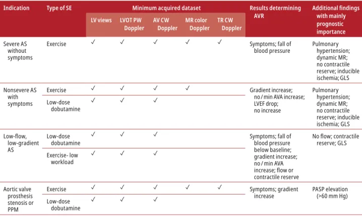
The recommended types of SE along with the minimum acquired dataset and most im‑
portant findings depending on indication are listed in TAbLE 1.
Asymptomatic severe aortic stenosis In patients with severe AS in whom symptoms have not developed yet, exercise SE is recommended to unmask symptoms or pathologic blood pres‑
sure responses.1 It also has remarkable prog‑
nostic value: an elevation of 18 to 20 mm Hg or higher in the mean aortic pressure gradi‑
ent, or a decrease / no change in LVEF and in‑
duced PH (≥60 mm Hg) are markers of poor prognosis.15‑18
the therapeutic approach to AS is based on echo‑
cardiographic assessment.9 Surgical repair or transcatheter aortic valve implantation is recom‑
mended in patients with severe AS with other‑
wise not explained symptoms and / or LV systolic dysfunction (class I).9 The criteria for severe AS are: peak transvalvular velocity of 4 m/s or high‑
er or the mean transvalvular pressure gradient of 40 mm Hg or higher without a high flow state present and aortic valve area (AVA) of less than 1 cm2.14 However, these criteria are fulfilled only in the case of the high ‑gradient AS. In several other clinical settings, when the valve morphol‑
ogy is suspicious of AS, the guidelines recom‑
mend SE to assess the severity of AS and to guide the therapeutic decision through risk stratifi‑
cation. Low ‑dose dobutamine SE is the chosen method if the assessment of the contractile or
D
GLS, –22.2%
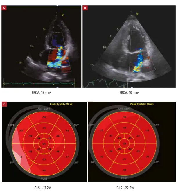
Figure 1 Stress echocardiography of a 56-year -old woman with mitral valve prolapse and uncertain symptoms. The mitral regurgitation severity did not increase during the exercise and preserved contractile reserve was observed (elevation in the left ventricular ejection fraction [LVEF] and global longitudinal strain [GLS]). The estimated pulmonary artery systolic pressure (PASP) did not increase significantly. During a 4‑year follow ‑up, cardiac disease (along with mitral regurgitation and LVEF) did not progress.
Abbreviations: EF, ejection fraction; EROA, effective regurgitant orifice area
A
EROA, 15 mm2
B
EROA, 10 mm2
EF, 61%
PASP, 28 mm Hg
C
GLS, –17.7%
EF, 69%
PASP, 34 mm Hg
is less than 1 cm2, indexedAVA is less than 0.6 cm2/m2, and the mean gradient is less than 40 mm Hg at rest. It is considered the most chal‑
lenging group. The diagnostic workup recom‑
mended by the guidelines is the same as de‑
scribed with the classical LFLG type. According to the guidelines, in these patients, AVR is recom‑
mended if symptoms are present and severe AS has been confirmed (class IIa, level of evidence C).9 Hypertrophic cardiomyopathy Sufficient data have now been collected showing that exer‑
cise testing is not only safe but also that it is a key element in the comprehensive evaluation of patients with HCM.19 Exercise ‑related symp‑
toms in patients with HCM are due to a number of factors including increased LV outflow tract (LVOT) gradients, MR, diastolic dysfunction, and myocardial ischemia in the absence of epi‑
cardial CAD. The recent ESC guidelines assign a IB class of recommendation to perform exer‑
cise SE in symptomatic patients without a rest‑
ing LVOT obstruction (if bedside maneuvers fail to induce LVOT gradient ≥50 mm Hg) to detect exercise ‑induced LVOT obstruction and MR.19 LVOT obstruction developing rapidly at lower levels of exercise is associated with greater im‑
pairment in functional capacity compared with Classic low ‑flow, low‑gradient aortic stenosis
In patients with low ‑flow, low‑gradient aortic ste‑
nosis (LFLG AS) with reduced systolic function (stroke volume [SV] index <35 ml/m², AVA <1 cm², aortic velocity <4 m/s, MG <40 mm Hg, and LVEF <50%) low ‑dose dobutamine SE is recom‑
mended to assess the stress ‑induced symptoms, blood pressure change, change in pressure gradi‑
ents or AVA, and to determine the flow (defined as SV increase ≥20%) and contractile reserve. Typi‑
cally, in true severe AS, a marked increase in gradi‑
ents can be observed with no or minimal increase in AVA. AVR is indicated in symptomatic patients with true severe AS (class I, level of evidence B) or in case of symptomatic patients, whose gradi‑
ents did not elevate but the AVA remained small‑
er than 1 cm2 andthe presence of flow / contrac‑
tile reserve has been proved (class I, level of evi‑
dence C).9 If the symptomatic patient’s AVA and gradient measurements have not changed during the SE and there is no contractile / flow reserve (which suggests an operative mortality of 30%–
50%, the therapeutic decision making is possible only after CT calcium scoring of the aortic valve.9 Paradoxical low ‑flow, low‑gradient aortic stenosis Paradoxical LFLG AS is present if LVEF is 50%
or higher, SV index is 35 ml/m2 or less, AVA
Table 1 The recommended types of stress echocardiography with the minimum acquired dataset and most important findings depending on indication. Adapted from Lancellotti et al.1
Indication Type of SE Minimum acquired dataset Results determining
AVR Additional findings
with mainly prognostic importance LV views LVOT PW
Doppler AV CW
Doppler MR color
Doppler TR CW Doppler Severe AS
without symptoms
Exercise ✓ ✓ ✓ ✓ ✓ Symptoms; fall of
blood pressure Pulmonary hypertension;
dynamic MR;
no contractile reserve; inducible ischemia; GLS Nonsevere AS
with symptoms
Exercise ✓ ✓ ✓ ✓ Gradient increase;
no / min AVA increase;
LVEF drop;
no increase
Pulmonary hypertension;
dynamic MR;
no contractile reserve; inducible ischemia; GLS Low ‑dose
dobutamine ✓ ✓ ✓
Low‑flow,
low‑gradient AS
Low ‑dose dobutamine
✓ ✓ ✓ Symptoms; fall of
blood pressure below baseline;
gradient increase;
no / min AVA increase;flowor
contractile reserve
Noflow;contractile
reserve; GLS Exercise‑ low
workload
✓ ✓ ✓
Aortic valve prosthesis stenosis or PPM
Exercise ✓ ✓ ✓ ✓ ✓ Symptoms; gradient
increase PASP elevation
(>60 mm Hg) Low ‑dose
dobutamine ✓ ✓ ✓
Abbreviations: AS, aortic stenosis; AV, aortic valve; AVA, aortic valve area; AVR, aortic valve repair; CW, continuous ‑wave; GLS, global longitudinal strain; LV, left ventricular;
LVOT,leftventricularoutflowtract;MR,mitralregurgitation;PASP,pulmonaryarterysystolicpressure;PPM,patient‑prosthesismismatch;PW,pulsed‑wave;TR,tricuspid
regurgitation
velocity which then persists throughout systo‑
le (“bell ‑shaped”).21 Key findings of worse prog‑
nosis are: limited exercise capacity, an abnormal blood pressure response (hypotensive or blunt‑
ed response), significant ST ‑depression, induc‑
ible wall motion abnormalities, blunted coro‑
nary flow reserve, LVOT obstruction (more than 50 mm Hg), and blunted systolic function. In‑
terestingly, some patients can show a paradox‑
ical decrease in LVOT obstruction during exer‑
cise, which is associated with a more favorable outcome and suggests alternative reasons for symptoms (FigurE 2b). Another paradoxical phe‑
nomenon can be also observed when the LVOT gradient starts to elevate after the stress test (FigurE 2C). The potential explanation is that in the recovery phase the preload and afterload rapid‑
ly drop down, and therefore the left ventricular volumes and dimensions decrease as well, which contributes to the LVOT obstruction (FigurE 2C).
Heart failure with preserved ejection frac- tion SE has been extensively validated in pa‑
tients with heart failure with reduced ejection fraction.22‑24 Left ventricular contractile reserve is associated with a greater chance of response to the cardiac resynchronization therapy.23,25 Shortness of breath, exertional fatigue, or poor exercise capacity has been increasingly recog‑
nized as the consequence of diastolic dysfunc‑
tion and considered to be the main cause in ap‑
proximately 40% of patients presenting with heart failure. HFpEF is a complex pathophysi‑
ological entity. Echocardiographic parameters provide a key tool for the diagnosis of the dis‑
ease, as indicated in the new ESC guidelines.26 HFpEF is defined as the presence of heart failure symptoms and signs with normal or preserved left ventricular EF and normal or small LV vol‑
umes. Structural heart disease can be also ob‑
served, which most often means LV hypertro‑
phy or left atrial enlargement. Furthermore, evi‑
dence of diastolic dysfunction (abnormal E/e’ ra‑
tio [averaged ≥13] and abnormal e’ <9 cm/s) can support the diagnosis too.26 A strong correlation between E/e’ and physical activity has been dem‑
onstrated in HFpEF.27 E/e’ has been compared to an invasive hemodynamic measurement during exercise and the correlation was in an acceptable range.28 However, it is not infrequent for patients with HFpEF to fall within a “grey zone” of E/e’
value.26 In the case of SE, multiparametric ap‑
proach is considered (FigurE 3). During the stress test, PASP should be measured. Stroke volume and its change during exercise should also be assessed.29 With diastolic heart failure during stress, lung ultrasound may show B ‑lines or ul‑
trasound lung comets, which is a simple, direct, semiquantitative sign of extravascular lung wa‑
ter accumulation.30 The absence of increased cardiac output during exercise, average E/e’ ra‑
tio higher than 14, the septal e’ velocity smaller later onset of the gradient.20 Posteriorly direct‑
ed MR is a secondary consequence of systolic an‑
terior motion. The MR jet may overlap and con‑
taminate the LV outflow jet, potentially resulting in an erroneous overestimation of the subaor‑
tic gradient. Doppler systolic flow shape typical of LV outflow gradients characteristically dem‑
onstrates a gradual increase in velocity in early systole with mid ‑systolic acceleration and peak‑
ing (“dagger ‑shaped”) (FigurE 2A). Whereas the MR signal begins suddenly at the onset of systo‑
le and rapidly establishes markedly increased
A
Peak gradient, 12 mm Hg Rest
Peak gradient, 107 mm Hg Peak stress
B Rest
Peak gradient, 179 mm Hg Peak gradient, 78 mm Hg Peak stress
C Peak stress
Peak gradient, 104 mm Hg Peak gradient, 138 mm Hg After 2 min of recovery
Figure 2 Continuous -wave Doppler echocardiography of 3 patients. A – in the first patient, obstruction of the left ventricular outflow tract (LVOT) was developed during exercise (12–107 mm Hg). B – in the second patient, the high peak gradient with the dagger shaped envelope measured in the LVOT decreased during exercise (179–78 mm Hg). C – in the third patient, the high LVOT gradient reached its peak not at peak stress, but 2 minutes after terminating the exercise (104–138 mm Hg).
B A
Cardiac output Key parameters
E/e’
PASP
B‑lines
Figure 3 Assessment of right ventricular pressure, left ventricular filling, left ventricular output, and extravascular lung water in patients with unexplained dyspnea and suspected heart failure with preserved ejection fraction
Abbreviations: E/e’, ratio of early mitral inflow velocity to mitral annular early diastolic velocity; others, see FigurE 1
PASP Key parameters
TAPSE Dynamic CO and PVR
E/e’
B A
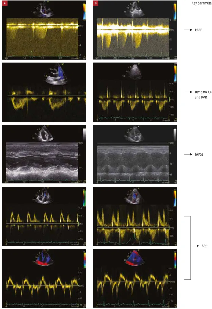
Figure 4 Assessment of right ventricular pressure, noninvasively measured dynamic pulmonary vascular resistance (PVR), tricuspid annular plane systolic excursion (TAPSE), and left ventricular filling pressure in patients at risk of pulmonary hypertension
Abbreviations: CO, cardiac output; others, see FigurES 1 and 3
ArtiCle informAtion
ConfliCt of interest None declared.
open ACCess This is an Open Access article distributed under the terms of the Creative Commons Attribution ‑NonCommercial ‑NoDerivatives 4.0 in‑
ternational License (CC bY ‑NC ‑ND 4.0), allowing third parties to download ar‑
ticles and share them with others, provided the original work is properly cited, not changed in any way, distributed under the same license, and used for non‑
commercial purposes only. For commercial use, please contact the journal office at kardiologiapolska@ptkardio.pl.
How to Cite Ágoston g, Morvai ‑illés b, Pálinkás A, Varga A. The role of stress echocardiography in cardiovascular disorders. Kardiol Pol. 2019; 77:
1011‑1019. doi:10.33963/KP.15032
referenCes
1 Picano E. Stress echocardiography. From pathophysiological toy to diagnostic tool. Circulation. 1992; 85: 1604‑1612.
2 Lancellotti P, Pellikka PA, Budts W, et al. The clinical use of stress echocardiog‑
raphy in non ‑ischemic heart disease: recommendations from the European Asso‑
ciation of Cardiovascular Imaging and the American Society of Echocardiography.
Eur Heart J Cardiovasc imaging. 2016; 17: 1191‑1229.
3 Płońska ‑gościniak E, Kasprzak JD, Olędzki S, et al. Polish Stress Echocardiogra‑
phy registry (Pol ‑STrESS registry) ‑ a multicentre study. Stress echocardiography in Poland: numbers, settings, results, and complications. Kardiol Pol. 2017; 75: 922‑930.
4 Picano E, Ciampi Q, Citro r, et al. Stress echo 2020: the international stress echo study in ischemic and non ‑ischemic heart disease. Cardiovasc ultrasound. 2017; 15: 3.
5 Picano E, Pellikka PA. Stress echo applications beyond coronary artery disease.
Eur Heart J. 2014; 35: 1033‑1040.
6 Chenzbraun A. Non ‑ischemic cardiac conditions: role of stress echocardiogra‑
phy. Echo res Pract. 2014; 1: r1‑r7.
7 Yared K, Lam KM, Hung J. The use of exercise echocardiography in the evalua‑
tion of mitral regurgitation. Curr Cardiol rev. 2009; 5: 312‑322.
8 Enriquez ‑Sarano M, Avierinos JF, Messika ‑Zeitoun D, et al. Quantitative deter‑
minants of the outcome of asymptomatic mitral regurgitation. N Engl J Med. 2005;
352: 875‑883.
9 baumgartner H, Falk V, bax JJ, et al. 2017 ESC/EACTS guidelines for the man‑
agement of valvular heart disease. Eur Heart J. 2017; 38: 2739‑2791.
10 Magne J, Pibarot P, Sengupta PP, et al. Pulmonary hypertension in valvular disease: a comprehensive review on pathophysiology to therapy from the HAVEC group. JACC Cardiovasc imaging. 2015; 8: 83‑99.
11 Magne J, Donal E, Mahjoub H, et al. Impact of exercise pulmonary hyper‑
tension on postoperative outcome in primary mitral regurgitation. Heart. 2015;
101: 391‑396.
12 Lancellotti P, Cosyns b, Zacharakis D, et al. importance of left ventricular lon‑
gitudinal function and functional reserve in patients with degenerative mitral re‑
gurgitation: assessment by two ‑dimensional speckle tracking. J Am Soc Echocar‑
diogr. 2008; 21: 1331‑1336.
13 Kusunose K, Popović Zb, Motoki H, Marwick TH. Prognostic significance of exercise ‑induced right ventricular dysfunction in asymptomatic degenerative mi‑
tral regurgitation. Circ Cardiovasc imaging. 2013; 6: 167‑176.
14 iung b, baron g, butchart Eg, et al. A prospective survey of patients with val‑
vular heart disease in Europe: the Euro Heart Survey on Valvular Heart Disease.
Eur Heart J. 2003; 24: 1231‑1243.
15 Lancellotti P, Lebois F, Simon M, et al. Prognostic importance of quantita‑
tive exercise Doppler echocardiography in asymptomatic valvular aortic stenosis.
Circulation. 2005; 112 (suppl 9): i377‑i382.
16 Maréchaux S, Hachicha Z, bellouin A, et al. usefulness of exercise ‑stress echocardiography for risk stratification of true asymptomatic patients with aortic valve stenosis. Eur Heart J. 2010; 31: 1390‑1397.
17 Maréchaux S, Ennezat PV, LeJemtel TH, et al. Left ventricular response to ex‑
ercise in aortic stenosis: an exercise echocardiographic study. Echocardiography.
2007; 24: 955‑959.
18 Lancellotti P, Magne J, Donal E, et al. Determinants and prognostic signifi‑
cance of exercise pulmonary hypertension in asymptomatic severe aortic stenosis.
Circulation. 2012; 126: 851‑859.
19 Task Force members; Elliott PM, Anastasakis A, borger MA et al. 2014 ESC Guidelines on diagnosis and management of hypertrophic cardiomyopathy:
the Task Force for the Diagnosis and Management of Hypertrophic Cardiomyopa‑
thy of the European Society of Cardiology (ESC). Eur Heart J. 2014; 35: 2733‑2779.
20 rowin EJ, Maron bJ, Olivotto i, Maron MS. role of exercise testing in hyper‑
trophic cardiomyopathy. JACC Cardiovasc imaging. 2017; 10: 1374‑1386.
21 Maron MS, Olivotto i, Zenovich Ag, et al. Hypertrophic cardiomyopathy is predominantly a disease of left ventricular outflow tract obstruction. Circulation.
2006; 114: 2232‑2239.
22 Stępniewski J, Kopeć g, Magoń W, Podolec P. ischemic aetiology predicts ex‑
ercise dyssynchrony in patients with heart failure with reduced ejection fraction.
Kardiol Pol. 2018; 76: 1450‑1457.
23 gilewski W, błażejewski J, Karasek D, et al. Are changes in heart rate, ob‑
served during dobutamine stress echocardiography, associated with a response
than 7 cm/s at baseline, peak tricuspid regurgi‑
tation velocity higher than 2.8 m/s with exer‑
cise usually indicate an abnormal stress test.1,31 Pulmonary hypertension Patients with certain types of pathologies are at significantly increased risk for pulmonary arterial hypertension (PAH).
Annual screening with rest echocardiography has been proposed for these patients (systemic sclerosis, heritable form of PAH, first degree relatives of a pa‑
tient with heritable PAH, portal hypertension, HIV infection, sickle cell disease). The stimulating con‑
cept of exercise ‑induced PH (as a possible transi‑
tional phase anticipating resting PH) has been de‑
veloped in several pathologies including system‑
ic sclerosis. Several studies report significant per‑
centages of patients with normal pulmonary pres‑
sure at rest but abnormal hemodynamic response during stress.32‑34 This high percentage of exercise‑
‑induced increase in pressure at SE clearly overesti‑
mates the subset of patients who will develop PAH.
Stress echocardiography should be performed on a semirecumbent cycle ergometer with an in‑
cremental workload of 25 every 2 minutes up to the symptom ‑limited maximal tolerated workload.35 The minimum acquired dataset in‑
cludes tricuspid regurgitant velocity and RV size and systolic function (tricuspid annular plane systolic excursion), lateral annular tissue Dop‑
pler, and RV free wall systolic strain, cardiac out‑
put, and depending on the referral indication, LV size and systolic, diastolic function (FigurE 4).
Doppler recordings should be obtained with‑
in 1 to 2 minutes of test completion.1 Postexer‑
cise imaging is less reliable since PASP is known to return to baseline very quickly.
During the examination, focusing on PASP with exercise is not enough. Studies have sug‑
gested that an assessment of pulmonary vas‑
cular resistance is more sensitive.36 A steeper slope of the dynamic pulmonary vascular re‑
sistance curve suggests a cohort at increased risk for the development of PAH.36 The inabili‑
ty to augment PASP with exercise, likely an in‑
direct surrogate of impaired contractile reserve, is associated with worse outcome.37
conclusion Stress echocardiography is an ef‑
fective, noninvasive, cost ‑efficient, radiation‑
‑free, and easily reproducible method, which plays an important role in the diagnostic and prognostic evaluation of nonischemic heart dis‑
eases. While the benefit of SE in the evalua‑
tion of several heart diseases (for example HCM and CAD) is well established, its role in other diseases, such as mitral valve pathologies, HF‑
pEF, or PH, has been for now more limited to the assessment of the prognosis. In these pa‑
thologies, the importance of SE in therapeu‑
tic decision making is not undoubtedly proven.
Further multicenter studies are needed to clar‑
ify the uncertainty.
to cardiac resynchronisation therapy in patients with severe heart failure? results of a multicentre ViaCrT study. Kardiol Pol. 2018; 76: 611‑617.
24 Pratali L, Otasevic P, Neskovic A, et al. Prognostic value of pharmacologic stress echocardiography in patients with idiopathic dilated cardiomyopathy: a pro‑
spective, head ‑to ‑head comparison between dipyridamole and dobutamine test.
J Card Fail. 2007; 13: 836‑842.
25 Ciampi Q, Carpeggiani C, Michelassi C, et al. Left ventricular contractile re‑
serve by stress echocardiography as a predictor of response to cardiac resynchro‑
nization therapy in heart failure: a systematic review and meta ‑analysis. bMC Car‑
diovasc Disord. 2017; 17: 223.
26 Ponikowski P, Voors AA, Anker SD, et al. 2016 ESC guidelines for the diagno‑
sis and treatment of acute and chronic heart failure: The Task Force for the diagno‑
sis and treatment of acute and chronic heart failure of the European Society of Car‑
diology (ESC). Developed with the special contribution of the Heart Failure Associ‑
ation (HFA) of the ESC. Eur J Heart Fail. 2016; 18: 891‑975.
27 Erdei T, Aakhus S, Marino P, et al. Pathophysiological rationale and diagnos‑
tic targets for diastolic stress testing. Heart. 2015; 101: 1355‑1360.
28 Donal E. The value of exercise echocardiography in heart failure with pre‑
served ejection fraction. J ultrason. 2019; 19: 43‑44.
29 Obokata M, Kane GC, Reddy YN, et al. Role of diastolic stress testing in the evaluation for heart failure with preserved ejection fraction: a simultaneous invasive ‑echocardiographic study. Circulation. 2017; 135: 825‑838.
30 Picano E, Frassi F, Agricola E, et al. Ultrasound lung comets: a clinically useful sign of extravascular lung water. J Am Soc Echocardiogr. 2006; 19: 356‑363.
31 burgess Mi, Jenkins C, Sharman JE, Marwick TH. Diastolic stress echocardiog‑
raphy: hemodynamic validation and clinical significance of estimation of ventricu‑
lar filling pressure with exercise. J Am Coll Cardiol. 2006; 47: 1891‑1900.
32 Collins N, Bastian B, Quiqueree L, et al. Abnormal pulmonary vascular re‑
sponses in patients registered with a systemic autoimmunity database: Pulmonary Hypertension Assessment and Screening Evaluation using stress echocardiography (PHASE ‑i). Eur J Echocardiogr. 2006; 7: 439‑446.
33 Alkotob ML, Soltani P, Sheatt MA, et al. reduced exercise capacity and stress‑
‑induced pulmonary hypertension in patients with scleroderma. Chest. 2006; 130:
176‑181.
34 Callejas ‑rubio JL, Moreno ‑Escobar E, de la Fuente PM, et al. Prevalence of exercise pulmonary arterial hypertension in scleroderma. J rheumatol. 2008; 35:
1812‑1816.
35 Ferrara F, Gargani L, Armstrong WF, et al. The Right Heart International Net‑
work (rigHT ‑NET): rationale, objectives, methodology, and clinical implications.
Heart Fail Clin. 2018; 14: 443‑465.
36 gabriels C, Lancellotti P, Van De bruaene A, et al. Clinical significance of dy‑
namic pulmonary vascular resistance in two populations at risk of pulmonary arte‑
rial hypertension. Eur Heart J Cardiovasc imaging. 2015; 16: 564‑570.
37 grünig E, Tiede H, Enyimayew EO, et al. Assessment and prognostic rele‑
vance of right ventricular contractile reserve in patients with severe pulmonary hy‑
pertension. Circulation. 2013; 128: 2005‑2015.