The Cytoplasmic Virus Diseases
KENNETH M. SMITH
Agricultural Research Council, Virus Research Unit, Huntingdon Road, Cambridge, England
I. Cytoplasmic Polyhedroses 457
A. Introduction 457
B. T h e Inclusion Bodies, or Polyhedra 459
C. Pathology 464
D. Morphology of Virus Particles 466
E. Ultrastructure of Virus Particles 470
F. Latency and Hereditary Transmission 472
G. Cross Transmission 475
H. List of Cytoplasmic Polyhedroses 477
II. T h e Tipula Iridescent Virus 479
A. Pathology 479
B. Host Range 481
C. Morphology and Ultrastructure of the T I V Particle 483
D. Replication 488
E. Purification and Chemical Studies 490
III. Other Types of Noninclusion Viruses 492
References 494
I . CYTOPLASMIC POLYHEDROSES A. Introduction
This chapter is divided into two main sections; the first deals with those cytoplasmic diseases which are characterized by the presence of inclusion bodies. T h e second is concerned with one particular cyto
plasmic disease without inclusion bodies. T h i s is dealt with at length because of its great scientific interest and because it has been studied in some detail. A third short section describes briefly a few further non- inclusion cytoplasmic diseases about which little is known.
457
14
T h e polyhedroses are virus diseases of insects that are characterized by the presence in the tissues of large numbers of many-sided crystals—
polyhedra—which contain the virus particles within them. T h e poly
hedroses have been known for many years and make up the bulk of the virus diseases of insects. It is, however, only recently that they were shown to consist of two distinct types with different disease symp
toms, different sites of virus multiplication, and viruses of different morphology. These two kinds are now divided into two large groups, the nuclear and the cytoplasmic polyhedroses, according to the site of virus multiplication. It is with the latter group that we are concerned in the first part of this chapter.
In the nuclear polyhedroses multiplication of the virus, which is usually rod-shaped, takes place in the skin, tracheae, fat body, and blood cells, the polyhedra occurring in large numbers in the cell nuclei.
In the later stages of the disease infection has been known to spread to the silk glands, nerve ganglia cells, and imaginal buds. Several cases are known of nuclear polyhedroses developing in the gut; these cases are known only in sawflies (Bird, 1953).
In the cytoplasmic polyhedroses the first site of multiplication is in the cell cytoplasm of the midgut, but as the disease progresses the poly
hedra occur also in the foregut and hindgut and they have been ob
served in some cases in practically every cell of the entire gut. They never occur in the cell nuclei.
T h e existence of a separate and distinct type of polyhedrosis virus, which was near-spherical instead of the usual rod-shape, was first dem
onstrated by Smith and Wyckoff (1950) in the larvae of Arctia caja (Linnaeus) and A. villica (Linnaeus), and the differential staining properties of this new type of polyhedra later investigated (Smith et ah, 1953).
Previously, other workers had observed polyhedra in the cytoplasm of midgut cells (Ishimori, 1934; Lotmar, 1941), but the fact that they were of an entirely different nature from the nuclear polyhedra was not realized.
In the decade since the first cytoplasmic polyhedrosis was described, the number of this type of disease recorded has become very large, as can be seen from the list given in Section I, H. Indeed in the writers opinion they probably outnumber the nuclear polyhedroses, although it is not possible to say as yet whether the many cytoplasmic polyhedroses recorded are all due to different viruses. Numerous examples of latent infection also occur. T h e cytoplasmic polyhedroses appear to be mainly confined to the Lepidoptera.
B. The Inclusion Bodies, or Polyhedra
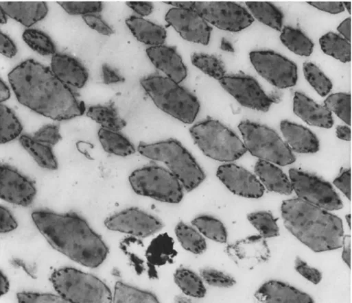
T h e cytoplasmic polyhedra in the different diseases vary greatly in size and shape; in some cases they tend to be very large as in the larvae of Bombyx mori (Linnaeus) and Ourapteryx sambucaria (Lin-
FIG. 1. Photomicrograph of a section through midgut cells of a larva of Estigmene acrea affected with a cytoplasmic polyhedrosis; note the large rounded polyhedra and the wide range of size ( χ 1500).
naeus); when large, the polyhedra lose their many-sided character and appear almost spherical (Fig. 1 ) . In some species such as Antheraea pernyi Guerin-Meneville, for example, polyhedra may be both very large and extremely small, the small polyhedra being at the limit of resolu-
tion of the optical microscope. Aruga and Israngkul (1961) have car
ried out some experiments on the factors controlling the size of the cytoplasmic polyhedra in the silkworm. They come to the conclusion that the size of the polyhedra depends upon the time interval between inoculation and appearance of symptoms; in other words, the longer the time, the larger are the polyhedra formed. In A. pernyi the poly
hedra occur in colonies surrounded by what appears to be a membrane (Fig. 2 ) . A somewhat similar phenomenon has been described by Bird
FIG. 2. Photomicrograph of a section through midgut cells of a larva of Antheraea pernyi affected with a cytoplasmic polyhedrosis; note the "colony" of polyhedra appar
ently enclosed in a membrane ( χ 1500).
and Whalen (1954) in the larvae of Choristoneura fumiferana (Clem
ens) .
It seems to be the general rule for polyhedral formation to start in the epithelial cells of the midgut and later to spread into both the foregut and the hindgut. Working with a cytoplasmic polyhedrosis of the silkworm, Tsujita (1955) considers that the polyhedra are formed in the cylindrical cells of the midgut epithelium, but not in the goblet or interstitial cells. In a similar disease of the spruce budworm, C.
fumiferana, the polyhedra occur in the cytoplasm of the midgut epi
thelium (Bird and Whalen, 1954).
It has long been debated whether the shape, but not the size, of the polyhedral crystal is characteristic of a given polyhedrosis; or, in
461 other words, does the virus govern the exact formation of the inclusion body? This certainly seems to be so with the nuclear polyhedroses, as exemplified by the cubic shape of the "polyhedra" in Panaxia dominula
(Linnaeus), the scarlet tiger moth, and by the crescent-shaped "poly
hedra" in Tipula paludosa Meigen. An interesting paper by Aruga et al.
(1961) on the cytoplasmic polyhedrosis of Bombyx mori (Linnaeus), the silkworm, suggests that the same thing holds for this type of disease.
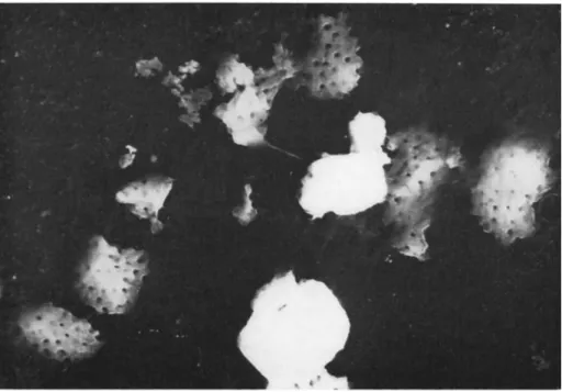
FIG. 3. Electron micrograph of cytoplasmic polyhedra from a larva of Phlogo- phora meticulosa after treatment with weak sodium carbonate; note the partially dissolved polyhedra with the empty sockets which had contained the near-spherical virus particles ( χ 28,000).
These workers observed that two types of cytoplasmic polyhedra oc
curred, a hexagonal and a tetragonal type. By isolating these, two viruses or virus strains were obtained, each with its characteristic polyhedral crystal. In the spruce budworm, the polyhedra are chiefly cuboidal and triangular forms have not been observed (Bird and Whalen, 1954). It seems, therefore, that in some cases the shape of the cytoplasmic poly
hedral crystal is associated with a particular virus.
Very little is known of the physicochemical properties or chemical nature of the cytoplasmic polyhedra. T h e i r reaction with weak alkali is very different from that of the nuclear polyhedra; instead of dissolv-
ing readily and leaving behind the virus particles enclosed in a mem
brane, the polyhedra dissolve only partially and a spongelike residue remains honeycombed with the sockets in which the virus particles have rested (Fig. 3 ) ; there is no encircling membrane. T h e propensity of the virus to dissolve before the polyhedral protein makes liberation of the virus a rather difficult matter, and this is dealt with in Section I D.
T h e reaction of cytoplasmic polyhedra to stains also differs sharply from that of the nuclear type. In a smear of nuclear polyhedra on a slide, fixed by mild heating and stained with Giemsa solution, the polyhedra remain unstained against a stained background. In contrast to this the cytoplasmic polyhedra take up the stain readily; provided the heating during fixation is not too severe, the two types of poly
hedra can be easily differentiated by this means.
There is already a certain amount of information regarding the macromolecular, paracrystalline lattice of the nuclear polyhedra ob
tained largely by the electron microscopy of ultrathin sections (Morgan et al., 1955, 1956; Day et al., 1956). Similar sections of cytoplasmic polyhedra have, in the writer's experience, failed to show the paracrys
talline lattice except in the case of a polyhedrosis in Hemerobius stigma Stephens (Neuroptera), and in this instance the origin of the poly
hedrosis is at present uncertain. However, it has been possible to reveal the paracrystalline lattice in cytoplasmic polyhedra by other means than thin sections. Figure 4 shows part of the crystalline protein of a cytoplasmic polyhedron from A. pernyi after treatment at an alka
line pH (Smith and Hills, 1962b).
T h e development of the polyhedra in a cytoplasmic disease of Thau- matopoea pityocampa Schiffermüller has been described by Xeros
(1956). Before the formation of the polyhedra, bodies which are apparently virogenic stroma appear in the epithelial cells of the gut.
Later, polyhedra of varying size, but mostly rather small, form sparsely over the virogenic stroma. Further development proceeds with the con
tinuing growth of the virogenic masses and with the increase in the number and size of the polyhedra at their surfaces. During this stage of the disease, large pores develop within the virogenic masses, and poly
hedra also arise and develop within these pores. In the final stages of the disease the polyhedra reach a size of 1.5 μ in diameter.
T h e occlusion of the virus particles inside the cytoplasmic polyhedra appears to be haphazard without any kind of ordered or symmetrical arrangement (e.g., see Wittig et al., 1960). This is in contrast to a nu
clear polyhedrosis of the crane fly (Tipula paludosa Meigen) larva, in wrhich the rod-shaped virus particles are occluded in rows which follow the outline of the crystal rather like growth rings in a tree (Smith, 1962).
Η Ο HFL I— > g < Μ
>
FIG. 4. Electron micrograph of part of the crystalline protein of a cytoplasmic polyhedron from a larva of Antheraea pernyi; note three virus cores and a single outer coat (χ 120,000).T h i n sections of cytoplasmic polyhedra reveal that the last occluded virus particles are each contained in a small depression on the surface of the crystal (Fig. 5 ) , and these depressions are shown by means of carbon replicas to be roofed over with a protein cover (see Section I D ) . Serological methods of study on the relationship of different inclu
sion bodies do not yet seem to have been applied to the cytoplasmic
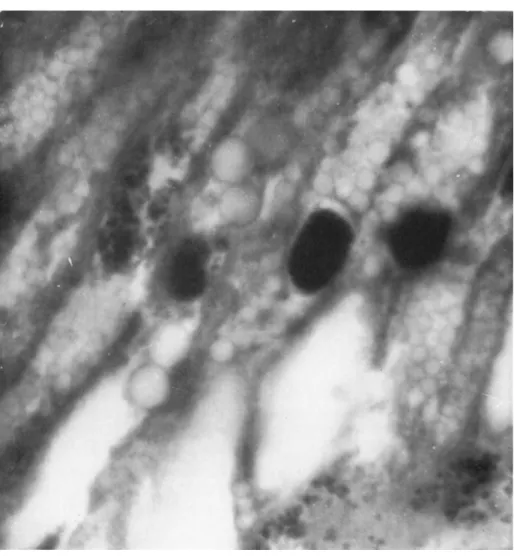
FIG. 5 . Electron micrograph of thin sections of very small cytoplasmic polyhedra from a larva of Hyloicus pinastri; note the virus particles stained black with osmic acid lying in sockets on the surface ( χ 26,000).
polyhedra although there is some information on the serological relation
ship of the nuclear polyhedra (Krywienczyk and Bergold, 1960).
C. Pathology
T h e outward appearance of lepidopterous larvae affected with nu
clear and cytoplasmic polyhedroses differ considerably, and this is mainly due to the respective sites of virus multiplication. In the
14. CYTOPLASMIC VIRUS DISEASES 465 nuclear polyhedroses the epidermis is one of the main organs attacked, and this is responsible for the characteristic limp cadavers which hang down from the food plant, to which they remain attached by the ab
dominal legs. Larvae infected with cytoplasmic polyhedroses lag greatly behind normal larvae in their development; there is no outstanding symptom at first, but affected individuals can be recognized by their small size, loss of appetite, and, sometimes, disproportionately large head
FIG. 6. Photomicrograph of a "colony" of very small cytoplasmic polyhedra from Antheraea pernyi ( χ 1500).
or long bristles. Later, the polyhedra developing in the gut can fre
quently be observed through the dorsal integument as a pale yellow or whitish area. In late stages of the disease, polyhedra are frequently regurgitated or voided in large quantities with the feces. A peculiar symptom apparently connected with a cytoplasmic polyhedrosis has been observed in full-grown larvae of A. pernyi in a late stage of the disease.
It occasionally happens that the hindgut is completely extroverted; this extroversion is possibly caused by a blocking of the alimentary canal accompanied by persistent effort on the part of the larva to void the polyhedra.
On opening a larva which has died of a cytoplasmic polyhedrosis, the abnormal state of the alimentary canal, especially the midgut, is at once apparent. Instead of the translucent, pale green organ of healthy larvae, the gut is opaque and pale yellow or milky in appearance owing to the huge numbers of polyhedra which become liberated as a milky fluid during the dissection procedure.
Histological examination of the gut in the early stages of disease reveals the formation of polyhedra in the midgut, mainly in the epi
thelial cells. As infection develops, however, polyhedra spread through the alimentary canal; in the case of a cytoplasmic polyhedrosis of Estig- mene acrea (Drury), the salt-marsh caterpillar, longitudinal sections of the entire caterpillar show the polyhedra present in almost every cell of the foregut, midgut, and hindgut.
T h e size of the polyhedra varies greatly, although not usually in the same cell (however, see Steinhaus and Dineen, 1959), and in some species, for example, the above-mentioned E. acrea and B. mori the variation is particularly great. In diseased larvae of A. pernyi many polyhedra are so small as to be only just within the resolution of the optical microscope (Fig. 6 ) . T h e reason for this great disparity in size is not clear; ultrathin sections of polyhedra of all sizes reveal no appar
ent difference and all contain virus particles.
When the disease is far advanced the polyhedra are liberated in large numbers in the lumen of the gut.
D. Morphology of Virus Particles
One of the difficulties of isolating the virus particles in any quantity from the cytoplasmic polyhedra has been to find a technique for liber
ating the virus. Unlike the rod-shaped viruses contained in nuclear polyhedra, the viruses from cytoplasmic polyhedra dissolve very readily in weak alkali; unless care is taken, all that remains after treatment is the polyhedral matrix, honeycombed with the sockets in which the virus particles have rested (Fig. 3 ) .
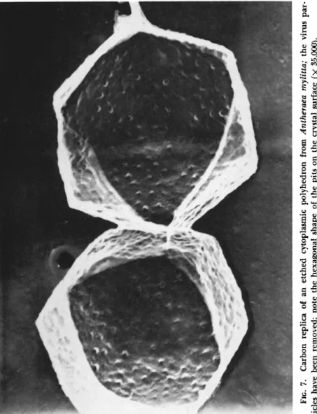
A method sometimes recommended for the extraction of the poly
hedra is simply to put the infected caterpillars into a flask of water and allow normal decomposition to proceed; in time the polyhedra sediment to the bottom of the flask. It has been shown (Hills and Smith, 1959), however, that this is an unsatisfactory method because the poly
hedra themselves deteriorate and become etched, while the virus parti
cles embedded in the surface of the polyhedra are lost (Fig. 7 ) . A better method is to open the larvae and plunge them immediately into distilled water; the guts are then removed under water and the polyhedra extracted. This method gives undamaged polyhedra with the individual
FIG. 7. Carbon replica of an etched cytoplasmic polyhedron from Antheraea mylitta; the viru tides have been removed; note the hexagonal shape of the pits on the crystal surface (χ 35,000).
virus particles on the surface of the crystal still covered with a layer of protein (Fig. 8 ) . T h e following techniques have proved effective in extracting the virus from several types of cytoplasmic polyhedra.
A suspension of polyhedra is dialyzed against weak sodium carbonate solution adjusted to pH 10.0 for 48 hours, then against water for 48 hours. T h e extract after cleaning at 10,000 rpm for 15 minutes on the centrifuge, and reduced in bulk, was subjected to gradient centrifugation through sucrose (Brakke, 1951). A clearly denned band was obtained, and a preparation of pure virus resulted.
An alternative is to dissolve 50 mg of polyhedra in 1.0 ml of 2 percent sodium carbonate solution for 30 seconds. T h e bulk of the liquid is increased to 30 ml by the addition of double-distilled and boiled water and centrifuged at 36,000 rpm for 30 minutes. T h e pellet is then suspended in distilled water and cleaned by centrif
ugation at 10,000 rpm for 15 minutes. T h e supernatant is reduced in bulk and subjected to gradient centrifugation through sucrose;
this gives a clearly defined band. T h e band is removed, increased in bulk to 5 ml with water, and centrifuged for 30 minutes at 36,000 rpm; this gives a pellet of pure virus.
This standardized technique has been applied to the cytoplasmic polyhedra of the larvae of the following insects, Arctia caja (Linnaeus), Antheraea mylitta (Drury), A. pernyi Guerin-Meneville, Nymphalis an- tiopa (Linnaeus), Vanessa cardui (Linnaeus), and Calophasia lunula
(Hufnagel). For the isolation of the virus from these species the opti
mum conditions appeared to be 0.5 percent sodium carbonate for 1.25 minutes for the polyhedra from A. caja, 2 percent for 30 seconds for A.
mylitta, 1.0 percent for 2 minutes for A. pernyi, 1.0 percent for 1.5 minutes for N. antiopa, and 1 percent for 1 minute for V. cardui and C. lunula (Hills and Smith, 1959).
It was first suggested by Kaesberg (1956) that the small plant viruses were not really spherical, but were icosahedral in shape possessing twenty sides, and it now seems as though icosahedron is the keyword in the contemplation of the "spherical" viruses. Further evidence on this is given in Section I, E, but at the moment we are concerned only with the external morphology of the virus particles.
Air-dried perparations of virus particles in the electron microscope are likely to show distortion and appear smaller than they actually are.
Accurate estimation of the size and contour, therefore, can only be made on frozen-dried specimens. Electron micrographs of a particle which is an icosahedron will show a silhouette of 5 or 6 sides, and this is more clearly demonstrated in the case of the Tipula iridescent virus ( T I V ) which is dealt with in a later section of this chapter. Some pictures, how-
FIG. 8. Similar to Fig. 7, but with the virus particles still in situ; note the protein cover to the sockets (χ 35,000).
ever, of virus particles from a cytoplasmic polyhedrosis affecting Anthe
raea mylitta show not only the six-sided contour of the particle but also a similar-shaped socket from which the virus has come (Smith, 1958). T h e polyhedral shape of the virus sockets in the crystal has been further dem
onstrated by means of carbon replicas of the polyhedra after removal of the virus particles (Fig. 7 ) .
Another method of demonstrating the contour of the virus particle is by means of shadow analysis. This is described in the section dealing with the Tipula iridescent virus; suffice it to say here that the technique has demonstrated that the cytoplasmic virus of A. mylitta is an icosahe
dron, although the issue is complicated by the very small size of the virus in comparison with the roughness of the substrate film on which the shadows are cast (Hills and Smith, 1959).
After observing a large number of cytoplasmic polyhedrosis viruses with the electron microscope, the question that inevitably arises is, are all these viruses the same? No difference whatever, on morphological grounds, can be observed between the viruses from Arctia caja, Antheraea mylitta, and A. pernyi. If these viruses are to be differentiated in some future scheme of classification, it must presumably be by their serology and ultrastructure and by amino acid analysis.
E. Ultrastructure of Virus Particles
T h e overall picture of a virus particle, derived from various methods of study, is of a thread of nucleic acid—ribonucleic acid (RNA) in plant viruses, deoxyribonucleic acid (DNA) and R N A in the animal viruses and phages—enclosed in a protein coat consisting of large numbers of identical protein subunits; in the small viruses these are the only com
ponents.
Crick and Watson (1956) suggested that all small viruses are built up on a framework of identical protein subunits packed together in a regular manner. Klug and Caspar (1960) point out that this means that a virus particle can be constructed out of subunits in only a limited number of ways. Determination of the symmetry gives considerable in
sight into the substructure of a virus particle.
In earlier electron micrographs of the spherical viruses, using the conventional shadow technique it was sometimes possible, if the resolu
tion was good, to observe that the surface of the particle was irregular and bumpy. T h e negative staining technique using phosphotungstic acid (Brenner and Home, 1959) has entirely changed the type of electron micrographs which can now be obtained of the small spherical viruses. It is now possible to count the protein subunits on the face of the particle,
471 and the number of morphological units can be found by adding some combination of the numbers twelve, twenty, thirty, and sixty or a multi
ple of sixty appropriate for the particular clustering arrangement (Klug and Caspar, 1960).
In studies of the cytoplasmic polyhedrosis of Antheraea pernyi the polyhedra have been split up by a minimum pH change into the virus and its protein in solution. These studies have shown that the virus par-
FIG. 9. "Cores" of three virus particles from a cytoplasmic polyhedrosis of An
theraea pernyi; each core is an icosahedron consisting of 12 subunits ( χ 120,000).
FIG. 10. T h e outer coats of the virus particles shown in Fig. 9 ( χ 100,000).
tides consist of an inner core and an outer protein shell. T h e inner core contains 12 large subunits and the outer core is made up of a large num
ber of much smaller subunits, but this number is not yet known (Figs. 9 and 1 0 ) . T h e crystalline lattice of the polyhedral protein is shown in Fig. 4 (Smith and Hills, 1962b).
As regards the nucleic acid content of the cytoplasmic polyhedrosis viruses, Xeros (1962) has carried out chromatographic and electropho
retic analysis of such polyhedra from Laothoe populi (Linnaeus). This showed that they contained 0.9 percent R N A and no DNA. Chromato
graphic analysis of similar polyhedra from a further five species showed the presence in them of similar quantities of R N A and the absence of DNA.
F. Latency and Hereditary Transmission
Latent virus infections are extremely frequent in most types of or
ganisms, and this is particularly true of the viruses affecting plants and insects. Indeed in some insects it is so common that entire populations of a species may be infected, as in the spruce budworm, C. fumiferana, and some stocks of the silkworm, B. mori. Anyone who has tried to rear large quantities of caterpillars in captivity will be aware of the rapidity with which a virus disease appears and spreads through insect colonies.
It is possible to stimulate these latent virus infections into virulence in a number of ways; these are called "stressors" by Steinhaus (1958a, b ) , who has made a preliminary study of stress as a factor in insect disease. Of natural stressors, crowding and temperature conditions are the commonest; the greater the number of insects confined in a given space, the greater the incidence of disease among them. High tempera
tures do not seem to have much effect on latent virus infections, but the writer has found that low temperatures and high humidity appear to stimulate into virulence a polyhedrosis of Cerura vinula (Linnaeus), the puss moth. Similarly with the larvae of Pieris rapae (Linnaeus), the small white butterfly, in Cambridge it is almost impossible to rear these in captivity owing to the invariable outbreak of a granulosis.
However, most of the information on the stimulation of latent virus infection in insects refers to the granuloses and particularly to the nu
clear polyhedroses. Some work has been done on the stimulation of la
tent cytoplasmic polyhedroses, and these are undoubtedly extremely widespread.
It has been shown by Japanese workers (Kurisu, 1955; Aruga, 1957;
Aruga et al., 1961) that low temperatures can stimulate into action a latent cytoplasmic polyhedrosis in the silkworm (see chapter 1 5 ) . Aruga
14. CYTOPLASMIC VIRUS DISEASES 473 and Hukuhara (1960) have experimented with various substances to try to induce nuclear and cytoplasmic polyhedroses in the silkworm, B.
mori. They tested eighteen chemicals by feeding them to fifth-instar larvae. Among these, six were found to be effective in the induction of cytoplasmic polyhedroses. These chemicals were sodium cyanide, sodium fluoride, arsenic acid, monoiodoacetic acid, sodium azide, ethylenedia- minetetraacetic acid ( E D T A ) , and its disodium salt. T h e last two chem
icals induced cytoplasmic polyhedroses with high frequency when they were fed to fifth-instar larvae, but only with low frequency when fed to fourth-instar larvae.
Aruga and Yoshitake (1961) found no significant increase in spon
taneous polyhedroses when silkworm larvae were exposed to ultraviolet radiation or X rays. T h e percentage of nuclear and cytoplasmic poly
hedroses was, however, markedly higher in larvae exposed to a double treatment of X rays and low temperature, than in those treated with low temperature alone. T h e cytoplasmic polyhedrosis of the alfalfa cater
pillar (Colias) was discovered in insects subjected to the stress of crowd
ing (Steinhaus and Dineen, 1959).
Another method of stimulation which, in the writer's experience, is frequently successful is inoculation of the larvae by feeding of a foreign virus; it seems necessary to use a nuclear polyhedrosis to stimulate a cytoplasmic one, but the reverse procedure does not seem to occur. Three examples of this type of stimulation are given here. One such case is the winter moth, Operophtera brumata (Linnaeus), the larvae of which were fed on foliage heavily contaminated with nuclear polyhedra from Vanessa cardui (Linnaeus). After a period of 10 to 20 days the larvae began to die of a virus disease, the final mortality being nearly 100 per
cent. Examination under the microscope revealed the presence of a cyto
plasmic polyhedrosis. All the control larvae remained healthy. Similarly with the larvae of Bupalus piniarius (Linnaeus) the pine looper; these were given the same virus with same result. It should be pointed out that these experiments wrhen repeated are not always positive; the result apparently depends upon whether or not a cytoplasmic virus is present
(Smith and Rivers, 1956). A more recent experiment on these lines is as follows; on April 18, 30 young larvae of Sphinx ligustri (Linnaeus), the privet hawk moth were colonized on lilac leaves heavily contaminated with nuclear polyhedra from Nymphalis to (Linnaeus), the peacock butterfly; 20 similar larvae were kept as a control. Between May 8 and May 13 five larvae died of a cytoplasmic polyhedrosis; all the control larvae remained healthy.
It is not known for certain what is the actual physical state of the
"latent virus"; it may be what is known as "occult virus," a definition
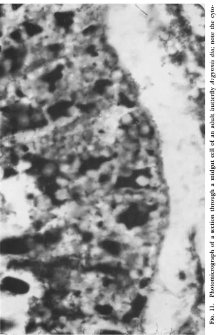
FIG. 11. Photomicrograph of a section through a midgut cell of an adult butterfly Argynnis dia; note the cyt lasmic polyhedra (χ 1200).
14. CYTOPLASMIC VIRUS DISEASES 475 suggested by Andrewes (1958) to cover those cases in which the state of the virus particles is not known and in which they cannot be detected. In their studies of two strains of a virus causing a cytoplasmic polyhedrosis in B. mori, Aruga et al. (1961) consider that the latent virus may be in an occult state from which it can be changed by cold treatment into an actively multiplying one.
W e come now to the question of hereditary transmission; so far as the viruses of the nuclear polyhedroses are concerned, there is a formidable body of evidence in favor of transmission of the virus through the egg.
This has recently been summarized by Bergold (1958).
There is little doubt also that the viruses of the cytoplasmic poly
hedroses are similarly transmitted. Cases occur fairly frequently of the adult insect containing cytoplasmic polyhedra within the body. A female privet hawk moth, Sphinx ligustri, reared from a batch of infected larvae was found to have the midgut filled with polyhedra. A female fritillary butterfly Argynnis dia (Linnaeus), similarly reared was observed to be voiding polyhedra; a section of the gut filled with polyhedra is shown in Fig. 11. Tanada and Chang (1960) also found that adults of the armyworm, Pseudaletia unipuncta (Haworth), developing from larvae exposed to a cytoplasmic polyhedrosis had polyhedra in the midgut epi
thelium. Most of such infected adults, however, usually die prematurely, and it is not known whether any of them are capable of laying viable eggs. Nevertheless, apart from this, there is considerable evidence of genetic transmission. At Cambridge in stocks of Hyloicus pinastri (Lin
naeus), the pine hawk moth, and of Phalera bucephala (Linnaeus), the buff tip moth, which are known to be infected with cytoplasmic poly
hedroses the young larvae succumb to the disease at a very early stage, sometimes immediately after emerging from the egg.
There are also many other examples, particularly in the Arctiidae, of larvae dying of cytoplasmic polyhedroses under conditions in which outside contamination with virus is excluded.
G. Cross Transmission
Although in the past there has been some controversy as to whether insect viruses as a whole are transmissible to other species, there is now no doubt whatever of the wide host range of one cytoplasmic virus, the Tipula iridescent virus ( T I V ) which is dealt with in the second half of this chapter. T h e situation with regard to the cross transmissi- bility of the cytoplasmic polyhedrosis viruses is still rather obscure, and it is, of course, intimately bound up with the question as to how far many of these viruses are identical. It is certainly true that cross trans-
mission seems to take place between various species of tiger-moth larvae (Arctiidae), but final judgment in these cases must wait upon the de
velopment of criteria of relationship, either by serological or chemical data.
It may be relevant here to give the results of one or two experiments out of a large number performed in the year 1961 by the writer on attempted cross transmission of cytoplasmic polyhedrosis viruses. In the first experiment the insect used was the comma butterfly Polygonia c-album (Linnaeus), and all the larvae were the progeny of a single female. On April 6, 30 young (second stage) larvae were colonized on nettle leaves contaminated with cytoplasmic polyhedra from Arctia caja, the tiger moth. Between April 17 and April 28 six larvae died of a cytoplasmic polyhedrosis, and on May 11 an adult butterfly emerged and died shortly afterward; the gut of this butterfly was found to be filled with cytoplasmic polyhedra. T h e remaining larvae pupated nor
mally. In the second part of this experiment, 30 similar larvae were fed with cytoplasmic polyhedra from Phalera bucephala, the buff-tip moth. On April 17, eleven days after the start of the experiment, seven larvae were dead of a cytoplasmic polyhedrosis and the rest were very much smaller and more backward than the controls, all of which ap
peared healthy, some having already pupated. By April 2 1 , all the larvae which had been fed with the cytoplasmic polyhedra from P.
bucephala were dead of a cytoplasmic polyhedrosis.
Of the control larvae all but two pupated normally; these two died of a cytoplasmic polyhedrosis. This experiment was repeated using the polyhedra which developed after feeding on the polyhedra of P. buceph
ala. In this case 13 larvae out of 24 died of a cytoplasmic polyhedrosis;
two were similarly affected in the controls.
In another experiment 35 second-stage larvae of Polygonia c-album were fed with cytoplasmic polyhedra from Sphinx ligustri, the privet hawk moth; these polyhedra had developed in the S. ligustri larvae after feeding on nuclear polyhedra from a larva of Nymphalts to, the peacock butterfly. This experiment was commenced on May 15 and by May 25, 27 larvae out of the 35 had died of a cytoplasmic polyhedrosis.
Of the 35 control larvae 12 died of a cytoplasmic polyhedrosis.
Experiments with the larvae of another species, Gonepteryx rhamni (Linnaeus), the brimstone butterfly, were commenced on April 11, again using cytoplasmic polyhedra from A. caja. By April 24 the ex
perimental larvae showed a most impressive difference from the con
trols, being only about half the size. By April 28, all the larvae had died of a cytoplasmic polyhedrosis. On April 25, the control larvae
477 were nearly full-grown and appeared healthy; 28 of these pupated nor
mally, but two died of a cytoplasmic polyhedrosis.
T h e obvious difficulty in assessing the results of these transmission experiments is the lack of means of differentiating between the viruses.
Were these genuine transmissions of virus or merely a stimulating of a latent virus infection? T h e mortality could not have been much higher if the larvae had been fed with a wild virus from their own species. It was known that both Polygonia c-album and G. rhamni do have cytoplasmic polyhedroses, and this is shown by the fact that some of the control larvae of the two species developed the disease sponta
neously. Nevertheless, the high rate of mortality does suggest that cross transmission had taken place and is further evidence that many of these cytoplasmic polyhedrosis viruses are one and the same.
Mention of an interesting cytoplasmic polyhedrosis of two insects belonging to the Neuroptera is relevant here although further investi
gation of the origin of the disease is wanted. It was shown by Sidor (I960) and Smith et al. (1959a) that when larvae of Chrysopa perla
(Linnaeus) and Hemerobius stigma Steven were fed upon pieces of larvae of Porthetria dispar (Linnaeus) ( = Lymantria dispar) which had died of a nuclear polyhedrosis, a number of these larvae themselves died of a polyhedrosis. Investigation of the disease showed that the polyhedra were present in large numbers in the cytoplasm of the epi
thelial cells of the mesenteron; it was therefore a cytoplasmic polyhe
drosis. However, examination of the polyhedra under the optical mi
croscope revealed what appeared to be virus bundles very similar to those visible in the nuclear polyhedra of P. dispar. This was confirmed on the electron microscope, and both carbon replicas and thin sections of the polyhedra showed the presence of bundles of rod-shaped virus particles, each bundle apparently enclosed in a membrane.
T h e fact that the polyhedra closely resemble the nuclear polyhedra from P. dispar and contain very similar bundles of rod-shaped virus particles suggests that this is a case of cross transmission. On the other hand, the disease is a cytoplasmic, not a nuclear, polyhedrosis, and this is more in line with a possible stimulation of a latent virus infection.
H. List of Cytoplasmic Polyhedroses
T h e following insects have been recorded at Cambridge as being affected with cytoplasmic polyhedroses. T h e list is not intended to be a complete record of all the cytoplasmic polyhedroses described but it serves to give an idea of the wide distribution of this type of virus dis
ease. [The reader may also consult lists by Hughes (1957) and by Mar
tignoni and Langston (1960) of insects reported to have virus diseases.]
L E P I D O P T E R A Rhopalocera
SATYRIDAE
Pararge aegeria (Linnaeus) Dira megera (Linnaeus) NYMPHALIDAE
Argynnis dia (Linnaeus) Vanessa cardui (Linnaeus) Aglais urticae (Linnaeus) Nymphalis io (Linnaeus) Nymphalis antiopa (Linnaeus) Polygonia c-album (Linnaeus) LYCAENIDAE
Lycaena phlaeas (Linnaeus) PIERIDAE
Aporia crataegi (Linnaeus) Pieris brassicae (Linnaeus) Pieris rapae (Linnaeus)
Euchloe cardamines (Linnaeus) Gonepteryx rhamni (Linnaeus)
Heterocera ARCTIIDAE
Hypocrita jacobaeae (Linnaeus) Phragmatobia fuliginosa (Linnaeus)
Cycnia mendica (Clerck) Spilosoma lutea (Hufnagel) Spilosoma lubricipeda (Linnaeus) Estigmene acrea (Drury)
Hyphantria cunea (Drury) Arctia villica (Linnaeus) Arctia caja (Linnaeus)
Parasemia plantaginis (Linnaeus) Rhyparia purpurata (Linnaeus) NOCTUIDAE ( = CARADRINIDAE)
Phlogophora meticulosa (Linnaeus)
AGROTINAE
Heliothis armigera (Hübner) Agrotis segetum (SchiffermüHer) Amathes glareosa (Esper) Triphaena pronuba (Linnaeus) Lampra fimbriata (von Schreber)
DASYPOLIINAE
Calophasia lunula (Hufnagel) Anchosceli helvola (Linnaeus) Agrochola lychnidis (Schiffermüller)
L l T H O P H A N E N A E
Lithophane leautieri (Boisduval) Antitype xanthomista (Hübner)
Heterocera (continued) Hada nana (Hufnagel) Hadena Serena (Schiffermüller) Heliophobus albicolon (Hübner) Xylomyges conspicillaris (Linnaeus) Diataraxia oleracea (Linnaeus) PLUSIIDAE
CATOCALINAE
Scoliopteryx libatrix (Linnaeus)
PLUSIINAE
Plusia gamma (Linnaeus) L Y M A N T R I I D A E
Orgyia antiqua (Linnaeus) Dasychira pudibunda (Linnaeus) Euproctis chrysorrhoea (Linnaeus) Porthetria (— Lymantria) dispar
(Linnaeus) HYDRIOMENIDAE
(— G E O M E T R I D A E ) Anaitis plagiata (Linnaeus) Operophtera brumata (Linnaeus) Operophtera fagata (Scharfenberg) SELIDOSEMIDAE ( = G E O M E T R I D A E )
Semiothisa liturata (Clerck) Bupalus piniarius (Linnaeus) Biston betularia (Linnaeus) Abraxas grossulariala (Linnaeus) Ourapteryx sambucaria (Linnaeus) Selenia lunaria (Schiffermüller) Crocallis elinguaria (Linnaeus) SPHINGIDAE
Sphinx ligustri Linnaeus Smerinthus ocellatus (Linnaeus) Dilina tiliae (Linnaeus)
Hyloicus pinastri (Linnaeus) Laothoe populi (Linnaeus) NOTODONTIDAE
Lophopteryx capucina (Linnaeus) Cerura vinula (Linnaeus) Phalera bucephela (Linnaeus) SATURNIIDAE
Samia (— Philosamia) cynthia (Drury) Actias selene (Hübner)
Antheraea (— Telea) polyphemus (Cramer)
Antheraea mylitta (Drury)
Antheraea pernyi Guerin-Meneville Automeris memusae (Walker)
479 Heterocera (continued)
DREPANIDAE Gastropacha quercifolia (Linnaeus)
Lasiocampa quercus (Linnaeus) Drepana lacertinaria (Linnaeus)
P H Y C I T I D A E BOMBYCIDAE
Ephestia cautella (Walker)
LASIOCAMPIDAE T I N E I D A E
Bombyx mori (Linnaeus)
Eriogaster lanestris (Linnaeus) Tineola bisselliella (Hummel) Malacosoma neustria (Linnaeus) Tinea pellionella (Linnaeus)
I I . T H E Tipula IRIDESCENT VIRUS
A. Pathology
This unusual and interesting virus was first discovered by the Virus Research Unit at Cambridge during routine examination of large numbers of the larvae of Tipula paludosa Meigen for the presence of another virus, that of a nuclear polyhedrosis. Brief descriptions were later given by Xeros (1954) and Smith (1955). T h e r e are no poly
hedra or similar inclusions formed in this disease, although isolated groups of virus particles, enclosed in a double or triple membrane, sometimes occur in the cells.
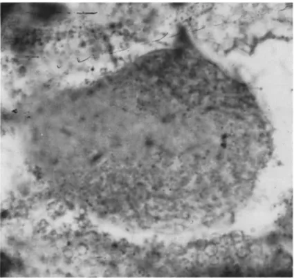
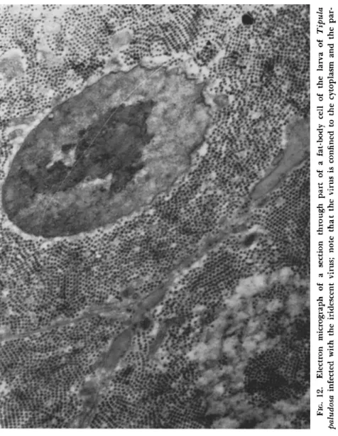
T h e initial site of virus multiplication appears to be the cytoplasmic regions of the fat-body cells, for it is here that the virus is found to be most heavily concentrated. T h e intracellular virus appears to avoid the nucleus completely, as may be seen from Fig. 12. T h i s is in marked contrast to the behavior of the virus of a nuclear polyhedrosis affecting the same insect, where the rod-shaped virus particles are found within the nuclei of the larval blood cells.
A larva of T. paludosa affected with the Tipula iridescent virus ( T I V ) can be recognized at once by the change in appearance that it induces in the body of the affected host. Whereas the normal appearance of these larvae is a dark tan, the color of the diseased ones is a somewhat opalescent blue-indigo. Examination by low-power microscopy shows that the color is particularly intense in the lobes of the hypertrophied fat body; as the disease approaches its termination the color is less intense and is more generally dispersed throughout the body. On open
ing a larva in an advanced stage of the disease, a thin white fluid is liberated together with a highly concentrated suspension of the virus which can be recognized by its iridescent colors. T h e thin white fluid consists of the disintegrated fat body.
In the early stages of the disease before the iridescence is well de
veloped, diagnosis is helped by wetting the surface of the larva in a tube held at an angle so that the light shows up the fat body.
Tipulid larvae affected with the nuclear polyhedrosis also appear
FIG. 12. Electron micrograph of a section through part of a fat-body cell of the larva of Tipula paludosa infected with the iridescent virus; note that the virus is confined to the cytoplasm and the par tiles are oriented (χ 10,000).
14. CYTOPLASMIC VIRUS DISEASES 481 white and opaque; they lack, however, the brilliant iridescence charac
teristic of T I V infection. A white fluid is also liberated from these larvae, but a smear stained with Giemsa solution and examined under
the microscope will reveal the presence in large numbers of the crescent- shaped "polyhedra."
T h i n sections of fat body from a larva infected with T I V have an unmistakable appearance when examined with the electron microscope.
T h e cell cytoplasm is completely filled with virus particles which, in an advanced stage of the disease, will already be oriented into a crystalline pattern (Fig. 1 2 ) . It is these microcrystals formed in the living insect which produce the iridescence.
Although the fat body is the initial site of multiplication of the virus, as the disease progresses other organs become infected. Anderson et al. (1959) showed the presence of T I V in the skin of T. paludosa by means of fluorescent staining, and this finding has been confirmed by electron microscopy. Indeed, as will be shown later, the virus seems able to multiply in many types of insect tissue including the muscles, wing buds, legs, and head.
B. Host Range
For a number of years the opinion has been held by some workers that the insect viruses were strongly species specific and that cross transmission did not occur. This view was challenged by Smith and Xeros (1953a, b ) , by Gershenson (1958), and by Steinhaus (1953) with the polyhedroses. T h e results of these studies seemed good evidence of genuine cross infection. T h e work on the polyhedroses, however, was hampered by the constant interference of latent virus infections, stimulated into activity by the injection of the foreign virus, and by the lack of a critical means of identification of the various viruses involved.
What was badly needed to study cross inoculation was a virus with definite characteristics and causing unmistakable symptoms; these would serve as a marker and differentiate the virus immediately from latent virus infections or casual contamination.
T I V is a virus of this type, and infection by it can be immediately recognized by the blue or violet iridescence of the affected organ, by the characteristic pentagonal or hexagonal outline of the virus particles under the electron microscope, and by the enormous quantities of virus produced in the diseased insect.
A systematic investigation of the host range of T I V was carried out by Smith et al. (1961), and much of the following information is from
that work. T h e easiest method of transmitting the viruses of polyhe
droses is to feed leaf material contaminated with polyhedra to the experimental insects. However, feeding as a method of transmitting T 1 V is unsatisfactory, and in many cases is quite unsuccessful. Instead, the insects were injected with a suspension of purified virus, together with an addition to the inoculum of 200 units of penicillin and 200 units of streptomycin per milliliter. T h e larvae of a few species reacted badly to injection, and these were induced to drink a drop of highly purified virus suspension.
T h e larvae of the following insects, in addition to the natural host Tipula paludosa Meigen have been experimentally infected with T I V : Diptera: Tipula oleracea Linnaeus; T. livida Van der Wulp; two uniden
tified species of Tipula; Bibio marci (Linnaeus), St. Mark's fly; Calli- phora vomitoria (Linnaeus); Mycetophila sp.
Lepidoptera: Porthetria (= Lymantria) dispar (Linnaeus) ; Bombyx mori (Linnaeus); Hepialus lupulinus (Linnaeus); H. humuli (Lin
naeus) ; Sphinx ligustri Linnaeus; Pieris brassicae (Linnaeus); P. rapae (Linnaeus); P. napi (Linnaeus) ; Gonepteryx rhamni (Linnaeus);
Nymphalis io (Linnaeus) ; Vanessa atalanta (Linnaeus) ; Galleria mel
lonella (Linnaeus).
Coleoptera: Tenebrio molitor Linnaeus; Agriotes obscurus (Linnaeus) ; Melolontha sp.
From the above list it will be seen that T I V has been transmitted experimentally to the larvae of seven species of Diptera, twelve species of Lepidoptera, and three species of Coleoptera.
T h e choice of experimental insects was governed largely by what insects were available, and there is little doubt that the experimental host range could be considerably widened. This is the first record of the transmission of an insect virus between different orders of insects.
T h e fat body appears to be the initial site of virus multiplication in all cases, but high concentration of virus, as shown by the degree of iridescence and by ultrathin sections under the electron microscope, develops in several other organs. In the small mycetophilid larvae the skin showed a bright blue iridescence and sections of the skin revealed the presence of great quantities of virus. It frequently happened that larvae of Porthetria dispar and Pieris brassicae, inoculated in a middle instar, did not die as a result of virus infection until after pupation.
In the case of Porthetria dispar this has the peculiar effect of causing the legs and wing buds to atrophy; at the same time they exhibit a bright blue iridescence indicating the presence of microcrystals of virus. Sections of the wing buds and legs when examined in the electron microscope
14. CYTOPLASMIC VIRUS DISEASES 483 reveal the presence of great concentrations of virus, and the same holds good for the head.
T h e effect of T I V on the prepupae and pupae of Pieris brassicae is to produce many monstrosities which may be due to disturbed wing formation. T h e virus is present in high concentration in the abdomen, wing shields, and fat body. Often the wing cases are abnormally swollen with a large droplet of blood and hang away from the body. When the pupal wing case is properly formed very small wings form after 10 days, but none of these pupae have survived to form adults.
Very high concentrations of virus were also found in the wing- buds of larvae of Hepialus humuli (Linnaeus), the swift moth. T h e virus accumulates in such quantities in the thorax of pupae of L. dispar and H. humuli that the aggregates are easily visible in sections with the optical microscope.
There is some evidence that the manufacture of the virus in some of these hosts is unsatisfactory insofar that the particles may be some
what fragile and easily ruptured by negative staining with phospho- tungstic acid. T h e virus particles from T. paludosa, the original host, show great uniformity in size, but those from P. brassicae are less uni
form and many were rather imperfect in shape.
Serological studies on T I V carried out by R . H. Stobbart show that to all intents and purposes the T I V preparations from four different insects T . paludosa, P. brassicae, Porthetria dispar, and Nymphalis io are practically identical.
C. Morphology and Ultrastructure of the TIV Particle
In the early days of the investigation of virus morphology by elec
tron microscopy, but after the advent of metal shadowing, it became apparent that all the virus forms were flattened. This flattening was found to be due to the forces of surface tension. T o eliminate this source of morphological artifact, the technique of freeze-drying was developed; by the use of this technique it was shown that the "heads"
of the T-even bacteriophage possessed a distinct polyhedral shape, the general appearance of which was that of a hexagonal prism with pyra
midal ends (Anderson, 1952).
Following upon this it was observed that the contours of some of the so-called spherical particles were not round but six-sided. Kaesberg
(1956) attempted to find the polyhedral form of some plant viruses, when frozen-dried, by an analysis of shadow shapes. A particular type of polyhedron, if it is regular, should cast a particular type of shadow, if it is cast with a specified orientation with respect to the positions of the vertices of the polyhedral particle. Kaesberg concluded that the
shapes of the small viruses investigated by him are best represented by regular icosahedra, figures with twenty sides. One difficulty here is due to the smallness of shadow detail in comparison with the roughness of the film upon which the shadows are cast. This became evident in our attempts to show the icosahedral shape of the virus from the cytoplas
mic polyhedrosis of Antheraea mylitta by the double shadowing tech-
FIG. 1 3 . A model of an icosahedron shadowed by two light sources and oriented so that an apex of the hexagonal contour points directly to each light source. This throws two shadows; one is four-sided and pointed, and the other is five-sided with a blunt end. (After Williams and Smith, 1 9 5 8 . )
nique (Hills and Smith, 1959). However, the difficulty brought about by the roughness of the substrate film is obviously reduced if the par
ticle and its associated shadow are large. T h i s is the case with T I V since it is a large virus measuring about 130 ηιμ in diameter.
FIG. 1 4 . A particle of the Tipula iridescent virus frozen-dried and shadowed in the same way; the similarity between the shadows thrown is evident ( χ 1 0 5 , 0 0 0 ) . (After Williams and Smith, 1 9 5 8 . )
In spite, however, of the flattening of the T I V particle on drying out of water, it still retains its distinctive noncircular contour. Six-sided contours are the most frequent, but occasional five-sided ones also occur.
(Fig. 1 5 ) .
In order to prove that the T I V particle is actually an icosahedron,
FIG. 15. Electron micrograph of particles of the Tipula iridescent virus, negatively stained with phosphotungstic acid note the hexagonal shape and apparent second membrane which is now known to be the inner row of protein subunit (χ 240,000).
487 the double shadowing was carried out as follows. A model of an ico
sahedron was made and shadowed by two light sources separated 60°
in azimuth and oriented so that an apex of the hexagonal contour points directly to each light source. T h i s throws two shadows, one is four-sided and pointed and the other is five-sided with a blunt end
(Fig. 1 3 ) . A particle of T I V frozen-dried and shadowed in the same way is shown in Fig. 14; the similarity between the shadows thrown is
FIG. 1 6 . Single particle of the Tipula iridescent virus at very high magnification ( χ 300,000); note the protein subunits, which are apparently hollow.
evident. This indicates with fair certainty that the T I V particle is an icosahedron (Williams and Smith, 1958).
T h e early attempts to investigate the ultrastructure of the T I V par
ticle were directed toward the study of thin sections of the particles under the electron microscope. These sections demonstrated vividly the six-sided appearance of the particles, but they also suggested that each particle was surrounded by a double membrane (Fig. 1 5 ) . In thin sections of T I V particles from Tipula paludosa stained with phospho- tungstic acid, and also in particles negatively stained, it is possible to make out individual units on the surface of the virus particle. High- resolution micrographs of virus from larvae of Bibio marci (Linnaeus),
negatively stained, do show a regular arrangement of the protein sub- units, the icosahedral faces being made up of small subunits. Similar subunits have been observed on virus particles obtained from larvae of the white butterfly, Pieris brassicae (Smith and Hills, 1959).
As with a number of other viruses, numerous empty particles of T I V occur and the significance of these is discussed in Section I I , D;
but further investigation of these empty particles, using the negative staining technique with phosphotungstic acid (PTA) (Brenner and Home, 1959) suggested that what appeared to be a second membrane was actually an inner row of protein subunits. Further high-resolution electron microscopy of the empty T I V shells with negative staining shows them to be composed of 812 protein subunits with which are lipids apparently helping to bind the subunits into a rigid structure.
T h e subunits measure 85 Ä by 140 Ä and are hollow and hexagonal when viewed end-on (Fig. 1 6 ) . They are arranged to form a 20-sided solid figure (icosahedron) each side being an equilateral triangle (Smith and Hills, 1962a).
D. Replication
From a survey of what is known on the replication of the viruses of plants and the higher animals, a general picture of biosynthesis emerges—an assembly rather than a multiplication.
In the replication of many viruses, especially the smaller entities, a dual process is involved in which the protein and nucleic acid are formed separately and then polymerized into the infectious particle.
This two-step process is supported by the so-called "eclipse phase,"
which is a lag period between the time of entry of the virus into the cell and the production of the mature infectious virus particles.
There is one outstanding phenomenon which has been observed in the replication of many spherical or near-spherical viruses, from both plants and animals, and that is the appearance of large numbers of empty protein shells of the same size as the virus. This phenomenon was first observed by Markham and Smith (1949) with the plant virus causing turnip yellow mosaic. These empty shells, called "top com
ponent" because they form the top layer during ultracentrifugation, do not contain ribonucleic acid and are therefore not infectious. Some
what similar empty shells have been described by H o m e and Nagington (1959) in studies on the structure and development of poliovirus. T h e highest count of empty shells they recorded was 11 to 14 percent at 51/4 hours after infection.
T h e presence of these empty shells in the early stages of infection
with T I V can be demonstrated in ultrathin sections of fixed fat body and in homogenates of infected fat body.
Careful study of the empty forms of T I V suggests very strongly that they are stages in the reproduction of the virus. This is borne out by the following facts: first, empty or partially empty shells occur predominantly in the early stages of the disease. In some sections,
FIG. 1 7 . Electron micrograph of sections of the Tipula iridescent virus particles showing apparent development stages ( χ 80,000).
indeed, practically no mature virus particles are visible but large num
bers of empty shells occur. Secondly, what appear to be stages in the development of the contents of the particles can be seen (Fig. 1 7 ) . In ultrathin sections of the shells, threads can be seen radiating from the center; the next apparent stage is the build-up of spherical accumula
tions in the center of the particle, which is considered to contain the deoxyribonucleic acid. As development proceeds, the space between the spheres and the inner surface of the protein subunit shell becomes filled up, and the mature virus particle is then complete (Smith and Hills, 1962a).
E. Purification and Chemical Studies
T h e quantity of T I V produced by a larva in a late stage of the disease is astonishingly large. T h e dry weights of the extracted, puri
fied virus and of the whole diseased larva have been compared; in the sample measured the virus weighed approximately 25 percent as much as the entire larva. This yield is easily a record for animal viruses; it is more than twice that reported for tobacco mosaic virus, where the dry weight of the purified virus may be as much as 10 per
cent of that of the diseased plant leaves. Only the yield of the T-even bacteriophage from infected cells of Escherichia coli (Migula) Castellani and Chalmers may be of comparable magnitude.
T h e purification of the virus particles is particularly simple owing to their large size and to the absence of particles of comparable size in extracts of diseased larval tissue. In order to obtain a purified virus suspension, the larvae are first cut up and placed in a beaker of water for several hours. After clarification of the resulting extract by low- speed centrifugation, the virus is obtained essentially pure by the ap
plication of two cycles of high- and low-speed centrifugation. Distilled water is a suitable suspending medium.
T h e pellets of virus resulting from centrifugation have fascinating optical properties. By transmitted light the pellet appears an orange or amber color. By reflected light it has an iridescent, turquoise ap
pearance. Within the pellet may be seen small regions reflecting the incident light quite brilliantly, giving the entire pellet the appearance of an opal.
When an embedded pellet is sectioned and examined in the electron microscope, the general appearance is that shown in Fig. 18. T h e pellet is seen to consist of regions of crystallinity, the average diameter of which is 5 to ΙΟιημ. Since the crystals are randomly oriented with respect to each other, a given section will cut them in different directions with respect to their faces, and the bizarre pattern shown in Fig. 18 will
result. T h e center-to-center distance of the virus particles in the closest packed arrays yet found is 1300 Ä. T h e origin of the reflection effects in the pellets is now evident. Bragg reflections result from the periodic particle arrays, and the scale of size of the interparticle spacing is such as to make visible the effects of the Bragg reflections. T h e change of the reflected color from turquoise to violet upon fixation and dehydra
tion of the pellet is due to shrinkage of the interparticle spacings. T h e
FIG. 1 8 . Electron micrograph of a thin section through a pellet of purified Tipula iridescent virus; note the patterns resulting from sections through small crystalline regions oriented at random ( χ 1 5 , 0 0 0 ) . (After Williams and Smith, 1 9 5 7 . )