Granuloses of Insects
ALOIS HUGER
Biologische Bundesanstalt für Land- und Forstwirtschaft, Institut für biologische Schädlingsbekämpfung, Darmstadt, Germany
I. Introduction 531
II. Diagnosis 532
A. Signs and Symptoms 532
Β. Microscopic Investigations 534
III. Isolation and Purification of Capsules and Viruses 536 IV. Morphology and Size of Capsules and Viruses 537
V. Physical Properties and Chemical Composition of Cap
sules and Viruses 541
VI. Serological Properties of Capsule Proteins and Viruses... 543 VII. Effect of Storage and Temperatures on the Infectivity of
Granulosis Viruses 544
VIII. Pathology 545
A. Host Species and Tissues Attacked by Granuloses . . . . 545
B. Cyto- and Histopathology 550
C. Susceptibility and Resistance of Hosts to Granuloses 556 D. Mixed Infections, Interference, and Synergism 558
E. Cross Infectivity 560
F. Latency and Stress 560
I X . Natural and Applied Control of Pests by Granuloses 563 A. Epizootiology—Natural Limitation 563
B. Biological Control 566
X . Taxonomy of Granulosis Viruses 567
References 569
I. INTRODUCTION
When Steinhaus published his comprehensive book on "Principles of Insect Pathology" in 1949 the granuloses had just been recognized to represent a distinct group of insect virus diseases which—like the polyhedroses—is characterized by a special type of virus inclusion body.
On observation with the light microscope, the bodies appear as minute 531
532 ALOIS HUGER
"granules" with dimensions of approximately 300 to 500 πιμ, almost at the limit of resolution. It was for this reason that diseases of this group were called "granuloses" by Steinhaus (1949a).
T h e granules had already been detected by Paillot (1926) in the first investigation of a disease of this type in caterpillars of the cabbage butterfly, Pieris brassicae (Linnaeus). A few years later, Paillot (1934) discovered a similar infection in larvae of the cutworm Agrotis segetum
(Schiffermüller). Shortly afterward, he described another disease of this type in the same host (Paillot, 1935, 1936), and finally a third one
(Paillot, 1937). From their differing pathologies Paillot considered the three diseases to be caused by distinct infectious agents and designated them pseudo-grasserie 1, 2, and 3.
It was not until 1947 that another disease of this type was discovered, this time by Steinhaus (1947), in the variegated cutworm, Peridroma margaritosa (Haworth). In 1948 Bergold confirmed the suspected virus nature of the causative agents of granuloses. In his investigation of a granulosis of the fir-shoot roller, Choristoneura murinana (Hübner), he isolated the rod-shaped viruses and demonstrated them with the electron microscope. Moreover, he showed that the granular virus in
clusions seen in the light microscope in reality are of capsular shape.
For this reason Bergold called them Viruskapseln, and the disease Kap- selvirus-Krankheit, the latter being synonymous with "capsular disease"
repeatedly used in the past.
Meanwhile, the term "granulosis," which is fairly descriptive of the gross appearance of diseased tissues, has been generally accepted. More
over, in referring to the virus, just as one would say "encephalitis virus"
we use the designation "granulosis virus." Electron microscope investi
gations performed so far have proved the capsulelike shape of the virus inclusion bodies typical of the granuloses. Hughes (1958), therefore, justly declared the term "capsules" to be more fitting for them than a nondescript word such as "granules," which derived from their light microscope appearance. Accordingly, in the following sections "cap
sules" is used to designate the virus inclusion bodies characteristic of granuloses.
In the last decade a series of granuloses have been reported upon, but only some have been more or less intensively studied. They usually become evident in the larval, and rarely the pupal, stages of Lepidoptera.
I I . DIAGNOSIS
A. Signs and Symptoms
During the period of lethal granulosis infection, generally the host larvae sooner or later exhibit external signs and symptoms especially as
to their color, condition, and behavior. T h e first indication of infection often is the loss of appetite; later on the larvae cease feeding. As a rule their surface—often especially on the ventral side—progressively changes from its usual color to a pale whitish or milky yellow appearance due to the development of vast numbers of capsules in the affected tissues, the integument often being mottled. A brownish discoloration occurs, for instance, with the buckeye caterpillar, Junonia coenia Hübner
(Steinhaus and Thompson, 1949), and, after a change to yellow, with the western grape leaf skeletonizer, Harrisina brillians Barnes and Mc- Dunnough (Smith et al., 1956). T h e change in color may also depend in part on the larval age at the time of infection (e.g., Wilson, 1960).
Infected larvae may continue to molt and may grow almost to the normal size or even larger, e.g., Pseudaletia unipuncta (Haworth)
(Tanada, 1959a), until they finally die. Others exhibit a marked re
duction in size, especially in an advanced stage of the disease, as, for example, larvae of H. brillians (Smith et aL, 1956). Several species be
come more-or-less swollen and develop intensified segmentation without, or with only slightly, increased turgidity, as, for instance, larvae of Pieris rapae (Linnaeus) (Tanada, 1953a; Todd, 1960), C. murinana (Bucher, 1953), Eucosma griseana (Hübner) (Martignoni, 1957), and Hyphantria cunea (Drury) (Schmidt, 1959) .
T h e change in color usually is accompanied by progressive weaken
ing, sluggishness, and naccidity of the larvae; in addition, they become increasingly less responsive to stimuli. If the disease has progressed far enough, the blood generally is turbid and milky owing to the presence of large numbers of capsules derived from disintegrated infected tissues.
Upon dissection of larvae, the diseased fat body appears opaque white throughout in heavy infections, and flecked opaque white in light in
fections.
Diarrheic symptoms may occur either as a result of a secondary bacteriosis induced by the granulosis (e.g., P. rapae; Tanada, 1953a; see Section V I I I , D) or almost regularly as reported from Harrisina brillians where, contrary to the case in other granuloses, the midgut is the prin
cipal seat of infection (Smith et aL, 1956). On drying, the diarrheic discharge frequently causes the moribund larvae to adhere to the sub
strate. Dead or paralyzed dying larvae usually are very flaccid and not infrequently remain suspended from leaves or elsewhere by their caudal or abdominal prolegs sometimes in the form of an inverted V. After death often they darken rapidly until they appear entirely black.
Death usually occurs in the larval, but sometimes in the prepupal or pupal, stage (e.g., see Section V I I I , C). T h e period of lethal granulosis infection varies considerably in different hosts as well as in a single
534 ALOIS HUGER
species, but, in general, it ranges between 4 and 20 to 25 days, taking up to 34 days with Pseudaletia unipuncta, according to Tanada (1959a).
Except for the host species, it may depend mainly on the decree of larval maturity at the time of infection, temperature, virus dose applied, virulence of the inoculum, and health condition of the host (e.g., T a nada, 1953a; Martignoni, 1957; Glass, 1958; Wittig, 1959a, b; Wilson, 1960).
In many species there is a marked liquefaction of the internal tissues after death. When, besides the fat body, the epidermis also is heavily infected by the virus, the skin is excessively fragile. It easily ruptures, thus liberating the liquefied body contents with myriads of capsules.
In such cases, the granuloses closely resemble the nuclear polyhedroses of Lepidoptera. However, when the epidermis is not strikingly affected by the granulosis virus, the integument remains relatively firm.
External symptoms may vary according to the larval age at infec
tion. Not infrequently they are displayed only in the final stage of the disease. Moreover, larvae may also die of granulosis without under
going the complete sequence of external signs and symptoms, or any at all. Altogether, external signs and symptoms are not very obvious;
in addition, they are nonspecific, and thus only indicative of the pres
ence of a possible granulosis virus infection. Therefore, microscopic examination is the only reliable method to use in identifying a granu
losis.
B. Microscopic Investigations
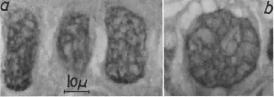
Although the capsules are very minute in size, they are still readily visible with the higher powers of an ordinary light microscope. For preliminary diagnosis of granulosis infections, therefore, they are a good diagnostic feature. However, with the normal light field illumination they can hardly be identified with any degree of certainty, whereas phase contrast and dark field illumination give good results. With the latter, capsules suspended in water or in the hemolymph of larvae with ad
vanced disease appear as highly refractive particles rapidly dancing with Brownian movement. T h e same is the case with capsules in diseased cells, especially when their contents are in the process of disintegration and liquefaction. In phase contrast the capsules appear dark, in the dark field they are bright-shining.
In the light field, fresh unstained fat cells occupied with capsules commonly display a slight cream or brownish coloration. T h e same tint may be seen in heavy concentrations of free capsules in a watery suspension. On observation of ordinary wet mounts of infected tissues
in the dark field, the areas densely packed with capsules appear light blue.
T h e investigation of diseased tissue in a fresh state in water mounts, with either dark field or phase contrast, reveals myriads of capsules emanating from ruptured cells. Thus also spherical vesicles—from a few microns up to about 20 μ in diameter—containing many strongly vibrating capsules are frequently discharged. These can also be observed in the liquefied body contents of moribund or dead larvae; they can fuse together. Paillot (1937) called them "boules hyalines." Apparently they occur in all granuloses (e.g., Bergold, 1948; Hughes and Thomp
son, 1951; Tanada, 1953a; Smith et ah, 1956; Martignoni, 1957).
It must be pointed out, however, that the recognition of capsules in the manner described above often becomes complicated, mainly either by cells which may contain minute granular material—such as ganglia and epidermis—or by the presence of large numbers of little uric acid crystals, both being of a refractivity similar to that of capsules. It needs some experience to distinguish capsules from such particles, espe
cially when the particles are present in abundance as compared to cap
sules. As to uric acid crystals, they differ best from capsules in that they are birefringent whereas capsules are not (Krieg, 1957). Unlike capsules, this granular material in normal epidermial and other cells is not uniform in size but is generally larger and consequently in less rapid motion.
Regardless of a positive or a doubtful diagnosis in the above man
ner, further confirmation must be obtained by the histopathological study of stained serial sections of diseased tissues. Infected cells com
monly exhibit characteristic pathological changes (see Section V I I I , B , 1 ) . Moreover, it is possible to demonstrate the capsules there by special staining methods. Martignoni (1957) used bromophenol blue after Mazia's method (also Tanada, 1959a), and the method for staining of polyhedra employed by Langenbuch (1955) and earlier authors. Huger
(1961) recently elaborated histological staining methods for capsules of C. mnrinana on the known principle of acidifying virus inclusion bodies prior to staining: An elective intense bright-red staining of capsules could be obtained, for example, by treatment of sections in 50 percent acetic acid at room temperature for 5 minutes prior to staining with Heidenhain's iron hematoxylin and counterstaining with an aqueous solution of 0.5 percent erythrosin for 2 minutes. Shvetsova (1961) also obtained good results in staining capsules with different dyes, such as eosin, methylene blue and especially carbol fuchsin, after pretreatment with alkali or acid.
T h e most important proof in original diagnosis of granulosis infec-
536 ALOIS HUGER
tions, however, is the demonstration of capsules and their occluded viruses with the electron microscope. Pertinent techniques are summa
rized in the following Section I I I .
I I I . ISOLATION AND PURIFICATION OF CAPSULES AND VIRUSES
For the isolation of capsules, larvae dead of granulosis are suspended in water and kept at room temperature until the larval tissues are pu
trefied and the capsules have sedimented at the bottom of the container in a whitish layer. T h e capsules remain unaffected by this treatment as they are indestructible by natural putrefaction processes occurring dur
ing this time. Separated in this way, they can easily be purified by differential centrifugation. Isolation of capsules naturally can be ex
pedited by grinding larvae in a mortar, separating out rough tissue debris by filtration, and subsequently centrifuging the nitrate at differ
ent speeds. A drop of the purified capsules suspended in redistilled water is then dried on an electron-microscope grid for study.
T h e liberation of virus particles from capsules is achieved by disso
lution of the paracrystalline proteinaceous capsule in weak alkalies. For several reasons Bergold (1953b, 1958) has suggested standardization of this process for polyhedra and capsules. T h e alkali solution employed consists of a mixture of a variable concentration of alkali ( N a2C 03 or K2C 03) —depending on the virus species—and a constant concentration of 0.05 Μ NaCl or KCl. For capsules of C. murinana 0.03 Μ N a2C 03 was used by Bergold. His standard method for capsules can be briefly summarized as follows:
Dissolve 5 mg (air dry) capsules per 1 ml alkali solution up to 3 hours at 20 °C. Centrifuge the capsule solution at about 4000 g for 5 minutes; discard the small brownish sediment consisting mainly of im
purities and partially dissolved capsules; spin the slightly turbid super
natant for about 1 hour at about 12,000 g to sediment the virus particles, which collect in a small bluish-white pellet. Pour off carefully the super
natant containing the capsule protein in solution; resuspend the pellet of virus in an equal amount of C02-free redistilled water; spin again for about 1 hour at 12,000 g. Discard the water-like supernatant; re
suspend the virus sediment in redistilled water, using one-seventh of the initial volume. T h e virus particles can now be mounted on grids for electron microscope examination. By further treatment with alkali they are decomposed to their constituents (see Section I V ) .
Of course, this standard method may be varied especially as to the concentration of alkali, and—depending on this—the period of capsule dissolution. By means of this method, in principle, granulosis viruses
from many hosts have been isolated and demonstrated by a series of workers (see T a b l e I).
IV. MORPHOLOGY AND SIZE OF CAPSULES AND VIRUSES
Virus inclusion bodies of granuloses consist of two main components, the proteinaceous capsular envelope and the rod-shaped virus particle occluded by this. Unlike a polyhedron which harbors many virus par
ticles, each capsule contains only one, occasionally two, lying side by side (Figs, la, l b , 3b, and 9 ) . T h e granulosis viruses very much re
semble the nuclear-polyhedrosis viruses. Since Bergold's (1948) first description of the structural composition of the C. murinana capsules, all further investigations of a series of capsules have led to the same result. In no case could evidence be obtained in support of the sug
gestion of Tokuyasu (1953), Smith and Xeros (1954), and Smith (1955) that in some granuloses the capsules regularly might contain two virus rods. This misinterpretation resulted from the "wheatlike" appearance of capsules, especially in an intermediate stage of dissolution (Hughes, 1958).
By means of electron-microscope studies, capsules of many hosts have been described to be oval, ellipsoidal, ovoid, or egg-shaped in outline.
Lower (1954), in his study of the granulosis of Persectania ewingii (Westwood), reported them to represent "subovoid, elongate bodies with more or less parallel sides." Indeed, in preparations of capsules of other hosts there occur forms which fit this description. T h e i r shape should be designated ovocylindrical instead of only uniformly ellipsoidal or ovoid. A certain variation in shape within an individual species seems to be a common feature.
Capsules generally are opaque to the electron beam. As shown by Bergold (1958, 1959a, b, c) in ultrathin sections, the capsular protein—
similar to the polyhedral protein—also consists of a macromolecular paracrystalline lattice (Fig. 1 ) . T h e molecules are in a cubic arrange
ment. In capsules of C. murinana they have dimensions of 57 χ 5 7 χ 229 Ä; the sides of the unit cell measure 57 A, and the angle (γ) is 120°
(Fig. l c ) . Surprisingly, there is not the least disturbance in the molecu
lar lattice by the virus rod.
Besides the normal capsules some aberrant long virus inclusions and also bizarre, angled, and branched elements repeatedly have been observed (Bergold, 1948; Steinhaus et aL, 1949; Hughes and Thompson, 1951; Tanada, 1953a, 1959a). It has been suggested that they are the result of the fusion of several capusles. A further interpretation is given in Section V I I I , B , 2. In addition, occasionally polyhedral-shaped
538 ALOIS HUGER
inclusion bodies are formed (Bergold, 1953a, c; Bird, 1959) that are about 1 μ in diameter.
T h e mean size of the capsules known to date ranges between 300 and 511 ηΐμ in length and 119 and 350 πιμ in width (Table I ) . They vary more or less in size also in the single species without regard to the quoted elongate elements measuring 1 to 2 μ and more.
T h e rod-shaped granulosis viruses are often slightly curved. T h e i r average width was stated (Table I) to range from 36 to 80 πιμ, their
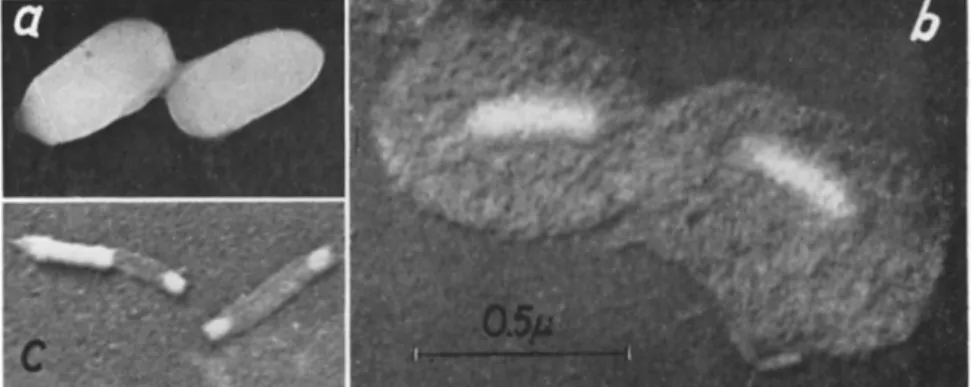
FIG. 1. Electron micrographs of ultrathin longitudinal (a) and cross (b) sections through capsules of Laphygma frugiperda (J. E . Smith) showing the macromolecular paracrystalline lattice of the capsule protein and the single virus rod occluded by this. On micrograph (c) the cubic arrangement of the molecular lattice of the capsule protein of Choristoneura murinana (Hübner) may be seen clearly. Magnifica
tion: a, χ 240,000; b, χ 270,000; c, χ 305,000. (Courtesy of G. H. Bergold.)
average length from 2 4 51 to 411 πΐμ. By treatment of the virus rods with weak alkali Bergold (1950, 1953b) was able to demonstrate that they are surrounded by two membranes like those of nuclear polyhedra. At first the outer one is shed, taking a spherical shape ("spherical mem
brane") (Fig. 2 ) . For this outer membrane Bergold (1953a, b) has introduced the term "developmental membrane" with reference to his life cycle theory for insect viruses (Bergold, 1950). Smith and Xeros (1954), like other workers, objected to this theory since it implied
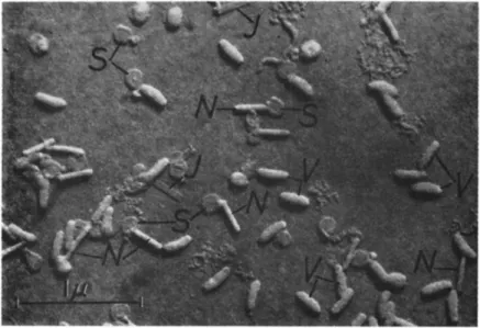
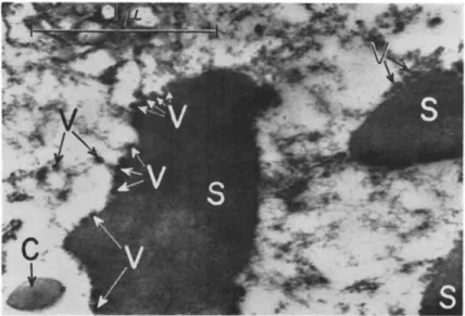
FIG. 2. Electron micrograph of virus rods isolated from capsules of Pieris rapae (Linnaeus) by alkali treatment. Note the various constituents of the viruses: V, virus rods still surrounded by the "developmental membrane"; S, so-called "spherical mem
branes" representing "developmental membranes" after being shed from the viruses.
Some "spherical membranes" are still in contact with their respective virus rods.
N, naked virus rods; / , empty tubular intimate membranes. Approximate magnifica
tion: χ 26,000. (Courtesy of Y. Tanada; from Tanada, 1953a.)
development of the virus particle from a sphere to a rod within this membrane; they substituted the term "inner capsule" for "developmental membrane." They based this designation on pictures where capsules had been partially dissolved in situ on screens, and thus the respective en
closed virus rod appeared capsulelike (Fig. 3b). Later on Smith (1956b) himself identified his "inner capsule" as a membrane without abandon-
l T h e mean length of 200 ιημ for the granulosis virus of P. brassicae reported by Vago et al. (1955) surely is to be regarded as invalidated by the later results by Vago (1959) (see Table I).
540 ALOIS HUGER
ing the term. Summing up, for the outer membrane of the virus rods, neither the term "inner capsule" is appropriate with regard to the real structure of the membrane and compared with the surrounding protein capsule nor is the designation "developmental membrane," which is fairly well established in the literature, justified, since the life-cycle theory (see above) cannot be maintained in the light of present knowl
edge (see Section V I I I , B , 2). Therefore, it would be better to speak only of an outer membrane of the virus rod.
T h e inner membrane, the so-called "intimate membrane," is closely applied to the virus rod but is slightly and evenly spaced from the outer membrane ("developmental membrane"). After disintegration of
FIG. 3. Electron micrographs of capsules and granulosis viruses of Natada nararia (Moore), (a) Untreated capsules; (b) Capsules after treatment with weak sodium carbonate, showing the partially dissolved and distended capsule protein and the virus rod ("inner capsule" of Smith, see Section IV) in the center, (c) T w o virus rods with partially dissolved contents, thus demonstrating the intimate mem
brane. Magnification: χ 50,000. (Courtesy of Κ. M. Smith and N. Xeros; from Smith and Xeros, 1954.)
its contents by weak alkali (Fig. 3c), the intimate membrane is visible as a straight, empty, and flattened tube (Fig. 2) (Bergold, 1950, 1953b, 1958;
Tanada, 1953a; Smith and Xeros, 1954).
A most important and interesting detection on the ultrastructure and nature of insect viruses recently was made by Smith and Hills (1960). Using the negative staining technique with phosphotungstic acid, they were able to demonstrate that the contents of the intimate membrane of virus rods of nuclear polyhedra show a wide-spaced helical structure comparable to that of the tobacco mosaic virus. When dis
charged from either end of the intimate membrane, these contents appear to uncoil. Smith and Hills consider this helix to be, in part, nucleic acid ( D N A ) . In the same way they detected a closely packed
helical structure of the intimate membrane of isolated granulosis virus rods (Fig. 4) which could not further be resolved on empty mem
branes. Nevertheless, the observations on virus rods of nuclear poly
hedra strongly suggest that the helical appearance of the intimate membrane of the granulosis virus is a replication of the helical struc
ture of its contents. Furthermore, Smith and Hills reported the pres
ence of a head capsule at either end of the intimate membrane of granulosis virus rods.
In preparations of granulosis viruses small oval or spherical bodies, as well as folded and V-shaped virus rods, have been observed repeatedly (Bergold, 1950, 1958; Hughes and Thompson, 1951; Wasser and Stein
haus, 1951; Hughes, 1952; Tanada, 1953a). Surely, they must not be
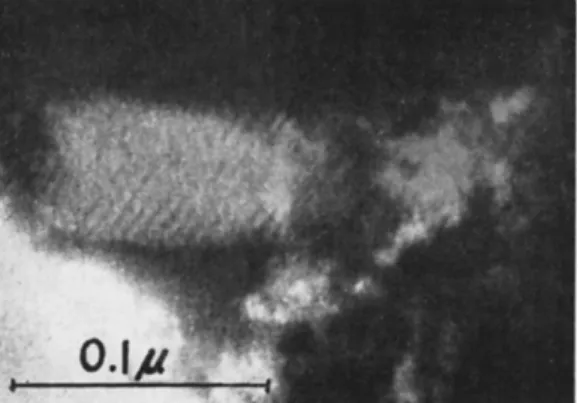
FIG. 4. Electron micrograph of a part of a naked granulosis virus of Pieris brassicae (Linnaeus) showing a helical structure on the intimate membrane. Magnifica
tion: χ 300,000. (Courtesy of Κ. M. Smith and G. H. Hills.)
interpreted as developmental forms in a complicated life cycle of insect viruses as Bergold (1950, 1953a, b, 1958) and Bird (1958, 1959) believe.
Furthermore, in granulosis-virus preparations long filaments or virus rods arranged in chainlike fashion have been observed (Steinhaus et aL,
1949; Schmidt and Philips, 1958; Steinhaus and Marsh, 1960). It would seem that they are in some way related to the long filaments observed by Bird (1958, 1959) in his electron microscope study of ultrathin sec
tions of the fat body of Choristoneura fumiferana (Clemens) infected with a granulosis virus.
V . PHYSICAL PROPERTIES AND CHEMICAL COMPOSITION OF CAPSULES AND VIRUSES
Capsules are insoluble in water, alcohol, acetone, xylene, and ether.
However, they are soluble in aqueous solutions of various acids and alkalis, such as N a2C Os, NaOH, K O H , N H4O H , H2S 04, and C H3C O O H .
542 ALOIS HUGER
For this reason, pretreatment with acids renders possible their distinct staining (e.g., Section I I , B ) . As already mentioned, capsules are not destroyed by natural putrefactive processes. They are heavier than water; for capsules of C. murinana, Bergold (1958) recorded a density of 1.279. After dissolution in weak alkali their inclusion-body protein gives a main component with a sedimentation constant of S2 0 = Svedberg units, and, calculated from this, a molecular weight of about 300,000. T h e split components have a sedimentation constant of S2 0 = 3.45, and a molecular weight of about 60,000.
Physical properties of the isolated virus rods include a sedimenta
tion constant of S20 = 1.324, a diffusion constant of D20 = 0.278 χ 1 07, a frictional ratio of f/f0 1.49, an axial ratio of 5.2, a particle weight of 460 χ 1 06 by calculation from sedimentation and diffusion constants, and of 435 χ 106 by calculation from the length and diameter of virus rods on electron micrographs. These determinations were made by Bergold (1948) with an analytical ultracentrifuge.
So far only the capsule protein of C. murinana and the granulosis- virus particles of C. murinana and C. fumiferana have been qualita
tively and quantitatively analyzed for their amino acid composition (Wellington, 1951, 1954). For the acid hydrolyzates of both purified virus rods and purified capsule protein the following amino acids have been determined by paper chromatography: alanine, arginine, aspartic acid, cysteine and/or cystine, glutamic acid, glycine, histidine, leucine and/or isoleucine, lysine, methionine, phenylalanine, proline, serine, threonine, tyrosine, and valine. Tryptophan was determined in un- hydrolyzed samples. Whether the considerable residue in each hydroly- zate was from unhydrolyzable material or was formed during hydrolysis, is not clear. T h e values for each amino acid recovered from the granulosis viruses of C. murinana and C. fumiferana are very similar, whereas both viruses differ in their nucleic acid composition (see below).
Simultaneous analyses on nuclear polyhedra of seven different hosts revealed that the proteins of insect virus inclusion bodies are very similar to one another in amino acid composition. In spite of this, many significant differences can be observed between them. It further became evident that the viruses of both the polyhedral and the capsule types are closely related with respect to the relative proportions of their various amino acids. Finally, comparing any two of the investigated viruses and their respective inclusion-body proteins it is observable that the viruses differ significantly in more amino acids than do the inclusion- body proteins (Wellington, 1954).
Investigations by Wyatt (1952a, b) revealed the presence of deoxy
ribonucleic acid (DNA) in granulosis viruses of C. murinana and C.
fumiferana. Like the viruses causing nuclear polyhedroses they only contain the purines adenine (A) and guanine ( G ) , and the pyrimi- dines cytosine (C) and thymine ( Τ ) , but no 5-methylcytosine or uracil.
Though both granulosis viruses originate from closely related hosts they differ in their nucleic acid composition, but have a similar ratio of A + T / G - f - C (1.67 and 1.87 respectively). This ratio, however, greatly differs between the polyhedrosis and granulosis virus of the same host, C. fumiferana (0.95 and 1.87, respectively). On the other hand, the A -f- T / G -\- C ratio of the C. murinana granulosis virus (1.67) is identical with that of the polyhedrosis virus of the widely separated hymenopteron Neodiprion sertifer (Geoffroy).
V I . SEROLOGICAL PROPERTIES OF CAPSULE PROTEINS AND VIRUSES
From earlier investigations we know that insect viruses are highly antigenic. Taking into account this fact, Krywienczyk et al. (1958) and Krywienczyk and Bergold (1960a) investigated the serological relation
ships of 17 different insect viruses after liberation from their inclusion bodies by the complement-fixation technique. They showed that, sero
logically, the tested viruses belong to three distinct groups: (1) "capsule"
(i.e., granulosis) viruses of Lepidoptera, (2) nuclear "polyhedron" (i.e., polyhedrosis) viruses of Lepidoptera, and (3) nuclear "polyhedron"
(i.e., polyhedrosis) viruses of Hymenoptera. Within each group most members could also be distinguished on the level of their serological titers. T h e first group includes the capsule viruses (indicated by the first C in the following parenthetical symbols) of C. murinana ( C C m ) , C. fumiferana (CCf), and Laphygma frugiperda ( J . E. Smith) (CLf).
Viruses of groups 2 and 3 did not cross-react with the antisera of capsule viruses [except a slight reaction of the polyhedron virus of Malacosoma americanum (Fabricius) when used as antigen]. Some weak cross re
actions were found when CLf and CCf served as antigens against seven polyhedron virus antisera. T h e CCm and CCf viruses which originate from phylogenetically very closely related hosts appeared to be serolog
ically identical. No cross reaction was obtained with the CCf virus and the polyhedron virus of the same host.
It is interesting to note that the capsule virus of Recurvaria milleri Busck (CRm) behaves differently from the other capsule viruses inves
tigated. As antigen it reacts with the CLf antiserum but not with CCf and CCm antisera. However, C R m reacted with 8 polyhedron virus antisera out of twelve tested. Using C R m antiserum no cross reactions occurred with CLf, CCm, and CCf antigens, but did with several polyhe
dron viruses.
544 ALOIS HUGER
Further investigations by Krywienczyk and Bergold (1960b) on the basis of the complement-fixation technique revealed that the inclusion- body proteins of the viruses employed above also fall into three serolog
ically distinct groups which correspond to those of their viruses (see above). From the capsule proteins only CCm and CCf, isolated from phylogenetically closely related hosts, gave a strong cross reaction; but apparently both are serologically not related to the capsule proteins CLf and CRm, whose host insects belong to phylogenetically distant families. Capsule protein CLf as antigen in complement fixation did not cross-react with any of the heterologous sera. Moreover, no sero
logical relation could be detected between the CCf capsule protein and the polyhedron protein of the same insect species.
T h e exceptional position of C R m (see above) again became evi
dent in that its capsule protein cross-reacts with all the polyhedron pro
teins available with reactions from 15 to 67 percent of homologous titers, whereas only weak nonreciprocal reactions with the other capsule proteins could be observed. This serological similarity between the cap
sule protein C R m and the lepidopterous polyhedron proteins became further evident when the agar diffusion technique was used (Krywien
czyk and Bergold, 1961).
These results are not directly comparable with earlier results of Tanada (1954) because of a different method of preparing the reactants.
Tanada observed that whole capsules of P. rapae used as antigen were agglutinated with antisera against whole polyhedra of P. rapae and Co
lias eurytheme Boisduval.
Subsequent studies of Krywienczyk and Bergold (1960c) strongly suggest that the insect viruses and their respective inclusion body pro
teins are only weakly, if at all, serologically related. Capsule virus antisera, for example, showed only weak, if any, reactions with their inclusion-body proteins and did not react with the polyhedron proteins.
Therefore, the significance of the insect virus inclusion bodies and their relation to the viruses serologically has not been clarified. Further studies are necessary to shed light upon this very interesting question.
V I I . E F F E C T OF STORAGE AND TEMPERATURES ON THE INFECTIVITY OF GRANULOSIS VIRUSES
For the use of biological control measures it is important to know the effect of storage and temperatures on the infectivity of viruses.
Polyhedra usually remain infective under room temperature for several years, some are reported even up to 10 to 20 years. Relatively little is known on the effect of storage on granulosis viruses. Smith (1959a) re
ports that the capsules of Pieris spp. "seem to lose much of their infec-
tivity after storage for one winter." Kelsey (1958), however, achieved 100 percent mortality of P. rapae in laboratory infection experiments with capsules of P. brassicae kept at ordinary room temperature for nearly 16 months. Rivers (1959) successfully used the same virus after one-year storage. Similarly, the granulosis virus of P. rapae remained infectious when kept "in a dry state under room conditions for over a year" (Tanada, 1956c). Müller-Kögler (unpublished) observed granu
loses in larvae of P. brassicae after peroral application of capsules stored on dried smears at room temperature for three years. No loss of infectivity was stated for the same capsules pressed with inert substances into tablets of 200 mg even after storage for 5 years (Vago et al., 1961). Capsules of C. murinana suspended in water also kept their infectivity at room temperature over a period of five years (Krieg, unpublished). In addi
tion, the infectivity after at least one-year storage is proved for the cap
sules of Pseudaletia unipuncta (Tanada, 1955), E. griseana (Martignoni and Auer, 1957), and Hyphantria cunea (Drury) (Schmidt, 1959).
Another important question is the effect of high and low tempera
tures. Tanada (1953a) found that the granulosis virus of Pieris rapae survived temperatures up to 70 to 75°C for 10 minutes, or at 70°C between 20 and 30 minutes within its capsule. Longer heating inacti
vated the virus. Similarly, the granulosis virus of Pseudaletia unipuncta was inactivated when heated at 75°C for 10 minutes or at 70°C for 40 minutes (Tanada, 1959a) (see Section V I I I , D ) .
No "noticeable loss in virulence" occurred with the granulosis virus of Pieris rapae after storage at 4 to 6°C in the refrigerator for over a year. Also by deep freezing at —32°C for 36 days "the virulence of the virus was hardly affected" (Tanada, 1953a). "Little or no loss of virulence" was recorded for capsules in diseased larvae of Argyrotaenia velutinana (Walker) after 2 years' storage in the freezing compartment of a household refrigerator (Glass, 1958). Under similar conditions the same was the case with capsules of C. murinana (Krieg, unpublished).
From the foregoing it seems that, in general, the capsules keep their infectivity for at least one, or up to two, years under room conditions.
Storage of the capsules at low temperatures surely is advantageous and possibly may lengthen the period of infectivity.
V I I I . PATHOLOGY
A. Host Species and Tissues Attacked by Granuloses
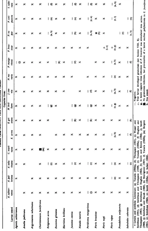
So far, about 35 species of Lepidoptera have been reported to suffer from a granulosis infection. Doubtless, their number is considerably higher in nature, and further cases will be detected in the future after
T A B L E I
DATA ON THE HISTOPATHOLOGY AND/OR THE DIMENSIONS OF CAPSULES AND VIRUSES OF A SERIES OF GRANULOSES
Host insect Family Tissues affected
Average size of capsules
(πιμ) Agrotis subterranea
(Fabricius) Phalaenidae Fat body 500 χ 300
Argyrotaenia velutinana
(Walker) Tortricidae Fat body 315 χ 160
Choristoneura fumiferana
(Clemens) Tortricidae
Fat body, epidermis, tracheal matrix(b)
350 χ 160(c)
Choristoneura murinana
(Hübner) Tortricidae
Fat body, epidermis, tracheal matrix(c)
360 χ 230(a)
Estigmene acrea (Drury) Arctiidae ρ 400 χ 250
Eucosma griseana (Hübner) Tortricidae Fat body,
epidermis(b) 397 χ 119(a) Eulype hastata (Linnaeus) Geometridae ? 360 χ 200 Euxoa ochrogaster (Guenee) Phalaenidae ? 440 χ 250 Harrisina brillians Barnes
and McDunnough Zygaenidae Midgut epithelium(b) 345 χ 256(a)
Hyphantria cunea (Drury) Arctiidae Fat body 500-600 χ 250-350
Junonia coenia Hübner Nymphalidae
Fat body, epidermis, tracheal matrix(b)
450 χ 300(a)
Natada nararia (Moore) Limacodidae Fat body,
epidermis 360 χ 200 Peridroma margaritosa
(Haworth) Phalaenidae Fat body 400 χ 250
Pieris brassicae (Linnaeus) Pieridae Fat body,
epidermis(a) 300 χ 120(b)
Pieris rapae (Linnaeus)
Pseudaletia unipuncta (Haworth)
Pieridae Phalaenidae
Fat body, epidermis(b, d), tracheal matrix(d)
Fat body
300 χ 200(b) 511.2 χ 253.7 Sabulodes caberata Guenee Geometridae Fat body 345 χ 170
546
Average size of virus rods
( π ΐ μ )
Site of development
of capsules
Name of the virus
Pertinent literature 350 x 60
250 x 50
272 x 36(a)
257 X 41(b)
270 x 40
306 x 49(a) 320 x 60 360 x 60 245 x 67(a)
276.8 x 52.9
300 x 40(b)
?
Cytoplasm(b)
Nucleus and cytoplasm(d)
p
Nucleus(b), cytoplasm(c)
?
?
?
?
Nucleus(b)
Bergoldiavirus clistor- habdion Wasser and Steinhaus
Bergoldiavirus calyp- tum Steinhaus
Bergoldiavirus thomp- sonium Steinhaus
Bergoldiavirus kovache- vici Schmidt and Philips
Bergoldiavirus latheti- cum Steinhaus
Steinhaus and Marsh (1960)
Wasser and Steinhaus (1951)
(a) Bergold (1953b), (b) Bird (1958), (c) Bird (1959)
(a) Bergold (1948), (b) Bergold (1953b), (c) W ittig (1959a), (d) Huger and Krieg (1961) Steinhaus (1949b) (a) Martignoni (1954),
(b) Martignoni (1957), (c) Bird (1958) Steinhaus (1959) Steinhaus (1959) (a) Steinhaus and Hughes
(1952), (b) Smith et al.
(1956)
Schmidt and Philips (1958)
(a) Steinhaus (1949b), (b) Steinhaus and T hom p
son (1949)
290 x 45 Nucleus — Smith and Xeros (1954)
340 x 40 Cytoplasm
270-300 x 75-80(d), Nucleus(c), 200 x 80(b) cytoplasm(a)
268 x 42(a), Nucleus(b), 293.6 x 46.9(c) cytoplasm(d) 411.6 x 62 Nucleus
275 x 65 Nucleus
Bergoldiavirus daboium Steinhaus
Bergoldiavirus brassicae (Paillot) Steinhaus
Bergoldiavirus virulen- tum Tanada
Bergoldiavirus nosodes Hughes and Thompson
Steinhaus et al. (1949) (a) Paillot (1926), (b)
Vago et al. (1955), (c) Smith and Rivers (1956), (d) Vago (1959) (a) Bergold (1953b), (b) Tanada (1953a), (c) Tanada (1956a), (d) Bird (1958)
Tanada (1959a)
Hughes and Thompson (1951)
547
548 ALOIS HUGER
increasing attention is paid to this group of insect-virus diseases. I n T a b l e I those granuloses are listed of which we have detailed informa
tion concerning their pathology and/or their viruses. Further granu
loses are known from the following hosts: Agrotis segetum (pseudo- grasserie 1, 2, and 3; Paillot, 1934, 1935, 1936, 1937); Amelia pallorana
(Robinson) (Steinhaus, 1957); Autographa californica (Speyer) (Hall, 1953); Chorizagrotis auxiliaris (Grote) (Steinhaus, 1957); Dendrolimus Sibiriens Tshetverikov (Lukyanchikov, 1962); Euplexia leucipara (Lin
naeus) (Smith and Rivers, 1956); Laphygma exigua (Hübner); Laphygma frugiperda, Megalopyge opercularis ( J . E. Smith) (Steinhaus, 1957);
Melanchra persicariae (Linnaeus) (Smith, 1956a, b; Smith and Rivers,.
1956); Nephelodes emmedonia (Cramer) (Steinhaus, 1957); Persectania ewingii (Lower, 1954); Pieris napi (Linnaeus) (Smith, 1956a, 1960);
Prodenia litura (Fabricius) (Smith, 1958); Recurvaria milleri (Steinhaus, 1957); Sesamia cretica Lederer (Vago, 1956; Steinhaus, 1960); Thaumeto- poea pityocampa (Schiffermüller) (Biliotti, 1959).
Very recently it could definitely be stated (Huger and Weiser, unpublished) that Weiser's (1948, 1949) "rickettsia-like organism" in the fat body of Camptochironomus tentans (Fabricius) really represents rickettsiae—not capsules as it was discussed (see Weiser, 1948, 1949; Stein
haus, 1949a, p. 514) on the basis of information available at that time.
With one exception (Harrisina brillians), in all granuloses studied so far the fat body is the principal tissue infected. As can be seen from T a b l e I, in about one-half of the hosts listed also the epidermis and, in some cases, additionally the tracheal matrix is reported to be affected.
Taking into account these apparent differences in tissue tropism, Mar
tignoni (1957) grouped the granuloses into two types, namely, monor- ganotropic diseases affecting tissues only of mesodermic origin (fat body), and polyorganotropic diseases affecting additional tissues of ec
todermal origin (epidermis, tracheal matrix). However, further careful investigations are necessary before the known granuloses can definitely be classified in this way; unfortunately the histopathology of most gran
uloses has not been thoroughly studied by means of many permanent histological serial sections. Omitting this, a light or irregular affection of the epidermis and tracheal matrix can easily be overlooked. There
fore, it is reasonable to doubt whether all the reports are complete in respect to the tissues actually affected. Martignoni (1954) himself re
ported that pathological changes in E. griseana were observed "with certainty" in the fat body only, and later (Martignoni, 1957) he stated its polyorganotropic nature. Moreover, we know that in the course of the disease, the epidermis and tracheal matrix become affected later
(Bird, 1958; Wittig, 1959a) or may even only occasionally be affected,
the latter being the case with / . coenia (Steinhaus and Thompson, 1949).
Tanada (1953a), studying the histopathology of the granulosis of P.
rapae, found that only the fat body and the epidermis showed ''distinct signs of pathology." Bird (1958), reinvestigating the same species later on, stated that in addition to this the tracheal matrix is affected. On the other hand, Smith and Rivers (1956) reported that in the granulosis of P. brassicae, where the epidermis is regularly attacked, there are lar
vae with infections apparently confined to the fat body.
Whereas the fat body in every case is affected, apparently there may occur different degrees of affection of ectodermal tissues. Therefore, from the above and other examples the question arises whether the affection of tissues perhaps is somehow correlated with the degree of susceptibility versus resistance of natural insect populations (see Section V I I I , C ) , or with the virulence of the virus. In this connection Paillot's
(1934, 1936, 1937) descriptions of three types of granuloses ("pseudo- grasserie 1," " 2 , " and " 3 " ) of Agrotis segetum are of special interest.
Although Paillot believed from their different pathologies that they are caused by three distinct infectious agents, one is inclined to assume that principally it was one and the same virus, and that only the susceptibility versus resistance of the populations (or the virulence of the virus?) was different (e.g., Section V I I I , C ) . This conclusion is supported by the fact that the characteristics of the three diseases progressively changed in a striking manner: For example, in granulosis 1 (Paillot, 1934) pathological changes are limited largely to the fat body. Since infec
tion experiments (peroral and by injection) led only to little success, the disease apparently is not very contagious. Granulosis 2 (Paillot, 1935, 1936) is more contagious than is granulosis 1; moreover, besides the adipose tissue, the epidermis and the tracheal matrix are affected.
T h e same tissues are attacked in granulosis 3 (Paillot, 1937), but the disease again is more malignant as compared with granulosis 1 and 2.
Moreover, the histopathological changes in the fat body in an advanced stage of the A. segetum granuloses are very similar.
Summing up, after considering the investigations performed so far one may say that apparently there exist monorganotropic and polyor- ganotropic granulosis infections. In certain cases (see above), however,
this typification is not always strictly realized insofar as there may occur variations in tissue tropism, depending apparently either on the condi
tion of the host or the virulence of the virus. T h e polyorganotropic granulosis infections, in general, are of a nature similar with respect to their tissue tropism, as are the nuclear polyhedroses of Lepidoptera which develop mainly in the fat body, epidermis, and tracheal matrix. Even the nuclear polyhedroses occurring in the midgut epithelium of sawflies have
550 ALOIS HUGER
a parallel in the granuloses: as yet, Harrisina brillians is the only host reported to have granulosis infection in the midgut cells.
B. Cyto- and Histopathology
1. Light Microscope Investigations
T h e granuloses of insects known so far generally are accompanied by striking pathological changes of the infected cells and tissues, the charac
teristics of which can be summarized as follows: One of the first visible reactions after infection may be a mitotic proliferation of the fat-body cells (Martignoni, 1957; Wittig, 1959a) already described by Paillot (1934, 1935, 1936) as "proliferation cellulaire." This cellular rejuvenation leads to more voluminous fat-body lobes. At the same time the nuclei begin to increase in size and the chromatin material may become pycnotic. As hypertrophy proceeds, the chromatin material undergoes karyorrhexis and karyolysis. T h e nucleoli mostly disappear. In this degenerating stage, portions of the chromatin often adhere in a more or less continu
ous layer just inside the nuclear membrane. T h e nucleus grows to such an extent that it fills a large portion of the entire cell and is surrounded by fat globules pressing against it. Meanwhile, the cell itself has in
creased in size. T h e highly enlarged nuclei now are filled with a cloudy, light-staining mass having a vaguely granular appearance and consisting of capsules. In addition, capsules also become visible in the cytoplasm.
T h e most characteristic feature observed in the better-studied gran
uloses, however, is the development of an intensively stainable (black or dark blue with Heidenhain's hematoxylin) network in the highly hypertrophied nuclear area as well as (but less close in texture) in the cytoplasm (Fig. 5) (Hughes and Thompson, 1951; Wittig and Franz, 1957; Bird, 1958, 1959; Wittig, 1959a, b; Huger, 1960a). Its strands measure about 0.5 to 1 μ in diameter. Finally, the nuclear boundary breaks down, thus allowing the nuclear and cytoplasmic constituents to intermingle and to form a unity. At this stage the whole cell is filled with capsules, fat globules at the periphery, a few chromatin remnants, and the said network which partially disintegrates. At the end of this path
ological process the cell membrane itself ruptures, liberating the cap
sules and capsule-filled vesicles (see Section I I , B) into the hemocoel of the host insect.
Often highly infected fat cells are directly adjacent to normal look
ing ones. A cellular reaction resulting in the formation of follicular nodules has been described by Paillot (1934, 1936, 1937) in the fat body of A. segetum.
Infected cells of the epidermis and tracheal matrix principally un
dergo the same pathological changes, but with more or less delay. By the hypertrophy of the epidermial cells the thickness of the epithelium is increased up to ten times or more. Often it forms numerous internal folds projecting into the body cavity (Paillot, 1936; Martignoni, 1957;
Wittig, 1959a). In larvae of C. murinana, Wittig (1959a, b) also ob
served degeneration of the muscles.
Only recently could the significance of the characteristic network be clarified. Contrary to Wittig (1959a, b), Huger (1960a) demonstrated
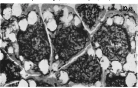
FIG. 5. Section (5 μ) through hypertrophied fat cells of a fifth-instar larva of Choristoneura murinana (Hübner) infected with granulosis virus. Note the character
istic black network (strands) in the highly hypertrophied nuclear area as well as in the cytoplasm. Stained with Heidenhain's iron hematoxylin and eosin. Magnification:
χ 1100. (Original.)
in the same host (Choristoneura murinana) that the network (directly after it forms) gives a positive Feulgen's nuclear reaction especially in that part occupying the hypertrophied nuclear area (Fig. 6 ) . In the course of the development of capsules the network becomes Feulgen negative. Huger's (1960a) suggestion that it might serve as virogenetic stroma was subsequently confirmed by electron microscope studies (Huger and Krieg, 1960, 1961) (see Section V I I I , B , 2).
T h e diversity of opinions as to whether the capsules develop in the nucleus or in the cytoplasm (see T a b l e I) mainly arose from the diffi
culty of staining the capsules in a convincing manner and from mis
interpretations of light and electron micrographs. T h e latter might
552 ALOIS HUGER
have been responsible for the opinion that the granulosis viruses and capsules of C. fumiferana, Eucosma griseana, and Pieris rapae develop only in the cytoplasm of cells, the nuclei of which are still intact (Bird,
1958, 1959) (see T a b l e I ) .
Bergold and Bird (1958, 1959) apply this opinion also to the blood cells which in an advanced stage of the disease—when masses of cap
sules from disintegrated tissues are floating in the hemolymph—show capsule-filled spherical vesicles with variable diameter (up to about 20 μ) in their cytoplasm. However, since in most cases the nuclei of blood cells do not exhibit characteristic changes in any way comparable to those of the infected cells described above, there is great probability that the capsules in the surrounding cytoplasm have mostly been phag-
FIG. 6. Section (5 μ) through three hypertrophied hyperdermis cells (a) and a hypertrophied fat cell (b) of a fifth-instar larva of Christoneura murinana (Hübner) infected with granulosis virus. Note the characteristic network positively stained with Feulgen's nuclear reaction (virogenetic stroma). Magnification: χ 980. (Original.)
ocytosed (Wittig, 1960). Nevertheless, a real development of viruses and capsules in blood cells must also be considered, but this question is still to be investigated.
By the elective staining method of Huger (1961) (see, e.g., Section II, Β ) , it was no problem to find out that the capsules of C. murinana develop in the nucleus as well as in the cytoplasm. This is not sur
prising with regard to the function of the network just mentioned and in view of the electron microscopic results dealt with in the next section.
2. Electron-Microscope Investigations
Parallel to light-microscope studies electron-microscope investigations of ultrathin sections are indispensable for a better understanding of the characteristic pathological changes in infected cells. T h e first investiga
tions of granuloses on this basis were carried out by Bird (1958, 1959) (cf., Section V I I I , Β , 1 ) . Contrary results have been obtained by Huger and Krieg (1960, 1961) studying ultrathin sections of infected
tissues of C. murinana, a host very closely related to C. fumiferana as studied by Bird. In agreement with the light-microscope observations
(see Section V I I I , B , 1 ) , they were able to follow the striking cytopath- ological changes. In the hypertrophied nuclei the accumulated chro
matic material aggregated to (intranuclear) strands (Fig. 7 ) . At the same time, there was an intensive production of cytomembranes by continuous renewal of the nuclear membrane (Fig. 7 ) . In this process
FIG. 7. Electron micrograph of an ultrathin section through a fat body cell of a larva of Choristoneura murinana (Hübner) infected with granulosis virus. Note the accumulation of chromatin material within the nucleus (N) partly aggregated to intranuclear stands (S), and the production of cytomembranes by renewal of the nuclear membrane (M, Mf), thus giving rise to perinuclear strands (S'), which spread over the cytoplasm (C). F, fat globules; Mi, mitochondria. Magnification: χ 13,800.
(Shown in part by Huger and Krieg, 1961.)
chromatic nuclear material is enclosed between the arising nuclear membranes and thus emitted into the cytoplasm. T h e layers of mem
branes progressively condense thus forming compact perinuclear strands which spread all over the cytoplasm. Together with the intranuclear strands they form the said typical network, from the surface of which virus rods are released (virogenetic stroma) (Fig. 8 ) .
By the disintegration of the network, virus rods and little protein particles are set free, both often surrounding the strands like a cloud
554 ALOIS HUGER
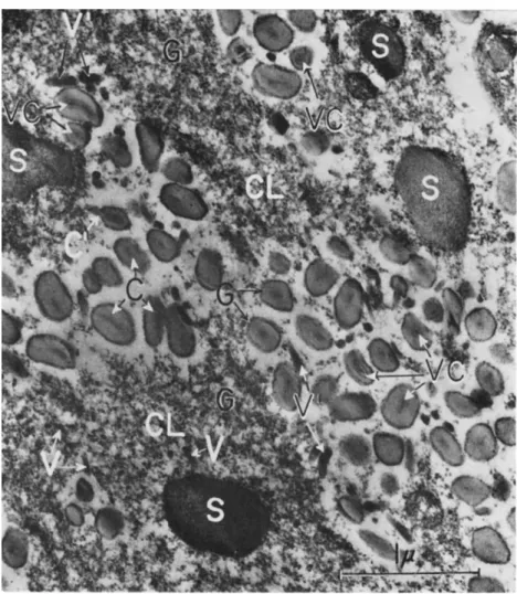
(Fig. 9 ) . Capsules are formed by progressive deposition of protein- aceous material around the virus rods. Sometimes the formation of capsules was observed to start at one end of the virus rod, thus devel
oping a cap which extended toward the opposite end until the capsule was completed (Fig. 9 ) . Hughes (1952) described this process for cap
sules of Sabulodes caberata Guenee. Furthermore, capsules can also be formed directly within the strands or they detach from their periphery.
In both cases strand material is transformed in situ to paracrystalline
FIG. 8. Electron micrograph of an ultrathin section through parts of strands (S) (network) in a larval fat body cell of Choristoneura murinana (Hübner) 10 days after infection with granulosis virus. Note the virus particles (V) being released from the surface of the disintegrating strands. C, capsule. Magnification: χ 39,700. (From Huger and Krieg, 1961.)
capsule protein. As the DNA-containing strands spread all over the cell, the formation of viruses and capsules takes place in the nuclear, as well as in the cytoplasmic, areas.
Furthermore, in the investigations by Huger and Krieg (1960, 1961) not the least evidence could be obtained for the development of granu
losis viruses from spheres to rods within the "developmental membrane'' as was suggested by Bergold (1950, 1953a, b, 1958) (life-cycle theory, see Section I V ) , and accepted by Bird (1958, 1959). On the contrary, the viruses are seen to be rod-shaped and naked when they become established and only subsequently do they acquire the "developmental membrane" and the capsular envelope (Fig. 9 ) . These observations become understandable in the light of the very recent findings on the
helical structure of insect virus rods by Smith and Hills (1960) (see Section I V ) . From this detection, and from the existence of eclipse phases (dissociation) of viruses (Yamafuji et al., 1954; Krieg, 1958;
Aizawa, 1959), it is reasonable to assume that the synthesis of the rod- shaped insect viruses is accomplished by the assembling of their com-
FIG. 9. Electron micrograph of an ultrathin section showing a part of a larval fat-body cell of Choristoneura murinana (Hübner) 12 days after infection with granulosis virus. T h e disintegrating strands (network) (S), are surrounded by clouds (CL) of granular particles (G) and naked virus rods (V). Note the virus rods with the
"developmental membrane" (V) and the encapsulated virus rods (VC). T h e formation of capsules (C) from the granular material (G) which is transformed to paracrystalline capsule protein is to be seen. Note further the encapsulation starting at one end of the virus rod (C). Magnification: χ 32,000. (From Huger and Krieg, 1961.)
556 ALOIS HUGER
ponents (nucleic acid and protein) comparable to that which occurs with the tobacco mosaic virus.
Not infrequently long filaments of the same thickness and density as virus rods are visible in ultrathin sections of infected cells (Bird, 1958, 1959; Huger and Krieg, unpublished). Apparently they break across to rods of about normal length. Micrographs of Huger and Krieg indicate that long virus threads lying either free in the cell or within strands may also be occluded by capsule protein, thus resulting in aber
rant long virus inclusion bodies referred to in Section IV.
C. Susceptibility and Resistance of Hosts to Granuloses
In general, young larvae are more susceptible to granuloses than are older ones (e.g., Lower, 1954; Tanada, 1953a, 1955, 1956b, c, 1959a; Mar
tignoni, 1957; Schmidt and Philips, 1958; Schmidt, 1959; Wittig, 1959a;
Sager, 1960). In peroral infection experiments with Pseudaletia uni
puncta, for example, the average mortality of the highly susceptible first- and second-instar larvae was 80.8 and 92.7 percent, respectively, compared to 23.4 percent in the considerably resistant sixth larval in- star (Tanada, 1956b). However, the susceptibility of the older larvae of this species is greatly increased when they are fed a mixture of gran
ulosis and polyhedrosis virus (see Section V I I I , D). Only for Harri
sina brillians it is suggested that larvae of the third instar are more susceptible to the granulosis than those of the first instar (Smith et al., 1956).
Tanada (1953a) showed that larvae of Pieris rapae succumbed to granulosis to a lesser extent when they were reared and infected at a high temperature (36 ° C ) . In an infection experiment conducted by Steinhaus and Dineen (1960) with larvae of Peridroma margaritosa, no deaths from granulosis occurred at 37 °C. High temperatures apparently are inhibitory to the activity of these and other insect viruses.
Larvae contracting granulosis in a late instar may pupate and ap
parently also develop to adults. However, infected pupae not infre
quently die (Bucher, 1953; Tanada, 1953a; Kovacevic, 1958; Wittig, 1959a; Raun, 1961). T h a t pupae, even healthy looking ones, may carry the virus is proved (1) by their being infectious when triturated and used as an inoculum in healthy larvae (Sager, 1960), and (2) by the demonstration of capsules in the electron microscope (Smith et al.,
1956). In addition, Vago and Atger (1961) found that production of the granulosis virus of Pieris brassicae is possible by infecting larvae perorally at the end of the feeding period and harvesting the capsules from the pupae.
Experimental infections of larvae with granulosis viruses usually
are accomplished by feeding of capsules, but they have also been achieved by direct inoculation of capsule suspensions into the body cavity, as is shown, for instance, with larvae of P. brassicae (Paillot, 1926), Agrotis segetum (Paillot, 1934, 1936, 1937), P. margaritosa
(Steinhaus, 1947), and Choristoneura murinana (Bucher, 1953). It is not proved whether in these cases the viruses have been liberated from their proteinaceous capsule in the hemolymph, and thus caused infec
tion, or whether infection has been brought about by free virus rods that were already present in the inoculum. T h e latter is more probable in view of the fact that capsules are not dissolved in the hemolymph of larvae with advanced disease and in view of the results obtained by Martignoni (1957) with larvae of Eucosma griseana which did not develop granulosis after injection of heavy doses of capsules into the hemocoel, whereas peroral applications of relatively small doses resulted in high infection rates.
Whereas our knowledge of the increase in resistance to virus infec
tions connected with the degree of larval maturity is well substantiated, there are only relatively few observations referring to the apparent oc
currence of resistant insect populations or on their acquired resistance to disease. As to the granuloses, David (1957) first reported on the development of "considerable resistance" to granulosis in a laboratory culture of P. brassicae bred through a severe epizootic of this disease which occurred in 1955, after the culture had been maintained healthy for five years. Subsequently further information on this interesting phenomenon was given by Rivers (1959) : at the height of the epidemic only about 1 percent of the larvae survived, but with these few it was possible to breed the culture up to its full strength again. Infection experiments with larvae of this recovered so-called Cambridge stock (using virus preparations from dead larvae) showed that their resistance to granulosis-virus infection was significantly increased as compared with P. brassicae larvae from other sources.
However, this acquired population resistance, apparently brought about by selection, can be overcome, especially during the first and sec
ond instars, when the larvae are fed very large doses of virus (Rivers, 1959). Moreover, Smith (1959a, 1960) noticed that granulosis viruses of P. brassicae are made capable of breaking the resistance by passage through Pieris napi. Special investigations (see Section V I I I , E) pos
sibly will show whether the P. brassicae virus really is able to multiply in P. napi or whether it only triggers off a latent granulosis in the latter.
When, in 1959, David and Gardiner (1960) conducted experiments with the resistant Cambridge stock, there had been no deaths from gran
ulosis in this culture for several generations, and the larvae looked