DEVELOPMENTAL BIOLOGY SUPPLEMENT 2 , 1 0 3 - 1 1 7 ( 1 9 6 8 )
The Biochemical Organization of Cytoplasmic Membranes
P H I L L I P S W . ROBBINS
Department 'of Biology, Massachusetts Institute of Technology, Cambridge, Massachusetts
INTRODUCTION
From the recent work on protein structure that has been summarized here by Doctor Anfinsen, it is clear that the secondary, tertiary, and quaternary structure of a protein is, in general, determined completely by the primary sequence of amino acids in the polypeptide chain.
Thus, given a sequence of amino acids and a complete understanding of the interactions of all side chains with each other and with the polypeptide backbone, it should be possible to predict accurately the structure of any protein. Extending this principle further, it should be possible to predict the structures of protein polymers such as the T4 bacteriophage tail fiber and bacterial flagella from the primary amino acid sequence in the monomeric protein subunits, since the structure of the polymer is determined by structural interactions among the protein subunits which, in turn, are determined by the primary amino acid sequence.
It is of interest to inquire to what extent this principle of self- assembly may be of help in understanding the structure of complex structural elements like the cytoplasmic membrane. Thompson (1965) has proposed that the structure of membranes is determined by self- assembly and states the case in the following way. "The different types of component lipids may be to membrane structure what amino acids are to protein structure, that is, the secondary, tertiary, and quaternary structure of membranes are determined by the structure and space
filling properties of the building blocks, their ratio, and their two- dimensional patterns." A contrary opinion was expressed by Stern (1965) at this Symposium three years ago. He said "whether the con
cept of self-assembly can be extended to all formed elements in a cell remains to be seen, but the prospect is . . . not bright. Unlike self-
103
104 PHILLIPS W . ROBBINS
assembled structures . . . membranes are transmitted as such from one cell generation to another; a membraneless cell is unknown. Where careful observations have been made of the formation of specific membranes, as in the chloroplast, evidence points to the growth of new membranes from pre-existing ones. The functional and structural heterogeneity of membranes (e.g., inner membranes of mitochondria, lamellae of chloroplasts, limiting membranes of cytoplasm) apparently makes self-assembly from a pool of individual molecules a rather hazardous mörphogenetic operation. The issue is whether the essen
tial genetic complement of a cell is reducible to sequence coding, or whether it must also contain other preformed elements that serve as frameworks, however small and undistinguished, for the reproduction of recognized structures." While this word of caution is in order, and it is certainly necessary to recognize the role of primer substances in the process of organizing membrane structures, I believe that the point of view expressed by Stern is overly pessimistic and that in the long run self-assembly will be found to play a major role in the deter
mination of membrane structure. In spite of its potential importance, however, I feel that at the present time the self-assembly principle has little value for understanding the structure of membranes. This is sim
ply because the overall picture is now so unclear that even the par
ticular lipid-lipid or lipid-protein interactions that would serve as the basis for self-assembly are not known. Thus, whether one supports the Danielli-Davson model of membrane structure (Fig. 1A) or the pro
tein subunit model of membrane structure (Fig. IB) it is clear that either model could be the result of a process of self-assembly. The choice depends on the relative aifinity of lipid for lipid or of lipid for protein and, of course, also on the structures of the component lipids and proteins.
Since the principle of self-assembly is at the present time of little help in predicting the structure of the complex cytoplasmic membrane, and since even data from electron microscopy may be interpreted to support either the lipid bilayer or protein subunit concepts of mem
brane structure, I feel it is of more value to select for review the three most pertinent biochemical systems in order to summarize what can be suggested from recent work about the real structure of membranes.
While it can only be considered a guess, it seems that in the long run the conclusion that may emerge is that the basic structure of mem
branes will be found to vary rather drastically from one system to
CYTOPLASMIC MEMBRANES 105
9ΎΥ??
PROTEIN
> LIPID
PROTEIN
-LIPOPROTEIN
FIG. 1. Hypothetical models of membrane structure. (A) The Danielli-Davson model as conceived by Thompson (1964) [see also Robertson (1964)]. (B) Inner mitochondrial membrane composed of lipoprotein repeating subunits as proposed by Green and Perdue (1966).
another, but that elements of both the lipid bilayer structure and the subunit structure will be found in almost every situation. My own feeling is that in most cases membrane structure involves a lipid bi
layer with protein and/or polysaccharide molecules incorporated into the matrix of the bilayer. I do not feel that it is clear how the problem of subunits will be resolved, but the detailed models proposed by Green and Perdue (1966) and Penniston et al. (1968) may have to be modified in light of further experimental work. While there is no doubt that some membranes such as those in the cristae of mitochon
dria and grana of chloroplasts must contain organized regions or domains, I feel that present evidence is insufficient for building de
tailed models of the molecular organization of these structures.
With this introductory statement, which presents my personal point of view on the present state of the problem of membrane structure, I would like to go on to review briefly the present situation in research on model membranes, the simple outer membrane of gram-negative enteric bacteria, and the inner membrane of the mitochondrion.
106 PHILLIPS W* ROBBINS
MODEL PHOSPHOLIPID-HYDROCARBON FILMS
During the last few years there has been considerable interest in the properties of thin films prepared from mixtures of phospholipids and neutral lipids. These studies have been carried out by Mueller and Rudin (1968) and by Thompson and co-workers (Thompson and Henn, 1968; Maddy et al., 1966). The films are prepared in a small chamber that is divided in half by a perforated polyethylene septum.
The chamber is filled with buffered aqueous medium, and a drop of organic solvent containing the lipid mixture to be studied is placed in the perforation. As the solvent diffuses into the aqueous medium, the drop thins and, if an appropriate mixture of lipids is present, finally undergoes a transition to give a film 50-100 Â thick.
Stable films have been prepared from mixtures of phospholipids and neutral lipids. Both mixed phospholipids and purified compounds such as phosphatidylcholine and phosphatidylethanolamine are active.
Even materials such as the mannolipid from Micrococcus lysodeikticus, which does not contain phosphate or other ionizing groups, will re
place the "phospholipid." This requirement therefore seems to be for a general amphipathic substance. A great many neutral lipids are active as second components including hydrocarbons such as n-decane and miscellaneous substances such as a-tocopherol and ^-carotene. The composition of the films formed depends on the ratio of neutral to phospholipid but is about 10:1, with the neutral component pre
dominating. The final composition of the membranes and their appear
ance in electron micrographs have led Thompson and Henn (1968) to postulate that much of the neutral lipid is present as "lenses" trapped in the membrane and that much of the rest is distributed unevenly throughout the structure. As far as the arrangement of the phospholipid molecules is concerned it seems very likely that they form the classical bilayer with the fatty acid chains in the hydrocarbon core and hydro- philic moieties in contact with the aqueous phase.
In spite of the predominance of neutral lipid and lack of protein in these artificial membranes, they have a great many characteristics that are similar to those of natural membranes. Table 1, taken from the review by Thompson and Henn (1968), lists a comparison of eleven properties of natural membranes and synthetic bilayers. The similarities in appearance in the electron microscope, thickness, capacitance, and surface tension make it seem plausible that phos-
CYTOPLASMIC MEMBRANES 107
TABLE 1
COMPARISON OF SOME PROPERTIES OF BILAYERS AND BIOLOGICAL MEMBRANES'1
1.
2.
3.
4.
5.
6.
7.
8.
9.
10.
11.
Property
Electron microscope image Thickness (A)
Capacitance (^mf/cm2) Resistance (Ω cm2) Dielectric breakdown (mV) Surface tension (dynes/cm) Water permeability (μ/sec) Activation energy for water tion (kcal/mole)
permea- Urea permeability (μ/sec X 102) Glycerol permeability (μ/sec Erythritol permeability (μ/?.-
: X 102) ec X 102)
Biological membranes
(20-25 °C)
Trilaminar 60-100 0 . 5 - 1 . 3 102-105 100 0.03-1 0.37-400
9.6<
0.015-280 0.003-27 0.007-5
Bilayer (36 °C)
Trilaminar 60-75 0.38-1.0
106-109 150-200 0.5-2
31.7C 12.7C
4.2b 4.6&
0.75&
Reference*
(1) (1) (2) (3) (4) (5) (6) (7) (8) (8) (8)
a Adopted from Thompson and Henn (1968).
b 20°C.
c 25°C.
d Key to references :
(1) ELBERS, P. F., in "Recent Progress in Surface Science" (J. F . Danielli, K. G. A.
Pankhurst, and A. C. Riddiford, eds.), Vol. 2, p . 443. Academic Press, New York, 1964.
(2) PAULY, H., AND PACKER, L., / . Biophys. Biochem. Cytol. 7, 603 (1960).
(3) COLE, K. C , in "Proceedings of the First National Biophysics Conference"
(H. Quastler and H. Morowitz, eds.). Yale Univ. Press, New Haven, Connecticut, 1959.
(4) SHANES, A. M., Pharmacol Rev. 10, 59 (1958).
(5) ACKERMAN, E., "Biophysical Science," pp. 236-239. Prentice-Hall, Englewood Cliffs, New Jersey, 1962.
(6) DICK, D . A. T., Intern. Rev. Cytol. 8, 387 (1959).
(7) HAMPLING, J. Gen. Physiol. 44, 365 (1960).
(8) VREEMAN, H. J , Proc. Koninkl. Ned. Akad. Wetenschap. Ser. B 69, 542 (1968).
pholipid bilayers could serve as the structural base or framework of natural membranes.
One striking difference in the properties of synthetic bilayers as compared to natural membranes is their high electrical resistance. As shown in Table 1, the resistance of synthetic lipid membranes is about 10,000 times greater than that of natural membranes. Recent work by Mueller and Rudin (1967) has shown that adsorption of the antibiotic valinomycin to bilayers will decrease this high resistance by several orders of magnitude. This is especially interesting in view
108 PHILLIPS W . ROBBINS
of the known relationship of valinomycin to cation transport in mitochondria. It was shown originally by Moore and Pressman (1964) that addition of valinomycin to a suspension of mitochondria caused an increase in respiration that depended on the addition of K+, Cs+, or Rb+. Direct measurement then demonstrated that the antibiotic caused a rapid uptake of K+ that was balanced by the extrusion of protons.
Chemically, valinomycin and the related antibiotics that show specific effects on cation transport are neutral molecules with a number of carbonyl residues that could potentially coordinate with monovalent cations. Lardy and co-workers (1967) have, in fact, built molecular models to determine whether specific chelation could take place. They found that specific coordination was a real possibility and that mono
valent cations could be bound firmly in the center of the antibiotic molecule. Since the peptide chain is relatively hydrophobic, it is likely that valinomycin associates with the lipid components of the membrane and there serves as a site for the specific binding of cations.
Once bound to the membrane in association with valinomycin, the cations could be transferred to another transport system or could simply use the antibiotic for facilitated transport across the lipid matrix.
In artificial membranes, valinomycin increases the conductance of potassium, cesium, and rubidium ion dramatically but has little effect on sodium and lithium ion conductance. It has also been demon
strated that a resting potential as high as 150 mV can be developed when NaCl and KCl solutions are placed on opposite sides of the membrane (Mueller and Rudin, 1967). The fact that valinomycin and related substances are able to recognize and interact with the lipid bilayer system in a fashion similar to their interaction with natural membranes provides support for the hypothesis that the phospholipid bilayer may be the basis of membrane structure.
SYNTHESIS AND ORGANIZATION OF THE LIPOPOLYSACCHARIDE OF THE INNER AND OUTER MEMBRANES OF THE
SALMONELLA CELL ENVELOPE
According to present models the cell envelope of gram-negative enteric bacteria consists of the following major components: (1) an inner cytoplasmic membrane; (2) an intermediate area, the "peri- plasmic" space, which contains the rigid mucopeptide polymer and
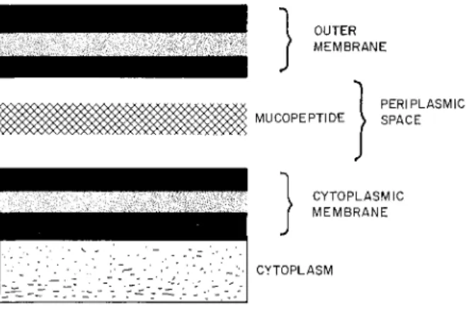
CYTOPLASMIC M E M B R A N E S 109 a number of enzymes that are released from the cell by treatment with EDTA; and (3) an outer membrane (de Pétris, 1967). When examined by negative staining in the electron microscope, the inner and outer membranes both share the triple-layered appearance characteristic of unit membranes. Although the composition of the membranes is not known in detail, it seems likely that both contain phospholipid, protein, and lipopolysaccharide. The principal phospholipid in the membranes of gram negative enteric bacteria is phosphatidylethano- lamine. The lipopolysaccharide component consists of a lipid (lipid A) and a "core" polysaccharide. The O-antigen polysaccharide, which contains galactose, mannose, and rhamnose in many Salmonella strains, is linked covalently to this core polysaccharide. A diagram of the organization outlined above is presented in Fig. 2.
OUTER MEMBRANE
PERIPLASMIC MUCOPEPTIDE \ SPACE
CYTOPLASMIC MEMBRANE
CYTOPLASM
FIG. 2. Organization of the cell envelope of gram-negative enteric bacteria.
This model is an interpretation of electron micrographs (de Pétris, 1967) taken of thin sections of Escherichia coli fixed in glutaraldehyde. Both inner and outer mem
brane appear as typical triple-layered "unit membrane" structures.
Recent work by Leive, Work, and their collaborators has shown that at least part of the outer membrane can be specifically released from the cell without affecting viability. Knox et al. (1966) have shown that cells deprived of lysine excrete into the growth medium particulate material that probably represents segments of outer mem
brane. Leive (1965) has found that treatment of cells with EDTA brings about a rapid release of outer membrane material. The particles released by lysine starvation and EDTA treatment are both rich in lipopolysaccharide, but both contain protein and phospholipid as
110 PHILLIPS W . ROBBINS
well. According to the proposal of Rothfield et al. (1966) lipopoly- saccharide and phospholipid molecules are able to anneal to form structures that contain both components, in varying proportions, all of which give the appearance of typical unit membranes when examined in the electron microscope. Possible molecular arrangements for the two components in these membranes have been suggested by Rothfield, and one possibility is shown in Fig. 3.
TTTTTTuTTTT^TTT^TTTT^u^^uTTTTT
LIPOPOLYSACCHARIDE MOLECULES
D = LI PID (" LIPID A " )
= POLYSACCHARIDE
OTHER AMPHIPATHIC MOLECULES (eg. PHOSPHATIDYL ETHANOLAMINE)
~~~ POLAR PORTION (Phosphoryl L· Ethanolamine )
' ^ N O N POLAR PORTION (Diglyceride)
FIG. 3. Hypothetical molecular arrangement of lipopolysaccharide and phos
pholipid molecules as a lipid bilayer (Rothfield et al., 1966). In this model the hydrophobic core is formed by interactions between lipid A of the lipopolysac
charide and fatty acid chains of the phospholipids. The polysaccharide of the lipopolysaccharide and phosphoryl residues of the phospholipids are in contact with the aqueous environment.
A number of models for biosynthesis and organization arise from considerations of the picture of the cell envelope presented above. On the one hand, it could be imagined that tiny complexes of inner membrane, outer membrane, and periplasmic area are organized in the cytoplasm and inserted as units or blocks into the preexisting cell envelope. A second possibility would be that enzymes and substrates concerned with the synthesis of the inner membrane are located in the cytoplasm while enzymes and substrates concerned with the synthesis of the outer membrane are located in the periplasmic space.
CYTOPLASMIC MEMBRANES 111 A third possibility would be that the components of the outer mem
brane are derived from the inner membrane by continuous accretion.
This process of accretion could be a one-way transfer of material from the inner membrane, the presumed site of synthesis, to the outer membrane, or there could be continuous exchange of mem
brane material across the periplasmic space in both directions. In order to investigate these questions, pulse labeling experiments have been carried out by Leive and similar experiments have been done in my own laboratory. The results in both cases have been very similar.
Leive's work (1968) is reported elsewhere, and our own experiments are reported here.
Salmonella newington was grown in 1.2 liters of L broth at 37°
with shaking to a density of 6 X 10s cells/ml. Glucose-14C was added to give a final concentration of 10~5 M and 1 /xC/ml. Preliminary experiments have shown that this amount of glucose is taken up almost completely in less than 30 seconds. After the pulse period of 30 or 60 seconds, unlabeled glucose was added to a final concentration of 0.2%. At various times after the beginning of the "chase," 150-ml samples were poured onto cracked ice. As soon as possible after completion of the chase, the chilled samples were centrifuged and washed with cold 0.9% NaCl. The washed cells were resuspended in 20 ml of 0.01 M EDTA, pH 8.0, and incubated at room temperature for 30 minutes with RNase and DNase (10 /Ag/ml each) to release lipopolysaccharide and reduce the viscosity of the suspension. The mixture was centrifuged for 15 minutes at 25,000 g, and the pellet was resuspended in 20 ml of 0.01 M EDTA, pH 8.O. Lipopolysaccharide was then isolated from each of the pellet and supernatant fractions.
The method used for lipopolysaccharide isolation has been described elsewhere (Robbins and Uchida, 1962) and involves phenol ex
traction at 68°, dialysis, acetone precipitation, and ultracentrifugation.
After the ultracentrifugation step, the lipopolysaccharide was hy- drolyzed overnight at 97°C in 0.2 N H2S04. The hydrolyzates were neutralized with a drop of pyridine and streaked directly on V& inches of Whatman No. 1 paper. After chromatography in butanol-pyridine -water (6:4:3), the strips were scanned for 14C. They showed radioactivity primarily in the galactose, mannose, and rhamnose areas.
The rhamnose areas were cut from the papers and eluted by suspen
sion in 4 ml of water. Aliquots (0.5 ml) were used for duplicate determinations of 14C by scintillation counting and rhamnose measure-
112 PHILLIPS W . BOBBINS
ments by the method of Dische. Since both methods are very accurate, it is possible to get a precise picture of the changes in rhamnose specific activity through the course of the experiment.
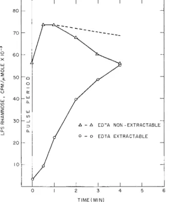
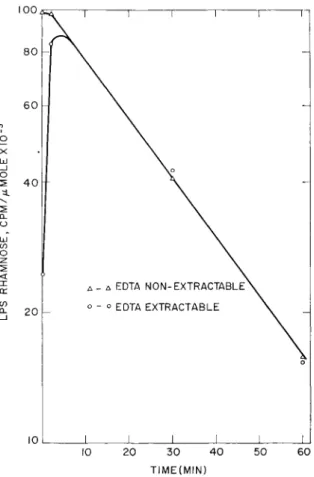
Figure 4 shows the results of an experiment in which samples were taken for 4 minutes after the beginning of the chase, and Fig. 5 shows the results of an experiment in which 30- and 60-minute samples
^ 60
^ Έ.
50
20
- -
— Q O cr
— LLI
CL ÜJ if)
— _ i
Q_
-
I i i i i ;
j ^\~---—...^_
cr —
/ Δ - Δ EDTA NON-EXTRACTABLE J O - o EDTA EXTRACTABLE
1 1 1 1 1
TIME(MIN)
FIG. 4. Change in specific activity of lipopolysaccharide rhamnose with time after a 30-second pulse of glucose-14C. The experiment was carried out as described in the text.
were taken. The picture that emerges is clear. After the pulse period there is little label in the EDTA-extractable fraction, whereas the EDTA nonextractable fraction is labeled maximally after 60 seconds.
Within 4 minutes the specific activity of rhamnose in the two fractions becomes the same, and it remains the same for at least 60 minutes. The decrease in specific activity shown in Fig. 5 corresponds to the dilution expected for a cell doubling time of 25 minutes after the
CYTOPLASMIC M E M B R A N E S 113
l±J _l
o 80
6 0
4 0
20
10
- \ I i i i i
1 \°
Λ - Δ EDTA N0N-EXTRACTABLE\
o - o EDTA EXTRACTABLE \
1 1 1 1 1 1
-
o
1 10 20 30
TIME(MIN)
40 50 60
FIG. 5. Change in specific activity of lipopolysaccharide rhamnose with time after a 60-second pulse of glucose-14C. The experiment was carried out as described in the text except that the pulse and chase were carried out on separate 300-ml aliquots of the same culture for each time point. An additional 300 ml of broth containing unlabeled glucose was added to the "60-minute" sample after 30 min
utes of growth so that logarithmic growth would continue throughout the chase period.
beginning of the chase period. If this same rate of dilution occurred after maximal labeling of the EDTA-nonextractable fraction during the early part of the chase period shown in Fig. 4, the curve would have followed the dotted line shown in the figure. Thus, during the time when the outer membrane is acquiring labeled lipopolysaccharide, the lipopolysaccharide of the inner membrane is losing label or be-
114 PHILLIPS W . ROBBINS
coming diluted to a greater extent than can be accounted for by growth alone.
All the data fit well with the proposal that lipopolysaccharide-bound O-antigen is synthesized in association with the inner cytoplasmic cell membrane and that lipopolysaccharide molecules exchange back and forth between the two membranes with a half-time of about 1.5 minutes at 37°. This model would account for the rapid labeling of the EDTA nonextractable fraction, the slower labeling of the EDTA extractable fraction, the loss of label from the nonextractable fraction during the early part of the chase, and the identity or near identity of the specific activities from 4 to 60 minutes. While this model may not be a unique explanation for the experimental results, the idea that the outer membrane of the cell envelope is synthesized by a process of accretion and that it remains in "contact" with the inner membrane by way of continuous material exchange across the periplasmic space deserves consideration and further experimental tests.
STRUCTURE OF MITOCHONDRIAL MEMBRANES
In contrast to model bimoleeular lipid leaflets and the relatively simple outer membrane of gram-negative enteric bacteria, membranes of the mitochondrion, and especially the inner membrane, are complex highly organized structures. Since excellent reviews have been pub
lished recently, I do not intend to review the subject of mitochondrial organization in detail. I wish only to contrast the obvious com
plexity of the mitochondrial membrane with the preceding subjects.
The outer mitochondrial membrane has a typical unit membrane appearance in electron micrographs and may be mainly phospholipid or have a phospholipid framework since it is readily dissolved by organic solvents. The inner membrane has a complex appearance, and when stained negatively with phosphotungstate shows particles 85 Â in diameter attached to the inner surface. Work by Racker (1967) has shown that these particles are probably identical to Fi, one of the "coupling factors" that has been extracted from mitochondria. A detailed examination of F1 has given important insights into structure- function relationships.
Submitochondrial particles prepared by sonication and differential centrifugation have the appearance of fragments of inner membrane when examined by negative staining in the electron microscope.
Successive treatment of these particles with Sephadex and urea leads
CYTOPLASMIC MEMBRANES 115 to a membrane preparation (SU particles) devoid of 85 Â particles and ATPase activity. (Fx has strong ATPase activity that is not sensitive to oligomycin. ) These SU particles contain cytochromes and oxidative enzymes but do not carry out oxidative phosphorylation.
Addition of Fi to SU particles restores the original morphology to the system. Membrane vesicles with 85 Â particles attached appear, and the ATPase activity of the added Fx is rendered sensitive to oligomycin. Although addition of other coupling factors is required for the restoration of oxidative phosphorylation, the simultaneous change in morphology and enzymatic properties provides a striking demonstration of the concept that structure and function are closely related in this system.
The specific components in SU particles that render Fi sensitive to oligomycin have been examined in some detail. It is found that if particles are dissolved in cholate and then fractionated with ammonium sulfate, a protein fraction (CFo) is obtained that will bind Fi. The CFo-Fi complex is amorphous and has little ATPase activity, but on addition of phospholipid to the system a moropho- logically normal particle is generated that has strong oligomycin- sensitive ATPase. Further examples could be cited of the strong relationship between structural integrity of the inner membrane of the mitochondrion and its enzymatic functioning in oxidative phos
phorylation. While it is clear that highly organized protein structures account for a major portion of the membrane, it is still possible that a basic phospholipid bilayer is present. Whether it is, in fact, present and its relationship to the organized protein components seem to be starting points for current research on complex membranes.
CONCLUSION
In spite of the fact that a great deal of research has already been done on membrane structure, it seems to me that we are just at the beginning of a period of acquiring firm information about real problems of organization and function. A great deal of what has been proposed to date could be classified as unwarranted generalization based on inconclusive evidence. It may be that new methods and approaches will be needed before solutions to existing problems can be found. However, work on model phospholipid bilayers, studies on simple membranes such as the outer membrane of gram-negative
116 P H I L L I P S W . ROBBINS
enteric bacteria, and detailed reconstruction experiments on complex membranes, like that of the mitochondrion, already look hopeful.
REFERENCES
DE PÉTRIS, S. ( 1967). Ultrastructure of the cell wall of Escherichia coli and chem
ical nature of its constituent layers. / . Ultrastruct. Res. 19, 45.
GREEN, D . E., and PERDUE, J. F . (1966). Membranes as expressions of repeating units. Proc. Natl. Acad. Sei. U. S. 55, 1295.
KNOX, K. W., VESK, M., and WORK, E. (1966). Relation between excreted lipo- polysaccharide -complexes and surface structures of a lysine-limited culture of Escherichia coli. J. Bacteriol. 92, 1206.
LARDY, H. A., GRAVEN, S. N., and ESTRADA-O, S. (1967). Specific induction and inhibition of cation and anion transport in mitochondria. Federation Proc. 26, 1355.
LEIVE, L. (1965). Release of lipopolysaccharide by EDTA treatment of E. coli.
Biochem. Biophys. Res. Commun. 2 1 , 290.
LEIVE, L. (1968). In preparation.
MADDY, A. H., HUANG, C., and THOMPSON, T. E . (1966). Studies on lipid bilayer membranes: a model for the plasma membrane. Federation Proc. 25, 933.
MOORE, C., and PRESSMAN, B. C. (1964). Mechanism of action of valinomycin on mitochondria. Biochem. Biophys. Res. Commun. 15, 562.
MUELLER, P., and RUDIN, D . O. (1967). Development of K+-Na+ discriminations in experimental bimolecular lipid membranes by macrocyclic antibiotics. Bio- chem. Biophys. Res. Commun. 26, 398.
MUELLER, P., and RUDIN, D . O. (1968). Action potentials induced in biomolecular lipid membranes. Nature 217, 713.
PENNISTON, J. T., HARRIS, R. A., ASAI, J., and GREEN, D . E. ( 1 9 6 8 ) . The con-
formational basis of energy transformations in membrane systems. I. Conforma- tional changes in mitochondria. Proc. Natl. Acad. Sei. U. S. 59, 624.
RACKER, E. ( 1967 ). Resolution and reconstruction of the inner mitochondrial mem
brane. Federation Proc. 26, 1335.
ROBBINS, P. W., and UCHIDA, T. (1962). Studies on the chemical basis of the phage conversion of O-antigen in the E-group Salmonellae. Biochemistry 1, 323.
ROBERTSON, J. D . (1964). Unit membranes: A review with recent new studies of experimental alterations and a new subunit structure in synoptic membranes.
In "Cellular Membranes in Development." Proc. 22nd Symp. Soc. Develop. Biol.
p. 1. Academic Press, New York.
ROTHFIELD, L., TAKESHITA, M., PEARLMAN, M., and HORNE, R. W . ( 1 9 6 6 ) . Role
of phospholipids in the enzymatic synthesis of the bacterial cell envelope. Fed- eration Proc, 25, 1495.
STERN, Η. (1965). Reproduction at the molecular level. In "Reproduction: Molecu
lar, Subcellular, and Cellular," Proc. 24th Symp. Develop. Biol. p . 5. Aca
demic Press, New York.
THOMPSON, T. E. (1964). The properties of bimolecular phospholipid membranes.
In "Cellular Membranes in Development," Proc. 22nd Symp. Soc. Develop. Biol.
p. 83. Academic Press, New York.
CYTOPLASMIC MEMBRANES 117
THOMPSON, T. E. (1965). Quoted in A. L. Lehninger, "The Mitochondrion,"
p. 223. Benjamin, New York.
THOMPSON, T. E., and H E N N , F . A. (1968). Experimental phospholipid model membranes. In "Structure and Function of Membranes of Mitochondria and Chloroplasts." ( E . Racker, e d . ) . Reinhold, New York, in press.
![FIG. 1. Hypothetical models of membrane structure. (A) The Danielli-Davson model as conceived by Thompson (1964) [see also Robertson (1964)]](https://thumb-eu.123doks.com/thumbv2/9dokorg/1117768.78341/3.664.175.487.108.440/hypothetical-membrane-structure-danielli-davson-conceived-thompson-robertson.webp)